TRIM21
W. A. McEwan,aF. Hauler,aC. R. Williams,bS. R. Bidgood,aD. L. Mallery,aR. A. Crowther,aand L. C. Jamesa MRC Laboratory of Molecular Biology, Cambridge, United Kingdom,aand British Antarctic Survey, Cambridge, United Kingdomb
Despite a central role in immunity, antibody neutralization of virus infection is poorly understood. Here we show how the
neu-tralization and persistence of adenovirus type 5, a prevalent nonenveloped human virus, are dependent upon the intracellular
antibody receptor TRIM21. Cells with insufficient amounts of TRIM21 are readily infected, even at saturating concentrations of
neutralizing antibody. Conversely, high TRIM21 expression levels decrease the persistent fraction of the infecting virus and
al-lows neutralization by as few as 1.6 antibody molecules per virus. The direct interaction between TRIM21 and neutralizing
anti-body is essential, as single-point mutations within the TRIM21-binding site in the Fc region of a potently neutralizing antianti-body
impair neutralization. However, infection at high multiplicity can saturate TRIM21 and overcome neutralization. These results
provide insight into the mechanism and importance of a newly discovered, effector-driven process of antibody neutralization of
nonenveloped viruses.
A
ntibody-mediated immunity forms a crucial part of the
anti-viral immune response, and its induction is a principal
objec-tive of vaccination. Reduced antibody (Ab) production, as occurs
in X-linked agammaglobulinemia, hypogammaglobulinemia,
and dysgammaglobulinemia, leads to persistent bacterial and viral
infection (
30
,
31
).
In vitro
, the binding of Abs to virus causes a
reduction in infectious titer, termed neutralization, which is
inde-pendent of effector mechanisms such as complement fixation or
Fc-mediated phagocytosis (
5
). Neutralizing antibodies (NAbs)
are thought to play an important role in antiviral immunity, since
the passive transfer of strongly neutralizing Abs is associated with
both antiviral protection (
10
,
12
) and the abrogation of disease (
7
,
34
). However, modeling and prediction of neutralization are not
straightforward (
29
). For instance, it is unclear how the binding of
one or a few Ab molecules per virus is sufficient for neutralization
(
4
). An average of 1.4 NAb molecules is capable of neutralizing
human adenovirus (AdV) type 2 (
39
), an apparently paradoxical
finding given that IgG molecules are considerably smaller than
adenovirus particles and occupy only a fraction of the viral surface
when bound. The binding of a single NAb was also reported to
neutralize poliovirus (
13
,
38
). A second neutralization
phe-nomenon that is poorly understood is the persistent fraction
(PF), i.e., the level of infection that remains at high NAb
con-centrations. The cause of the PF was previously attributed to
aggregated virus, low-affinity Abs, viral heterogeneity, and
polyclonal interference (
2
,
19
).
Recently, we showed that Abs can mediate neutralization
in-tracellularly by recruiting the cytosolic Ig receptor TRIM21 (
23
).
The engagement of NAb-virus complexes by TRIM21 promotes
the degradation of both Ab and virus by the proteasome, a process
termed antibody-dependent intracellular neutralization (ADIN)
(
25
). In this study, we describe the mechanistic requirements for
ADIN. We quantitatively examine both NAb-virus stoichiometry
and TRIM21 levels in order to probe their influence on the
effi-ciency of neutralization and the PF of viral infection. We find that
in cells depleted of TRIM21, neutralization is inefficient, requiring
a higher NAb-virus stoichiometry, and is accompanied by a higher
level of remaining infectivity. Conversely, interferon
(IFN)-stim-ulated cells have robust TRIM21 expression, efficient
neutraliza-tion, and low levels of persistent infection. The importance of
ADIN in virus neutralization is confirmed by the genetic
knock-out (KO) of TRIM21 in mouse cells and antibody site-directed
mutagenesis to ablate TRIM21 binding, either of which is
suffi-cient to substantially diminish the potency of a strongly
neutral-izing monoclonal Ab. However, while ADIN provides efficient
protection at low levels of viral infection, coinfection experiments
reveal that it can be overcome at high levels of virus exposure.
MATERIALS AND METHODS
Cell lines and viruses.Mouse embryonic fibroblast (MEF) cells were ob-tained from the ATCC (ATCC SCRC 1008) or prepared from wild-type (WT) C57BL/6 embryos or embryos with TRIM21 replaced by an en-hanced green fluorescent protein (GFP) construct (42). The level of ex-pression of GFP in cells from knockout mice was sufficiently low to permit the detection of adenovirus-delivered GFP. MEF and HeLa cells were maintained in Dulbecco’s modified Eagle’s medium (DMEM) with 10% fetal calf serum, 100 U/ml penicillin, and 100g/ml streptomycin at 37°C in a 5% CO2atmosphere. For stable TRIM21 knockdown, HeLa cells were
transduced with retroviral particles encoding small hairpin RNA (shRNA) directed to the human TRIM21 sequence GCAGCACGCTTGA CAATGA, and transduced cells were selected with puromycin at 2g/ml as previously described (23). For transient knockdown by small interfer-ing RNA (siRNA), cells were transfected with equal amounts of siRNA oligonucleotides T21siRNA1 (UCAUUGUCAAGCGUGCUGC; Dhar-macon) and T21siRNA2 (UGGCAUGGAGGCACCUGAAGGUGG; In-vitrogen) by using Oligofectamine (InIn-vitrogen) as directed by manufac-turer and as previously described (23). Replication-deficient, E1-deleted GFP- and red fluorescent protein (RFP)-expressing human adenovirus 5 stocks (3) were prepared by two rounds of CsCl centrifugation as previ-ously described (23).
Quantification of adenovirus.In order to determine virus stock con-centrations, samples were diluted 1:20 in 0.1% SDS and heated to 98°C for
Received25 March 2012Accepted15 May 2012
Published ahead of print30 May 2012
Address correspondence to L. C. James, [email protected].
Copyright © 2012, American Society for Microbiology. All Rights Reserved.
doi:10.1128/JVI.00728-12
on November 7, 2019 by guest
http://jvi.asm.org/
2 min, and the absorbance at 260 and 280 nm was measured. The number of particles per ml was calculated by multiplying the absorbance at 260 nm by the dilution factor and dividing by the extinction coefficient for AdV (9.09⫻10⫺13ml cm virion⫺1) (22,27). The ratio of the absorbance at 260
nm to the absorbance at 280 nm was 1.3, as expected for pure, intact AdV preparations. In order to calculate the concentration of hexon trimers, the virus concentration was multiplied by 240. To determine virus titers by quantitative PCR (qPCR), DNA was extracted by using a DNeasy blood and tissue kit (Qiagen) and assayed by using primers and a probe directed to the GFP transgene of AdV-GFP (see “Quantitative PCR”). A dilution series of virus stocks of known concentrations was used as a calibration curve in order to calculate the virus concentration.
Antibodies.Anti-AdV hexon antibody 9C12 (32,37) was purified from the hybridoma supernatant on a protein G column (GE Healthcare). 9C12 was labeled with Alexa Fluor 488 (9C12-A488) by using a protein-labeling kit as directed by the manufacturer (Invitrogen). Absorbance measurements were taken at 495 and 280 nm, permitting the calculation of the concentration in mg/ml by using the formula [A280⫺(0.11⫻
A495)]/1.4. For the cloning of mouse monoclonal IgG 9C12, cDNA was
prepared from 9C12 hybridoma cells (Developmental Studies Hybridoma Bank) and was subjected to PCR amplification by using a strategy based on that described previously by Li et al. (21). Mixtures of 5=degenerate primers that target the signal peptide-coding regions were used with re-verse primers that target the 3=end of the constant domains. The light-chain amplicon was digested with SalI and XbaI for cloning into the cy-tomegalovirus (CMV) promoter of pBUD CE4.1 (Invitrogen), while the heavy chain was digested with NotI and KpnI for cloning into the EF1␣ promoter of the same vector. For all primers, R is A or G; Y is C or T; M is A or C; K is G or T; S is C or G; W is A or T; V is A, C, or G; and N is A, C, G, or T. For the light chain, the 5=primers used were VL1S (acgtGTCGA
CCCACCATGGAGACAGACACACTCCTGCTAT), VL2S (acgtGTCGA
CCCACCATGGATTTTCAAGTGCAGATTTTCAG), VL3S (acgtGTCG
ACCCACCATGGAGWCACAKWCTCAGGTCTTTRTA), VL4S (acgtG
TCGACCCACCATGKCCCCWRCTCAGYTYCTKGT), and VL5S (acgt
GTCGACCCACCATGAAGTTGCCTGTTAGGCTGTTG) (the SalI site is underlined, and the Kozak sequence is in boldface type). For the light chain, the 3=primer used was CLX (catgtctagaCTAACACTCATTCCTG TTGAAGC) (the XbaI site is underlined, and the stop codon is in boldface type). For the heavy chain, the 5=primers used were VH1N (aataGCGGC
CGCCACCATGGRATGSAGCTGKGTMATSCTCTT), VH2N (aataGC
GGCCGCCACCATGRACTTCGGGYTGAGCTKGGTTTT), VH3N (aat
aGCGGCCGCCACCATGGCTGTCTTGGGGCTGCTCTTCT), and VH4N (aataGCGGCCGCCACCATGATRGTGTTRAGTCTTYTGTRC CTG) (the NotI site is underlined, and the Kozak sequence is in boldface type). Lowercase type denotes stuffer sequences that are not complemen-tary to the target DNA. The reverse primer used was CHKpn (catgGGTA CCTCATTTACCAGGAGAGTGGGAG) (the KpnI site is underlined, and the stop codon is in boldface type). Point mutations were introduced by using QuikChange site-directed mutagenesis (Stratagene) according to the manufacturer’s instructions. Protein was produced by the transfection of plasmids into 293F cells (Invitrogen), using FreeStyle Max transfection reagent. The supernatant was harvested after 6 days and purified on a protein G-Sepharose column (GE Healthcare). Purified goat anti-Ad5 hexon polyclonal IgG preparations were obtained from AbD Serotec (pAb1) and Millipore (pAb2). Immunoblotting for TRIM21 was per-formed, as previously described (23), with mouse monoclonal antibody D12 (Santa Cruz Biotechnology). The-actin level was detected with rabbit polyclonal serum (Cell Signaling). Immunoblot bands were quan-tified by using ImageQuant TL software (GE Healthcare).
Quantitative PCR.For quantitative real-time PCR (qPCR), cDNA was prepared from target cells concurrently with infection by using a TaqMan Gene Expression Cells-to-Ct kit (Applied Biosystems) according to the manufacturer’s instructions, including a DNase treatment step to remove genomic DNA. For human cells, the absolute quantification of TRIM21 was performed by using a TRIM21 TaqMan gene expression
assay (Hs00172616_m1; Applied Biosystems) with primers that span the boundary of exons 4 to 5 against a serial dilution of a plasmid encoding human TRIM21 cDNA. Mouse TRIM21 mRNA was detected with prim-ers designed to span the boundary of exons 2 to 3, mT21F (TGGGTGTG TGCCCAGTCT) and mT21R (CATCGTGAGATCCATTTCCA), and probe mT21P (6-carboxyfluorescein [FAM]–ACCAGGAGAAGATCCA CGTG– 6-carboxytetramethylrhodamine [TAMRA]). The TRIM21 locus is disrupted by a GFP transgene in TRIM21 KO mice (42). mRNA from this transgene was detected by using primers directed to GFP, GFPF (CA ACAGCCACAACGTCTATATCAT) and GFPR (ATGTTGTGGCGGAT CTTGAAG) and probe GFPP (FAM-CCGACAAGCAGAAGAACGGCA TCAA-TAMRA). The control-actin gene was quantified by using an ACTB gene expression assay (catalog number 4387430; Applied Biosys-tems). Thermal cycling and fluorescent measurements were performed by using a 7900HT Realtime PCR system (Applied Biosystems), and cycling conditions were chosen according to gene expression assay instructions.
Virus neutralization assays.Cells were seeded into 6-well plates at 1⫻105cells/well and allowed to adhere overnight. Where appropriate,
human or mouse IFN-␣(Sigma-Aldrich) was added at the time of plating at 1,000 U/well unless otherwise indicated. Virus was diluted to 3⫻104IU
in 10l in phosphate-buffered saline (PBS), mixed with an equal volume of antibody at the stated concentration, and incubated for 1 h to allow binding to reach equilibrium. NAb-virus mixes were diluted 100-fold into cell culture medium and added to cells. Siliconized reaction tubes and pipette tips were used throughout to minimize nonspecific interactions of protein and plasticware. GFP-positive cells were analyzed by flow cyto-metry after 24 h of incubation at 37°C. Levels of infection in the absence of NAb were in the range of 30 to 50%. Infectivity was calculated by convert-ing the percent GFP-positive cells to a multiplicity of infection (MOI) by using the equation MOI⫽ ⫺ln{[100⫺(% GFP-positive cells)]/100}. The remaining infectivity (I/I0) was calculated by dividing the infectivity by
that of a PBS-treated control infection.x-axis values are expressed as
g/ml of the input Ab (the concentration in the reaction mixture is half the stated concentration).
Quantitative electron microscopy (EM) and neutralization assays.A total of 9⫻108particles of AdV-GFP and antibody 9C12 at the
appropri-ate concentration were mixed 1:1 in a 20-l reaction mixture for 1 h. Ten microliters of the reaction mixture was removed for the infection of HeLa or MEF cells after the appropriate dilution in PBS. The remaining 10l of the virus-Ab reaction mixture was incubated with 1l undiluted 10-nm-gold-labeled anti-mouse secondary antibody (Sigma-Aldrich) for 1 h be-fore the loading of 5l of ternary complexes onto non-glow-discharged carbon-coated grids. Samples were stained with 1% uranyl acetate and examined with a Philips model EM208S microscope. Seven images, with an average of 12.6 virions per image, were taken at each primary Ab con-centration at a⫻28,000 magnification. Virions were scored for the num-ber of gold particles per virion. To calculate the numnum-ber of primary Ab sites per virion, the background level of labeling in the absence of 9C12 was subtracted (0.503 gold particles per virus) and divided by the average number of gold particles per labeled site (1.403 gold particles per site). To calculate the number of NAb molecules required to neutralize a virion (), the number of NAb molecules at the lowest concentration of 9C12 (0.5
g/ml) was chosen for downstream calculations, as any effects of the saturation of the secondary antibody are minimized. The number of NAb molecules per virion was assumed to increase linearly with the 9C12 con-centration. This is an approximation, since each virus will eventually be-come saturated, and the curve is therefore not linear. However, within the range of 9C12 concentrations used, the linear approximation lies within 3% of those predicted by equilibrium dynamics. The number of NAb molecules per virus associated with 1 natural logarithm of neutralization on IFN-stimulated cells was then calculated by linear regression.
Quantitative fluorescent pelleting assay.9C12-A488 was mixed 1:1 with 2⫻109virus particles in a 200-l reaction mixture and permitted to
reach equilibrium by incubation at room temperature for 2 h. Extended incubations (overnight) were also performed but showed no greater
on November 7, 2019 by guest
http://jvi.asm.org/
ing or significant impact on theKd(dissociation constant) (data not
shown). As a control, 9C12-A488 was incubated with PBS only. Virus– 9C12-A488 or control mixtures were carefully overlaid onto a 600-l 30% sucrose cushion and centrifuged at 21,000⫻gfor 4 h. The uppermost 300
l was removed, and the top of the sucrose cushion was washed twice with 300l PBS to reduce the amount of potentially contaminating unbound 9C12-A488. The remaining liquid was carefully removed, and the pellet was resuspended by periodic vortexing in 100l PBS over 2 h. The fluo-rescence at 520 nm was measured after excitation at 485 nm on a BMG Pherastar FS plate reader. The concentration of 9C12-A488 in the resus-pended pellets was quantified by comparison to a calibration curve of 9C12-A488 at known concentrations. The amount of 9C12-A488 that was specifically pelleted by the interaction with AdV was calculated by sub-tracting the nonspecific 9C12-A488 –PBS control values. The molar con-centration of 9C12-A488 was calculated by using a molecular mass of IgG of 150,000 g mol⫺1, andx-axis values inFig. 2Frepresent nanomolar
concentrations of 9C12 in the NAb-virus-binding reaction. The concen-tration of virus in the pellet was calculated by qPCR for GFP, calibrated against a dilution series of virus at known titers in terms of particles per milliliter (see above). These data permitted the calculation of the stoichi-ometric ratio of 9C12-A488 and adenovirus.
Prediction of NAb-virus stoichiometry by equilibrium dynamics. We used the law of mass action to predict the binding of antibody to virus at equilibrium. The corresponding equations follow a previously reported methodology (18).
Viral antigen (V) and antibody (A) are proposed to bind, to give a complex (VA):
V⫹Aº
koff
kon
VA (1)
The rate of the forward reaction is governed by a constant,kon, while the
rate of the reverse reaction is governed bykoff. If the concentration of
the viral antigen is [V] and the concentration of the NAb is [A], then the concentration of complex [VA] will be governed by the dissociation con-stant,Kd:
Koff Kon⫽Kd⫽
关
V兴关
A兴
关
VA兴
(2) At equilibrium, the concentrations of viral antigen and antibody will de-pend on the initial concentrations, denoted [V0] and [A0], respectively. Aconservation condition is applied, such that
关
V兴
⫽关
V0兴
⫺关
VA兴
(3)and
关
A兴
⫽关
A0兴
⫺关
VA兴
(4)Substituting the conservation equations into equation 1, we can state the rate of change of virus-NAb complex formation as
⭸
关
VA兴
⭸t ⫽
kon
共关
V0兴
⫺关
VA兴兲共关
A0兴
⫺关
VA兴兲
⫺koff关
VA兴
(5)At equilibrium, the relationship between free and complexed virus and antibody takes the form of the quadratic equation
0⫽
关
VA兴
2⫺关
VA兴共关
V0
兴
⫹关
A0兴
⫹Kd兲
⫹关
V0兴关
A0兴
(6)Solving the quadratic equation permits the calculation of the concentra-tion of the antibody-virus complex at equilibrium:
关
VA兴
⫽共关
A0兴
⫹关
V0兴
⫹Kd兲
2⫾
冑冉
共关
V0兴
⫹关
A0兴
⫹Kd兲
2
冊
2
⫺
关
A0兴关
V0兴
(7)The proportion of viral antigen bound () is equal to
⫽
关
关
VAV兴
0兴
⫽关
A0兴
⫹Kd 2关
V0兴
⫹1 2⫺
冑冉
关
A0兴
⫹Kd 2关
V0兴
⫹1 2
冊
2 ⫺
关
A0兴
关
V0兴
(8) We can then calculate the average number of antibodies per virus (m) asm⫽ S (9)
whereSis the number of bound antigens per virus at maximum capacity. For these calculations, we let [V] equal the concentration of hexon trim-ers, as measured by the optical density. A value of 205 was used forS, and a value of 28 nM was used forKd, parameters which were obtained from
the quantitative fluorescence pelleting assay.
Saturation and postadsorption neutralization assays.For saturation experiments, AdV-GFP at 1.6⫻109IU/ml was incubated 1:1 with 9C12 at
1g/ml for 1 h before the addition of increasing volumes of Ab-virus complexes to HeLa cells in 6-well plates, as described above. The MOI for AdV-GFP is expressed as the equivalent of unneutralized infectious units per cell. Concurrently, AdV-RFP was incubated 1:1 with 9C12 at 1g/ml for 1 h and added to wells (MOI of⬃0.1) immediately after the addition of AdV-GFP. For postadsorption neutralization (PAN) assays, cells were cooled to 4°C before the addition of virus to the medium. Cells were incubated at 4°C for 1 h, before 3 cold-PBS washes. One milliliter of cold complete DMEM with 9C12 was then added to cells for a further 1 h, before washing as described above and the addition of warm complete DMEM. Cells were incubated at 37°C for a further 24 h before analysis of infection by flow cytometry.
All statistical analyses were performed with Prism 5 (GraphPad Soft-ware).
RESULTS
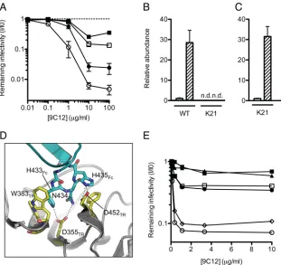
ADIN is an important component of antibody neutralization.
To investigate the importance of ADIN in virus neutralization,
mouse embryonic fibroblast (MEF) cells were prepared from
wild-type (WT) (TRIM21
⫹/⫹) and TRIM21 knockout (K21)
(TRIM21
⫺/⫺) mice (
42
). Cells were infected with
replication-de-ficient human adenovirus type 5-GFP that had been preincubated
with a titration of a mouse monoclonal antihexon IgG1, 9C12 (
32
,
37
). 9C12 neutralized the infection of WT cells but had only a
limited effect on the infection of K21 cells (
Fig. 1A
). We have
previously shown that interferon (IFN) upregulates TRIM21
ex-pression, resulting in an enhanced ADIN phenotype (
23
).
Repeat-ing the neutralization in the presence of IFN resulted in a highly
potent block to infection in WT cells, while K21 cells remained
readily infected. The quantification of TRIM21 mRNA transcripts
reveals a 30-fold increase in the transcript abundance after IFN
stimulation of WT cells (
Fig. 1B
). We confirmed that K21 cells
remained responsive to IFN by measuring the abundance of
transcripts from the transgene that disrupts the TRIM21 locus
(
Fig. 1C
). Thus, in the absence of TRIM21, cells are poorly
protected from infection by 9C12 and, moreover, do not
dis-play an enhanced ADIN phenotype after IFN stimulation.
To demonstrate that neutralization by 9C12 depends on a
spe-cific interaction between TRIM21 and NAb, we performed
struc-ture-guided mutagenesis of the binding interface. The previously
solved crystal structures of human and mouse TRIM21-IgG
com-plexes and the accompanying isothermal titration calorimetry
data show that TRIM21 residues W381, W383, D355, and D452
bind to the “HNH” (positions 433 to 435) motif within the Fc (
Fig.
1D
), and the mutation of any one of these TRIM21 residues
ab-lates binding (
14
). Here we made single-point mutants of the
an-tibody HNH motif, replacing each residue with alanine as well as
replacing N434 with aspartic acid. As can be seen in
Fig. 1E
, each
on November 7, 2019 by guest
http://jvi.asm.org/
single-point mutant reversed neutralization, whereas a mutation
outside the TRIM21-binding domain (K219R) did not impair
neutralization. Indeed, the mutation of H435 (a residue that
makes hydrogen bond interactions with TRIM21) to alanine
in-creased the remaining infectivity from 8% to 79% at 1
g/ml
9C12. Meanwhile, the mutation of N434 to aspartic acid, which
introduces a repulsive charge opposite TRIM21 residue D355,
im-paired neutralization beyond that observed for the mutation of
the same residue to alanine. This finding confirms that a specific
interaction between NAb and TRIM21 is required for
neutraliza-tion and demonstrates that neutralizaneutraliza-tion by this monoclonal
NAb is severely diminished when its TRIM21-binding motif is
mutated.
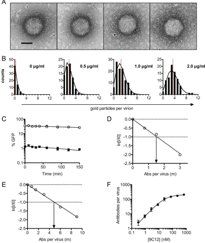
ADIN mediates neutralization by few antibody molecules
per virus.
The existence of an effector mechanism for
neutraliza-tion, such as ADIN, may explain why neutralization can
some-times be observed with very few antibody molecules. To
deter-mine how many NAb molecules are required for ADIN-mediated
neutralization, we quantified NAb-virus stoichiometry by two
in-dependent quantitative techniques: an immunogold electron
mi-croscopy (EM) assay and a dye-conjugated NAb pelleting assay.
The former technique provides information concerning the
quan-tity and distribution of NAb molecules on the virus and can detect
phenomena such as virus aggregation, while the latter technique
provides a robust measure of NAb-virus stoichiometry over a
wide range of NAb concentrations. With both approaches, parallel
infection experiments permit the quantification of the number of
NAb molecules (
) required to neutralize a virus. We define
as
the number of NAb molecules bound at 1 natural logarithm of
neutralization. Assuming a Poisson distribution of antibody
bind-ing, this means that the maximum potential neutralization is a
value of 1, with the remaining infectivity equal to the fraction of
unbound virus (
19
). We chose 9C12 as the neutralizing Ab, since
it does not block entry (
37
) and therefore permits the specific
study of ADIN-mediated neutralization.
For the EM approach, virus was incubated with 9C12, followed
by incubation with an excess of gold-labeled secondary Ab, and
the number of Ab molecules per virion was calculated (
Fig. 2A
).
The binding of NAb to virus was found to closely approximate a
Poisson distribution at all concentrations of NAb assayed
(
Fig. 2B
). We confirmed that the dilution of preformed NAb-virus
complexes does not result in the dissociation of complexes within
the time frame of the experiment (
Fig. 2C
). Finally, and in
agree-ment with previous findings (
37
), 9C12 was not found to
aggre-gate virus, since virions remained largely monodispersed when
9C12 was present. Parallel infection experiments were performed
FIG 1ADIN is essential for efficient neutralization of adenovirus. (A) Antihexon monoclonal antibody 9C12 was titrated against adenovirus on clonal fibroblast (MEF) lines derived from wild-type (WT) (n⫽8) (circles) or TRIM21 knockout (K21) (n⫽8) (squares) mice. Cells were either untreated (closed symbols) or treated with IFN-␣(open symbols). Data represent mean remaining infectivities (I/I0) from the addition of antibody versus a PBS-treated control infection⫾
standard errors of the means. (B) TRIM21 mRNA levels were quantified by qPCR in unstimulated (white bars) or IFN-␣-stimulated (hatched bars) MEF cells. n.d., not detected. (C) Levels of the transgene that disrupts the TRIM21 locus in K21 cells were measured by qPCR. Shading is as described above for panel B. (D) The previously solved structure of the TRIM21-IgG Fc complex (14) showing interactions between TRIM21 (yellow) (residues labeled TR) and the IgG Fc HNH motif (blue) (residues labeled Fc). (E) Antihexon monoclonal antibody 9C12 was mutated at three positions or expressed as the wild type. Antibodies were titrated against adenovirus on HeLa cells. Open circles, WT 9C12; closed circles, H433A mutant; open squares, N434A mutant; closed squares, N434D mutant; triangles, H435A mutant; diamonds, K219R mutant.
on November 7, 2019 by guest
http://jvi.asm.org/
[image:4.585.137.448.65.358.2]on IFN-stimulated HeLa and wild-type MEF cells, allowing
neu-tralization to be plotted as a function of the number of NAb
mol-ecules per virus. The data showed that in MEF and HeLa cells, 1.6
and 4.8 molecules of 9C12, respectively, are sufficient to
neu-tralize adenoviral infection by 1 natural logarithm (
Fig. 2D
and
E
). Thus, when using a mouse Ab on mouse cells and in the
presence of abundant TRIM21, neutralization approaches its
theoretical maximum.
FIG 2ADIN mediates neutralization by few antibody molecules. (A) 9C12-adenovirus complexes were immunogold labeled and examined by electron microscopy. Gold particles are visible as black dots around the AdV capsid and show 0, 1, 2, and 3 regions labeled, respectively (from left to right). Scale bar, 50 nm. (B) Virions were scored for the number of gold particles per virus at increasing concentrations of 9C12. The distribution of gold particles per virus (black bars) was found to approximate a Poisson distribution (black lines), given the mean number of gold particles per virus (red dashed line). These data were used to calculate the average number of NAb molecules per virion. (C) PBS (circles) or 9C12 at 1.5g/ml (squares) was added to AdV for 1 h before 100-fold dilution into complete DMEM and incubation for various amounts of time before addition to cells. No increase in titer was observed for NAb-labeled virus over time, confirming that complexes do not dissociate upon dilution. (D and E) The infectivity of diluted NAb-virus complexes was assayed with MEF (D) and HeLa (E) cells treated with IFN-␣, permitting a direct quantification of the average number of NAb molecules per virion at 1/eneutralization. Data represent the natural logarithm of remaining infectivity, ln(I/I0). (F) Stoichiometry of 9C12-A488 –adenovirus after pelleting of complexes. Virus and antibody concentrations were
quantified by qPCR and fluorescence spectroscopy, respectively. Data are fitted to a Michaelis-Menten curve (R2⫽0.94), giving aK
dof 28 nM and a maximum binding of 205 antibody molecules per virus (standard error of the mean,⫾10). Data are represented as means⫾standard errors of the means. A parallel infection assay with IFN-treated HeLa cells estimated the number of antibody molecules required for 1 natural logarithm of neutralization () to be 5.2.
on November 7, 2019 by guest
http://jvi.asm.org/
[image:5.585.86.496.70.555.2]To confirm that low Ab-virus stoichiometry is sufficient for
ADIN, we also measured the number of Abs per virus by
complex-ing virus with Alexa Fluor 488-labeled 9C12 (9C12-A488) at a
range of concentrations and pelleting through a sucrose cushion.
Pelleted complexes were resuspended, the amount of bound dye
was quantified by measurements of the fluorescence intensity, and
the number of viruses was measured by qPCR, permitting the
calculation of the stoichiometric ratio of Ab to virus. The resulting
data were fit to a Michaelis-Menten curve (
Fig. 2F
), to give a
dis-sociation constant (
K
d) for the 9C12 interaction with adenovirus
of 28 nM. We also observed that a maximum of 205 NAb
mole-cules (standard error,
⫾
10) were able to bind per virion. Given
that there are 240 hexon trimers per virion, this finding suggests
that the majority of hexon trimers can be occupied by 9C12 at
saturation. Parallel neutralization experiments on IFN-treated
HeLa cells revealed that 5.2 NAb molecules per virion were
suffi-cient for neutralization, comparable to the value of 4.8 NAb
mol-ecules per virion obtained by EM. Together, the data show that
high TRIM21 levels resulting from IFN stimulation permit
neu-tralization at a low Ab-virus stoichiometry.
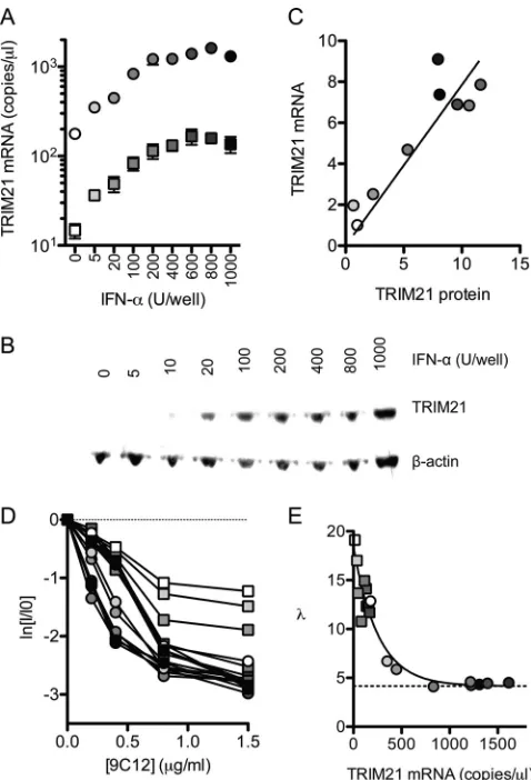
TRIM21 levels influence neutralization efficiency and the
level of the persistent fraction.
Next, we sought to analyze the
impact of TRIM21 levels on the efficiency of ADIN. TRIM21
ex-pression was manipulated by IFN-
␣
titration and the retroviral
transduction of HeLa cells with a TRIM21-directed shRNA. Using
this approach, we were able to obtain cells with TRIM21 mRNA
levels that varied over 2 orders of magnitude and with a
corre-sponding distribution of protein levels (
Fig. 3A
to
C
).
Neutraliza-tion experiments were carried out with these cells by using 9C12 at
concentrations that yielded neutralization but not a saturation of
binding (less than 20% maximum occupancy). Cells with the least
amounts of TRIM21 gave a shallow gradient, reflecting a
require-ment for higher levels of NAb binding in order to elicit an
equiv-alent level of neutralization (
Fig. 3D
). In contrast, high levels of
TRIM21 permitted more efficient neutralization, reflecting a
lower NAb-virus stoichiometry being sufficient for neutralization.
We used equilibrium dynamics to calculate the number of
anti-bodies per virus, using the parameters obtained from the
fluores-cence pelleting method, and plotted the number of Ab molecules
required for 1 natural logarithm of neutralization against the
TRIM21 transcript levels (
Fig. 3E
). At the lowest concentration of
TRIM21, 18 Ab molecules were required for 1 natural logarithm
of neutralization. As TRIM21 levels were increased, the number of
Ab molecules required decreased rapidly until reaching a plateau
corresponding to
⬃
4.2 NAb molecules per virus. Importantly, the
data suggest that TRIM21 levels, rather than other effects of IFN,
dictate the efficiency of neutralization, since IFN stimulation of
TRIM21 knockdown cells restores the neutralization efficiency
only to the level of unstimulated HeLa cells. Thus, where NAbs
operate via ADIN, the number of Ab molecules required for
neu-tralization can be described as a function of the TRIM21
concen-tration,
(TR), which varies according to an exponential decay
(
Fig. 3E
):
共
TR
兲
⫽
Ae
⫺cTR⫹
B
(10)
where TR is the TRIM21 concentration,
B
is the value of
at the
asymptote,
A
is the value of
at a TR of 0 minus the asymptote
value
B
, and
c
is the decay parameter. These findings imply that at
low levels of cellular TRIM21, large gains in the efficiency of ADIN
can be made by small increases in TRIM21 levels. However, as
TRIM21 concentrations increase further, gains in neutralization
efficiency will be reduced.
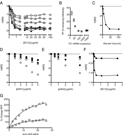
The above-described data show that at a low NAb-virus
stoi-chiometry, robust TRIM21 expression is essential for efficient
neutralization. To determine the importance of TRIM21 at a high
NAb-virus stoichiometry, we performed neutralization
experi-ments over a wide range of 9C12 concentrations with cells with
various TRIM21 expression levels. In HeLa cells, independent of
TRIM21 levels, we observed that the addition of 9C12 beyond a
concentration of 2
g/ml (equivalent to 6.7 nM 9C12 in the
bind-ing reaction) yielded no further increase in neutralization
(
Fig. 4A
). The PF remained constant even after a further 50-fold
increase in the Ab concentration to 100
g/ml (
Fig. 4A
). A similar
FIG 3TRIM21 levels determine neutralization efficiency. (A) IFN-␣was added to HeLa cells (circles) or HeLa cells transduced with TRIM21 shRNA (squares), and TRIM21 mRNA levels were quantified by RT-qPCR. (B) TRIM21 protein levels were quantified by immunoblotting and normalized to
-actin levels. (C) Relative TRIM21 mRNA and protein levels are closely cor-related (P⬍0.001 by Pearson’s product-moment correlation;R2⫽0.83). (D)
Virus neutralization assays were performed on these cells, which display an initial gradient of neutralization that varies with TRIM21 levels. (E) The num-ber of antibody molecules per virus at each concentration of 9C12 was calcu-lated by using binding parameters obtained as described above (Fig. 2F). Lin-ear regression was used in order to calculate the number of molecules of 9C12 bound at 1/eneutralization ().was found to decrease exponentially with the TRIM21 concentration, reaching a plateau at 4.2 antibody molecules per virus (dashed line). For panels D and E, circles indicate HeLa cells, and squares indicate HeLa cells expressing TRIM21-directed shRNA. Points for panels A and C to E are shaded white to black according to IFN levels.
on November 7, 2019 by guest
http://jvi.asm.org/
[image:6.585.301.540.65.416.2]phenomenon was observed for MEF cells, where the addition of
9C12 above 10
g/ml gave little further neutralization (
Fig. 1A
).
These extremes of 9C12 concentrations (100
g/ml) are
equiva-lent to a 12-fold excess over the
K
dand are predicted by
equilib-rium dynamics to provide a 93% saturation of hexon binding
sites. In both HeLa and MEF cells, the level of remaining
infectiv-ity at which the PF occurs is dependent on the cellular TRIM21
concentration. In HeLa cells depleted of TRIM21, the PF occurs
when the remaining infectivity is 32%, and increasing the level of
TRIM21 results in a proportional decrease in the PF (
Fig. 4B
).
Interestingly, there appears to be a threshold after which
increas-ing TRIM21 levels further fails to reduce the PF further. We
hy-pothesize that this represents the point at which other ADIN
com-ponents become limiting. The onset of the PF occurs before the
FIG 4Infection and persistence are influenced by the neutralization mechanism and viral MOI. (A) Neutralization data fromFig. 3Dare shown with thexaxis extended to show the persistent fraction range. The PF remains approximately constant within the range of 1.5 to 100g/ml 9C12, but its level is determined by the cellular TRIM21 concentration. (B) The average PF, calculated from all data atⱖ5g/ml 9C12 (n⫽7) (⫾standard errors of the means), decreases with TRIM21 levels before remaining constant at⬃6% remaining infectivity. Symbols and shading as forFig. 3D(C) Remaining infectivity of adenovirus, assayed alongside a fluorescence pelleting assay, permitting quantification of the number of antibody molecules per virus in the persistent fraction. (D and E) Two polyclonal sera, pAb1 (D) and pAb2 (E), were titrated against AdV on HeLa cells (circles) or HeLa cells treated with siRNA directed toward TRIM21 (squares), which were either IFN stimulated (open symbols) or untreated (closed symbols). (F) Remaining infectivity after AdV was permitted to adsorb to the surface of HeLa cells before the addition of neutralizing monoclonal antibody 9C12. Symbols are as described above for panels D and E. (G) AdV-GFP, preincubated with 9C12, was added to HeLa (circles) or TRIM21 shRNA knockdown (squares) cells at a range of MOIs. A high MOI of AdV-GFP relieved the neutralization of 9C12-labeled AdV-RFP in HeLa cells but to a lesser extent in TRIM21 knockdown cells. Michaelis-Menten curve fitting (black lines) reveals that the maximal saturation values are significantly different between the two conditions (P⫽0.0008 byFtest).
on November 7, 2019 by guest
http://jvi.asm.org/
[image:7.585.82.506.64.525.2]saturation of virus with Ab (
Fig. 4C
), coinciding with fewer than
50 NAb molecules per virus, indicating that even though virus
particles can accommodate more NAb molecules, an increasing
NAb concentration does not result in higher levels of
neutraliza-tion. Taken together, the data suggest that TRIM21 levels remain
important at a high NAb-virus stoichiometry to lower the
persis-tent fraction. Furthermore, increasing the Ab concentration
fur-ther yields diminishing returns and cannot compensate for low
levels of TRIM21.
A feature of 9C12 neutralization in the above-described
exper-iments is that it is linear and commences without a discernible lag
(
Fig. 2D
and
E
). However, in a previous experiment with
poly-clonal sera, we observed evidence of a lag when TRIM21 was
de-pleted (
23
). We repeated these experiments using two different
polyclonal sera against adenovirus and over an extended range of
antibody concentrations. As before, potent neutralization was
ob-served at high TRIM21 expression levels and was markedly
dimin-ished when TRIM21 was depleted (
Fig. 4D
and
E
). In addition to
a drop in the neutralization potency, the depletion of TRIM21
results in a clear change in the shape of the neutralization curve: in
IFN-stimulated cells, infectivity decreases linearly with the NAb
concentration, whereas a lag or shoulder is observed for
TRIM21-depleted cells. Classically, the presence of a lag has been
inter-preted as evidence that neutralization depends on multiple
NAb-binding events, with a threshold number of NAb molecules being
required to bind the virus before infection can be prevented (
19
).
Threshold-based neutralization mechanisms are likely to include
entry blocking, which typically requires that available receptor
interaction sites are masked. The data therefore suggest that while
neutralization by polyclonal sera is mediated principally by ADIN,
other mechanisms of neutralization are also present. The data also
imply that the number of Abs required for a given level of
neutral-ization may depend upon the mechanism of neutralneutral-ization. For
example, the addition of 2
g/ml pAb2 gives nearly 2 natural logs
of neutralization on IFN-stimulated cells but almost no
observ-able neutralization in cells depleted of TRIM21, presumably
be-cause in the latter case, the critical number of NAb molecules
required to block entry has not been reached (
Fig. 4E
).
Examination of neutralization profiles is not the only way of
differentiating between neutralization mechanisms. Abs have
been reported to neutralize without preventing viral attachment
to cells, a phenomenon referred to as postadsorption
neutraliza-tion (PAN). In a previous study by Emini et al., it was found that
six out of seven monoclonal Abs against poliovirus failed to
pre-vent viral entry but were nevertheless potently neutralizing (
8
).
This kind of phenomenon may be indicative of neutralization by
an intracellular process such as ADIN. To determine whether
ADIN provides a mechanism for PAN, we added AdV-GFP to
target cells at 4°C, a temperature that permits virus attachment but
not uptake. Antibody 9C12 was then introduced, and the
temper-ature was increased to 37°C to allow infection. We observed
effi-cient PAN, in that the Ab neutralized infection despite being
un-able to block receptor binding (
Fig. 4F
). Furthermore, PAN
occurred without a lag and was dependent, in terms of both the
neutralization efficiency and the level of persistence, on TRIM21.
High levels of infection can overcome ADIN.
In the
above-described experiments, we have shown how Ab and TRIM21 levels
operate synergistically to influence the efficiency of neutralization
and the persistent fraction. The third important component that
determines cellular infection is the viral dose or multiplicity of
infection (MOI). We hypothesized that at a high MOI, TRIM21
and the ADIN machinery may become saturated. To test this, we
simultaneously infected cells with two NAb-bound adenoviruses
encoding GFP or RFP reporter genes (
Fig. 4G
). NAb-labeled
AdV-GFP was added to cells in increasing amounts at a high MOI, while
NAb-labeled AdV-RFP was added at a constant, low MOI (0.1).
We observed a dose-dependent rescue of AdV-RFP infection with
increasing AdV-GFP levels. To confirm that increased infection is
due specifically to the saturation of TRIM21, we also performed
the experiment with cells depleted of TRIM21 by shRNA. The
ability of AdV-GFP to rescue AdV-RFP was significantly reduced
in cells with reduced TRIM21 levels. The data therefore support a
model in which TRIM21 can be titrated by virus-NAb complexes
at high MOIs and provide a parallel to other antiviral restriction
factors, TRIM5
␣
and Lv1, which are also saturated by high levels
of infection (
11
).
DISCUSSION
This study suggests that ADIN provides an important component
of viral neutralization, to the extent that neutralization can be
substantially impaired in cells that lack the ADIN receptor
TRIM21 or when an antibody is mutated to prevent TRIM21
binding. We have determined the mechanistic requirements of
ADIN in terms of how antibody, TRIM21, and virus contribute to
the efficiency of neutralization and the level of the persistent
frac-tion. These mechanistic determinants are useful in distinguishing
ADIN from other forms of neutralization. In particular, ADIN
generates characteristic linear neutralization profiles and is
detect-able in postadsorption neutralization assays.
Our data indicate that ADIN is remarkably efficient, capable of
mediating neutralization in the presence of as few as 1.6 Ab
mol-ecules per virus in a cognate murine TRIM21-IgG system. Such
low-stoichiometry neutralization may be particularly important
in vivo
when NAb concentrations are low, such as during the
ini-tial stages of an adaptive immune response or in the intercellular
spaces of infected tissue. The difference observed for human cells,
where a minimum of 5 Ab molecules is required, may reflect the
⬃
10-fold-lower affinity of human TRIM21 for mouse IgG (
15
).
The conservation of ADIN demonstrated here with human and
mouse cells, as well as the conservation of the TRIM21-IgG
mo-lecular interaction between distantly related mammalian species
(
15
), suggests that ADIN is broadly conserved as an antiviral
mechanism. Further studies are required to determine the
preva-lence of ADIN among other mammalian species as well as the
in
vivo
contribution of ADIN to antiviral immunity.
Neutralizing antibodies can fail to completely prevent
infec-tion, even at a stoichiometric excess. We find that this persistent
fraction of infection can be dependent on cellular levels of
TRIM21, since reductions in TRIM21 levels increase the level of
the PF. However, neutralization also becomes unresponsive to
increases in TRIM21 expression levels beyond a threshold level.
This “double-plateau” region, where neutralization is
unrespon-sive to changes in both antibody and TRIM21 levels, is suggestive
of a maximum rate of virus disposal within the cell, possibly
be-cause other factors become limiting. This interpretation is favored
over a model in which the plateau is a result of the saturation of the
NAb binding of the virus surface, since our data suggest that the
PF commences at Ab concentrations substantially lower than
those required for saturation. This in turn implies that the
incom-ing virions could accommodate more TRIM21 molecules under
on November 7, 2019 by guest
http://jvi.asm.org/
higher levels of NAb labeling, given that TRIM21 dimers bind IgG
at a 1:1 ratio (
14
). We therefore predict that the PF will be sensitive
to the pharmacological or biological modulation of ADIN
com-ponents acting downstream of TRIM21. Moreover, limiting
con-centrations of TRIM21 and its cofactors provide a potential
mech-anism to explain why the PF has been observed to vary between
cell lines (
16
). ADIN may also help to explain other neutralization
phenomena such as postadsorption and postentry neutralization.
For instance, the ability of an Ab to neutralize infection after virus
adsorption to the cell surface or after entry has been documented
for many nonenveloped viruses, including adenovirus,
papillo-mavirus, and poliovirus (
1
,
6
,
8
,
9
,
24
,
32
,
36
,
37
,
40
), but has
remained largely unexplained. Furthermore, we speculate that
an-tibodies are not the only extracellular antiviral molecules to exert
their effects within cells. Indeed, the postentry neutralization of
adenovirus has been demonstrated by the antimicrobial peptides
of the defensin family, which prevent virus uncoating (
28
,
33
).
Despite the efficiency of the ADIN system, our data suggest
that high levels of infection are capable of saturating TRIM21 and
overcoming neutralization. Thus, a high multiplicity of infection
may promote escape from neutralization
in vivo
. Given that the
burst yield of an AdV-infected cell is typically more than 1,000 IU
(an infected cell may contain in excess of 10,000 particles [
41
])
and that virions are released relatively synchronously upon cell
lysis, localized virus titers may result in effective multiplicities of
infection that exceed those required for saturation in this study.
Future
in vivo
studies are needed to confirm whether the
satura-tion of TRIM21 represents a mechanism of evasion of
neutraliza-tion.
We observed the most efficient neutralization when cells were
activated by IFN. This emphasizes the importance of immune
stimulation in the process of neutralization and is an example of
synergy between innate and adaptive immunity. Synergy between
IFN and neutralization was previously noted for other
nonenvel-oped viruses (enterovirus 70, coxsackievirus A24, and adenovirus
type 3) (
20
). Of note, such synergy was not observed for an
envel-oped virus, herpes simplex virus. Viruses will differ in their
sus-ceptibilities to ADIN according to their specific entry
mecha-nisms, capsid compositions, and life cycles. Most significantly,
viruses with envelopes are likely to avoid ADIN altogether by
shedding both their lipid coats and attached Ab molecules during
entry. It is therefore important to note that unusual neutralization
phenomena have been observed for enveloped viruses, including
variations of the PF between cell lines (
17
) and low-stoichiometry
neutralization (
26
,
35
). While TRIM21 is unlikely to be directly
involved, our data suggest that considering neutralization, at least
in part, as an effector-driven process may be important. Thus,
where evidence for such phenomena exists, we do not rule out that
other receptors may mediate or facilitate the neutralization of
these pathogens.
REFERENCES
1.Booy FP, Roden RB, Greenstone HL, Schiller JT, Trus BL.1998. Two antibodies that neutralize papillomavirus by different mechanisms show distinct binding patterns at 13 A resolution. J. Mol. Biol.281:95–106. 2.Burton DR, Saphire EO, Parren PW.2001. A model for neutralization of
viruses based on antibody coating of the virion surface. Curr. Top. Micro-biol. Immunol.260:109 –143.
3.de Martin R, Raidl M, Hofer E, Binder BR.1997. Adenovirus-mediated expression of green fluorescent protein. Gene Ther.4:493– 495. 4.Dimmock NJ.1984. Mechanisms of neutralization of animal viruses. J.
Gen. Virol.65(Pt 6):1015–1022.
5.Dimmock NJ.1995. Update on the neutralization of animal viruses. Rev. Med. Virol.5:165–179.
6.Dorner AJ, Dorner LF, Larsen GR, Wimmer E, Anderson CW.1982. Identification of the initiation site of poliovirus polyprotein synthesis. J. Virol.42:1017–1028.
7.Edghill-Smith Y, et al.2005. Smallpox vaccine-induced antibodies are necessary and sufficient for protection against monkeypox virus. Nat. Med.11:740 –747.
8.Emini EA, Kao SY, Lewis AJ, Crainic R, Wimmer E.1983. Functional basis of poliovirus neutralization determined with monospecific neutral-izing antibodies. J. Virol.46:466 – 474.
9.Emini EA, Ostapchuk P, Wimmer E.1983. Bivalent attachment of anti-body onto poliovirus leads to conformational alteration and neutraliza-tion. J. Virol.48:547–550.
10. Emini EA, et al.1992. Prevention of HIV-1 infection in chimpanzees by gp120 V3 domain-specific monoclonal antibody. Nature355:728 –730. 11. Hatziioannou T, Cowan S, Goff SP, Bieniasz PD, Towers GJ. 2003.
Restriction of multiple divergent retroviruses by Lv1 and Ref1. EMBO J.
22:385–394.
12. Hessell AJ, et al.2007. Fc receptor but not complement binding is im-portant in antibody protection against HIV. Nature449:101–104. 13. Icenogle J, et al.1983. Neutralization of poliovirus by a monoclonal
antibody: kinetics and stoichiometry. Virology127:412– 425.
14. James LC, Keeble AH, Khan Z, Rhodes DA, Trowsdale J.2007. Struc-tural basis for PRYSPRY-mediated tripartite motif (TRIM) protein func-tion. Proc. Natl. Acad. Sci. U. S. A.104:6200 – 6205.
15. Keeble AH, Khan Z, Forster A, James LC. 2008. TRIM21 is an IgG receptor that is structurally, thermodynamically, and kinetically con-served. Proc. Natl. Acad. Sci. U. S. A.105:6045– 6050.
16. Kjellen L.1985. A hypothesis accounting for the effect of the host cell on neutralization-resistant virus. J. Gen. Virol.66(Pt 10):2279 –2283. 17. Kjellen LE, Schlesinger RW. 1959. Influence of host cell on residual
infectivity of neutralized vesicular stomatitis virus. Virology7:236 –239. 18. Klasse PJ, Moore JP.1996. Quantitative model of antibody- and soluble
CD4-mediated neutralization of primary isolates and T-cell line-adapted strains of human immunodeficiency virus type 1. J. Virol.70:3668 –3677. 19. Klasse PJ, Sattentau QJ.2002. Occupancy and mechanism in antibody-mediated neutralization of animal viruses. J. Gen. Virol.83:2091–2108. 20. Langford MP, Villarreal AL, Stanton GJ.1983. Antibody and interferon
act synergistically to inhibit enterovirus, adenovirus, and herpes simplex virus infection. Infect. Immun.41:214 –218.
21. Li J, Wang Y, Wang Z, Dong Z.2000. Influences of amino acid sequences in FR1 region on binding activity of the scFv and Fab of an antibody to human gastric cancer cells. Immunol. Lett.71:157–165.
22. Maizel JV, Jr, White DO, Scharff MD.1968. The polypeptides of ade-novirus. I. Evidence for multiple protein components in the virion and a comparison of types 2, 7A, and 12. Virology36:115–125.
23. Mallery DL, et al. 2010. Antibodies mediate intracellular immunity through tripartite motif-containing 21 (TRIM21). Proc. Natl. Acad. Sci. U. S. A.107:19985–19990.
24. Mandel B.1967. The interaction of neutralized poliovirus with HeLa cells. I. Adsorption. Virology31:238 –247.
25. McEwan WA, Mallery DL, Rhodes DA, Trowsdale J, James LC.2011. Intracellular antibody-mediated immunity and the role of TRIM21. Bioessays33:803– 809.
26. McLain L, Dimmock NJ.1994. Single- and multi-hit kinetics of immu-noglobulin G neutralization of human immunodeficiency virus type 1 by monoclonal antibodies. J. Gen. Virol.75(Pt 6):1457–1460.
27. Mittereder N, March KL, Trapnell BC.1996. Evaluation of the concen-tration and bioactivity of adenovirus vectors for gene therapy. J. Virol.
70:7498 –7509.
28. Nguyen EK, Nemerow GR, Smith JG.2010. Direct evidence from single-cell analysis that human alpha-defensins block adenovirus uncoating to neutralize infection. J. Virol.84:4041– 4049.
29. Reading SA, Dimmock NJ.2007. Neutralization of animal virus infectiv-ity by antibody. Arch. Virol.152:1047–1059.
30. Rezaei N, Hedayat M, Aghamohammadi A, Nichols KE.2011. Primary immunodeficiency diseases associated with increased susceptibility to vi-ral infections and malignancies. J. Allergy Clin. Immunol.127:1329 – 1341.e2; quiz 1342-1343. doi:10.1016/j.jaci.2011.02.047.
31. Sanna PP, Burton DR.2000. Role of antibodies in controlling viral dis-ease: lessons from experiments of nature and gene knockouts. J. Virol.
74:9813–9817.
on November 7, 2019 by guest
http://jvi.asm.org/
32. Smith JG, Cassany A, Gerace L, Ralston R, Nemerow GR.2008. Neu-tralizing antibody blocks adenovirus infection by arresting microtubule-dependent cytoplasmic transport. J. Virol.82:6492– 6500.
33. Smith JG, Nemerow GR.2008. Mechanism of adenovirus neutralization by human alpha-defensins. Cell Host Microbe3:11–19.
34. Takada A, Ebihara H, Jones S, Feldmann H, Kawaoka Y.2007. Protec-tive efficacy of neutralizing antibodies against Ebola virus infection. Vac-cine25:993–999.
35. Taylor HP, Armstrong SJ, Dimmock NJ.1987. Quantitative relationships be-tween an influenza virus and neutralizing antibody. Virology159:288–298. 36. Toogood CI, Crompton J, Hay RT.1992. Antipeptide antisera define
neu-tralizing epitopes on the adenovirus hexon. J. Gen. Virol.73(Pt 6):1429 –1435. 37. Varghese R, Mikyas Y, Stewart PL, Ralston R.2004. Postentry neutraliza-tion of adenovirus type 5 by an antihexon antibody. J. Virol.78:12320 –12332.
38. Wetz K, Willingmann P, Zeichhardt H, Habermehl KO.1986. Neutral-ization of poliovirus by polyclonal antibodies requires binding of a single IgG molecule per virion. Arch. Virol.91:207–220.
39. Wohlfart C.1988. Neutralization of adenoviruses: kinetics, stoichiome-try, and mechanisms. J. Virol.62:2321–2328.
40. Wohlfart CE, Svensson UK, Everitt E.1985. Interaction between HeLa cells and adenovirus type 2 virions neutralized by different antisera. J. Virol.56:896 –903.
41. Wold WSM, Horwitz MS.2007. Adenoviruses, p 2395–2436.InKnipe DM, et al (ed), Fields virology, 5th ed, vol 2. Lippincott Williams & Wilkins, Philadelphia, PA.
42. Yoshimi R, et al.2009. Gene disruption study reveals a nonredundant role for TRIM21/Ro52 in NF-kappaB-dependent cytokine expression in fibroblasts. J. Immunol.182:7527–7538.
on November 7, 2019 by guest
http://jvi.asm.org/