ORIGINAL ARTICLE
Suppression of in
fl
ammation and tissue damage by a
hookworm recombinant protein in experimental colitis
Ivana B Ferreira
1, Darren A Pickering
1, Sally Troy
1, John Croese
1,2, Alex Loukas
1and Severine Navarro
1Gastrointestinal parasites, hookworms in particular, have evolved to cause minimal harm to their hosts when present in small numbers, allowing them to establish chronic infections for decades. They do so by creating an immunoregulatory environment that promotes their own survival, but paradoxically also benefits the host by protecting against the onset of many inflammatory diseases. To harness the therapeutic value of hookworms without using live parasites, we have examined the protective properties of the recombinant protein anti-inflammatory protein (AIP)-1, secreted in abundance by hookworms within the intestinal mucosa, in experimental colitis. Colitic inflammation assessed by weight loss, colon atrophy, oedema, ulceration and necrosis, as well as abdominal adhesion was significantly suppressed in mice treated with a single intraperitoneal dose of AIP-1 at 1 mg kg−1. Local infiltration of inflammatory cells was also significantly reduced, with minimal goblet cell loss and preserved
mucosal architecture. Treatment with AIP-1 promoted the production of colon interleukin (IL)-10, transforming growth factor (TGF)-βand thymic stromal lymphopoietin (TSLP), resulting in the suppression of tumour necrosis factor (TNF)-α, IL-13 and IL-17 A cytokines and granulocyte macrophage colony-stimulating factor (GM-CSF), CX motif chemokine (CXCL)-11 and cyclooxygenase synthase (COX)-2 mRNA transcripts. AIP-1 promoted the accumulation of regulatory T cells in the colon likely allowing rapid healing of the colon mucosa. Hookworm recombinant AIP-1 is a novel therapeutic candidate for the treatment of inflammatory bowel diseases that can be explored for the prevention of acute inflammatory relapses, an important cause of colorectal cancer.
Clinical & Translational Immunology(2017)6,e157; doi:10.1038/cti.2017.42; published online 6 October 2017
Inflammatory bowel diseases (IBD) are a group of chronic auto-immune diseases affecting the digestive track, primarily represented by Crohn’s disease (CD) and ulcerative colitis (UC).1Both types of IBD are caused by an inappropriate immune response in genetically susceptible individuals to intestinal microbial species, however, the site and nature of inflammation differ between the two diseases.2,3CD
can affect the entire intestinal track from the mouth to the anus, whereas UC mainly affects the colon and the rectum. Although the role of innate cells is pivotal in CD, both conditions are T-cell-mediated and characterised by increased levels of interleukin (IL)-6, IL-17, interferon (IFN)-γ and tumour necrosis factor (TNF)-α.4–6 However, the immune response in UC appears to be more skewed towards a T-helper cell type 2 (Th2) response, with increased levels of IL-4 and IL-13 production in the tissue.6The current treatments for IBD rely on nonspecific immunosuppressive drugs, such as steroids, antibiotics, and immunomodulators targeting the TNF pathway or the gut-homing integrinα4β7.7–11However, the repetitive cycles of acute inflammation followed by temporary remission in IBD result over time in severe impairment of gut function, motility and tissue remodelling.12–14 Despite encouraging clinical trial end points, TNF-αinhibitors are not effective in all patients and do not prevent
relapse.9–11One of the major consequences of UC progression is the development of colorectal cancer, which is the third most common malignancy in humans.15,16The rising incidence of IBD parallels the trend of other autoimmune and allergic diseases.17,18 However, for
reasons that are still unclear, the rate of childhood-onset IBD has been the highest observed over the past two decades.19–21In addition to the debilitating symptoms associated with the disease, children affected by early-onset IBD suffer significant malabsorption and nutritional deficiencies resulting in growth failure, skeletal impairment, and significant psychological and developmental delays.22–25These recent observations underscore the urgent need for novel therapeutic approaches to be developed.
A promising new avenue of research using live helminth therapy has seen encouraging levels of success for the management of autoimmune diseases, such as IBD and Celiac disease.26–28 Indeed, experimental infection with ova of the pig whipworm Trichuris suis (TSO) successfully improved both UC and CD Disease Activity Index.29
However, because humans are not the natural host and infection resulted in rapid parasite clearance, repeated administrations were required30 and recent phase 2 clinical trials in IBD failed to meet clinical end points.31 Interestingly, hookworms, such as 1Centre for the Biodiversity and Molecular Development of Therapeutics, Australian Institute of Tropical Health Medicine, James Cook University, Cairns, Queensland, Australia
and2Department of Gastroenterology and Hepatology, The Prince Charles Hospital, Brisbane, Queensland, Australia
Correspondence: Professor A Loukas or Dr S Navarro, Australian Institute of Tropical Health and Medicine, James Cook University, E4 McGregor Road, 1 Centre for the Biodiversity and Molecular Development of Therapeutics, Smithfield, Queensland 4878, Australia.
E-mail: [email protected] or [email protected]
Necator americanus and Ancylostoma caninum,have co-evolved with their mammalian hosts where they establish chronic infections over many years.28The tolerability of iatrogenicN. americanusinfection has
been assessed in patients with autoimmune gastrointestinal diseases. Hookworm infection coupled with escalating oral gluten challenge resulted in remarkably improved gluten tolerance in Celiac patients,32
and a non-significant trend towards reduced CD activity scores in a small number of CD patients after hookworm infection.33
Nematodes, and hookworms in particular, have been shown to ameliorate chronic inflammatory diseases by promoting regulatory immune circuits, particularly the induction of regulatory T cells (Tregs)
and the modification of the intestinal microbiota.28,32,34–37We, and others, have shown that much of the immunomodulatory prowess of helminths can be attributed to the release of excretory/secretory (ES) products into host tissues.36,38–40This complex mix of proteins41and
other molecules (unpublished) has been shown to ameliorate colitis in numerous mouse models,38,42,43 and denaturation of the protein
component of ES products ablated the anti-colitic properties.38
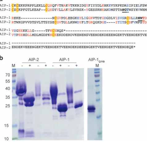
Proteomic analysis of A. caninum excretory/secretory (ES) proteins revealed the relative abundance of two Tissue Inhibitor of Metallo-protease (TIMP)-like proteins, anti-inflammatory protein (AIP)-1 and AIP-2,41 neither of which appear to have the protease inhibitory
properties that characterise the TIMP family.44We recently showed
that AIP-2 induced the expansion of Tregsthat promoted long-term
protection against allergic responses in both mice and humans.40
A similar rationale was used earlier to investigate Ac-AIP-1 (also referred to as tissue inhibitor of metalloprotease (TIMP)-1) as a potential modulator of dendritic and T-cell function.45However, its
efficacy as a therapeutic approach to suppress inflammatory disease was never tested. In this study, haptenating agent 2,4,6-trinitrobenzene sulphonic acid (TNBS) was used to evaluate the therapeutic validity of Ac-AIP-1 for treating acute colitis. Despite its limitations, the TNBS-model of colitis is T-cell mediated and skewed towards a Th2 phenotype comparable to human UC.46–48 Recombinant Ac-AIP-1 protected against all the hallmark parameters of inducible colitis and promoted a regulatory immune environment in treated mice.
RESULTS
RecombinantAc-AIP-1 protects against TNBS-induced intestinal inflammation
The immunoregulatory properties of Ac-AIP-1 have previously been explored using anin vitroT-cell suppression assay and protein-pulsed bone-marrow-derived dendritic cells.45Suppression of inflammation using the related hookworm protein, AIP-2, was shown to be optimal at 1 mg kg−1in a mouse model of asthma40and colitis (unpublished).
Therefore, the same dose for administering AIP-1 herein was used. To determine the therapeutic potential of AIP-1 in a model of acute inflammation, mice were treated with 1 mg kg−1of AIP-1 or vehicle
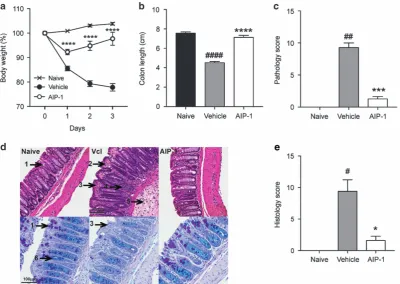
[image:2.595.106.507.68.352.2]via the intraperitoneal (i.p) route. Five hours later, mice were administered 2.5 mg trinitrobenzoylsulfonic acid (TNBS) in 50%
ethanol via intrarectal injection, resulting in a 15 to 20% weight loss in the vehicle group over the course of the study (Figure 1a). Interest-ingly, mice treated with AIP-1 displayedo10% weight loss on thefirst day post-TNBS injection, and recovered most of their initial weight by day 3 (Figure 1a). Cellular enumeration of peritoneal lavages showed that i.p treatment with AIP-1 did not induce eosinophil infiltration at the site of injection (data not shown). In comparison to the naive control mice, colon lengths were significantly decreased in the vehicle group (Po0.0001), while AIP-1-treated mice remained unaltered by the administration of TNBS (Figure 1b). Macroscopic analysis of the colons revealed a significant reduction of tissue inflammation as seen by minimal adhesion, oedema, wall thickening and ulceration (Figure 1c). Haematoxylin and eosin (H&E) staining of distal colon sections from the vehicle group showed mucosal erosion and epithelial hyperplasia, pronounced cellular infiltration in the lamina propria and intraepithelial compartments, evidence of oedema and ulceration, and loss of healthy goblet cells (Figure 1d, top panels, Figure 1e). However, mice treated with AIP-1 displayed an overall mucosal architecture similar to that of healthy controls (Figures 1d and e). Mucin secretion and goblet cell numbers following periodic acid-Schiff-alcian blue (PAB) staining of the colons further illustrated that AIP-1 treatment promoted the maintenance of mucosal barrier integrity (Figure 1d, bottom panels). In contrast, vehicle-treated mice displayed a signifi -cant decrease in mucin production, loss of goblet cells and pro-nounced mucosal barrier remodelling (Figure 1d, bottom panels). Together, these results show that AIP-1 is highly efficient at suppres-sing TNBS-induced intestinal inflammation.
Production ofN-glycan-deficientAc-AIP-1
Yeast-based expression of recombinant proteins is associated with high
N-linked glycosylation, which can interfere with the immune system.46,49–52In addition, helminth excretory/secretory (ES) products contain glycan moieties that have been shown to skew the immune system, particularly towards the Th2 phenotype.52The native AIP-1 protein sequence contains an asparagine (Asn) at position 119, which appears to be glycosylated byPichia(Figures 2a and b). To eliminate addition of theN-glycan we substituted Asn-119 for glutamine (Gln), and termed the mutant recombinant protein AIP-1Q119. SDS-PAGE
profile of AIP-1Q119showed an absence of smearing compared to wild
type recombinant AIP-1, indicative of an absence ofN-glycosylation (Figure 2c).
AIP-1-induced protection against intestinal inflammation is independent of yeast-derived glycan modification
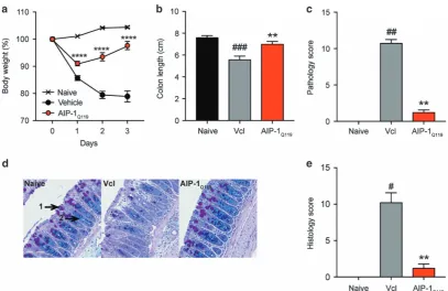
To assess whether Asn-119 substitution to Gln affected the anti-inflammatory properties of the recombinant wild type protein, mice were treated as described previously with AIP-1Q119 (1 mg kg−1) or
vehicle prior to TNBS injection (Figures 3a–e). The level of protection in AIP-1Q119-treated mice against weight loss was comparable to that
of AIP-1 (Figure 3a). Colon shortening and pathology were also significantly inhibited with AIP-1Q119when compared to the vehicle
control (Figures 3b and c). Finally, mucosal architecture and integrity were maintained upon treatment in comparison to the vehicle group as shown by the significant reduction of histology score in AIP-1Q119-treated mice (Figures 2d and e).
Treatment with AIP-1 or AIP-1Q119suppresses systemic
inflammation
In vitroT-cell receptor stimulation of splenocytes revealed a significant systemic inhibition of tumour necrosis factor (TNF)-αproduction in both AIP-1- and AIP-1Q119-treated mice, which correlates with the
suppression of TNBS-induced pathology observed previously (Figure 4a). Interestingly, both recombinant proteins seemed to restore systemic production of IL-10 to a level comparable to that seen in healthy controls, suggesting the promotion of pro-regulatory responses by AIP-1 (Figure 4b).
AIP-1Q119promotes colon immune regulation and tissue repair
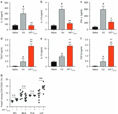
To address the impact of AIP-1Q119treatment on the production of
cytokines at the site of inflammation, colons of mice exposed to TNBS were homogenised and analysed by ELISA (Figures 5a–c). In line with our previous observations, AIP-1Q119 treatment significantly
suppressed inflammatory cytokines IL-13, IL-17 A and IFN-γ (Figures 5a–c). Interestingly, AIP-1Q119 administration also signifi
-cantly increased the levels of thymic stromal lymphopoietin (TSLP) in the colon suggesting mucosal healing (Figure 5d). As seen previously in the spleen, IL-10 production in the colon of AIP-1Q119-treated mice
was also markedly increased as well as TGF-β, suggesting the promotion of regulatory responses (Figures 5e and f). To reveal the potential involvement of regulatory T cells (Treg) in the protection
against TNBS-induced colitis by AIP-1Q119, colons and peripheral
tissues were collected and cells analysed byflow cytometry. While no significant differences were seen in the peripheral tissues, colons of mice treated with AIP-1Q119 displayed a significantly increased
frequency of CD4+CD25+Foxp3+cells (Figure 5g).
AIP-1Q119affects the proinflammatory processes induced by
TNBS in the colon
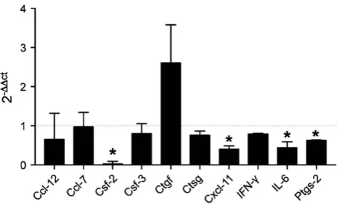
To reveal suppression of inflammation by AIP-1Q119in gut tissue, total
RNA was extracted from colon sections of mice exposed to TNBS. Differentially expressed genes were identified by comparison of expression levels with vehicle-treated samples that served as baseline (Figure 6). Mice treated with AIP-1Q119 displayed a significant
decrease in the expression of mRNAs in the colon encoding the proinflammatory mediators granulocyte macrophage colony-stimulating factor (GM-CSF)- 2, cyclooxygenase (COX)-2, IL-6 and CXC-motif chemokine 11. Although not significant, mice treated with AIP-1Q119 also displayed increased expression of connective tissue
growth factor (CTGF)-encoding mRNA, indicating potential enhanced tissue repair processes. Together, this profile suggests that AIP-1Q119
affects the expression of factors responsible for the migration of activated inflammatory cells such as neutrophils and lymphocytes, likely contributing to the reduction of tissue damage and promoting repair.
DISCUSSION
We38,42 have previously demonstrated that A. caninum excretory/
secretory (AcES) products significantly alleviated intestinal pathology in a mouse model of UC. Protease digestion and heat denaturation showed the protective compound(s) ofAcES were of protein moieties. Interestingly, despite AcES retaining characteristics of inducing Th2 cytokines, pro-regulatory and repair mechanisms were also observed.38
Indeed, proinflammatory cytokines IFN-γ, IL-6, IL-17 A and TNF-α were dramatically suppressed, and the recruitment of alternatively activated macrophages and IL-10/IL-4-producing cells were seen in the mucosa. As previously, shown by Mulvenna and colleagues,AcES is a complex mixture of over 100 proteins, the function of which remain mostly unknown.41 Recently, one of the most abundant proteins produced by A. caninum, AIP-2, was shown to suppress allergen-induced inflammation both in experimental asthma and PBMCs from confirmed allergy patients.40 AIP-2 administration
retinaldehyde dehydrogenase activity, resulting in a significant accu-mulation of Tregsat mucosal sites. Interestingly,A. caninumAIP-1 is
also an abundant protein found in AcES; however, its function remains unclear. Ac-AIP-1, like Ac-AIP-2, contains a signal peptide followed by a TIMP-like netrin domain.Ac-AIP-2 has a C-terminal tail that appears to be absent fromAc-AIP-1 (Figure 2). Cuellar and colleagues showed that AIP-1 induced a systemic state of T-cell unresponsiveness as seen byex vivoT-cell receptor (TCR) activation of splenocytes with anti-CD3.45 The protein was further shown (in a
non-diseased state) to modulate major histocompatibility complex (MHC)-II expression on bone-marrow-derived dendritic cells which were capable of inducing IL-10-producing CD4 and CD8 Foxp3+T
regs.
In this study, we have demonstrated that Ac-AIP-1 significantly protected mice against TNBS-induced weight loss, as well as the cardinal features of colitis. Indeed, colon mucosal barrier integrity was maintained despite the chemical assault resulting in the absence of colon shortening, oedema, ulceration and necrosis. A mutation on the glycosylation site of the recombinant protein demonstrated that the protection induced by AIP-1 was not a bystander immunomodulatory effect of glycans added during the protein expression and folding process in Pichia.51,52 When administered systemically in
TNBS-treated mice, both native and glycosylation mutant AIP-1 significantly suppressed the production of IL-13, IL-17 A, IFN-γ and TNF-α, known to play a central role in IBD.53,54 Although IL-13 is directly
implicated with increased colon epithelial permeability and apoptosis, therapeutic intervention using IL-13 blockade, Anrukinzumab, failed to show any improvement of clinical response or remission.55,56
Evidence undeniably supports that colitis is a multifactorial disease, which cannot be suppressed with the neutralisation of a single component, such as IL-13. However, inhibition of signal transducer and activator of transcription (STAT)-6, for which the phosphoryla-tion status is highly elevated in UC patients, seemed to prevent IL-13-induced apoptosis and improved transepithelial resistance.57 Because of the complexity of IBD pathology, cytokine neutralisation therapy has shown mixed results suggesting that the development of novel treatments should focus on different targets. Indeed, the use of Infliximab or Adalimumab, both monoclonal antibodies directed against TNF-α, were shown to be highly effective against acute colitis, although efficacy was strongly dependent on the severity of the disease.58,59 In addition, resistance to anti-TNF-α therapy has been observed over time, supporting the notion that single cytokine therapy is insufficient for the treatment of IBD. Indeed, the suppression of proinflammatory mediators without promoting mucosal barrier repair will not prevent relapse and disease progression. Interestingly, despite the short timeline of the TNBS model, AIP-1 not only suppressed key pathogenic cytokines important to colitis, but also significantly prevented colonic mucosal damage.
As seen previously with AcES treatment in experimental colitis, AIP-1 promoted the production of IL-10, suggesting a potential mechanism of systemic regulation of inflammation.38 A similar
observation was made upon analysis of the colonic mucosa in which the levels of IL-10 were significantly elevated in comparison to vehicle-treated mice but also in comparison with naïve control. This further supports the notion that IL-10 production seems to be an important suppressive mechanism induced by AIP-1. A genetic-linkage analysis of patients with colitis revealed distinct mutations in the IL-10 gene, demonstrating a central role for this cytokine in the negative feedback necessary to maintain mucosal homeostasis.60,61In our study, TGF-β was also found to be elevated in the colonic mucosa upon AIP-1 treatment. Not only is TGF-β pivotal for the suppression of gut inflammation and enhancing barrier function, but it also suppresses tumour progression in colon cancer and promotes the induction of functional Tregsfrom naive CD4+T-cell precursors.62,63Interestingly,
treatment with AIP-1 resulted in a significant increase in the frequency of CD4+CD25+Foxp3+T cells in the colonic lamina propria. Together, this data suggests that AIP-1 modulates the mucosal cytokine environment by enhancing regulatory processes.
Despite its involvement in the development of allergy and key role in the induction of Th2 responses, TSLP has been shown to have protective effects in experimental colitis.64,65The levels of TSLP in the
colon of AIP-1-treated mice were strikingly higher than both naïve and vehicle control mice. Interestingly, TSLP was shown to be important for protective immunity following infection with the gastrointestinal nematode Trichuris muris by limiting Th1- and Th17-induced mucosal damage.65–67 In addition, TSLP can induce Foxp3+ T
reg by influencing plasmacytoid dendritic cell function in
both mice and humans.68,69 AIP-1 administration modulated colon
expression of a combination of mediators involved in inflammation; expression of mRNAs encoding GM-CSF, CXCL11, IL-6 and PGE-2 were significantly downregulated in AIP-1-treated mice in comparison to vehicle control. Reduced expression of these particular mediators implies that AIP-1 may affect the expansion and migration of activated Th1/Th17 cells induced by TNBS by interfering with PGE-2/IL-6 signalling and GM-CSF/CXCL11 production.70Although not signifi
[image:4.595.57.295.74.303.2]-cant, the expression level of connective tissue growth factor (CTGF)
trended towards elevated levels in AIP-1-treated mice, further supporting cell turnover, wound healing and tissue repair.71 While
the direct action of AIP-1 on the epithelium, the production of TSLP and the inhibition of PGE-2 has yet to be fully demonstrated, AIP-1 seems to induce a multifactorial response beneficial for the suppression of inflammation and tissue damage induced by TNBS.
In like fashion to our findings with AIP-2 in a mouse model of asthma,40AIP-1 seems to promote regulatory cells in the mucosa and
suppress inflammation.40However, the upregulation of genes involved
in mucosal turnover observed herein for AIP-1 was not described for AIP-2. Indeed, AIP-1 seems to have a role in modulating local and systemic production of pro-regulatory cytokines, such as IL-10 and TGF-β, likely allowing tissue repair. Another cytokine important for mucosal repair in IBD is IL-22, which was significantly elevated in the colon of human volunteers experimentally infected with human hookworms.37However, the involvement of IL-22 in AIP-1-induced protection in colitis has yet to be determined. Considering that both AIP-1 and AIP-2 are found abundantly inAcES, one can postulate that both proteins act concertedly to increase the number of regulatory cells and allow the tissue to rapidly heal from parasite-induced injury. While it would be pertinent to assess the combined therapeutic role of AIP-1 and AIP-2 in colitis, we have shown here that on its own, AIP-1 seems to be a good therapeutic candidate for the treatment of colitis by supressing inflammatory responses, preventing tissue remodelling and promoting gut healing.
METHODS
Mice
[image:5.595.86.494.66.330.2]Five-week-old male C57BL/6 were purchased from the Animal Resources Centre (Perth, Western Australia, Australia) and were housed according to Australian code for the care and use of animals for scientific purposes under specific pathogen-free conditions. Mice received food and waterad libidum. All procedures were approved by the James Cook University Animal Ethics Committee under projects A1484 and A2012.
Figure 3AIP-1Q119retains the protective properties against TNBS-induced inflammation. (a–e) Mice received a single intraperitoneal injection (i.p) of 20μg AIP-1Q119in PBS or vehicle, followed 5 h later by an enema with 2.5 mg of TNBS in 50% ethanol. (a) Body weight was recorded daily for the indicated groups. Data show means±s.e.m. of a representative experiment out offive withn=5. Two-way ANOVA with Tukey’s comparisons test used to compare vehicle vs AIP-1Q119over time. (b,c) Colons were removed and measured; adhesion, oedema, mucosal wall thickening and ulceration were scored on a scale of 0–3, with 3 indicating highest degree of damage. (d,e) Colons were opened longitudinally, washed in PBS, and a 1 cm section from the distal colon was fixed in 4% paraformaldehyde. Data show histological micrographs of periodic-acid Schiff (PAS) (×200) obtained from a representative mouse from each group. Histological score was performed by assessing epithelial changes (presence of goblet cells (1), hyperplasia (2), erosion), cell infiltrate, and mucosal architecture. Data show means±s.e.m. of a representative experiment out offive, withn=5. Mann–WhitneyU-test performed comparing naïve vs vehicle groups (#) or vehicle vs AIP-1Q119groups (*); *Po0.05; **Po0.01; ***Po0.001; ****Po0.0001.
[image:5.595.38.279.448.554.2]Reagents and protein expression
Recombinant Ac-AIP-1 and the glycosylation mutant Ac-AIP-1Q119 were expressed as secreted proteins in the yeast Pichia pastoris using methods described elsewhere.72Mutation of Asn-119 to Gln was achieved using PCR as described elsewhere.73The cDNAs encoding the mature sequences ofAc-AIP-1 (amino acids 17-140) andAc-AIP-1Q119were cloned in frame into pPICZαA (Invitrogen, CA, USA) usingXhoI andXbaI restriction sites. The recombinant plasmids were linearized bySacI digestion and transformed intoP. pastoris
strain X-33 by electroporation according to the manufacturer’s instructions (Invitrogen). Transformants were selected on yeast extract-peptone-dextrose plates containing zeocin and assessed for expression of recombinant protein via western blot with monoclonal anti-6 × His antibody. A western blot-positive clone for each protein was grown in a shakerflask, and expression of the recombinant 6 × His tagged Ac-AIP-1 and Ac-AIP-1Q119 were induced with methanol, as per the manufacturer’s instructions (Invitrogen). The recombi-nant fusion proteins were purified with a nickel affinity column and eluates containingAc-AIP-1 andAc-AIP-1Q119were concentrated using Amicon Ultra Centrifugal concentrators and buffer exchanged into phosphate-buffered
saline (PBS) pH 7.4. Lipopolysaccharide contents in Ac-AIP-1 and
Ac-AIP-1Q119 were below 5 ng mg−1 as determined by the Limulus Amoebocyte Lysate (LAL) assay (Pierce Thermo Fisher Scientific, MA, USA).
Induction of colitis
[image:6.595.101.512.68.462.2]Mice were randomly assigned to each group. Recombinant proteins were administered via the intraperitoneal (i.p) route in sterile phosphate-buffered saline at a dose of 1 mg kg−1. Five hours later, mice were anaesthetised with xylazine (5 mg kg−1, Rompun 2%, Bayer, Germany) and ketamine (50 mg kg−1, Ketavest; Pfizer Inc., NY, USA). 2,4,6-Trinitrobenzenesulfonic acid (TNBS; Sigma-Aldrich, MI, USA) was prepared by dissolving 2.5 mg in 50% ethanol. Once unresponsive, mice received an enema with a 125 mg kg−1 dose of TNBS using a lubricated 20-G soft catheter (Terumo, Tokyo, Japan) as previously described.47,48,74Animals were monitored daily for weight loss and general wellbeing over 4 days. Colitis experiments were repeatedfive times with a sample size (n) offive mice per experimental group.
Clinical assessment of colitis
To eliminate bias, mice were assessed in a blinded fashion and de-identified at end point. Mice were weighed daily, and their overall appearance (piloerection), activity level and posture were recorded. No animals were excluded from the study. On day 3 following TNBS injection, mice were killed by CO2 asphyxiation, and colons were collected for observation, characterisation by flow cytometry, cytokine measurements and RNA extraction. When dissecting, the level of tissue adhesion was scored from 0 to 3, with 0 corresponding to absence of adhesion and 3 corresponding to severe adhesion. Colons were measured, cut longitudinally, washed in saline, and observed under an Olympus SZ61 microscope (Notting Hill, VIC, Australia) (×0.67–4.5). Scoring of clinical pathology included adhesions (0–3), mucosal oedema (0–3), ulceration (0–3) and bowel wall thickening (0–3), for a maximum total score of 12 as previously described.38
Tissue preparation and cell culture
Mesenteric lymph nodes (MLN), peripheral lymph nodes (brachial, inguinal, and popliteal) (PLN), spleens and colons were processed in RPMI 1640 media containing 2% foetal bovine serum (FBS), 400 U type I collagenase and 1 mg ml−1DNase I (Life Technologies, Thermo Fisher Scientific, MA, USA) using GentleMACS (Miltenyi Biotec, Germany) and incubated for 15 min at 37 °C. Cells were strained through a 70μm cell strainer (BD Biosciences, NJ, USA). Erythrocytes were lysed with red blood cell lysis buffer (ACK). Colon lamina propria (cLP) were obtained after digestion in RPMI containing 5% FCS, 5 mMEDTA, and 2 mMdithiothreitol (DTT) as described previously.75 Briefly, colons were washed in ice-cold PBS, minced and incubated under agitation for 30 min at room temperature. Intestinal epithelial lymphocytes were discarded byfiltration and the remainder was further incubated in RPMI containing 5% FBS, 400 U type I collagenase and 1 mg ml−1 DNAse I for 30 min at 37 °C. Cells werefiltered and stained with anti-mouse CD3, CD4, CD25 and Foxp3 monoclonal antibodies (BD Biosciences, eBiosciences Thermo Fisher Scientific) and analysed on a BD FACSCanto IIflow cytometer. Treg enumeration experiments in which naïve mice were treated with a daily i.p injection of AIP-1Q119or vehicle for 5 days were repeated twice with a sample size (n) of nine mice (vehicle) and ten mice (AIP-1Q119).
Histology
Distal colons were collected on day 3 post-TNBS injection andfixed overnight in 4% paraformaldehyde, dehydrated with 70% ethanol and embedded in paraffin. Sections were stained with haematoxylin and eosin (H&E) for morphology or periodic acid Schiff for detection of mucopolysaccharide accumulation as described previously.76
Cytokine quantification
Splenocytes were cultured in triplicate inflat-bottom 96-well plates (106cells per well) either with complete RPMI 1640 medium alone or in medium supplemented with 1μg ml−1anti-CD3 antibody (BD Biosciences) for 72 h at 37 °C and 5% CO2. Colon samples were homogenized in calcium- and magnesium-free Hank’s Balanced Salt Solution and phosphatase and protease inhibitor cocktail (Roche, Basel, Switzerland). IFN-γ, TNF-α, IL-10, TGF-β (latent and active form), TSLP, IL-13 and IL-17 A were quantified by ELISA (BD Biosciences) from splenocyte supernatants and colon homogenates.
RNA extraction and gene array
A colon section (0.5 cm) was washed in PBS, placed in 1 ml of TRIzol and dissociated using a TissueLyser (Qiagen, Hilden, Germany) for 10 min with the use of metal beads. Total RNA extraction was performed by phenol–chloroform separation according to the manufacturer’s instructions. After treatment of RNA with RQ1 DNase (Promega, WI, USA),first-strand cDNA was produced with random hexamers and SuperScript III reverse transcriptase (Invitrogen). Samples were tested in 1:100 dilution using a custom wound healing RT2 profiler PCR array and SYBR green (Qiagen). A Rotor-Gene 6000 (Qiagen) was used for real-time thermal cycling. Melting curve analysis was used to confirm that single products had been amplified. All genes were normalised for levels of transcription relative to the housekeeping genes beta-glucuronidase (Gusb), Hypoxanthine guanine phosphoribosyl transferase (Hprt), Glyceraldehyde-3-phosphate dehydrogenase (Gapdh) and beta-actinActb.
Statistical analyses
All data were analysed with GraphPad Prism (version 7; San Diego, CA, USA). Sample size (n=5) was determined by using a power of 80%, one-sided test, representing the probability offinding significant differences between vehicle and AIP-1 or AIP-1Q119-treated groups, with an acceptable Type 1 error of 0.05 and an expected effect size of 1.8. Data are expressed as the mean±s.e.m. Body weight values were analysed using two-way analysis of variance (ANOVA) followed by the Tukey’spost-hoctest. Comparisons for all pairs were performed by unpaired two-tailed Mann–WhitneyU-test. Significance levels were set at a
Pvalue of 0.05.
CONFLICT OF INTEREST
AL and SN have jointly invented a certain invention, entitled METHOD FOR TREATING INFLAMMATION, as described in the following patent applica-tions: PCT Patent Application No. PCT/AU2013/000247,filed on 13 March 2013 with priority of 13 March 2012; and subsequent national phase patent applications in Australia, US, Europe, Canada, China, Japan, India, New Zealand and South Africa. The remaining authors declare no conflict of interest.
ACKNOWLEDGEMENTS
We thank A. Susianto for animal husbandry at JCU and C. Winterford and M. Christensen from the Histotechnology Unit at QIMR Berghofer Medical Research Institute for their help with histology. We thank Bin Zhan from Baylor College of Medicine for provision of the AIP-1 (formerly known as
Ac-TMP-1) expression construct. This work was supported by the National Health and Medical Research Council of Australia (NHMRC) program (grant 1037034 to AL), NHMRC Principal Research Fellowship (to AL). SN was supported by a research grant from the Children’s Hospital Foundation, a research grant from the Faculty of Medicine, Health and Molecular Sciences, a Capacity Building Grant from the Australian Institute of Tropical Health and Medicine, JCU. IBF was supported by an Australian Postgraduate Award scholarship. Additionalfinancial support was provided by the Australian and Queensland governments via the establishment of the Australian Institute of Tropical Health and Medicine at JCU.
[image:7.595.42.279.67.210.2]Author contributions:IBF and SN performed the experiments. SN and AL conceived the study, designed the experiments and wrote the manuscript. DAP and ST expressed the proteins, generated the protein mutant and proofread the manuscript. JC gave advice on experimental design and proofread the manuscript.
Figure 6AIP-1Q119 modulates colon gene expression profile in TNBS-exposed mice. Total medial colon mRNA transcript levels of 10 inflammatory genes were measured at day 4. Data show the average relative gene expression levels (2ΔΔCT) of AIP-1
1 Bouma G, Strober W. The immunological and genetic basis of inflammatory bowel disease.Nat Rev Immunol2003;3: 521–533.
2 Saleh M, Trinchieri G. Innate immune mechanisms of colitis and colitis-associated colorectal cancer.Nat Rev Immunol2011;11: 9–20.
3 Van Limbergen J, Radford-Smith G, Satsangi J. Advances in IBD genetics.Nat Rev Gastroenterol Hepatol2014;11: 372–385.
4 Bollrath J, Phesse TJ, von Burstin VA, Putoczki T, Bennecke M, Bateman Tet al.
gp130-mediated Stat3 activation in enterocytes regulates cell survival and cell-cycle progression during colitis-associated tumorigenesis.Cancer Cell2009;15: 91–102. 5 Grivennikov S, Karin E, Terzic J, Mucida D, Yu GY, Vallabhapurapu Set al.IL-6
and Stat3 are required for survival of intestinal epithelial cells and development of colitis-associated cancer.Cancer Cell2009;15: 103–113.
6 Neurath MF. Cytokines in inflammatory bowel disease.Nat Rev Immunol2014;14: 329–342.
7 Furfaro F, Bezzio C, Ardizzone S, Massari A, de Franchis R, Maconi G. Overview of biological therapy in ulcerative colitis: current and future directions.J Gastrointestin Liver Dis2015;24: 203–213.
8 Coskun M, Vermeire S, Nielsen OH. Novel targeted therapies for inflammatory bowel disease.Trends Pharmacol Sci2017;38: 127–142.
9 McLean MH, Neurath MF, Durum SK. Targeting interleukins for the treatment of inflammatory bowel disease-what lies beyond anti-TNF therapy?Inflamm Bowel Dis
2014;20: 389–397.
10 Musch E, Andus T, Kruis W, Raedler A, Spehlmann M, Schreiber Set al. Interferon-beta-1a for the treatment of steroid-refractory ulcerative colitis: a randomized, double-blind, placebo-controlled trial.Clin Gastroenterol Hepatol2005;3: 581–586. 11 Danese S, Colombel JF, Peyrin-Biroulet L, Rutgeerts P, Reinisch W. Review article: the
role of anti-TNF in the management of ulcerative colitis–past, present and future.
Aliment Pharmacol Ther2013;37: 855–866.
12 Bitton A, Dobkin PL, Edwardes MD, Sewitch MJ, Meddings JB, Rawal Set al.Predicting relapse in Crohn's disease: a biopsychosocial model.Gut2008;57: 1386–1392. 13 Ruffolo C, Scarpa M, Faggian D, Basso D, D'Inca R, Plebani Met al.Subclinical
intestinal inflammation in patients with Crohn's disease following bowel resection: a smolderingfire.J Gastrointest Surg2010;14: 24–31.
14 Peyrin-Biroulet L, Loftus EV Jr, Colombel JF, Sandborn WJ. Long-term complications, extraintestinal manifestations, and mortality in adult Crohn's disease in population-based cohorts.Inflamm Bowel Dis2011;17: 471–478.
15 Eaden JA, Abrams KR, Mayberry JF. The risk of colorectal cancer in ulcerative colitis: a meta-analysis.Gut2001;48: 526–535.
16 von Roon AC, Reese G, Teare J, Constantinides V, Darzi AW, Tekkis PP. The risk of cancer in patients with Crohn's disease.Dis Colon Rectum2007;50: 839–855. 17 Shanahan F, Bernstein CN. The evolving epidemiology of inflammatory bowel disease.
Curr Opin Gastroenterol2009;25: 301–305.
18 Ananthakrishnan AN. Epidemiology and risk factors for IBD.Nat Rev Gastroenterol Hepatol2015;12: 205–217.
19 Malmborg P, Hildebrand H. The emerging global epidemic of paediatric inflammatory bowel disease–causes and consequences.J Intern Med2016;279: 241–258. 20 Munyaka PM, Khafipour E, Ghia JE. External influence of early childhood establishment
of gut microbiota and subsequent health implications.Front Pediatr2014;2: 109. 21 Bager P, Simonsen J, Nielsen NM, Frisch M. Cesarean section and offspring's risk of
inflammatory bowel disease: a national cohort study.Inflamm Bowel Dis2012;18: 857–862.
22 Bousvaros A, Sylvester F, Kugathasan S, Szigethy E, Fiocchi C, Colletti R et al.
Challenges in pediatric inflammatory bowel disease.Inflamm Bowel Dis2006;12: 885–913.
23 Van Limbergen J, Russell RK, Drummond HE, Aldhous MC, Round NK, Nimmo ER
et al.Definition of phenotypic characteristics of childhood-onset inflammatory bowel disease.Gastroenterology2008;135: 1114–1122.
24 Moeeni V, Day AS. Impact of inflammatory bowel disease upon growth in children and adolescents.ISRN Pediatr2011;2011: 365712.
25 Gasparetto M, Guariso G. Crohn's disease and growth deficiency in children and adolescents.World J Gastroenterol2014;20: 13219–13233.
26 Weinstock JV, Elliott DE. Helminths and the IBD hygiene hypothesis.Inflamm Bowel Dis2009;15: 128–133.
27 Helmby H. Human helminth therapy to treat inflammatory disorders-where do we stand?
BMC Immunol2015;16: 12.
28 Loukas A, Hotez PJ, Diemert D, Yazdanbakhsh M, McCarthy JS, Correa-Oliveira Ret al.
Hookworm infection.Nat Rev Dis Primers2016;2: 16088.
29 Summers RW, Elliott DE, Urban JF Jr, Thompson RA, Weinstock JV. Trichuris suis therapy for active ulcerative colitis: a randomized controlled trial.Gastroenterology
2005;128: 825–832.
30 Summers RW, Elliott DE, Urban JF Jr, Thompson R, Weinstock JV. Trichuris suis therapy in Crohn's disease.Gut2005;54: 87–90.
31 Scholmerich J, Fellermann K, Seibold FW, Rogler G, Langhorst J, Howaldt Set al.
A randomised, double-blind, placebo-controlled trial of trichuris suis ova in active Crohn's disease.J Crohns Colitis2017;11: 390–399.
32 Croese J, Giacomin P, Navarro S, Clouston A, McCann L, Dougall Aet al.Experimental hookworm infection and gluten microchallenge promote tolerance in celiac disease.
J Allergy Clin Immunol2015;135: 508–516.
33 Croese J, O'Neil J, Masson J, Cooke S, Melrose W, Pritchard Det al.A proof of concept study establishing Necator americanus in Crohn's patients and reservoir donors.Gut
2006;55: 136–137.
34 Zaiss MM, Rapin A, Lebon L, Dubey LK, Mosconi I, Sarter Ket al.The intestinal microbiota contributes to the ability of helminths to modulate allergic inflammation.
Immunity2015;43: 998–1010.
35 Wilson MS, Maizels RM. Regulation of allergy and autoimmunity in helminth infection.
Clin Rev Allergy Immunol2004;26: 35–50.
36 Maizels RM, McSorley HJ. Regulation of the host immune system by helminth parasites.
J Allergy Clin Immunol2016;138: 666–675.
37 Gaze S, McSorley HJ, Daveson J, Jones D, Bethony JM, Oliveira LMet al.Characterising the mucosal and systemic immune responses to experimental human hookworm infection.PLoS Pathog2012;8: e1002520.
38 Ferreira I, Smyth D, Gaze S, Aziz A, Giacomin P, Ruyssers Net al.Hookworm excretory/ secretory products induce interleukin-4 (IL-4)+ IL-10+CD4+ T cell responses and
suppress pathology in a mouse model of colitis.Infection and immunity2013;81: 2104–2111.
39 Mosconi I, Dubey LK, Volpe B, Esser-von Bieren J, Zaiss MM, Lebon Let al.Parasite proximity drives the expansion of regulatory T cells in Peyer's patches following intestinal Helminth Infection.Infect Immunity2015;83: 3657–3665.
40 Navarro S, Pickering DA, Ferreira IB, Jones L, Ryan S, Troy Set al.Hookworm recombinant protein promotes regulatory T cell responses that suppress experimental asthma.Sci Transl Med2016;8: 362ra143.
41 Mulvenna J, Hamilton B, Nagaraj SH, Smyth D, Loukas A, Gorman JJ. Proteomics analysis of the excretory/secretory component of the blood-feeding stage of the hookworm, Ancylostoma caninum.Mol Cell Proteomics2009;8: 109–121. 42 Ruyssers NE, De Winter BY, De Man JG, Loukas A, Pearson MS, Weinstock JVet al.
Therapeutic potential of helminth soluble proteins in TNBS-induced colitis in mice.
Inflamm Bowel Dis2009;15: 491–500.
43 Cancado GG, Fiuza JA, de Paiva NC, Lemos Lde C, Ricci ND, Gazzinelli-Guimaraes PH
et al. Hookworm products ameliorate dextran sodium sulfate-induced colitis in BALB/c mice.Inflamm Bowel Dis2011;17: 2275–2286.
44 Cantacessi C, Hofmann A, Pickering D, Navarro S, Mitreva M, Loukas A. TIMPs of parasitic helminths-a large-scale analysis of high-throughput sequence datasets.Parasit Vectors2013;6: 156.
45 Cuellar C, Wu W, Mendez S. The hookworm tissue inhibitor of metalloproteases (Ac-TMP-1) modifies dendritic cell function and induces generation of CD4 and CD8 suppressor T cells.PLoS Neglect Trop Dis2009;3: e439.
46 Kiesler P, Fuss IJ, Strober W. Experimental models of inflammatory bowel diseases.Cell Mol Gastroenterol Hepatol2015;1: 154–170.
47 Scheiffele F, Fuss IJ. Induction of TNBS colitis in mice.Curr Protoc Immunol2002; Chapter 15: Unit 15 9.
48 Wirtz S, Neufert C, Weigmann B, Neurath MF. Chemically induced mouse models of intestinal inflammation.Nat Protoc2007;2: 541–546.
49 Ahmad M, Hirz M, Pichler H, Schwab H. Protein expression in Pichia pastoris: recent achievements and perspectives for heterologous protein production.Appl Microbiol Biotechnol2014;98: 5301–5317.
50 Yu P, Zhu Q, Chen K, Lv X. Improving the secretory production of the heterologous protein in Pichia pastoris by focusing on protein folding.Appl Biochem Biotechnol
2015;175: 535–548.
51 Schnaar RL. Glycans and glycan-binding proteins in immune regulation: a concise introduction to glycobiology for the allergist.J Allergy Clin Immunol2015; 135: 609–615.
52 Kuijk LM, van Die I. Worms to the rescue: can worm glycans protect from autoimmune diseases?IUBMB Life2010;62: 303–312.
53 Greten FR, Eckmann L, Greten TF, Park JM, Li ZW, Egan LJet al.IKKbeta links inflammation and tumorigenesis in a mouse model of colitis-associated cancer.Cell
2004;118: 285–296.
54 Ullman TA, Itzkowitz SH. Intestinal inflammation and cancer.Gastroenterology2011; 140: 1807–1816.
55 Reinisch W, Panes J, Khurana S, Toth G, Hua F, Comer GMet al.Anrukinzumab, an anti-interleukin 13 monoclonal antibody, in active UC: efficacy and safety from a phase IIa randomised multicentre study.Gut2015;64: 894–900.
56 Tilg H, Kaser A. Failure of interleukin 13 blockade in ulcerative colitis.Gut2015;64: 857–858.
57 Rosen MJ, Frey MR, Washington MK, Chaturvedi R, Kuhnhein LA, Matta Pet al.STAT6 activation in ulcerative colitis: a new target for prevention of IL-13-induced colon epithelial cell dysfunction.Inflamm Bowel Dis2011;17: 2224–2234.
58 Thorlund K, Druyts E, Mills EJ, Fedorak RN, Marshall JK. Adalimumab versus infliximab for the treatment of moderate to severe ulcerative colitis in adult patients naive to anti-TNF therapy: an indirect treatment comparison meta-analysis.J Crohns Colitis
2014;8: 571–581.
59 Shen C, de Hertogh G, Bullens DM, Van Assche G, Geboes K, Rutgeerts Pet al.
Remission-inducing effect of anti-TNF monoclonal antibody in TNBS colitis: mechan-isms beyond neutralization?Inflamm Bowel Dis2007;13: 308–316.
60 Glocker EO, Kotlarz D, Boztug K, Gertz EM, Schaffer AA, Noyan Fet al.Inflammatory bowel disease and mutations affecting the interleukin-10 receptor.N Engl J Med2009; 361: 2033–2045.
61 Kotlarz D, Beier R, Murugan D, Diestelhorst J, Jensen O, Boztug Ket al.Loss of interleukin-10 signaling and infantile inflammatory bowel disease: implications for diagnosis and therapy.Gastroenterology2012;143: 347–355.
62 Fu S, Zhang N, Yopp AC, Chen D, Mao M, Chen Det al.TGF-beta induces Foxp3+
T-regulatory cells from CD4+CD25−precursors.Am J Transplant2004;4: 1614–1627. 63 Becker C, Fantini MC, Schramm C, Lehr HA, Wirtz S, Nikolaev Aet al.TGF-beta suppresses tumor progression in colon cancer by inhibition of IL-6 trans-signaling.
64 Dieleman LA, Palmen MJ, Akol H, Bloemena E, Pena AS, Meuwissen SGet al.Chronic experimental colitis induced by dextran sulphate sodium (DSS) is characterized by Th1 and Th2 cytokines.Clin Exp Immunol1998;114: 385–391.
65 Spadoni I, Iliev ID, Rossi G, Rescigno M. Dendritic cells produce TSLP that limits the differentiation of Th17 cells, fosters Treg development, and protects against colitis.
Mucosal Immunol2012;5: 184–193.
66 Taylor BC, Zaph C, Troy AE, Du Y, Guild KJ, Comeau MRet al.TSLP regulates intestinal immunity and inflammation in mouse models of helminth infection and colitis.J Exp Med2009;206: 655–667.
67 Massacand JC, Stettler RC, Meier R, Humphreys NE, Grencis RK, Marsland BJet al.
Helminth products bypass the need for TSLP in Th2 immune responses by directly modulating dendritic cell function. Proc Natl Acad Sci USA2009; 106: 13968–13973.
68 Hanabuchi S, Ito T, Park WR, Watanabe N, Shaw JL, Roman Eet al.Thymic stromal lymphopoietin-activated plasmacytoid dendritic cells induce the generation of FOXP3+
regulatory T cells in human thymus.J Immunol2010;184: 2999–3007. 69 Watanabe N, Wang YH, Lee HK, Ito T, Wang YH, Cao Wet al.Hassall's corpuscles
instruct dendritic cells to induce CD4+CD25+regulatory T cells in human thymus.
Nature2005;436: 1181–1185.
70 Stenson WF. Prostaglandins and epithelial response to injury.Curr Opin Gastroenterol
2007;23: 107–110.
71 Ryseck RP, Macdonald-Bravo H, Mattei MG, Bravo R. Structure, mapping, and expression of fisp-12, a growth factor-inducible gene encoding a secreted cysteine-rich protein.Cell Growth Differ1991;2: 225–233.
72 Ranjit N, Zhan B, Stenzel DJ, Mulvenna J, Fujiwara R, Hotez PJet al.A family of cathepsin B cysteine proteases expressed in the gut of the human hookworm, Necator americanus.Mol Biochem Parasitol2008;160: 90–99.
73 Krauer K, Buck M, Flanagan J, Belzer D, Sculley T. Identification of the nuclear localization signals within the Epstein-Barr virus EBNA-6 protein.J Gen Virol2004;85: 165–172.
74 Kim YS, Ho SB. Intestinal goblet cells and mucins in health and disease: recent insights and progress.Curr Gastroenterol Rep2010;12: 319–330.
75 Weigmann B, Tubbe I, Seidel D, Nicolaev A, Becker C, Neurath MF. Isolation and subsequent analysis of murine lamina propria mononuclear cells from colonic tissue.
Nat Protoc2007;2: 2307–2311.
76 Erben U, Loddenkemper C, Doerfel K, Spieckermann S, Haller D, Heimesaat MMet al.
A guide to histomorphological evaluation of intestinal inflammation in mouse models.
Int J Clin Exp Pathol2014;7: 4557–4576.
This work is licensed under a Creative Commons Attribution 4.0 International License. The images or other third party material in this article are included in the article’s Creative Commons license, unless indicated otherwise in the credit line; if the material is not included under the Creative Commons license, users will need to obtain permission from the license holder to reproduce the material. To view a copy of this license, visit http:// creativecommons.org/licenses/by/4.0/