Int. J. Electrochem. Sci., 9 (2014) 6834 - 6842
International Journal of
ELECTROCHEMICAL
SCIENCE
www.electrochemsci.org
Study on the Antioxidant Capacities of Four Antioxidants Based
on Oxidizing Guanine in a Composite Membrane
Xueliang Wang1, Rong Chen2, Li Sun2, Zhangyu Yu1,2,*
1
Department of Chemistry and Chemical Engineering, Heze University, Heze 274015, China
2
College of Chemistry and Chemical Engineering, Qufu Normal University, Qufu 273165, China;
*
E-mail: [email protected]
Received: 30 July 2014 / Accepted: 7 September 2014 / Published: 29 September 2014
The antioxidant capacities of four antioxidants (ascorbic acid, caffeic acid, coumaric acid and resveratrol) were studied herein on a guanine biosensor. The guanine was immobilized on a glassy carbon electrode (GCE) by constant potential deposition, and then a composite membrane of poly(dimethyldiallylammonium chloride) (PDDA), Fe@Fe2O3 core–shell nanonecklace and glucose
oxidase (GOX) was formed on its surface. In glucose solution, the glucose oxidase (GOX) catalyzed glucose to generate hydrogen peroxide (H2O2). Then H2O2 reacted with Fe@Fe2O3 core–shell
nanonecklace and produced hydroxyl radical (·OH) in the membrane to oxidize guanine. As antioxidants were introduced in this system, these antioxidants clear away •OH and alleviate the guanine oxidation degree. By recording the square wave voltammetric signal change of the guanine, the antioxidant capacities of the four antioxidants were evaluated. The sensor exhibited good reproducibility and stability for rapid assessment of the antioxidant capacity.
Keywords: Guanine; glucose oxidase; antioxidants; biosensor; antioxidant capacity
1. INTRODUCTION
Reactive oxygen species (ROS), such as superoxide radical (O2·-), hydroxyl free radicals
(·OH), hydrogen peroxide (H2O2) and so on, can be generated in organisms via normal aerobic
against ROS damage by scavenging free radicals with enzymes such as alpha-1-microglobulin, superoxide dismutases, catalases, lactoperoxidases, glutathione peroxidases and peroxiredoxins, or with small molecule antioxidants such as ascorbic acid (vitamin C), tocopherol (vitamin E), uric acid, and glutathione [5, 6].
In recent years, the electrochemical DNA biosensors based on DNA damage induced by ROS have become important tools in evaluation of DNA damage and antioxidant capacity of antioxidants because the ROS can induce several types of DNA damage and antioxidants can effectively scavenge ROS [7-10]. Among of the bases of DNA, the guanine is the most easily oxidized due to it's the lowest oxidation potential. In addition, the oxidation product of the guanine, 8-oxoguanine is considered an important biomarker of DNA oxidative damage [11, 12]. Therefore, guanine is often used as a DNA model molecule to study the DNA damage by ROS and repair mechanisms [13].
One of the key factors to develop an antioxidant biosensor is the generation of the ROS which is used to oxidize biomolecules because the ROS can’t be purchased from market. At present, there are five means to obtain ROS in fabricating electrochemical antioxidant biosensors. They have been summarized by the literature of [10] as follows (1) Generation via Fenton reaction [14], i.e., hydroxyl radical (OH·) and hydroxide anion (OH
-) are generated by the reaction between transition metals, such as Fe (Ⅱ), Cu (Ⅰ), Cr (Ⅱ), with H2O2; (2) Using photocatalytic reaction to generate ROS. The
photocatalytic reaction of TiO2 is the most usually used among of all photocatalytic reactions to
generate ROS [15]; (3) Utilizing xanthine oxidase (XOD) to catalyze the oxidation of xanthine or hypoxanthine. In this reaction, O2 was reduced to H2O2, and O2•- is formed as an intermediate of the
reaction [16]; (4) Adding NaOH to dimethylsulphoxide (DMSO) to generate O2•-. The production of
O2•- is inversely proportional to the concentration of water in DMSO solution [17]; (5) Producing O2•-
by injecting KO2 into aprotic organic solvents, especially in DMSO [18,19].
Among of the antioxidant biosensor based on DNA damage, Fenton reaction system is one of the most common ways to produce ROS [20, 21]. Most of these sensors are conducted in solution and the unstable H2O2 must be used, which was inevitable to lead a bad reproducibility. To circumvent this
problem, Liang et al. developed a DNA damage biosensor by embedding glucose oxidase into a composite film of sensors in which the ROS was generated in a composite membrane via the catalyzing reaction of glucose oxidase for glucose and following the Fenton reactions [22]. Wang et al. set up an electrochemical DNA damage biosensor by utilizing electro-generated H2O2 to react with
nano Fe@Fe2O3 to generate OH· and damage DNA in situ in a composite membrane [23-25].
In this research, an antioxidant biosensor was developed on the basis of the works of Liang [22] and Wang [23-25]. The sensor showed good reproducibility and stability for rapid evaluation of antioxidant capabilities of ascorbic acid and phenolic compounds (caffeic acid, coumaric acid, resveratrol).
2. EXPERIMENTAL
2.1 Materials and apparatuses
co., Ltd. (China). The GOX solution was prepared with Tris-maleate buffer solution of pH 7.0 (0.2 mol L-1 three hydroxymethyl aminomethane + 0.2 mol L-1 maleic acid + 0.2 mol L-1 NaOH). Fe@Fe2O3
core–shell nanonecklace was synthesized as our previous work [24], and scattered by 2.0 mg mL-1 PDDA into 2.0 mg mL-1 suspension. K3[Fe(CN)6] and K4[Fe(CN)6], were purchased from Shanghai
No. 1 Reagents Factory (China). Ascorbic acid (AA) was obtained from Xilong chemical co., Ltd. (China). Resveratrol was obtained from Sahn chemical technology co., Ltd. (China). Caffeic acid and coumaric acid were purchased from Shanghai source biological technology co., Ltd. (China); 0.1 mol L-1 phosphate buffer solution (PBS) was prepared with 0.1 mol L-1 NaH2PO4 and 0.1 mol L-1
Na2HPO4. All other chemicals were of analytical grade and ultrapure water was used in all the
experiments.
The electrochemical experiments were carried out on a CHI 660 C electrochemical workstation (Shanghai CH Instruments Co., Ltd., China) with three-electrode system a glassy carbon electrode (GCE) or modified GCE working electrode, a saturated calomel reference electrode (SCE) and a platinum wire counter electrode.
2.2 Construction of the biosensor
The GCE was firstly pretreated to mirror as previously reported work [26], and then it was activated under the potential of +1.7 V for 160 s in PBS of pH 3.5. After that, the GCE was immersed into a 0.1 mol L-1 PBS of pH 3.5 containing 4 mg L-1 of guanine, so the guanine was immobilized onto the activated GCE surface via an adsorption step which was conducted with a potential of +0.4 V for 180 s under constant stirring [27-29]. Finally, the guanine modified electrode was sequentially covered with 2.0 mg mL-1 Fe@Fe2O3 of 4.0 µL and 3.0 mg mL-1 GOX of 4.0 µL. The resulted electrode was
named as GOX/PDDA-Fe@Fe2O3/G/GCE.
2.3 Detection
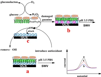
In the composite membrane, the hydroxyl radical (·OH) was generated via a series of reactions between the GOX, glucose and Fe@Fe2O3 to oxidize guanine in situ. The mechanism of this sensor
and the detection method were depicted in the Scheme 1. First, the square wave voltammetry (SWV) of the GOX/PDDA-Fe@Fe2O3/G/GCE was performed in PBS of pH 3.5 from 0.4 to 1.4 V with
frequency of 15 Hz, step potential of 4.0 mV and amplitude of 25 mV (Scheme 1a) and the peak current was recorded as i0. Then the modified electrode was incubated in the PBS of pH 5.0 containing
50 mmol L-1 glucose in the absence (Scheme 1b) or presence (Scheme 1c) of antioxidant for a certain time, after that, the SWV was performed again under the same conditions as above, and the peak current was recorded as it. The guanine oxidative damage and the antioxidant capacity were detected
Scheme 1. Mechanism of the biosensor and the detection method. SWV:square wave voltammetry.
3. RESULTS AND DISCUSSION
3.1 Optimal pH value for oxidizing guanine
3.5
4.0
4.5
5.0
5.5
6.0
6.5
0.0
0.2
0.4
0.6
0.8
1.0
1.2
1.4
i p
[image:4.596.121.468.71.336.2]pH
Figure 1. The influence of pH of the incubation solution on the Δip. Δip was the difference of the SWV
peak currents of GOX/PDDA-Fe@Fe2O3/G/GCE before and after it was incubated in the PBS
By investigating the difference of the SWV peak current (Δip = i0 - it) of the
GOX/PDDA-Fe@Fe2O3/G/GCE after this sensor was oxidized in PBS of different pH containing 50 mmol L-1
glucose, the influence of the pH value the on the guanine damage was studied (Figure 1). The experiments indicated that in the pH range from 3.5 to 6.5, the Δip increased with the pH values at first
and the biggest value of Δip was obtained at about pH 5.0 and then declined. Therefore, PBS of pH 5.0
was used as optimal buffer solution for oxidizing guanine.
3.2 The influence of the volume ratio of Fe@Fe2O3 and GOX on the Δip
The volume ratio of Fe@Fe2O3 and GOX in fabricating the modified electrode had great
influence on the Δip. On the premise of keeping the total volume as 8.0 µL, the influence of the volume
ratio of 2.0 mg·mL-1 Fe@Fe2O3 and 3.0 mg·mL-1 GOX on the Δip was investigated (Figure 2). It was
found that the Δip increased with the increase of VFe@Fe2O3 VGOX, and reached the maximum value at
the VFe@Fe2O3 VGOX of 4 4 and then declined. At ratio of 0 8 or 8 0, Δip value was close to zero,
indicating that Fe@Fe2O3 core-shell nanonecklace or GOX in the membrane played synergistic effect
for guanine oxidation. The optimal ratio of VFe@Fe2O3 VGOX was 4 4.
0:8 1:7 2:6 3:5 4:4 5:3 6:2 7:1 8:0 0.0
0.2 0.4 0.6 0.8 1.0 1.2 1.4
ip
/ 1
10
-6 A
[image:5.596.144.458.365.610.2]VFe@Fe2O3:VGOX
Figure 2. Effect of the ratio of VFe@Fe2O3 VGOX in composite membrane on the Δip. The Δip has the
same significance as in the figure 1. Incubation time was 2.0 min.
3.3 Detection of the antioxidant activity
The GOX/PDDA-Fe@Fe2O3/G/GCE was incubated in the pH 5.0 of PBS containing 50
mmol·L-1
time, and no more change as the incubation time was 4 min. That was to say the guanine of on the electrode surface was oxidized completely in 4 min.
0
1
2
3
4
2.0
2.5
3.0
3.5
4.0
4.5
5.0
5.5
6.0
0.4 0.6 0.8 1.0 1.2 0.2 0.4 0.6 0.8 1.0 1.2 1.4 1.6 1.8 2.0 4 min ip / 10 -5A
E / V
[image:6.596.120.473.129.388.2]0 min i p / 10 -6 A time/min
Figure 3. The relationship between ip and the incubation time. Inset: SWV of
GOX/PDDA-Fe@Fe2O3/G/GCE in pH 3.5 of PBS after it was incubated in pH 5.0 of the PBS containing 50
mmol L-1 glucose for different time.
0 1 2 3 4 5
0.0 0.5 1.0 1.5 2.0 2.5 3.0 3.5 a b i p / 1 10 -6 A t/min
Figure 4. The relationship between Δip in the absence of AA (a) and presence of AA (b) with the
[image:6.596.133.468.463.697.2]
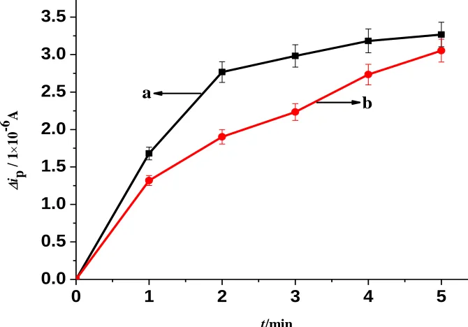
Ascorbic acid (AA) is well-known radical scavenger. Its antioxidant capacity for against guanine damage was studied first. As can be seen from the Figure 4, curve a and b showed the effect of incubation time on Δip in the absence of AA and presence of 10 mg·L-1 AA, respectively. It was found
that the slope of curve b was smaller than that of curve a, indicating as 10 mg·L-1 AA was added into the incubation solution, the increase trend of Δip was slowed. This phenomenon indicated that AA, had
some effects on the protection guanine from oxidation.
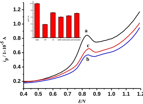
Then antioxidant capacity of 10 mg L-1 caffeic acid was studied by SWV and the results were showed in Figure 5. In pH 3.5 of PBS, GOX/PDDA-Fe@Fe2O3/G/GCE had a sensitive SWV peak of
guanine at about 0.82 V (curve a). It was the oxidation peak of guanine. After it was incubated in pH 5.0 of the PBS containing 50 mmol L-1 glucose for 2 min, the SWV peak decreased apparently (curve b), indicating that partial guanine has been oxidized. However, when the modified electrode was incubated in pH 5.0 of the PBS containing 50 mmol L-1 glucose and 10 mg L-1 caffeic acid for 2 min, the SWV peak (curve c) was bigger than that of curve b. The results indicated that caffeic acid could effective against the guanine oxidation.
Under the same concentration (10 mg L-1) condition, the antioxidant capacity of ascorbic acid, caffeic acid, coumaric acid and resveratrol was investigated respectively (Figure 5 inset). The AOT % of ascorbic acid, resveratrol, coumaric acid and caffeic acid were calculated as 73.5%, 71.6%, 64.4% and 60.7%, respectively. The results showed that different antioxidants have different antioxidant capacity. The highest values was obtained for ascorbic acid and followed by resveratrol. Caffeic acid has the lowest antioxidant capacity. The conclusions are consistent with the reference of [30]. Compared with the other electrochemical biosensors [27, 28, 31], the guanine damage processes and the protection of antioxidants for it in this biosensor were not conducted directly in Fenton solutions, but via a series of biochemical reactions in a composite membrane. These processes are more similar with those processes in vivo. The proposed biosensor had potential use in determination of the total antioxidant capacities in fruit juices.
0.4 0.5 0.6 0.7 0.8 0.9 1.0 1.1 1.2
0.2 0.4 0.6 0.8 1.0 1.2
blank .OH AA caffeic acidcoumaric acid resveratrol
0 20 40 60 80 100
AOT
/ %
ip
/ 1
10
-5 A
E/V
a
b c
Figure 5. SWV of GOX/PDDA-Fe@Fe2O3/G/GCE in pH 3.5 of PBS before (a) and after it was
[image:7.596.180.409.515.682.2]
3.4 Repeatability and stability of the biosensor
The repeatability and stability of the biosensor was investigated by measuring the SWV peak current before and after it was incubated in the incubation solution. For a batch of five biosensors fabricated in the same way, the relative standard deviation (RSD) of Δip was 5.6%, suggesting that that
the sensor shows good repeatability. By comparing the difference of Δip between a new fabricated
biosensor and a stored biosensor in the air for 7 days at room temperature, the results showed that the mean deviation was less than 3% ( n = 5), showing the good stability of the biosensor.
4. CONCLUSION
The antioxidant capacity of ascorbic acid, resveratrol, coumaric acid and caffeic acid was investigated using a guanine electrochemical biosensor. The antioxidant capacities of the four antioxidants were detected by recording the SWV signal change and results showed that ascorbic acid had the highest antioxidant capacity and followed by resveratrol. Caffeic acid had the lowest antioxidant capacity. The sensor showed good reproducibility and stability for rapid evaluation of antioxidant ability of scavenging free radicals.
ACKNOWLEDGEMENTS
The work was supported by the National Natural Science Foundation of China (No. 21105023) and the Natural Science Foundation Committee of Shandong Province, China (Nos. BS2013HZ027 and ZR2009BM003).
References
1. M. L. Urso, Y. W. Chen, A. G. Scrimgeour, P. C. Lee, K. F. Lee and P. M. Clarkson, J. Physiol., 579 (2007) 877
2. R. A. Bachorz, G. Lupica, M. Gutowski and M. Haranczyk, J. Mol. Model., 15 (2009) 817 3. N. R. Perron, J. N. Hodges, M. Jenkins and J. L. Brumaghim, Inorg. Chem., 47 (2008) 6153 4. N. Qu, L. H. Guo, B. Z. Zhu, Biosens. Bioelectron., 26 (2011) 2292
5. M. Moretti, J. Budni, S. D. Bonfanti dos, A. Antunes, J. F. Daufenbach, L. M. Manosso, M. Farina and A. S. Rodrigues, J. Mol. Neurosci., 49 (2013) 68
6. S. Guleria, A. K. Tiku, G. Singh, A. Koul, S. Gupta and S. Rana, J. Plant Biochem. Biotech., 22 (2013) 9
7. M. S. Cooke, M. D. Evans, M. Dizdaroglu and J. Lunec, FASEB J., 17 (2003) 1195
8. Y. Wang, H. Y. Xiong, X. H. Zhang and S. F. Wang, Sensor Actuat B-Chem., 161 (2012) 274 9. P. Bellion, J. Digles, F. Will, H. Dietrich, M. Baum, G. Eisenbrand and C. Janzowski, J. Agr. Food
Chem., 58 (2010) 6636
10.B. Prieto-Simón, M. Cortina, M. Camps, C. Calas-Blanchard, Sensor Actuat B-Chem., 129 (2008) 459
11.L. D. Mello and L. T. Kubota, Talanta, 72 (2007) 335
14.F. Miroslav, K. Tatiana and P. Emil, Biosens. Bioelectron., 15 (2000) 107
15.K. Nagaveni, M. S. Hegde, N. Ravishankar, G. N. Subbanna and G. Madras, Langmuir, 202 (2004) 900
16.I. Fridovich, Science, 201 (1978) 875
17.K. Hyland and C. Auclair, Biochem. Bioph. Res. Commun., 102 (1981) 531 18.L. Vadim and S. Alexander, Anal. Chem., 69 (1997) 454
19.B. Ge and F. Lisdat, Anal. Chim. Acta, 454 (2002) 53 20.A. Mugwerua and J. Rusling, Electroanalysis, 18 (2006) 327
21.O. Korbut, M. Bučková, J. Labuda and P. Gründler, Sensors, 3 (2003) 1
22.M. M. Liang, S. P. Jia, S. C. Zhu and L. H. Guo, Environ. Sci. Technol., 42 (2008) 635 23.X. L. Wang, T. Yang and K. Jiao, Biosens. Bioelectron., 25 (2009) 668
24.X. L. Wang and K. Jiao, Anal. Chim. Acta, 664 (2010) 34-39
25.X. L. Wang, K. Jiao and G. C. Li, Electrochim. Acta, 56 (2011) 2828
26.Y. Y. Feng, T. Yang, W. Zhang, C. Jiang and Jiao K, Anal. Chim. Acta, 616 (2008) 144
27.M. F. Barroso, C. Delerue-Matos and M. B. P. P. Oliveira, Biosens. Bioelectron., 26 (2011) 3748 28.M. F. Barroso, C. Delerue-Matos and M. B. P. P. Oliveira, Food Chem., 132 (2012) 1055
29.R. Chen, X. L. Wang and Z.Y. Yu, Sensor Actuat B-Chem., 192 (2014) 234
30.B. M. Fátima, C. Delerue-Matos and M. B. P. P Oliveira, Food Chem., 132 (2012) 1055 31. Y. Yang, J. Zhou, H. Zhang, P. Gai, X. Zhang and J. Chen, Talanta, 106 (2013) 206