PAPER
Enhanced laser-energy coupling to dense plasmas driven by
recirculating electron currents
R J Gray1
, R Wilson1
, M King1
, S D R Williamson1
, R J Dance1
, C Armstrong1,2
, C Brabetz3
, F Wagner3 , B Zielbauer3
, V Bagnoud3
, D Neely1,2
and P McKenna1
1 SUPA Department of Physics, University of Strathclyde, Glasgow G4 0NG, United Kingdom
2 Central Laser Facility, STFC Rutherford Appleton Laboratory, Oxfordshire OX11 0QX, United Kingdom
3 Plasma Physics Department, GSI Helmholtzzentrum fuer Schwerionenforschung GmbH, D-64291 Darmstadt, Germany
E-mail:[email protected]
Keywords:laser–plasma interactions, laser absorption, relativistic plasma
Abstract
The absorption of laser energy and dynamics of energetic electrons in dense plasma is fundamental to
a range of intense laser-driven particle and radiation generation mechanisms. We measure the total
re
fl
ected and scattered laser energy as a function of intensity, distinguishing between the in
fl
uence of
pulse energy and focal spot size on total energy absorption, in the interaction with thin foils. We
con
fi
rm a previously published scaling of absorption with intensity by variation of laser pulse energy,
but
fi
nd a slower scaling when changing the focal spot size. 2D particle-in-cell simulations show that
the measured differences arise due to energetic electrons recirculating within the target and
undergoing multiple interactions with the laser pulse, which enhances absorption in the case of large
focal spots. This effect is also shown to be dependent on the laser pulse duration, the target thickness
and the electron beam divergence. The parameter space over which this absorption enhancement
occurs is explored via an analytical model. The results impact our understanding of the fundamental
physics of laser energy absorption in solids and thus the development of particle and radiation sources
driven by intense laser
–
solid interactions.
Introduction
Laser energy absorption by electrons is central to high intensity laser–plasma interaction physics. It strongly
influences the optical properties of the plasma[1,2]and the characteristics of beams of high energy photons [3,4]and ions[5,6]produced. Given that the energy coupling to electrons is highly sensitive to a number of interrelated and evolving laser and plasma parameters, absorption remains one of the most challenging areas of thefield to understand. Addressing this challenge is essential for the many applications which require efficient energy transfer to particles and radiation, such as advanced ignition schemes for inertial confinement fusion [7,8]and isochoric heating of matter[9].
In the case of dense plasma, the dominant laser energy coupling mechanisms, such asJ×Bacceleration
[10], vacuum heating[11]and resonance absorption[12], are sensitive to parameters including the laser polarisation and intensity, its incidence angle onto target and the density scale length of the target plasma [13–15]. In addition, the occurrence of multiple absorption mechanisms and transitions between mechanisms
as the laser–plasma interaction evolves, make it difficult to investigate the coupling dynamics experimentally. In
the absence of experimental techniques to resolve the various coupling processes occurring on ultrashort time-scales, we can progress our understanding of the overall absorption physics by determining thetotallaser absorption from measurements of the reflected and scattered laser light[16,17], and its dependency on laser and plasma parameters. The resulting, essentially empirical, insight feeds into the future design of experiments and is ultimately essential for the verification of theoretical and numerical models of absorption physics.
To date, there are few experimental measurements of total absorption in the relativistic laser–dense plasma
interaction regime. Results reported in Pinget al[18], for 0.8μm wavelength laser pulses with duration equal to OPEN ACCESS
RE CE I VED
29 September 2017
RE VI S ED
25 January 2018
A CC E P T E D FO R P UB LI C A T I O N
19 February 2018
P UB L IS H ED
28 March 2018
Original content from this work may be used under the terms of theCreative Commons Attribution 3.0 licence.
Any further distribution of this work must maintain attribution to the author(s)and the title of the work, journal citation and DOI.
150 fs and energy between 0.02 and 20 J, demonstrate an intensity dependent scaling of laser absorption which extends beyond 90% at∼2×10²⁰W cm−2. These results were utilised to develop an empirical scaling law[19] and to develop a theoretical model which identifies upper and lower bounds on laser-coupling for a given laser intensity[20]. Whilst this demonstrates that absorption depends strongly on laser intensity by variation of pulse energy, the influence of focal spot size and pulse duration on total energy absorption in the relativistic regime is not well characterised. Changes to theflux and temperature of the electrons inferred from measurements of laser-driven proton acceleration and x-ray generation indicate that there are more complex coupling dynamics at work when the focal spot size is varied and this merits further investigation[21–23].
In this article, we report on thefirst measurements of total absorption scaling as a function of laser intensity in which the pulse energy and focal spot size are separately varied, for the case of relativistic laser–foil
interactions. We demonstrate that the scaling of the absorption with intensity by variation of focal spot size is significantly slower than by variation of laser pulse energy. Particle-in-cell(PIC)simulations reveal that enhanced absorption observed with relatively large focal spot sizes is a consequence of additional heating of the relativistic electrons which recirculate within the target between the sheathfields formed on the surfaces[24]. Using a simple analytical model, we explore the parameter space for which this effect occurs and show that it is a function not only of the focal spot size but also the laser-pulse duration, target thickness and the electron spectral and divergence distributions. The results highlight the importance of considering changes to absorption caused by the recirculating population of relativistic electrons in experiment design and ultimately in the development of laser-driven particle and radiation sources.
Methodology
The experiment was performed using the high power PHELIX laser at the GSI laboratory near Darmstadt. Linearly polarised pulses with duration,τL, equal to(700±100)fs(full width at half maximum, FWHM), with
energy,EL, varied between 4 and 180 J and a central wavelength of 1.053μm, were incident at 0°(i.e. along the
target normal axis)onto aluminium foil targets with thickness,l, equal to 6 and 20μm. The laser intensity contrast was 10−12at 1 ns and 10−4at 10 ps, prior to the peak of the pulse[25]. The laser energy was changed by
the rotation of a calibrated wave plate between two crossed polarisers between the front end and preamplifier sections. The energy range from 10 to 200 J can be selected without any additional change to the laser or beamline configuration, and consequently the contrast does not change significantly over that range. This was verified via test shots with extremely thin foil targets at full power and the use of a third-order scanning autocorrelator at low energies. Anf/1.5 off-axis parabola was used to focus the laser pulse to a focal spot diameter,fL, which was varied in the range 4–270μm(FWHM)by movement of the parabola toward afixed
target position. By separately varyingELandfL, we investigate laser absorption over the intensity range IL=1017–10
20
W cm−2 .
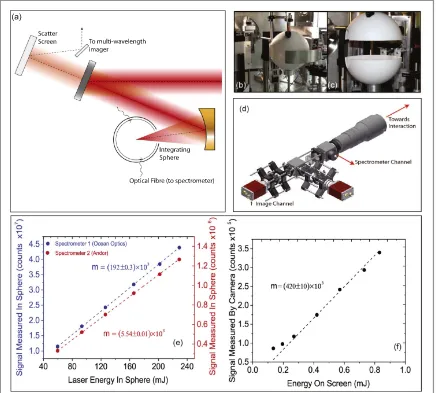
We developed and deployed two novel diagnostics which operate simultaneously to measure the total laser absorption. As shown infigure1, a custom 180 mm diameter integrating(Ulbricht)sphere was developed to make measurements of the total unabsorbed laser pulse energy, from which the absorbed fraction is determined [26]. The sphere was divided into two hemispheres to enable access for a focal spot and target alignment camera prior to laser irradiation of the target foil positioned at the centre of the sphere. After the target was aligned the two hemispheres were moved together forming a tight seal at the interface. A 70 mm diameter aperture at the equator of the sphere accommodates the full focusing laser cone and a 30 mm diameter aperture at the bottom pole enables the target mounting and positioning. Two small 2 mm diameter apertures, 50°above the equator
(60°anticlockwise along the equator from laser axis)and 45°below the equator(120°clockwise along the equator from laser axis), were used for multimode opticalfibre connections. The other ends of the opticalfibres were connected to Ocean Optics(Maya2000Pro)and Andor Shamrock(303i)optical spectrometers. The response of the sphere was calibrated byfirst measuring the energy throughput of the laser system, including all optics up to the sphere entrance, using a calibrated large diameter calorimeter. The sphere response was characterised by directly irradiating the sphere walls with the laser pulse at relatively low energy levels(up to a maximum of 1 J). For full power laser pulses thefibre optic cables werefiltered using well-characterised neutral densityfilters prior to thefibre terminal to avoid the possibility of optical damage being induced inside thefibre. The absolute energy of the scattered light in the sphere was then calculated by scaling the measured signal by a factor of the neutral density response. The unabsorbed component of the laser pulse was measured by
integrating around the central laser wavelength peak by±40 nm(the range over which the spectrum decreases to the background level). At all energies the spectrometers werefiltered such that only the 1st harmonic could be observed. In order to measure the backscattered and specularly reflected light from the target, which escapes the sphere and is collected by the parabola, a scatter screen was placed behind thefinal turning mirror before the parabola(seefigure1(a)). The backscattered light was imaged using a custom dual wavelength imaging system,
as shown infigure1(d). An absolute calibration for the energy response of the imager was made by directly irradiating the scatter screen with light split from the incoming laser beam. A calibrated 50:50 beam-splitter was used, with one half directed onto a calorimeter and the other onto the scatter screen, thereby enabling a direct calibration of the imager response for laser pulse energy. These calibrations are shown infigures1(e)and(f).
Measurements of laser absorption
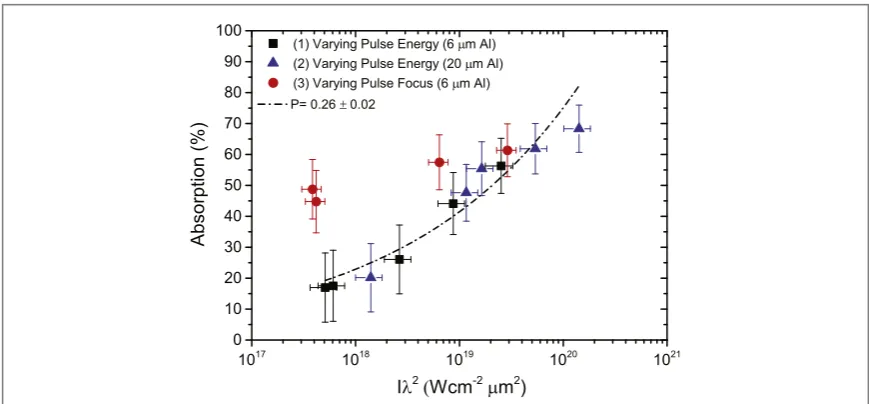
We investigate the scaling of absorption with intensity in a similar range as the detailed investigation reported in Pinget al[18](for otherwise different laser pulse parameters)and then extend our measurements to include variation of focal spot size. Specifically, we measured the change in absorption as a function of intensity by varyingELwhilst keepingfLconstant(at best focus;fL=4μm). The results are shown infigure2as blue
triangles forl=20μm and as black squares forl=6μm targets. We also plot the empirical model presented in [19]and afit to our data with the same form:
fabs=(ILl2 A)P, ( )1
wherefabsis the absorption fraction,ILλ2is the irradiance in units of W cm−2μm2,Pis afitting parameter and A=3.37×10²⁰
W cm−2
μm2(from Davies[19]).
[image:3.595.119.555.62.455.2]Wefind that our measurements are in good agreement with the predictions of the Davies model, and thus by extension the measurements reported in Pinget al[18](upon which that model was based). This is despite the different laser pulse duration and other parameters explored, as detailed above. We note that for significantly
Figure 1.(a)Schematic of the experimental set-up showing the positions of the integrating sphere and the backscatter screen;(b),(c)
thinner targets(sub-micron), which may become subject to hole-boring due to the laser radiation pressure or expand to the point that relativistic self-induced transparency occurs during the interaction, a different scaling with intensity is expected due to a transition from surface-dominated to volumetric interaction processes(see for example[27–30]).
Our measurements show good agreement, in terms of scaling withIL, with previously published values for
whichfLisfixed(at a few microns)andELvaried. Published experimental measurements of the intensity scaling
of the maximum energy of laser-accelerated protons andflux of x-rays suggests a different scaling when the intensity is varied by changing the laser focal spot size[21,23,31,32]. We extend our direct measurements of total energy absorption to investigate this. The red circle data points infigure2are the measured absorption values by varyingfL, forfixedEL. Firstly, it is clear that this scaling is considerably slower than for the case of
varyingEL. Secondly, we note that forIL∼5×10¹⁷W cm−2there is a factor∼3 higher total absorption when
using a higher energy and large focal spot. The enhanced case has considerably more energy in the pulse(157 J compared to 4 J)and a considerably larger focal spot 270μm compared to 4μm.
PIC simulations of laser-energy coupling
We performed simulations using the fully relativistic PIC code EPOCH(in 2D)to investigate changes to the underlying absorption physics arising from variation offL. The laser light was linearly polarised and delivered in
aτL=550 fs(FWHM)Gaussian pulse(aτL=30 fs case is considered later). The target was Al11
+
with an initial, neutralizing electron density equal to 30nc(wherencis the critical density; the density at which the plasma
frequency is equal to the laser frequency)and thickness varied between 6 and 50μm. The initial electron and ion temperatures were 10 keV and 40 eV, respectively. A box size of up to 84μm×140μm was used, with
12600×11520 cells and 10 particles per cell.
In thefirst instance, we selectedILandfLvalues to be the same as two of the experimental data points, to
enable comparison. We consider a peak intensity of∼1×10¹⁹W cm−2, withf
L=50μm andfL=4μm.
Since it is not possible to deconvolve the incoming and reflected laserfields from thefields associated with the expanding electron population, the energy content and temperature of the electrons is considered in lieu of an absorption measure. Example electron spectra at a timet=334 fs after the peak of the pulse interaction(i.e. the time prior to the fastest electrons leaving the simulation box)are shown infigure3(a). This exhibits behaviour consistent with the experiment, namely that the larger spot case results in a higher total energy of relativistic electrons compared to the smaller spot case. The inset offigure3(a)shows the normalised energy difference in the total electron energy above the initial thermal energy forfL=50μm compared tofL=4μm(ò50μmand ò4μm, respectively)as a function of time. The energy gain increases as the intensity increases on the rising edge of
[image:4.595.118.553.60.262.2]the laser pulse and saturates towards the peak of the laser pulse interaction. The gain primarily comes from a non-uniform heating of electrons at the higher end of the spectrum, which changes the temperature of the high energy electrons. This result cannot be accounted for by established electron temperature scaling laws(for
Figure 2.Measured total laser energy absorption as a function of irradiance. The black square and blue triangular data points are measurements made when the intensity is changed by varyingELforl=6μm andl=20μm, respectively. The dot–dash line is afit to this data using the empirical model presented in[19]and shown in equation(1), with the parametersP=0.26 and
A=3.37×10²⁰W cm−2μm2. The red circular data points are measurements made by varyingfLfor constantEL.
example, those presented in[33,34])or the empirical scaling of Davies[19]discussed earlier, becauseILis
constant.
In order to establish if this is a surface interaction effect(e.g. changes in the absorption mechanism or deformation of the critical density surface)or if it arises due to volumetric effects, we investigated the effect of changinglfor bothfL=50μm andfL=4μm. The target thicknesses investigated werel=6, 20 and 50μm.
The results of these simulations are shown infigures3(b)and(c), again by extracting the electron spectrum sampled 334 fs after the peak of the pulse. ForfL=4μm the electron spectrum and temperature are essentially
independent of target thickness(figure3(b)). By contrast, for thefL=50μm case(figure3(c)), there is a
significant change in the electron spectra aslis changed, with the thinnest targets resulting in the highest temperature and electron energies. For largefL, decreasinglleads to significant changes in the measured
electron spectra. For smalll, increasingfLalso leads to significant changes in the spectra for constantIL. This
change(and the correlated change in the total absorption)therefore cannot be a purely surface,field driven, effect and must be related to the relativistic electron dynamics within the target bulk.
Mackinnonet al[35]shows via PIC simulations that the recirculation of relativistic electrons between the sheathfields[24]which build up on the surfaces can enhance the overall electron density and temperature at the rear and thereby enhance the energy of sheath-accelerated protons. Here, we propose that this recirculating electron population is in fact extracting additional energy from the laser pulse upon returning to the front surface, resulting in a higher overall laser-to-electron coupling efficiency. We test this hypothesis by reducingτL
[image:5.595.122.554.62.398.2]such that an electron close to the average energy of the spectrum(≈5 MeV)could only make one pass within the target during the pulse duration and therefore gain no(or limited)additional energy from this recirculating process. As shown infigure3(d), we measure essentially no difference in the electron spectra between the two focal spot sizes. This shows that the recirculation of relativistic electrons within the target is playing an important role in the laser-energy coupling dynamics in the interaction.
Figure 3.Electron energy spectra for:(a)focal spot sizesfL=4μm andfL=50μm, forfixedl=6μm andτL=550 fs;(b)
variation in target thickness froml=6–50μm, forfixedfL=4μm andτL=550 fs;(c)variation in target thickness from l=6–50μm, forfixedfL=50μm andτL=550 fs;(d)focal spot sizesfL=4μm andfL=50μm, forfixedl=6μm and
τL=30 fs. The inset in(a)shows the total energy difference as a function of time(for electrons above the initial electron thermal
energy)between thefL=4μm andfL=50μm cases, normalised to the energy forfL=4μm(forfixedl=6μm andτL=550
It is difficult to directly observe the temporal dynamics of this change in the simulations after a large number of passes, given that there is a population of electrons constantly being injected while the pulse is present, in addition to the recirculating population. Since there is a large range of electron energies, after a certain distance or a number of passes through the target, the two populations become spatially indistinct. However, in the early stages of the interaction it is possible to observe the recirculating population and the additional energy gain at the front surface. This is shown infigure4as a time-space map of the density of electrons with energy greater than 150 keV and propagating counter to the incoming laser pulse. While the energy gain and its dependence on the number of round-trips(given a sufficiently large spot size, pulse length and electron energy)is clear from the simulations, the precise mechanism of the energy exchange is more difficult to extract. The process appears similar to that proposed in Krygieret al[36], where electrons leave the target propagating toward the laser pulse and are turned around by a loop magneticfield, extracting additional energy from the laser pulse via direct laser acceleration(DLA)[37]. That process typically takes place in a long density scale length plasma. For a short density scale lengths, the recirculating electrons are reflected by the sheathfields building up on the target front and rear surfaces and extract additional energy during each interaction with the laserfield at the target front.
A geometric model of recirculation-enhanced absorption
These results highlight that laser intensity alone is not sufficient to predict changes in the absorption or the internal electron spectrum. WhereasILcan be changed by varyingEL,τLorfL, the coupling dynamics can be
radically different depending on which parameter is changed. There is a multi-dimensional parameter space in which the recirculation-enhanced absorption can occur. In order for additional energy to be extracted from the laser pulse the recirculation time(i.e. the transit time for a round trip)must be shorter thanτLand the
divergence angle of the electrons must be small enough that a sufficient number of electrons remain within the focal spot region upon their return to the front surface. The recirculation time and degree to which electrons stay within the region of the focal spot is also affected by the target thickness.
To explore this multi-dimensional parameter space in a tractable way, we construct an analytical model based purely on a ballistic(i.e not affected by self-generatedfields or scattering within the target)approach to electron transport and recirculation, whereby an electron will gain a constant fraction of its initial energy upon re-interacting with the laser at the target front surface. Electron energies above 1 MeV are considered and therefore the effect of collisions is ignored, given that the distance travelled by the electron during pulse duration is small compared to the stopping range. The number of passes an electron will make during timeτLcan be
shown to be
n
l cos
2 , 2
e L div
n t q
=
t ( ) ( )
[image:6.595.121.553.61.239.2]whereνeis the relativistically corrected electron velocity andθdivis the angle of divergence of the electron trajectory with respect to the central axis. For a givenθdivit is also possible to show that the number of passes an electron will make before it leaves the laser focal spot region is
Figure 4.Temporal evolution of electron density along the laser propagation axis, for electrons with energy greater than 150 keV(to avoid sampling the background electron population)and travelling in the negativeXdirection(i.e. toward the laser)forfL=50μm
and(a)l=6μm and(b)l=20μm. The dashed red lines show the initial boundaries of the target, whereX=0 corresponds to the front surface.
n
l
2 tan . 3
L
spot
div
f q
=
( ) ( )
If we take the energy gained,Egain, by an electron of energyEefrom the laser to be a constant fractionαof the
energy, then it can be shown that the overall energy gained via recirculation will be the minimum ofnτornspot. In other words, either the electron will gain energy until it leaves the region of the focal spot(due to its trajectory)
or until the laser pulse switches off. This can be expressed as
E 1 n n n n E
2 e. 4
gain= ( spot+ t-∣ spot- t∣)a ( )
There is of course not simply a single electron energy or divergence angle, but in fact a distribution of both. We take thenN E( )e =N e0 (-Ee kTe)as the energy spectrum of the electron population, whereTeis the electron
temperature andN0is the initial electron number. The energy dependent divergence angle can be calculated using the formula of Mooreet al[38]. This givesqdiv( )Ee =tan-1( ¯q 2 (g-1) ), whereq¯is the average
divergence angle.
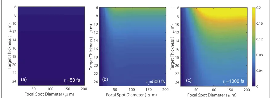
Using this model, the total energy gained over the spectrum due to recirculation as a function offLandlcan
be examined. Infigures5(a)–(c)this parameter is plotted forlin the range 6–25μm andfLin the range
4–200μm, forτLequal to 50, 500 and 1000 fs. The total energy gain is normalised to the total energy of the input
spectrum. It can be seen that forτLof the order of the electron transit time(i.e tens of femtoseconds)there is no
additional energy gain, because the recirculating electron population does not re-interact with the laser pulse a large number of times. For longer pulse durations, as infigures5(b)and(c), there is significant energy gain for l<20μm, but only whenfLis greater than∼50μm. The total energy gain is principally as a result of an increase
in the hot tail of the spectrum, as observed in the simulations. This occurs because the highest energy electrons have the smallest divergence in the distribution and thus shortest recirculation time, and therefore interact more times the laser pulse. The higher divergence of the lower energy electrons means they move out of the region of the focal spot in a fewer number of passes and also return to the front surface a fewer number of times.
It is important to note that the range oflandτLfor which absorption enhancement occurs is highly
dependent on the choice of energy spectrum and divergence-energy functions, as well as the value of the gain coefficientα. Furthermore, the temporal evolution of the forward going electron population, as a result of the temporal evolution of the laser heating at the front surface, is not considered. Instead, we are assuming that a high energy electron population exists and allow this population to freely recirculate. Also, the population of relativistic electrons which escape the target(although often only on the order of a few percent)is not included in the model. These complexities, especially in regard to the self-consistent dynamics of the laser-injected and recirculating populations, quickly make this a complex problem to solve analytically. To that end, this simple model should be seen not as a calculation of the overall change in absorption, but illustrative of the parameter space in which recirculation-enhanced absorption is relevant.
Summary
In summary, these results demonstrate the significance of laser focal spot size in the absorption of energy into thin foils in the relativistic laser–plasma interaction regime. Complementary diagnostic techniques have been
[image:7.595.117.554.63.222.2]applied to determine the total energy absorption via calibrated measurements of the total laser energy specularly Figure 5.l–fLparameter space plots. The colour maps corresponds to the difference in total energy between the input and output
reflected and scattered from the target foil. Good agreement is found with previously published results[18]of absorption scaling with laser intensity when varying the pulse energy. However, a slower scaling is measured when varying the focal spot size for afixed laser energy, which is consistent with the picture emerging from investigations of secondary particle and radiation source properties as a function of focal spot size. PIC simulation results show that this slower absorption scaling with intensity is driven by additional heating of the relativistic electron population which recirculates within the foil between the sheathfields formed on the surfaces. This occurs when the focal spot size and pulse duration are sufficiently large and the electron beam divergence is sufficiently small to enable the recirculating population to interact multiple times with the laser field. The effect is diminished or does not occur in tight focal spot geometries or for femtosecond-scale laser pulse durations(with micron-thick targets).
The results are not only of fundamental interest, but also impact on the optimisation of laser energy coupling in relativistic laser–foil interactions and therefore influence the development of the laser-driven ion
acceleration, high harmonic generation, x-ray production and other sources involving foil targets. They show that the method of varying the laser intensity(i.e. by variation of pulse duration, energy or focal spot size)can significantly change the overall energy coupling into the plasma, and by extension key properties of the secondary beams of particles and radiation produced.
Acknowledgments
We acknowledge the support of the PHELIX laser group and the use of the ARCHIE-WeST and ARCHER high performance computers. The work was supported by EPSRC(grant numbers EP/J003832/1, EP/R006202/1, EP/M018091/1, EP/K022415/1 and EP/L000237/1), STFC(Grant No. ST/K502340/1)and the European
Union Horizon 2020 research and innovation program(Grant Agreement No.654148 Laserlab-Europe).
EPOCH was developed under EPSRC Grant No. EP/G054940/1. Data associated with research published in this paper can be accessed at:https://doi.org/10.15129/16b2e4c8-e1ae-4673-b7d5-93898ba578ad.
ORCID iDs
P McKenna https://orcid.org/0000-0001-8061-7091
References
[1]Borghesi Met al1997Phys. Rev. Lett.78879–82
[2]Bin J Het al2015Phys. Rev. Lett.115064801
[3]Norreys P Aet al1999Phys. Plasmas62150–6
[4]Cipiccia Set al2011Nat. Phys.7867–71
[5]Snavely R Aet al2000Phys. Rev. Lett.852945–8
[6]Macchi A, Borghesi M and Passoni M 2013Rev. Mod. Phys.85751–93
[7]Tabak Met al1994Phys. Plasmas11626–34
[8]Roth Met al2001Phys. Rev. Lett.86436
[9]Patel P Ket al2003Phys. Rev. Lett.91125004
[10]Kruer W L and Estabrook K 1985Phys. Fluids28430–2
[11]Brunel F 1987Phys. Rev. Lett.5952–5
[12]Ginzburg V L 1964The Propagation of Electromagnetic Waves in Plasmas(Oxford: Pergamon)
[13]Eidmann K, Rix R, Schlegel T and Whitte K 2001Europhys. Lett.55334
[14]Nishimura Het al1981Phys. Rev.A232011–9
[15]Singh P Ket al2015Sci. Rep.517870
[16]Gumbrell E Tet al1998Phys. Plasmas53714–21
[17]Teubner Uet al1996Phys. Rev.E544167–7177
[18]Ping Yet al2008Phys. Rev. Lett.100085004
[19]Davies J R 2008Plasma Phys. Control. Fusion51014006
[20]Levy M Cet al2014Nat. Commun.54149
[21]Coury Met al2012Appl. Phys. Lett.100074105
[22]Robson Let al2007Nat. Phys.358–62
[23]Chen L Met al1998Phys. Plasmas114439–45
[24]Sentoku Y, Cowan T E, Kemp A and Ruhl H 2003Phys. Plasmas102009–15
[25]Bagnoud Vet al2016High Power Laser Sci. Eng.4e39
[26]Buckley H 1921J. Inst. Electr. Eng.59143–52
[27]Gray R Jet al2014New. J. Phys.16093027
[28]Gonzalez-Izquierdo Bet al2016Nat. Phys.12505–12
[29]Gonzalez-Izquierdo Bet al2016Nat. Commun.712891
[30]Powell H Wet al2015New J. Phys.17103033
[31]Brenner C M, McKenna P and Neely D 2014Plasma Phys. Control. Fusion56084003
[32]Quinn M Net al2011Plasma Phys. Control. Fusion53025007
[33]Wilks S C, Kruer W L, Tabak M and Langdon A B 1992Phys. Rev. Lett.691383–6
[34]Beg F Net al1997Phys. Plasmas4447–57
[35]Mackinnon A Jet al2002Phys. Rev. Lett.88215006
[36]Krygier A Get al2014Phys. Plasmas21023112
[37]Pukhov A, Sheng Z-M and Meyer-ter-Vehn J 1999Phys. Plasmas62847
[38]Moore C I, Knauer J P and Meyerhofer D D 1995Phys Rev. Lett.742439