Copyright O 1989, American Society for Microbiology
Integrated Proviral Human
Immunodeficiency
Virus
Type
1
Is
Present in CD4+
Peripheral Blood
Lymphocytes
in
Healthy Seropositive Individuals
M. C.
PSALLIDOPOULOS,1*
S. M. SCHNITTMAN,2 L.M. THOMPSON III,1 M. BASELER,3 A. S. FAUCI,2 H. C. LANE,2AND N. P. SALZMAN'Division of Molecular Virology and Immunology, Georgetown UniversitySchoolof Medicine, Washington, D.C. 200071; National Institute of Allergy andInfectious Diseases, Bethesda, Maryland208922; andProgram
ResourcesIncorporated, Frederick, Maryland
217013
Received 7 April 1989/Accepted 17 July 1989
Evidence of a latent human immunodeficiency virus type 1 (HIV-1) infection in healthy, seropositive
individuals who do not have viral antigens in their sera and from whom virions cannot be rescued in
cocultivationexperimentswasexamined.ProviralDNAwasdetectedby amplification bythepolymerase chain reaction procedure. In each of 10 seropositive individuals, the presence ofHIV-1 proviral sequences was
demonstrated in their peripheral bloodmononuclear cells. By using fluorescence-activated cell sorting, we
obtainedhighly enriched subpopulations ofperipheralblood mononuclear cells and found that the CD4+T-cell subset isthe cell subset thatconsistently harbors the HIV-1 proviralsequences.The numberofHIV-1-infected CD4+ Tcellswas variableamongthe 10healthy individuals, rangingfrom 1 in 100 to 1 in40,000. While in vitroinfection of CD4+ Tcellscausesdownregulation and eventual loss of CD4surfacemolecules,this is not trueinvivo where it isonly the CD4+ population that harbors the virus.Thisdisparitymayreflectdifferences betweenalatentinfectioninvivowiththelyticresponseof cellsinfected invitro.
Infection ofTlymphocytes by humanimmunodeficiency virus type 1 (HIV-1) involves binding of the virus to the surface CD4 molecule(2, 6, 15,21), and individuals infected with HIV-1 who develop acquired immunodeficiency syn-drome (AIDS) show a profound and selective depletion of CD4+lymphocytes(13, 19, 27). However, on thebasis of in situ hybridization studies, the fraction of CD4+ lympho-cytes thatactively express HIV-1 isless than0.01% ofthe total number ofmononuclear cells (10). The evidence that cells of themonocyte/macrophage lineage harbor HIV-1 in AIDSpatientssuggests thattheymay serve as areservoir for the virus in the body and also play a role inAIDS-related dementias (16). Similarly, the presence of HIV-1 in bone marrow myeloidprecursors(3) andtheabilitytoinfectand propagatevirus innormalhuman bone marrow suggests that myeloidprecursor cells canbe avirus reservoir duringthe courseofthedisease (7).
While cells expressing the CD4 surface molecule harbor HIV-1 in AIDS patients (S. Schnittman, H. C. Lane, M. Baseler,F.Massari, C. Fox, and A. S. Fauci, Abstr. 4th Int. Conf.onAIDS,abstr. no. 1,p. 168, 1988), to understand the etiology of the diseaseit isimportant toestablishwhichcells harbor virus attheearlierstages ofinfection. This informa-tionwillidentify the primary target for effective chemother-apeutic intervention at a time when the total body load of virus may still be small. We have examined 10 healthy individuals who have antibodies against HIV-1 (as deter-minedbyenzyme-linked immunosorbent assay (ELISA) and Westernblot[immunoblot] assay) but are negative for HIV-1 production bycocultivation and for viral antigen.
Inthisstudy, subsets of each of the individuals' peripheral blood mononuclear cells were obtained by fluorescence-activated cellsortingusingCD4, CD8,CD14, and CD19 cell surface markers and DNA from each highly purified cell
*Correspondingauthor.
subpopulationwas examinedby DNA geneamplificationby the polymerase chain reaction (PCR) method. We have found that the CD4+ cell subpopulation(Tlymphocytes) is the population that consistently harbors HIV proviral se-quences and that there are striking differences between individuals in the number of T cells (CD4+) infected with HIV-1. In 2 of the 10 individuals studied, integratedHIV-1 sequences have also been detected in the CD14+(monocyte/ macrophage)subpopulations. The significanceof these find-ings in thepathogenesis ofthedisease is discussed.
MATERIALS AND METHODS
Cells and cell preparation. Jurkat is an HIV-1-negative T-cell leukemia line (9). ACH2 is a chronically HIV-1-infected T-cell line containing one integrated copy of the HIV-1genome(5).
Whole
blood wascollectedby leukapher-esis or in preservative-free sodium heparin syringes from HIV-1-seropositive volunteers (who had provided written consent). Peripheral blood mononuclear cells(PBMC)were obtained by Ficoll-Hypaque density gradient centrifugation of whole blood, washed twice inphosphate-buffered saline (PBS), suspended at 107 cells/ml in PBS containing 3% human ABsera, andincubatedfor 30min at4°C to block Fc receptors on monocytes. One half of the unfractionated PBMC were sorted directly to obtain CD4+ and CD8+ T lymphocytes,and theother halfofthe cells underwent T-cell depletion byrosettingwith ,-aminoethylisothiuronium bro-mide-treated sheep erythrocytes. The cells were then cen-trifuged at 200 x g for 5 min and gently suspended in 20 ml ofRPMI 1640. Rosetted and nonrosetted cells were sepa-rated by Ficoll-Hypaque density gradient centrifugation. The nonrosetted cells enriched formonocytes and B cells werewashed twotimesin PBS andsubjected tocell sorting. Monoclonal antibodies. PBMC were stained with either fluorescein isothiocyanate (FITC)-conjugated anti-Leu 3a (anti-CD4)orFITC-conjugatedanti-Leu 2a(anti-CD8)(Bec-4626
on November 10, 2019 by guest
http://jvi.asm.org/
HIV-1 IN CD4+ T CELLS 4627 ton Dickinson Immunocytometry Systems, Mountain
View,
Calif.). Rosette-negative PBMC were stained with FITC-conjugated anti-Leu M3 (anti-CD14) or FITC-FITC-conjugated anti-Leu 12 (anti-CD19; Becton Dickinson Immunocytome-try Systems). All cell preparations were stained according to the instructions of the manufacturer. The background con-trols for each sort consisted of PBMC stained with a nonre-active monoclonal antibody isotype control.
Cell sorting. Fluorescence-labeled PBMC were sorted with an EPICS C flow cytometer (Coulter Electronics, Hialeah, Fla.) equipped with a 2W argon laser with an excitation wavelength of 488 nm. Sorting logic was con-trolled by the use of forward angle light scatter versus log green fluorescence bit map gating. Cellular debris, erythro-cytes, and cell aggregates were eliminated from sorting by forward angle light scatter. Cells that were reactive with each monoclonal antibody were determined by comparing the fluorescence of cells stained with specific antibody and of cells that had been incubated with FITC-labeled isotype controls. Cells were sorted at a rate of approximately 2,000 cells per second with coincidence abort engaged. Both positively and negatively stained cells meeting the appropri-ate sort criteria were collected into separappropri-ate tubescontaining sterile PBS. Upon completion of cell sorting, both positive and negative cell populations were analyzed for purity by using machine parameters identical to those forsorting.
Sample preparation. For PCR, highly purified subpopula-tions of sorted PBMC and unfractionated PBMC from the same individual were washed twice in PBS and the cell pellets were stored at -70°Cuntil use. The cells werelysed in freshly prepared lysis buffer containing 10 mM Tris hydrochloride (pH 8.0), 1 mM EDTA, 0.001% TritonX-100, 0.0001% sodium dodecyl sulfate (SDS), and 600 ,ug of proteinase K (Boehringer Mannheim Biochemicals, India-napolis, Ind.) per ml. The lysate was digested at55°C for 1 h, and the enzyme was heat inactivated by boiling for 15 min. Highly enriched sorted subpopulations were lysed at afinal concentration of 2 x 10⁶cells per ml, unfractionatedPBMC at
107
cells per ml, Jurkat at107
cells per ml, and a mixture of Jurkat and ACH2 at cell concentrations of107
and 2 x 10⁴ cells per ml, respectively. The cell lysates were storedat4°C for short periods or frozen at-20°Cwithout anyappreciable loss of PCR reactivity.Oligonucleotides and dNTP. The oligodeoxynucleotides used in this study were synthesized with a DNAsynthesizer (Applied Biosystems, Foster City, Calif.) and purified on 20% polyacrylamide gels (18). For amplification ofHIV-1 DNA sequences, primer pairsrepresentingthelongterminal repeat (LTR) (SK 29/30), gag (SK 38/39),andenvelope (SK 68/69) from conserved regions of the HIV-1 genome were employed (17, 20). For the detection of the amplified prod-ucts, their corresponding probes (SK31, SK19,andSK70, respectively) were used. The HLA-DQalphageneservedas an internal control to monitorthe efficiencyofDNA ampli-fication using oligonucleotide primers (QH26/27) from the second exon (22). Deoxyribonucleotides and T4 polynucle-otide kinase were obtained from Boehringer Mannheim Biochemicals, and
[_y-32PIATP
(4,500Ci/mmol)was obtained from ICNPharmaceuticals, Inc. (Irvine, Calif.). The probes (1pRg)
were endlabeledwithT4polynucleotidekinase and 50,uCi
of[_y-32PIATP
(18). The unincorporated label was re-moved by passing the kinase reaction mixture through an Elutip column (Schleicher & Schuell, Inc., Keene, N.H.), and labeledoligonucleotides were elutedwith 500RI
of 1 M NaCl in TE buffer.PCR. The PCR was runessentially asdescribedby Saiki et
al., with slight modifications (23). Briefly, 50
pI
from each celllysate (-1pug of genomicDNA) wasdirectlyamplifiedin a100-pdfinal volume of the PCRmixture. The PCR mixture contained 10 mMTris hydrochloride (pH8.3), 50 mMKCl, 10mMMgCl2,0.2 mMdGTP,0.2 mMdATP,0.2 mMdTTP, 0.2 mMdCTP, 50 pmolofeachprimer,200pug
ofgelatineper ml, and 2U of Taq DNApolymerase (PerkinElmerCetus, Norwalk,Conn.). Thesamples wereoverlaid with 100pI
of mineral oilandamplified for 30cycles inaThermal Cycler apparatus(PerkinElmerCetus)thatprovided2-minperiods
ofincubationat94,55, and72°Cpercycle. Atthe endof 30 cycles, the apparatus was programmed to add one extra cycleandpolymerizationat72°Cwasextended for10minto ensurecompletion ofthe amplified products. When shorter primers were used (SK 29/30), two extra cycles were per-formedinitiallyatannealingtemperaturesof
37°C
insteadof 55°Cincubation. This allowsthe shorterprimers
toannealto the target DNA moreefficiently, andthesubsequent
run was performed asdescribed above.Afteramplification,
the aque-ous phase was extracted with 100 pI of chloroform and amplifiedproductswere analyzed for the sequence of inter-est. TEbuffer (10pI)
containing0.02pmolof theappropriate
32P-labeled
probe and NaClto give afinal concentration of 0.15 MNaCl was added to 30p1
of the PCR mixture. The sampleswereoverlaidwith 50pI ofmineral oil andheatedat 94°C for 5 min. The probe was allowed to anneal to the amplified DNA sequences at 56°C for 10 min(1,
24).
The hybridization mixture was extracted with 50 pI of chloro-form, and 10 ,u of the aqueousphase
wassubjected
to electrophoresis on 10%polyacrylamide
gels (18).
Amplified
HIV-1 sequences were visualizedby
autoradiography.
Al-ternatively, 20RI
ofthePCRproducts
ofthe HLA amplifi-cationwereanalyzed afterelectrophoresis
oncomposite
gels
containing 3% NuSieve-1% SeaKem agaroses(FMC
Corp.,
Marine ColloidsDiv.,
Rockland,
Maine).
Theamplified
products werevisualizedby ethidium bromidestaining.
HIV-1 culture. PBMC (5 x
106 cells)
orpurified
lympho-cyte subpopulations(106 cells)
fromHIV-1-seropositive
in-dividuals were cultured for 3days
in 5 ml of medium containingphytohemagglutinin
(PHA)
andwere then mixed with 5 x10⁶
PBMC fromhealthy
HIV-1-negative
blood donors that were cultured in the same way. The cultures were maintained at 37°C in a 5%CO2
atmosphere
for 28 days. NormalPBMC(5
x106)
wereaddedtotheculturesondays 7 and 14. Every second
day,
cells in theculture wereallowedtosettletothebottom oftheflaskand 5ml of culture medium was removed and
replaced
with fresh medium lackingphytohemagglutinin
(PHA)
(12,
26).
Virusgrowth
was monitored by reverse
transcriptase
and p24 HIV-1 antigen capture assays incell-freeculturesupernatants
three times aweek.Virologic and immunologic
profile.
Serumsamples
wereassayed forHIV-1antibodies
by
ELISA(Abbott
Laborato-ries, North Chicago,Ill.),
and theantibody
reactivity
wasconfirmed
by
Western blot(Dupont,
NEN Research Prod-ucts,Wilmington,
Del.).
The HIV-1p24
antigen
wasassayed
in a solid-phase
immunoassay
(Abbott
Laboratories,
Chi-cago,Ill.).All assays wereperformed
following protocols
of themanufacturers. Thehematology
testswereperformed by
standard
laboratory
methods.RESULTS
Clinical status of the
participants.
Allparticipants
in this study are male homosexuals who arepositive
for HIV-1 antibodies as determinedby
ELISA and Western blotas-VOL.63, 1989
on November 10, 2019 by guest
http://jvi.asm.org/
says. The total CD4+ counts were normal in 7 of 10
individuals (see Table 3), and remaining laboratory tests
were normal. Physical examination was normal with no
evidence oflymphadenopathy. The clinical and laboratory
picture is ofstage HIV infection, asjudged by the CDC
classification system (4). These individuals were chosen
because of these characteristics and were not randomly
selected fromapopulation of HIV-1 seropositive patients.
Viral expression. Although these 10 individuals had high
titers of antibodies to HIV-1 by ELISA and Western blot
assays,nop24viralantigensweredetected in theirsera,nor
wasHIV-1expressedincocultivationassays.Toaddress the
possibilitythatautologousCD8+ cells inhibit HIV-1
produc-tion in these culture-negative patients (14, 25), we
coculti-vated either PHA-treated unfractionated PBMCor
fraction-ated CD4+ orCD8+lymphocyteswith PHA-blasted normal
PBMC. Portions of culture fluidswere removed three times
aweek and assayed for reverse transcriptase and p24. The
cultures were terminated on day 28. Both reverse
tran-scriptaseandp24assayswere negative in allof the samples (data not shown). On the basis of the results, we conclude
that neither unfractionated nor CD4+ or CD8+ cells were
able to express HIV-1 when cultured in the presence of
lymphocytesfromhealthyindividuals.Thus, CD8+cells do not suppress HIV-1 expression by CD4+ infected cells in
individuals whoareatanearlystageof thedisease,although
in other studiesautologous CD8+ cells have been shownto
affect therecoveryof virus (14, 25).
PCR. To determine if HIV-1 proviral sequences were
presentin lymphocytes of the 10individuals, cell lysatesof
Ficoll-Hypaque-preparedPBMCweresubjectedto30cycles
ofpolymerasechainamplificationwith HIV-i-specific
prim-ers, and the presence of HIV-1 was detected by
HIV-1-specific probes. All 10 individuals contained proviral se-quencesin their PBMC when tested forenvelopesequences
(Fig. There are significant differences in the signal intensity between the individuals, even though equal
num-bersof PBMCwereamplifiedineachcase. Samples 1,7, 8,
9, and 10gave strong signals, whereas samples 2 through 6
gaveweak signals, indicatingthatasmaller numberof cells
are harboring the HIV-1 provirus. This may also reflect
variabilitybetween theprimer-probe sequencesanddiffering
viral sequence present in these individuals. Presumably,
because of thisvariability, only those sequencesthat share
close homology with the primer-probe sequences will be
detected andgenerateastrong signal. Suchaneffectcanbe
partiallyexcluded, since similar resultswereobtained when
differentprimersetsfrom the LTR(Fig. iB) andgagregion
of HIV-1(Fig. 1C)wereused. Thedifferences inintensityof
theamplified productsmorelikelyreflectatruedifference in
the number of infected cells within the populationsthat are
being compared. This issue is addressed below.
Cell sorting. In order to answer the question of which
subpopulation of PBMC harbors the HIV-1 proviral se-quences, PBMC from the 10 individuals were sorted by
fluorescence-activatedcellsortingwithmonoclonal antibod-iestoCD4,CD8, CD14,andCD19 cell surface markers. The
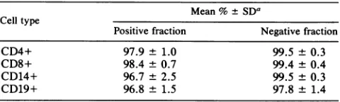
purityofCD4+, CD8+, CD14+ and CD19+ cellpopulations
obtained by fluorescence-activated cell sorting was 97.9,
98.4, 96.7, and96.8%, respectively (Table 1). The purityof the reciprocal subpopulations that were unstained by
anti-bodywereallgreater than 99%. The sorted cellswerelysed
togiveapproximately 2 x 10⁶cellsperml, whereas
unfrac-tionated cells were lysed at 10 x 106 cells per ml and
contained approximately the same number of CD4+ cells
per milliliter as in the CD4+ sorted pool. PCR was
per-A 1 234 5678919 0
4.
B 1 2 34567 89110
C
1 2 3 4 5 6 7 8 9 10
FIG. 1. Analysis of PCR-amplified HIV-1 sequences from
healthy, seropositiveindividuals.Celllysatescorrespondingto5 x
i0'PBMCwere amplifiedwith HIV-1-specific primersfrom
enve-lope(SK68/69) (A), LTR(SK 29/30)(B), andgag(SK38/39) (C).
Thespecificamplifiedsequencesweredetectedby liquid
hybridiza-tion with12P-labeledprobesSK70,SK31,andSK19,respectively.
The heteroduplex product representing less than 10% of the PCR materialwasanalyzedon10%polyacrylamide gels,and autoradiog-raphywasperformedat -70'Cfor 4 h.
formeddirectlyon50-pdlportionsof celllysateswithprimers
from the LTR, and envelope genes. As seen previously,
HIV-1 DNA sequences were present in eachof the
unfrac-tionated cell populations and in every case in the CD4+ T
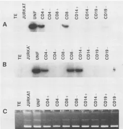
lymphocyte subpopulations. Representative data with the
env probe are shown in Fig. 2A and B, and the complete
results with env and LTR are presented in Table 2. The
absence ofapositive signalin the CD4- population
demon-stratesthat the CD4+ cell is the subpopulation infectedby
HIV-1. When cells were sorted for CD8+, it was the
negatively stained CD8- population that was positive by
PCR; it mainly contained CD4+ Tcells. Similarly, no HIV
viral sequences were detected in B lymphocytes (CD19+).
TheCD19-subpopulationwasoccasionallypositivewhen T
cellswere notremoved efficiently by rosetting (Fig. 2A). In
two cases, however, CD14+ (monocyte/macrophage) cells
were positive by PCR(arepresentative sample is shown in
Fig.2B). Therewasnoevidence ofanyT-cellcontamination
in the CD14+ subsets when these cellswererestained. Most
T cells were removed prior to fluorescence-activated cell
sorting analysis by sheep erythrocyte rosetting and by ex-cludingTcells duringthe sorting procedure. Therefore, the
positivePCRsignalinCD14+ cells is indicative of HIV that
TABLE 1. Purity of sorted PBMCsubpopulations
Mean%± SDa
Celltype
Positive fraction Negativefraction
CD4+ 97.9 ±1.0 99.5 ±0.3
CD8+ 98.4 ±0.7 99.4 ±0.4
CD14+ 96.7 ±2.5 99.5 ±- 0.3
CD19+ 96.8 ±1.5 97.8 ±+ 1.4
aMeanpercentage ±standarddeviation basedon10,000cellsanalyzedper
samplefor 10participants.
on November 10, 2019 by guest
http://jvi.asm.org/
[image:3.612.317.557.644.716.2]HIV-1 IN CD4+ T CELLS 4629
etr It cn)
0.
0-
0-
0 [image:4.612.59.309.79.343.2]0 0 00
TABLE 2. PCR resultsonunfractionatedPBMC and sorted subsets testedwith primers fromthreeconserved regions
ofHIV-1
a0
Positive (+)ornegative(-)resultsforindicated cells
cc
w =
LI--
LLz a.q:C 0
1-U Z
C~
~iEZ,.
~-
..3Mb,,,,
-, C
Krq d
D Q a D D
C) CD
.-llrqr cc cc
a 0
0 0 0 0
0
0)
0
0)
Icr
_r
_ CY0
0 0 0 0
FIG. 2. Amplification of sorted PBMC. Unfractionated PBMC ([UNF]5x 10⁵cellsperreaction) and their respective sorted subsets
(105 cells per reaction) were amplified by using HIV-1 envelope primers (SK 68/69) (A and B) and HLA-DQ alpha primers (QH
26/27) (C). HIV-1-amplified DNA was detected with SK70 32p_
labeled probe by liquid hybridization and analyzed on 10%
poly-acrylamide gels. Autoradiography was performed at -70°C for 16
(A) and 72 (B) h. All 10 individualswerestudied inthisway. Gel A
representsoneindividual (subject 8) with onlyaCD4+
subpopula-tionpositive for HIV-1, and gel Brepresentsoneindividual (subject
5) with both the CD4+ and CD14+ subsets positive. Gel C
repre-sents the analysis of 1 in 10 of the PCR product (for subject 8) amplified for HLA-DQ alpha sequences and fractionated on 3%
Nuseave-1%agarosegelstained with ethidium bromide. Jurkat isan
HIV-1-negative cell line, and TE represents an amplification
con-tainingallreagents exceptDNAthatserves as anegative control.
1 2
Subject Unfractionated CD4+ CD4- CD14+a
envb LTR gag env LTR env LTR env LTR
1 + + + + + - - +
-2 + + + + + + _ _
-3 + + + + + - _ _ _
4 + + + + + _ _ _ _
5 + + + + + _ _ + _
6 + + + + + _ _ _
-7 + + + + + - _ _ _
8 + + + + + - _ _ _
9 + + + + + - _ _ _
10 + + + + +
aThereciprocalCD14- populationwasoccasionallypositivebecause this subsetoften contains CD4+ cellsthatremained afterthe sheep redblood cell procedure.
benv,Enveloperegionof HIV-1.
ispresent in a population ofmonocyte/macrophage subpop-ulations ofthese two individuals. For each sample that was examined,the quality ofDNAlysates and the efficiency of PCR amplification was evaluated by measuring the HLA amplification (Fig. 2C).
Viral load. In order to estimate the number of cells infected with HIV-1, crude lysates of PBMC from the 10 individualswere serially diluted in TEbuffer and PCR was performed (Fig. 3A). Purified CD4+ T cells were similarly analyzed forfour of these individuals(Fig. 3B). The signal intensity fromeach amplificationwas compared with serial dilutions of a mixture of Jurkat cells and ACH2 cells (1 x
107:2
x104
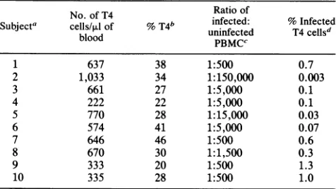
cells per ml). The latter serves as a standard in which each ACH2 cell contains one integrated copy of HIV-1 (5). The results demonstrate that there is indeed a differencein theintensitiesreflectingthenumberof infected cells in each individual(Table3). In theanalyses of PBMC, the viral load ranged from 1 infected cell in 500 to 1 in 150,000.Fourindividualshad aviralburdenof 1:500infected cells, which would correspondto 1in 100CD4+ cells(Table 3), a value thatis similarto the burdenof individuals with AIDS. Values forthe numberof infected CD4+ cells were3 4 5
A a b c a b c a b c d a b c d
_
_-F
B
a b c a b c a b c d a b c d*_I_
a b c d
a b c d
abc
FIG. 3. PCRamplification of envelope sequencesofrepresentative 10-fold dilutions of PBMC and theirrespectiveCD4+ T cells. (A) Lysates ofPBMC(107cells perml) wereserially dilutedinTEtorepresent 5 x 10⁵(a),5 x 104(b),5 x 103 (c), and 5 x 10² (d) cellsper
amplification (gels 1 through 4). A mixture of Jurkat:ACH2 (1x 10⁷:2x 10⁴cellsperml)werealsoincludedascontrolanddiluted torepresent 103(a), 102 (b), 10 (c), and 1 (d) ACH2 cellsperamplification (gel 5). (B) Lysatesfrompurified CD4+ T cells (2 x 10⁶cellsperml) diluted
in TEtorepresent105(a), 104(b),103(c), and 102 (d)cellsperamplification (gels1through 4).Gel5representsdilution ofJurkat:ACH2as
describedabove. All PCRamplfications and detection of amplified envelope sequences wereperformedas previouslydescribed. Thecell
lysateswerefromsubjects 5, 6, 7, and 10(gels1through 4,respectively). Autoradiographywasperformedat -70°Cfor 18 h.
%-cc
WI
-A
LL Lt L a:
z Do a
D 000
B
VOL.63, 1989
_._...
on November 10, 2019 by guest
http://jvi.asm.org/
[image:4.612.173.457.551.663.2]TABLE 3. Estimated number of latently infectedTcells
No.of T4 Ratio of
Subjecta cells/pAlof %T4" uninfected %Infected
blood
PBMCC
1 637 38 1:500 0.7
2 1,033 34 1:150,000 0.003
3 661 27 1:5,000 0.1
4 222 22 1:5,000 0.1
5 770 28 1:15,000 0.03
6 574 41 1:5,000 0.07
7 646 46 1:500 0.6
8 670 30 1:1,500 0.3
9 333 20 1:500 1.3
10 335 28 1:500 1.0
For each subject, p24 antigen capture,ELISA,and Westernblotanalyses werecarried out. In all subjects, p24 was not detectedand strongpositive Western blots and ELISAs wereobserved. Virus couldnotbe rescued from
lymphocytesafter cocultivation with normalPBMC.
bPercentage of T4 cells in the totalTlymphocytecount.
cRatio of cells estimated to beinfected by PCR from serial dilutions of PBMCcelllysatestocontrol cell line ACH2. Resultsrepresent the averageof twoindependentexperiments.
d The estimated percentage of infected T4 cells, assuming that 75% of PBMC contain Tlymphocytes.
similar when PCR was carried out with either PBMC or
purified CD4+ cells.
DISCUSSION
We have studied 10 HIV-1-seropositive homosexual males, all of whom are healthy as judged by
physical
examinations and clinical andlaboratorytests. We surmise thatthese individualsdemonstratean
early
HIV-1infection,
althoughthetime of infectioncannotbedetermined. On the basis of the lack of HIV-1 viral antigens (p24) and the inability of their PMBC to infecttarget
leukocytes
intissue culture, they are not activelyexpressing
virus or viral expression is below the level detected by cocultivation assays. By PCR, a powerful and sensitive technique, wehave demonstrated that each of these individuals has inte-gratedproviralHIV-1 sequencesinPBMC. Ofthe 10people studied,5 gavestrongPCRsignals comparableinintensityto that seen inpatients with AIDS. The otherfive individuals gaveweakersignalintensities,andinonecase(Fig. 1A, lane 2) the signal was just detectable. Sincean equal numberof cells were amplified from each person, this represents dif-ferences in therelative number of infected cells in each of the individuals. In view of the findings that similar results wereobtained when three differentprimers orprobeswere usedfor the PCRamplification (Table 2), it is unlikelythat the difference in intensities between individuals can be attributed togeneticvariabilityof HIV-1. Prospective stud-ies on larger numbers ofindividuals will be necessary to establish if thevirusload detectedearly in the course of the disease is of prognostic significance with respect to devel-opmentof clinical symptoms.
The observationthatvirus could not be rescuedfrom the PBMC of these individuals in the cocultivation assays is further evidencethat either none or only an extremely low numberofinfectedcells areactively expressing HIV-1. The absence of clinicalsymptoms maybedirectlyrelated to what appears to be a quiescent infection. In contrast to other studies (14, 25), ourfailure to detect virus is not because suppressor cells (CD8+) were inhibiting viral production, since we were also unable to rescue virus when highly
enriched CD4+ cells (more than 98%
pure)
were coculti-vated with normal PHAblasts.HIV-1 infection ofCD4+ Tcells in vitrohasbeen
previ-ously
describedto causedownmodulationand eventualloss of the CD4 surface molecule(13,
19, 27).
This in vitro observation does notappearto betrue invivo,
whereonly
the CD4+populations consistently
harbor theHIV-1 provi-ral sequences. If the infected CD4+ T cells hadlost their CD4 surfacemarker,
thenegatively
stained CD4- cellpopulation
would have beenpositive by
PCR.Similarly,
the CD8-population
which containspredominantly
CD4+ T cells is alsopositive
by PCR,
and thesignal
intensity
of thispopulation
isequivalent
tothatoftheunfractionatedPBMC andCD4+ populations. Wecanconclude thatCD4+ Tcells harboringHIV-1 in vivo show noapparentloss of the CD4 surfacemolecule, unlike the invitro observations (13). Our finding isconsistent with thenotion oflatentinfectionwhere HIV-1replication
doesnotoccur,andit is inagreementwith ournegative cocultivation results.Monocyte/macrophage subsets expressing the CD4 sur-face molecule can be infected and express HIV-1 in vitro. Thesecellsmayserve an
important
roleas areservoirforthe HIV-1 virus in the body and in the pathogenesis ofHIV-1 infection (3, 7, 8, 10, 11, 16). However, in thisstudy,
utilizing
highly enrichedCD14+ monocytes thatwere exam-ined by PCR, only 2of10 individuals have HIV-1-infected monocytes, and in both cases there was a weakpositive
signal. From our data, it is evident that the CD4+ T cell is the predominant cell population that harbors the HIV-1 provirus in the peripheral blood circulation of healthy,
seropositive
individuals early in the disease.Comparing PCR results obtained with serial dilutions of PBMC from these healthy seropositive individuals and the control cell line ACH2, a quantitative estimation of the numberof infected cells is possible. Assuming that 75% of PBMC are T lymphocytes, we can calculate that there is a rangefrom0.003 to 1%of the CD4+ Tcells thatharbor the HIV-1
provirus,
compared with results with sera from pa-tientswith AIDS, where 1% oftheCD4+ Tcells is infected (unpublished results). While some healthy HIV-1seroposi-tive individuals have values comparable to those seen in patients with AIDS, others have a 10- to 500-fold-lower number of infected cells. Low levels of infected CD4+ T cells and low levels of HIV-1 expression may primarily be attributedtointeractionsofviral regulatory genes withother viral or cellular factors. At an early stage of the disease,with a low frequency of infected cells, the cellular and humoral immunologic surveillance system of the body may be able to limit the viral infectionforextended periodsof time. Factors that activatelatent virustogenerate cyclesof lytic infection remain to be defined.
ACKNOWLEDGMENTS
Wethank J.Sninsky for helpfuldiscussions, A.Buckler-Whitefor the synthesis ofoligonucleotides, and A. Mannarelli for assistance in preparing the manuscript.
Thisproject has been funded in part with federal funds from the Department of Health and Human Services under contract number NO1-CO-74102 withProgram Resources, Inc.
LITERATURE CITED
1. Abbott, M. A., B. J. Poiesz, B. C. Byrne, S.Kwok, J. J.Sninsky, and G. D. Ehrlich. 1988. Enzymatic geneamplification: qualita-tive and quantitaqualita-tive methods for detecting proviral DNA
am-plifiedin vivo. J. Infect. Dis. 158:1158-1169.
2. Barre-Sinoussi, F., J. C. Chermann, F. Rey, M. T.Nugeyre, S.
on November 10, 2019 by guest
http://jvi.asm.org/
HIV-1 IN CD4+ T CELLS 4631 Chamaret, J. Gruest, C. Dauguet, C. Axier-Blein, F.
Brun-Vezinet, C. Rozioux, W. Rozenbaum, and L. Montagnier. 1983. Isolation of a T-lymphotropic retrovirus from a patient at risk foracquiredimmunodeficiency syndrome (AIDS). Science 220: 868-871.
3. Busch, M., D.Beckstead, D. Gantz, and G. Vyas. 1986. Detec-tion of human immunodeficiency virus infection of myeloid precursors in bone marrow samples from AIDS patients. Blood 68(Suppl. 1):122a.
4. Centersfor DiseaseControl. 1987.Revision of CDC surveillance casedefinition for acquiredimmunodeficiency syndrome. Mor-bid. Mortal. Weekly Rep. 36(Suppl. 1S):3S.
5. Clouse, K.A., D.PoweUl,I.Washington, G. Poli, K. Strebel, W. Farrar, P. Barstad, J. Kovacs, A. S. Fauci, and T. M. Folks. 1989.Monokine regulation of humanimmunodeficiency virus-1 expression in a chronically infected human T cell clone. J. Immunol. 142:431-438.
6. Dagliesh, A. G., P. C. Beverly, P. R. Clapham, D. H. Crawford, M. F.Greaves, and R. A.Weis. 1984. The CD4 (T4) antigen is an essential component of the receptor for the AIDS retrovirus. Nature(London)312:763-767.
7. Folks, T. M., S. W. Kessler, J. M.Orenstein, J. S. Justement, E. S. Jaffe, and A. S. Fauci. 1988. Infection and replication of HIV-1in purified progenitor cells of normal human bone mar-row. Science242:919-922.
8. Gartner, S., P. Markovitz, D. M. Markovitz, M. H. Kaplan, R. C. Gallo, and M. Popovic. 1986. The role of mononuclear phagocytes inHTLV-IIIVLAVinfection. Science 233:215-219. 9. Hansen, J.A.,P. J. Martin, and R. C. Nowinski. 1980.
Mono-clonal antibodies identifying a novel T-cell antigen and Ia antigens of human lymphocytes. Immunogenetics 10:247-260. 10. Harper,M.E., L. M.Marselie,R. C.Gallo, and F. Wong-Staal.
1986. Detection of lymphocytes expressing human T-lympho-tropic virus type III inlymph nodes and peripheral blood from infected individuals by in situ hybridization. Proc. Natl. Acad. Scid. USA 83:772-776.
11. Ho,D. D., T. R. Rota, and M. S. Hirsch. 1986. Infection of monocyte/macrophages by human T lymphotropic virus type III. J. Clin. Invest. 77:1712-1715.
12. Hoffman,A.D.,B.Banapour,andJ.A.Levy. 1985. Character-ization of the AIDS-associated retrovirus reverse transcriptase and optimal conditions for its detection of virions. Virology 147:326-335.
13. Hoxie,J. A., J. D. Alpers, J. L. Rackowski, K. Huebner, B. S. Haggarty,A.J. Ledarbaum,andJ.C. Reed.1986.Alterations in T4(CD4) protein and mRNA synthesis in cells infected with HIV.Science234:1123-1127.
14. Kannagi, M., L. V. Chalifoux, C. I. Lord, and N. L. Letvin. 1988.Suppression of simian immunodeficiency virusreplication invitro by CD8+ lymphocytes. J. Immunol. 140:2237-2242. 15. Klatzmann, D.,F. Barre-Sinoussi, M. T.Nugeyre, C. Dauguet,
E. Vilmer, C. Griscelli, F. Brun-Vezinet, C. Rouzioux, J. C. Gluckman, J.C.Chermann,and L.Montagnier. 1984. Selective
tropism oflymphadelopathy-associated virus (LAV) for helper-induced Tlymphocytes. Science 225:59-63.
16. Koenig, S., H. E. Gendelman, J. M. Orenstein, M. C. Dal Canto, G. H. Pezeshkpour, M. Yungbluth, F. Janotta, A. Aksamit, M.A.Martin, and A. S. Fauci. 1986. Detection of AIDS virus in macrophagesinbrain tissue from AIDS patients with encepha-lopathy. Science 233:1089-1093.
17. Kwok, S., D. H.Mack, K. B. Mullis, B. Poiesz, G. Ehrich,D. Blair, A. Friedman-Kien,andJ. J.Sninsky. 1987.Identification of human immunodeficiency virus sequences by using in vitro enzymatic amplification and oligomer cleavage detection. J. Virol. 61:1690-1694.
18. Maniatis, T.,E. F. Fritsch,andJ. Sambrook. 1982. Molecular cloning: alaboratorymanual. ColdSpringHarborLaboratory, ColdSpring Harbor,N.Y.
19. McDougal, J. S., A.Mawle, S.P.Cort, J. K.A.Nicholson, G.D. Gross, J.A.Sheppler-Campbell,D.Hicks,andJ.S.Sligh. 1985. Cellular tropism of the human retrovirus HTLVIII/LAV. 1. Role ofTcell activation and expression of theT4antigen. J. Immunol. 135:361-363.
20. Ou, C.-Y.,S.Kwok, S.W.Mitchell,D.H. Mack,J. J. Sninsky, J. W. Krebs, P. Feorino, D. Warfield, and G. Schochetman. 1988. DNAamplification for direct detection of HIV-1 inDNA peripheral blood mononuclear cells. Science 239:295-297. 21. Popovic,M.,M.G.Sarngaradharan, E. Read, andR.C.Gallo.
1984. Detection, isolation, and continuous production of cyto-pathic retroviruses (HTLV-III) from patients with AIDS and pre-AIDS. Science 224:497-500.
22. Saiki, R. K., T. L. Bugawan, G. T. Horn, K. B. Mullis, and H. A. Erlich. 1986.Analysis of enzymatically amplifiedP-globin and HLA-DQ alpha DNA with allele-specific oligonucleotide probes. Nature(London) 324:163-166.
23. Saiki,R.K.,D.H.Gelfand,S.Stoffel,S.J. Scharf,R.Higuchi, G. T. Horn, K. B. Mullis, and H. A. Erlich. 1988. Primer-directedenzymatic amplification ofDNA withathermostable DNApolymerase. Science 239:487-491.
24. Saiki,R.K., S.Scharf,F. Faloona, K.B. Mullis, G. T. Horn, H.A.Erlich,and N.Arnheim. 1985.Enzymaticamplificationof ,-globin genomic sequences and restriction site analysis for diagnosis of sickle cell anemia. Science230:1350-1354. 25. Walker,C.M.,D.J. Moody,D.P.Stites,andJ.A.Levy. 1986.
CD8+lymphocytescancontrolhumanimmunodeficiency virus infection in vitro by suppressing virus replication. Science 234:1563-1566.
26. Willey,R.L.,D.H.Smith, L.A.Lasky,T.S.Theodore,P. L. Earl,B. Moss,D. J. Capon, and M. A. Martin. 1988. In vitro mutagenesis identifies aregion within the envelope geneof the humanimmunodeficiency virusthat iscriticalforinfectivity. J. Virol. 62:139-147.
27. Yuille,M. A. R.,M. Hugunin,P. John, L. Peer, L.V. Sacks, B.J. Poiesz,R. H. Tamar,and A. E.Silverstone. 1988. HIV-1 infection abolishes CD4 biosynthesis but not CD4 mRNA. J. AIDS1:131-137.
VOL.63, 1989