White Rose Research Online URL for this paper:
http://eprints.whiterose.ac.uk/88383/
Version: Accepted Version
Article:
Baker, Daniel Hart orcid.org/0000-0002-0161-443X, Karapanagiotidis, Theodoros
orcid.org/0000-0002-0813-1019, Coggan, David et al. (2 more authors) (2015) Brain
networks underlying bistable perception. Neuroimage. pp. 229-234. ISSN 1053-8119
https://doi.org/10.1016/j.neuroimage.2015.06.053
[email protected] https://eprints.whiterose.ac.uk/ Reuse
Items deposited in White Rose Research Online are protected by copyright, with all rights reserved unless indicated otherwise. They may be downloaded and/or printed for private study, or other acts as permitted by national copyright laws. The publisher or other rights holders may allow further reproduction and re-use of the full text version. This is indicated by the licence information on the White Rose Research Online record for the item.
Takedown
If you consider content in White Rose Research Online to be in breach of UK law, please notify us by
Brain networks underlying bistable perception
Daniel H. Baker, Theodoros Karapanagiotidis, David D. Coggan,
Kirstie Wailes-Newson & Jonathan Smallwood
Department of Psychology, University of York, YO10 5DD, UK
: [email protected]
Abstract
Bistable stimuli, such as the Necker Cube, demonstrate that experience can change in the absence of changes in the environment. Such phenomena can be used to assess stimulus-independent aspects of conscious experience. The current study used resting state functional magnetic resonance imaging (rs-fMRI) to index stimulus-independent changes in neural activity to understand the neural architecture that determines dominance durations during bistable perception (using binocular rivalry and Necker cube stimuli). Anterior regions of the Superior Parietal Lobule (SPL) exhibited robust connectivity with regions of primary sensorimotor cortex. The strength of this regionÕs connectivity with the striatum predicted shorter dominance durations during binocular rivalry, whereas its connectivity to pre-motor cortex predicted longer dominance durations for the Necker Cube. Posterior regions of the SPL, on the other hand, were coupled to associative cortex in the temporal and frontal lobes. The posterior SPLÕs connectivity to the temporal lobe predicted longer dominance during binocular rivalry. In conjunction with prior work, these data suggest that the anterior SPL contributes to perceptual rivalry through the inhibition of incongruent bottom up information, whereas the posterior SPL influences rivalry by supporting the current interpretation of a bistable stimulus. Our data suggests that the functional connectivity of the SPL with regions of sensory, motor and associative cortex allows it to regulate the interpretation of the environment that forms the focus of conscious attention at a specific moment in time.
Highlights
¥ Isolated distinct networks from adjacent regions of parietal cortex with rs-fMRI
¥ Specific parts of these networks determine dominance durations for bistable stimuli
¥ Resting state approach avoids confound of non-causal correlations in brain activity
¥ Parietal cortex integrates low and high level data to interpret the environment
Keywords: bistable perception, resting state fMRI, connectivity, parietal cortex
1 Introduction
A remarkable feature of the human mind is its capacity to process information that is independent of environmental input. We often lose ourselves in our thoughts, decoupling experience from the here and now (Baird et al., 2014). At other times we suddenly realize that we having been looking at the world from the wrong perspective: many famous visual illusions depend upon the resolution of uncertain perceptual input (Sterzer et al., 2009). These examples demonstrate that our conscious experience of our
compete for awareness, and the Necker cube (see Figure 1b), in which a wire-frame cube can be perceived from multiple perspectives. Understanding the neural architecture that mediates these alternations is considered a critical step in uncovering neural correlates of consciousness (Crick and Koch, 1998).
Identifying the neural correlates of bistable perception by recording online activity, however, may confound activity that is causally linked to bistable alternations with activity that is merely correlated with it. The finding that activity in a given brain region (e.g. frontoparietal cortex Kleinschmidt et al., 1998; Lumer and Rees, 1999;
[image:3.595.114.482.252.634.2]Sterzer and Kleinschmidt, 2007; Weilnhammer et al., 2013) correlates with perception does not reveal whether that region drives the alternations, or reflects a consequence of processes occurring elsewhere. Carefully controlling for the character of perceptual transitions has been shown to dramatically reduce the number of brain regions that are viable candidates for determining alternations (Knapen et al., 2011). Consequently, it is still a matter of debate whether different forms of bistable stimuli depend on bottom-up or top down influences, and which specific neural systems support these aspects of conscious experience.
The current work capitalizes on the fact that
stimulus-independent changes in neural processing occur naturally during wakeful rest. Neuroimaging has revealed that almost all functional networks that support aspects of task related processing have a comparable resting state network (Smith et al., 2009), and the integrity of these networks varies across individuals in a manner that is predictive of complex forms of cognition such as meta cognitive accuracy (Baird et al., 2013), spontaneous thought (Gorgolewski et al., 2014), reading comprehension (Smallwood et al., 2013) and executive control (Reineberg et al., 2015). As the neural activity at rest is uncontaminated by external input, it provides a relatively pure method to explore stimulus-independent neural processes and so a useful tool to understand the functional architecture of bistable perception.
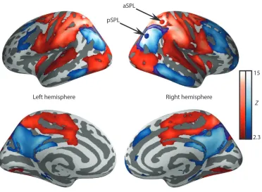
The current study explores the functional connectivity of two regions of the superior parietal lobule (SPL) that have been shown to play a key role in bistable perception in studies using repetitive transcranial magnetic stimulation (rTMS). Application of TMS to the anterior region of the superior parietal lobule (aSPL) in the right hemisphere causes binocular rivalry and bistable motion alternations to speed up (Carmel et al., 2010; Kanai et al., 2011). By contrast, a more posterior region of the same structure (pSPL) slows down the alternations of a bistable structure from motion stimulus when targeted with TMS (Kanai et al., 2010). The apparently opposing functions of these two regions of the SPL is surprising given their physical proximity (see Figure 2). Recent fMRI work has demonstrated that the regions interact with visual areas involved in processing motion to resolve bistable motion percepts, and the strengths of connections are able to predict individual alternation rates accurately (Megumi et al., 2015).
In Experiment One, we examined the functional architecture associated with these regions in a large group of participants with the aim of identifying the large-scale networks that these areas participate in. Building on prior work linking individual differences in perception to neural organisation (Kanai et al., 2011, 2010; Megumi et al., 2015; Song et al., 2013a; 2013b; van Loon et al., 2013; Watanabe et al., 2014), in Experiment Two we probed the functions of these networks by examining whether modulations in the connectivity across individuals contains explanatory information on their rate of perceptual alternations. We did this for two different bistable stimuli, selected for their reliance on either lower-level stimulus features
(binocular rivalry between two orthogonal gratings) and higher-level stimulus features (alternations between two interpretations of the Necker cube). By selecting contexts in which bistability occurs at either lower or higher levels of analysis we hoped to be able to reveal neural processes that contribute to top down and bottom up processing in bistable perception. We were especially interested whether the different regions of the SPL had different functional architectures at rest, and whether such differences could explain the heterogeneous relationship between rivalrous experience in this region of cortex.
2 Experimental Procedures
2.1 Resting state scans
The dataset from the Nathan Kline Institute (NKI) / Rockland contained 141 subjects. The resting state fMRI data were acquired with the following parameters: TR 2500 ms, TE 30 ms, 120 volumes, matrix size 72x72, 38 slices, flip angle 80¡, 0.3 mm spacing between slices, voxel size 3x3x3 mm and an interleaved slice acquisition order. A high-resolution anatomical image was also acquired for each subject using the MPRAGE sequence (Magnetization Prepared Rapid Acquisition Gradient Echo).
Data from Experiment Two were acquired using an eight-channel phased array head coil (GE) tuned to 127.4 MHz on a GE 3 Tesla Signa Excite HDx MRI scanner at York Neuroimaging Centre, at the University of York. 31 participants were recruited. 1 was excluded due to insufficient brain coverage, 2 for excessive movement and 2 were identified as outliers based on their rivalry or MRI data falling more than ±3SD from the group mean. Blood oxygen level-dependent (BOLD) contrast images with fat saturation were acquired using a gradient single-shot echo planar imaging (EPI) sequence with the following acquisition parameters: repetition time (TR) 2000 ms, echo time (TE) minimum full (~19 ms), 210 volumes, flip angle 90¡, matrix 64 x 64, field of view (FOV) 192 mm, slice thickness 3 mm with a 0.5mm gap and 32 slices with an interleaved (bottom -> up) acquisition order.
high-resolution T1-weighted in-plane anatomical picture was also acquired for all participants, using a fluid attenuated inversion recovery (FLAIR).
2.2 Pre-processing
All fMRI pre-processing and analyses for Experiment One and Two were performed using FSL. We extracted the brain from the skull using the BET toolbox for both the FLAIR and the structural T1 weighted images and these scans were registered to standard space using FLIRT (Jenkinson and Smith, 2001). Prior to conducting the functional connectivity analysis the following pre-statistics processing was applied to the resting state data; motion correction using MCFLIRT (Jenkinson et al., 2002); slice-timing correction using Fourier-space time-series phase-shifting; non-brain removal using BET (Smith, 2002); spatial smoothing using a Gaussian kernel of FWHM 6mm; grand-mean intensity normalisation of the entire 4D dataset by a single multiplicative factor; highpass temporal filtering (Gaussian-weighted least-squares straight line fitting, with sigma = 100 s); Gaussian lowpass temporal filtering, with sigma = 2.8s.
2.3 Low level analysis
We created two spherical seed ROIs, 2 mm in radius, one centered in aSPL and the other in pSPL. The time series of these regions were extracted and used as explanatory variables in a separate subject level functional connectivity analysis for each seed. In these analyses, we also included eight nuisance regressors: white matter (WM), cerebrospinal fluid (CSF), and six motion parameters. The WM and CSF covariates were generated by segmenting each individualÕs high-resolution structural image (using FAST in FSL). The default tissue probability maps, referred to as Prior Probability Maps (PPM), were registered to each individualÕs high-resolution structural image (T1 space) and the overlap between these PPM and the corresponding CSF and WM maps was identified. These maps were then thresholded (40% for the SCF and 66% for the WM) and binarized. Finally, these masks were used to calculate the average time course for each aspect of the brain for each individual. The six motion parameters were calculated in the motion-correction step during pre-processing. Movement in each of the three Cartesian directions (x, y, z) and rotational movement around three axes (pitch, yaw, roll) were included for each individual.
At the group-level the data were processed using FEAT version 5.98 part of FSL (FMRIB's
Software Library, www.fmrib.ox.ac.uk/fsl) and the analyses were carried out using FMRIB's Local Analysis of Mixed Effects (FLAME) stage 1 with automatic outlier detection. Mean displacement was included at the group level as a covariate of no interest. The Z statistic images were then thresholded using clusters determined by Z>2.3 and a (corrected) cluster significance threshold of p=0.05 (Worsley, 2001).
2.4 Bistable alternation rate experiment
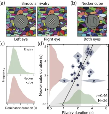
We showed bistable stimuli to participants for trials of 120 seconds in duration and asked them to report their percepts using a computer mouse. One stimulus consisted of oblique gratings (2c/deg, 50% contrast, ±45 deg, 2.3 deg in diameter, smoothed by a raised cosine envelope) shown to opposite eyes (see Figure 1a). The other was a wireframe drawing of a cube, with a face width of two degrees (see Figure 1b), shown to both eyes with no binocular disparity. All stimuli were presented on a gamma-corrected Iiyama VisionMaster Pro 510 CRT monitor with a mean luminance of 32cd/m2, and were viewed through a mirror stereoscope to permit presentation of different images to the left and right eyes (where required). The stimuli were surrounded by a dark ring, and a binocular Voronoi texture to promote binocular vergence and fusion (Baker and Graf, 2009).
Participants were instructed to hold down one mouse button whilst they perceived a particular percept (e.g. a left-oblique grating, or the lower face of the cube appearing closer) and the other button when they perceived the alternative percept (e.g. a right-oblique grating, or the upper face of the cube appearing closer). If they could not decide (e.g. because they experienced a piecemeal percept) they were instructed to release both buttons. Each participant completed four 120-second trials for each of the two stimuli, which were displayed in a randomized order across trials. We also switched the orientations of the rivalling stimuli between the eyes on each alternate trial to avoid adaptation and balance any eye dominance effects. Because the dominance durations followed the typical positively skewed gamma distribution (Figure 1c), we calculated the geometric (rather than arithmetic) mean dominance durations and standard deviations, which are shown in Figure 1d.
3 Results
3.1 Experiment One
scans collected from 141 participants (Nooner et al., 2012). The network associated with the anterior seed (red button in the upper right panel of Figure 2) involved the occipital lobe, regions of the parietal lobe, and the dorsal surface of the temporal lobe. The network associated with the posterior seed (blue button in the upper right panel of Figure 2) involved lateral regions of temporo-parietal cortex, elements of the temporal lobe, and dorsolateral regions of the prefrontal cortex. We verified the distinctness of these two networks using a large meta-analytic resting state database (www.neurosynth.org; Yarkoni et al., 2011), that produced similar (though less extensive) networks, shown in Supplementary Figure S1. It is noteworthy that the connectivity of the aSPL seed was targeted at primary
sensori-motor regions, whereas the connectivity of the pSPL seed region connected with regions of associative cortex (e.g. the temporal lobe and lateral/medial regions of the dorsal prefrontal cortex). These contrasting patterns of connectivity support the hypothesis that these parietal regions have distinct influences on bistable perception because they are functionally connected to different aspects of cortex. In particular, these data suggest that the aSPL connectivity to basic sensory motor information would allow it to integrate lower level sensory information while the connections between the pSPL and regions of associative cortex suggest that it could be important in the top-down coordination of experience.
Figure 2: Resting state networks derived from 141 participants. Seed regions were in the anterior superior parietal lobule (aSPL, red button, MNI coordinates: x=36, y=-45, z=51) and in the posterior superior parietal lobule (pSPL, blue button, MNI coordinates: x=38, y=-64, z=32) of the right hemisphere.
3.2 Experiment Two
To understand the function these networks play in bistable perception, we analyzed resting state scans from a further 26 individuals who also took part in an experiment outside of the scanner to measure their dominance durations for two bistable stimuli (Figure 1a,b). We verified that the same networks were produced from our two parietal seed regions for this independent group
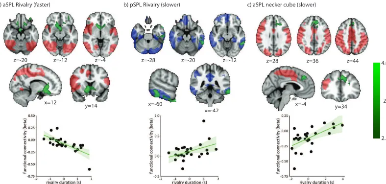
[image:6.595.117.489.301.574.2]We regressed mean dominance durations against resting state connectivity from our two seed regions to determine which areas of cortex were involved in determining the dynamics of bistable alternations. We found that the coupling between the aSPL seed and regions of the striatum (green regions in Figure 3a) showed a greater association with faster alternations for the binocular rivalry stimulus (i.e. shorter durations). The striatum is known to be important in relaying prediction error signals to primary regions of cortex (Daw et al., 2011) and might play this role in perceptual decision making (Forstmann et al., 2010; though see also Boekel et al., 2015).
Slower alternations during binocular rivalry were associated with stronger connectivity between the pSPL and regions of the left temporal lobe
primarily in the inferior temporal gyrus (ITG, Figure 3b). The ITG constitutes the most anterior aspect of the ventral stream and is hypothesized to code relatively high-level information about stable properties of a stimulus, specifically in the visual modality (Kravitz et al., 2013).
Finally, relatively longer dominance durations in the Necker Cube were associated with stronger coupling between the aSPL and a region of medial cortex in the left hemisphere, at the boundary between the pre-supplementary motor area and the dorsal anterior cingulate cortex (see Figure 3c). This region of cortex is implicated in the coordination of the selection of goal related actions (Domenech and Koechlin, 2015) under conditions when stimulus input is stable.
Figure 3: Regions associated with alternation rates. In each panel the red or blue shading shows the resting state networks derived in Figure 2, and the green shading shows regions that correlate with alternation rates. Panel (a) shows significant clusters where connectivity with the aSPL was associated with faster alternation rates during binocular rivalry. Panel (b) shows clusters from the pSPL seed associated with slower rivalry alternations. Panel (c) shows significant clusters where connectivity with the aSPL was associated with slower Necker cube alternations. Each subplot shows three axial slices, one saggital slice and one coronal slice, with MNI coordinates given below each image. The scatterplots show the relationship between dominance durations and connectivity (y-values are fMRI beta parameter estimates). In axial and coronal slices the left hemisphere is shown on the right of the image.
4 Discussion
We found evidence that anterior and posterior regions of superior parietal cortex which influence bistable perception in opposing directions do so because of differences in their functional architecture. Our seed region in the pSPL exhibited connectivity with regions of parietal and lateral prefrontal cortex whose specialism is in the extraction of higher order features of sensory input (Duncan, 2010; Goldman-Rakic, 1988). This functional network is consistent with prior fMRI studies that have
found a relationship between alternations and activity in similar regions of fronto-parietal cortex (Kleinschmidt et al., 1998; Knapen et al., 2011; Lumer and Rees, 1999; Sterzer and Kleinschmidt, 2007; Watanabe et al., 2014; Weilnhammer et al., 2013). Importantly, Experiment Two found that connectivity between pSPL and ventral aspects of the temporal lobe was enhanced for individuals who experienced slower rivalry alternations. This brain region is important in visual aspects of semantics (Squire et al., 2004; Visser et al., 2010) and activity there is suppressed when an image is rendered invisible by rivalry suppression (Fang and He, 2005).
a) aSPL Rivalry (faster)
z=-20 z=-12 z=-4
y=14 x=12
b) pSPL Rivalry (slower)
z=-28 z=-20 z=-12
y=-42 x=-60
2.3 4.0
Z c) aSPL necker cube (slower)
z=28 z=36 z=44
[image:7.595.95.489.310.498.2]Together these data are consistent with the claim that the pSPL is part of a distributed functional system that helps stabilize a specific interpretation of low-level perceptual input.
In contrast to the pSPL, the aSPL was connected to regions of sensori motor cortex that are thought to serve functions more closely tethered to sensory input (Buckner and Krienen, 2013). Importantly, our data show that strong aSPL connectivity with the striatum was linked to shorter rivalrous durations. Given the potential role of the striatum in prediction error (Forstmann et al., 2010), this pattern of heightened connectivity suggests that during binocular rivalry, a specific percept may remain dominant as long as the competing low-level input is suppressed. Consistent with this interpretation of our data, recent work has shown that increasing the reward associated with a particular stimulus, a manipulation that increases the magnitude of a prediction error signal, increases the dominance of that particular stimulus (Marx and Einhauser, 2015).
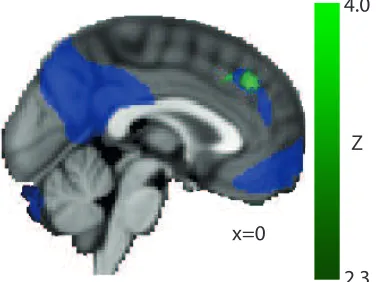
Figure 4: Overlap between the pSPL resting state network from Figure 2 (purple) and the connectivity between the aSPL region associated with slower Necker cube reversals (green; see Figure 3c).
Compared with resolving binocular rivalry, viewing a Necker cube involves overcoming incongruency in perceptual input at a relatively higher level. Connectivity between the aSPL and a region of dorso-medial prefrontal cortex was enhanced for participants with relatively longer dominance durations for the Necker cube. This region of pre-frontal cortex is implicated in the top-down coordination of cognition and action (Botvinick et al., 2004). Importantly, this region overlaps with the connectivity patterns of the pSPL (see Figure 4) suggesting that it is functionally connected to information arising in a bottom up manner from sensori-motor cortex and in a top down manner from information in associative cortex (see also Watanabe et al., 2014). This convergence of information would be
especially important in allowing perceptual incongruity to be resolved at relatively complex levels such as that which occurs while viewing the Necker Cube.
Recently, a study using dynamic causal modelling delineated a network involving the right hemisphere parietal regions studied here (aSPL and pSPL) and motion area V5 (Megumi et al., 2015). The connectivity in this network accurately predicted individual alternation rates for a bistable structure from motion stimulus (see also Watanabe et al., 2014). We did not find significant connectivity with V5 using our resting state method, though this is likely because our stimuli were static, and so are unlikely to activate this motion-sensitive region of visual cortex. That we found no significant connectivity to early visual areas might suggest that the contribution of these regions to bistable alternation rates is determined by factors other than connectivity, such as neurotransmitter levels (van Loon et al., 2013).
4.1 Summary and conclusions
Building on prior investigations using techniques such as TMS, our study demonstrates the complex role that the SPL plays in maintaining a unified perceptual experience in the face of perceptual ambiguity. Anterior aspects are important in regulating low-level visual incongruity through interactions with sensori motor regions as well as limbic structures (such as the striatum). Posterior aspects, by contrast, may stabilize perceptual input through connectivity to regions of associative cortex including ventral aspects of the temporal lobe and elements of domain general prefrontal cortex. Importantly, these two systems may act in tandem to facilitate a unified perceptual interpretation in the face of more complex perceptual uncertainty (such as that which occurs while viewing the Necker Cube).
The participation of the larger SPL in a diverse and heterogeneous set of functional networks as our data shows indicates that it acts as a hub
regulating the balance of neural processing. It is already known that this region of cortex is a member of a rich ÔclubÕ of regions whose connectivity allows them to integrate neural processing in an influential manner (van den Heuvel and Sporns, 2011). The heterogeneous nature of this pattern, coupled with its distinct psychological consequences, explains why disrupting adjacent regions of the SPL using TMS can have different and apparently contradictory consequences on rivalry durations. More generally, its role as a hub would allow the
2.3
4.0
Z
[image:8.595.98.286.369.510.2]SPL to stabilize perceptual experience on one of several possible perceptual interpretations that a complex environment can afford.
5 Author Contributions
DHB: Conceived the experiments, collected data, analysed data, wrote the paper
TK: Analysed data, wrote the paper DDC: Analysed data, wrote the paper KWN: Collected data, analysed data
JS: Conceived the experiments, collected data, analysed data, wrote the paper
6 Acknowledgements
Supported by Royal Society Research Grant RG130121 (awarded to DHB) as well as a Templeton Science of Prospection Award to JS. Thanks to Michael Millham and Cameron Craddock for their help with the Nathan Kline institute, Daniel Margulies and Beth Jefferies for their input on the interpretation of the data and to Hao Ting, Florence Ruby and Mahiko Konishi for their help with the data collection.
7 References
Baird, B., Smallwood, J., Gorgolewski, K.J., Margulies, D.S., 2013. Medial and lateral networks in anterior prefrontal cortex support metacognitive ability for memory and perception. J. Neurosci. 33, 16657Ð16665. doi:10.1523/JNEUROSCI.0786-13.2013 Baird, B., Smallwood, J., Lutz, A., Schooler,
J.W., 2014. The Decoupled Mind: Mind-wandering Disrupts Cortical Phase-locking to Perceptual Events. J. Cogn. Neurosci. 26, 2596Ð2607. doi:10.1162/jocn_a_00656 Baker, D.H., Graf, E.W., 2009. Natural images
dominate in binocular rivalry. Proc. Natl. Acad. Sci. U. S. A. 106, 5436Ð5441. doi:10.1073/pnas.0812860106
Boekel, W., Wagenmakers, E.-J., Belay, L., Verhagen, J., Brown, S., Forstmann, B.U., 2015. A purely confirmatory replication study of structural brain-behavior correlations.
Cortex 66, 115Ð133.
doi:10.1016/j.cortex.2014.11.019
Botvinick, M.M., Cohen, J.D., Carter, C.S., 2004. Conflict monitoring and anterior cingulate cortex: an update. Trends Cogn. Sci. 8, 539Ð 546. doi:10.1016/j.tics.2004.10.003
Buckner, R.L., Krienen, F.M., 2013. The evolution of distributed association networks in the human brain. Trends Cogn. Sci. 17, 648Ð665. doi:10.1016/j.tics.2013.09.017 Carmel, D., Walsh, V., Lavie, N., Rees, G., 2010.
Right parietal TMS shortens dominance
durations in binocular rivalry. Curr. Biol. 20, R799ÐR800. doi:10.1016/j.cub.2010.07.036 Carter, O.L., Pettigrew, J.D., 2003. A common
oscillator for perceptual rivalries? Perception 32, 295Ð305.
Crick, F., Koch, C., 1998. Consciousness and neuroscience. Cereb. Cortex 8, 97Ð107. Daw, N.D., Gershman, S.J., Seymour, B., Dayan,
P., Dolan, R.J., 2011. Model-Based Influences on HumansÕ Choices and Striatal Prediction Errors. Neuron 69, 1204Ð1215. doi:10.1016/j.neuron.2011.02.027
Domenech, P., Koechlin, E., 2015. Executive control and decision-making in the prefrontal cortex. Curr. Opin. Behav. Sci. 1, 101Ð106. doi:10.1016/j.cobeha.2014.10.007
Duncan, J., 2010. The multiple-demand (MD) system of the primate brain: mental programs for intelligent behaviour. Trends Cogn. Sci. 14, 172Ð179. doi:10.1016/j.tics.2010.01.004 Fang, F., He, S., 2005. Cortical responses to
invisible objects in the human dorsal and ventral pathways. Nat. Neurosci. 8, 1380Ð 1385. doi:10.1038/nn1537
Forstmann, B.U., Anwander, A., Schafer, A., Neumann, J., Brown, S., Wagenmakers, E.-J., Bogacz, R., Turner, R., 2010. Cortico-striatal connections predict control over speed and accuracy in perceptual decision making. Proc. Natl. Acad. Sci. 107, 15916Ð15920. doi:10.1073/pnas.1004932107
Gallagher, R.M., Arnold, D.H., 2014. Interpreting the temporal dynamics of perceptual rivalries. Perception 43, 1239Ð1248. doi:10.1068/p7648 Goldman-Rakic, P.S., 1988. Topography of cognition: parallel distributed networks in primate association cortex. Annu. Rev. Neurosci. 11, 137Ð156. doi:10.1146/annurev.ne.11.030188.001033 Gorgolewski, K.J., Lurie, D., Urchs, S., Kipping,
J.A., Craddock, R.C., Milham, M.P., Margulies, D.S., Smallwood, J., 2014. A Correspondence between Individual Differences in the BrainÕs Intrinsic Functional Architecture and the Content and Form of Self-Generated Thoughts. PLoS ONE 9, e97176. doi:10.1371/journal.pone.0097176 Jenkinson, M., Bannister, P., Brady, M., Smith,
S., 2002. Improved optimization for the robust and accurate linear registration and motion correction of brain images. NeuroImage 17, 825Ð841.
Jenkinson, M., Smith, S., 2001. A global optimisation method for robust affine registration of brain images. Med. Image Anal. 5, 143Ð156.
20, 1626Ð1630. doi:10.1016/j.cub.2010.07.027
Kanai, R., Carmel, D., Bahrami, B., Rees, G., 2011. Structural and functional fractionation of right superior parietal cortex in bistable perception. Curr. Biol. 21, R106ÐR107. doi:10.1016/j.cub.2010.12.009
Kleinschmidt, A., BŸchel, C., Zeki, S., Frackowiak, R.S., 1998. Human brain activity during spontaneously reversing perception of ambiguous figures. Proc. R. Soc. B 265, 2427Ð2433. doi:10.1098/rspb.1998.0594 Knapen, T., Brascamp, J., Pearson, J., van Ee, R.,
Blake, R., 2011. The Role of Frontal and Parietal Brain Areas in Bistable Perception. J. Neurosci. 31, 10293Ð10301. doi:10.1523/JNEUROSCI.1727-11.2011 Kravitz, D.J., Saleem, K.S., Baker, C.I.,
Ungerleider, L.G., Mishkin, M., 2013. The ventral visual pathway: an expanded neural framework for the processing of object quality. Trends Cogn. Sci. 17, 26Ð49. doi:10.1016/j.tics.2012.10.011
Lumer, E.D., Rees, G., 1999. Covariation of activity in visual and prefrontal cortex associated with subjective visual perception. Proc. Natl. Acad. Sci. 96, 1669Ð1673. doi:10.1073/pnas.96.4.1669
Marx, S., Einhauser, W., 2015. Reward modulates perception in binocular rivalry. J. Vis. 15(1): 11, 1Ð13. doi:10.1167/15.1.11 Megumi, F., Bahrami, B., Kanai, R., Rees, G.,
2015. Brain activity dynamics in human parietal regions during spontaneous switches in bistable perception. NeuroImage 107, 190Ð 197. doi:10.1016/j.neuroimage.2014.12.018 Nooner, K.B., Colcombe, S.J., Tobe, R.H.,
Mennes, M., Benedict, M.M., Moreno, A.L., Panek, L.J., Brown, S., Zavitz, S.T., Li, Q., Sikka, S., Gutman, D., Bangaru, S., Schlachter, R.T., Kamiel, S.M., Anwar, A.R., Hinz, C.M., Kaplan, M.S., Rachlin, A.B., Adelsberg, S., Cheung, B., Khanuja, R., Yan, C., Craddock, C.C., Calhoun, V., Courtney, W., King, M., Wood, D., Cox, C.L., Kelly, A.M.C., Di Martino, A., Petkova, E., Reiss, P.T., Duan, N., Thomsen, D., Biswal, B., Coffey, B., Hoptman, M.J., Javitt, D.C., Pomara, N., Sidtis, J.J., Koplewicz, H.S., Castellanos, F.X., Leventhal, B.L., Milham, M.P., 2012. The NKI-Rockland Sample: A Model for Accelerating the Pace of Discovery Science in Psychiatry. Front. Neurosci. 6. doi:10.3389/fnins.2012.00152
Reineberg, A.E., Andrews-Hanna, J.R., Depue, B.E., Friedman, N.P., Banich, M.T., 2015. Resting-state networks predict individual differences in common and specific aspects of executive function. NeuroImage 104, 69Ð78. doi:10.1016/j.neuroimage.2014.09.045
Smallwood, J., Gorgolewski, K.J., Golchert, J., Ruby, F.J.M., Engen, H., Baird, B., Vinski, M.T., Schooler, J.W., Margulies, D.S., 2013. The default modes of reading: modulation of posterior cingulate and medial prefrontal cortex connectivity associated with comprehension and task focus while reading. Front. Hum. Neurosci. 7, 734. doi:10.3389/fnhum.2013.00734
Smith, S.M., 2002. Fast robust automated brain extraction. Hum. Brain Mapp. 17, 143Ð155. doi:10.1002/hbm.10062
Smith, S.M., Fox, P.T., Miller, K.L., Glahn, D.C., Fox, P.M., Mackay, C.E., Filippini, N., Watkins, K.E., Toro, R., Laird, A.R., Beckmann, C.F., 2009. Correspondence of the brainÕs functional architecture during activation and rest. Proc. Natl. Acad. Sci. 106, 13040Ð13045. doi:10.1073/pnas.0905267106 Song, C., Schwarzkopf, D.S., Lutti, A., Li, B.,
Kanai, R., Rees, G., 2013. Effective Connectivity within Human Primary Visual Cortex Predicts Interindividual Diversity in Illusory Perception. J. Neurosci. 33, 18781Ð 18791. doi:10.1523/JNEUROSCI.4201-12.2013
Song, C., Schwarzkopf, D.S., Rees, G., 2013. Variability in visual cortex size reflects tradeoff between local orientation sensitivity and global orientation modulation. Nat. Commun. 4. doi:10.1038/ncomms3201 Squire, L.R., Stark, C.E.L., Clark, R.E., 2004.
The medial temporal lobe. Annu. Rev. Neurosci. 27, 279Ð306. doi:10.1146/annurev.neuro.27.070203.144130 Sterzer, P., Kleinschmidt, A., 2007. A neural basis for inference in perceptual ambiguity. Proc. Natl. Acad. Sci. 104, 323Ð328. doi:10.1073/pnas.0609006104
Sterzer, P., Kleinschmidt, A., Rees, G., 2009. The neural bases of multistable perception. Trends Cogn. Sci. 13, 310Ð318. doi:10.1016/j.tics.2009.04.006
Van den Heuvel, M.P., Sporns, O., 2011. Rich-Club Organization of the Human Connectome. J. Neurosci. 31, 15775Ð15786. doi:10.1523/JNEUROSCI.3539-11.2011 Van Loon, A.M., Knapen, T., Scholte, H.S., St
John-Saaltink, E., Donner, T.H., Lamme, V.A.F., 2013. GABA shapes the dynamics of bistable perception. Curr. Biol. 23, 823Ð827. doi:10.1016/j.cub.2013.03.067
Visser, M., Jefferies, E., Lambon Ralph, M.A., 2010. Semantic processing in the anterior temporal lobes: a meta-analysis of the functional neuroimaging literature. J. Cogn. Neurosci. 22, 1083Ð1094. doi:10.1162/jocn.2009.21309
dynamics of brain activity during human bistable perception. Nat. Commun. 5, 4765. doi:10.1038/ncomms5765
Weilnhammer, V.A., Ludwig, K., Hesselmann, G., Sterzer, P., 2013. Frontoparietal cortex mediates perceptual transitions in bistable perception. J. Neurosci. 33, 16009Ð16015. doi:10.1523/JNEUROSCI.1418-13.2013
Worsley, K.J., 2001. Testing for signals with unknown location and scale in a χ2 random field, with an application to fMRI. Adv Appl Prob SGSA 33, 773 Ð 793.
Supplementary materials
[image:12.595.108.487.107.362.2]Figure S1: Networks derived from the Neurosynth dataset (www.neurosynth.org) with seeds placed in the same regions of the superior parietal lobule as detailed in the main paper (red and blue buttons). Colour intensity here corresponds to PearsonÕs r values indicating the strength of connectivity with the seed regions.
Figure S2: Networks derived from a second data set of 26 participants. Seeds were placed in the same regions of the superior parietal lobule as detailed in the main paper (red and blue buttons). The networks have the same layout as those shown in Figure 2.
Left hemisphere Right hemisphere
r
0.1 0.6
Left hemisphere Right hemisphere
Z
[image:12.595.116.491.444.703.2]