CORRELATION OF CLINICAL PARAMETERS AND
OBJECTIVE ASSESSMENT TOOLS IN ACTIVE
THYROID EYE DISEASE
Submitted in partial fulfillment of requirements of
M.S. OPHTHALMOLOGY
REGIONAL INSTITUTE OF OPHTHALMOLOGY
MADRAS MEDICAL
DR. M.G.R. MEDICAL UNIVERSITY
A Dissertation on
CORRELATION OF CLINICAL PARAMETERS AND
OBJECTIVE ASSESSMENT TOOLS IN ACTIVE
THYROID EYE DISEASE
Submitted in partial fulfillment of requirements of
M.S. OPHTHALMOLOGY
BRANCH – III
REGIONAL INSTITUTE OF OPHTHALMOLOGY
MADRAS MEDICAL COLLEGE
CHENNAI – 600 003
THE TAMILNADU
DR. M.G.R. MEDICAL UNIVERSITY
CHENNAI.
APRIL 2013
CORRELATION OF CLINICAL PARAMETERS AND
OBJECTIVE ASSESSMENT TOOLS IN ACTIVE
CERTIFICATE
This is to certify that this dissertation titled “CORRELATION OF
CLINICAL PARAMETERS AND OBJECTIVE ASSESSMENT
TOOLS IN ACTIVE THYROID EYE DISEASE” is a bonafide record
of the research work done by DR.G.SENTHAMARAI, post graduate in
the Regional Institute of Ophthalmology & Government Ophthalmic
Hospital, Madras Medical College and Research Institute, Chennai – 03, in
partial fulfillment of the regulations laid down by The Tamil Nadu Dr.
M.G.R. Medical University for the award of M.S. Ophthalmology Branch
III, under my guidance and supervision during the academic years 2010 –
2013.
PROF.DR.M.SUBHASHINI.M.S.,D.O.,
H.O.D OF ORBIT AND OCULOPLASTY,
Regional Institute of Ophthalmology,
Madras Medical College,
Chennai – 600 008.
PROF.DR.K.MARAGATHAM..M.S.D.O.,
DIRECTOR AND SUPERINTENDENT,I/C
Regional Institute of Ophthalmology,
Madras Medical College,
Chennai – 600 008.
PROF. DR. V. KANAGASABAI. M.D., Ph.D., DEAN,
Madras Medical College & Government General Hospital,
ACKNOWLEDGEMENT
I express my sincere thanks and gratitude to
Prof. Dr. V. Kanagasabai. M.D., PH.D., Dean, Madras Medical College, for permitting me to conduct this study. I have great pleasure in thanking
Prof. Dr. M. Subhashini. M.S., D.O., H.O.D of Orbit and Oculoplasty Department, RIOGOH who was my unit Chief and guide in this study for her valuable guidance and constant support at every stage throughout the
period of this study. With profound gratitude.
I thank Prof. Dr. M. Radhakrishnan. M.S., D.O., and Prof. Dr. K. Vasantha. M.S., FRCS., former Directors of the RIOGOH for guiding me in my initial stages at this institute and instilling an interest in the field of
Ophthalmology. I am deeply indebted for their words of advice and pearls
of wisdom
I am very grateful to my unit assistant professors, Dr.A.Yogeswari, M.S.,Oph., Dr.A. Samarapuri.. M.S., Oph., Dr. P. Ashok kumar. M.S. Oph., for their constant support and guidance throughout my period of study at this Institute. They have been responsible for all that I have learnt
during this period. Their suggestions were invaluable additions to this
study.
I am also indebted to all Professors and assistant Professors of this
institute for the help and guidance rendered during my period of study at
this Institute. I also place on record my thanks and appreciation of the
work and support received from all my colleagues during my study period.
Finally, I am indebted to all my patients for their consent and sincere
DECLARATION BY THE CANDIDATE
I hereby declare that this dissertation entitled “CORRELATION
OF CLINICALPARAMETERS AND OBJECTIVE ASSESSMENT
TOOLS IN ACTIVE THYROID EYE DISEASE” is a bonafide and
genuine research work carried out by me under the guidance of
Prof. Dr. M. Subhashini, M.S., D.O.,
DATE :
PLACE : DR.G.SENTHAMARAI
CONTENTS
S.NO TITLE PAGE NO.
PART I
1. INTRODUCTION 1
2. HISTORY 2
3. PHYSIOLOGY OF THYROID HORMONE 3
4. ANATOMY OF ORBIT 6
5. PATHOGENESIS 11
6. IMMUNOLOGY OF THYROID EYE DISEASE 14
7. CLASSIFICATION 15
8. CLINICAL MANIFESTATION 18
9. EVALUATION 23
10. MEDICAL MANAGEMENT 26
11. SURGICAL MANAGEMENT 30
PART II
13. AIMS AND OBJECTIVES 35
14. INCLUSION CRITERIA 36
15. MATERIALS AND METHODS 37
16. OBSERVATION AND DISCUSSION 41
17. RESULTS 71
18. CONCLUSION 75
PART III
19. REFERENCES 76
20. PROFORMA 82
21. KEY TO MASTER CHART 86
ABBREVIATION
TED- Thyroid Eye disease
TPO- Thyroid peroxidase
MIT - Mono iodo tyrosine
DIT- Di Iodo Tyrosine
TBG- Thyroid Binding Globulin
IGF- 1-Insulin Like Growth Factor-1
TSHR-Thyroid Stimulating Hormone Receptor
GAG- Glycosaminoglycans
IOP- Intraocular Pressure
SR- LPS-Superior Rectus-Levator Palpebrae Superioris complex
IR- Inferior Rectus
MR- Medial Rectus
SO- Superior Oblique
EOM- Extraocular Muscles
VA- Visual Acuity
CAS- Clinical Activity score
IV MP- Intravenous Methyl Prednisolone
INTRODUCTION
Thyroid eye disease (TED) also known as Graves Orbitopathy, is
typically self-limiting autoimmune process associated with dysthyroid
status. Incidence of TED is 90% in graves disease,3% in Hashimoto’s
Thyroiditis,1% in primary Hypothyroidism, and 6% in Euthyroid status
and at least 50% of these patients develop clinically evident symptomatic
TED. The patients may present as mild disease to severe irreversible
disease. In 5-10% of patients vision loss occurs due to corneal
decompensation or optic nerve compression.TED affects women 2.5-6
times more frequently than men, in older age female : male ratio decreases.
The peak incidence is in second to fifth decade, severity increases with
older than 50 years. Smoking is strongly associated with TED due to
generalized stimulation of autoimmune disease and effect of hypoxia on
HISTORY
Thyroid-related orbitopathy has been recognized by the medical
community since 200 years. Caleb Parry showed the association between
eyeball enlargement and goiter in the year 1786 .In 1835, Robert J Graves
described the symptom complex of thyroid enlargement, protrusion of eyes
and palpitation. Adolph von Basedow also described these features in the
PHYSIOLOGY OF THYROID HORMONE
Secretion of thyroid hormones Approximately 100 µg of thyroid
hormones is secreted each day, mostly in the form of T4 with about 10%
as T3. Eighty percent of the T4 undergoes peripheral conversion to the
more active T3 in the liver and kidney (T3 is ten times more active than
T4) or to reverse T3 (rT3) that has little or no biological activity. Excess
iodine given to a person with normal thyroid gland activity leads to an
Uptake and organification of iodine by the thyroid gland
1. Active iodide uptake. (I-) in exchange for Na+.
2. Iodide discharged from the follicular cell by competing ions such as
perchlorate, bromide or chlorate.
3. Iodide uptake is stimulated by TSH (the main control point for
hormone synthesis).
4. TPO causes Oxidation of iodide by hydrogen peroxide (H2O2) to
form active iodine.
5. Active transport of iodine across the apical surface of the follicular
cell.
6. Formation of mono- and diiodotyrosines (MIT and DIT) by
incorporation of active iodine into thyroglobulin molecule.
7. Uptake of the thyroglobulin into the lumen of the follicle
8. About 1% of stored colloid is removed each day. When the gland is
very active this may rise to nearly100% and colloid stores are
depleted.
THYROID HORMONE TRANSPORT:
Over 99% circulating thyroid hormones are bound to plasma
and 20-15% to albumin. Only a tiny fraction is in the ‘free’ form.
Comparison of the serum concentrations of T4 and T3
T4 T3
Serum concentration T3 T4
Total 100nmol/l 2nmol/l
Free 20pmol/l 5pmol/l
Serum half lives: T4 — 7 days, • T3 — 1 day, rT3 — 4 hours.
FUNCTIONS OF THYROID HORMONE:
Every cell in the body is acted upon by thyronine. increase in basal
metabolic rate, regulation of long bone growth in synergy with growth
hormone and neuronal maturation, affects protein synthesis, increase the
body’s sensitive response to catecholamines by permissiveness. Hormones
regulate protein, carbohydrate and fat metabolism. Thyroid hormones
ANATOMY OF ORBIT
The complex of neurosensory, vascular, motor, and secretory
structures within the orbit are confined to 30 cm3, bounded anteriorly by
the lids, and surrounded by bone, nasal sinuses, intracranial contents, and
deep facial structures. The orbit is pyramidal in shape with an overall
volume of 30 cm3, of which the eye constitutes 7 cm3.
DIMENSIONS:
Height of orbital opening - 35mm
Width of orbital opening - 40mm
Depth of orbit - 40mm
Interorbital distance - 25mm
The Orbital walls are composed seven bones:
Ethmoid, frontal, lacrimal, maxillary, palantine, sphenoid and zygomatic.
THE ORBITAL ROOF:
It is triangular in shape Formed by lesser wing of the sphenoid and
the orbital part of frontal bones, which may have within it a posterior
extension of the frontal sinus. Apically, the lesser wing contains the optic
has an axis of 36 to the sagittal plane. Thus, the optic canals are separated
anteriorly by 3 cm and posteriorly by 2.5 cm.
THE LATERAL WALL:
It is made up of the greater wing of the sphenoid, frontal, and
zygomatic bones, and is at an angle of 45 to the medial wall. It is 4.5 cm to
5.0 cm long, and is the strongest orbital wall. Posteriorly, it is separated
from the roof by the superior orbital fissure and from the floor by the
inferior orbital fissure. Laterally, it forms a portion of the temporalis fossa
and is thinnest at the suture line between the greater wing of the sphenoid
and the zygomatic bone (where it can be fractured easily at surgery).
Posteriorly, the inferior orbital fissure communicates with the
THE MEDIAL WALL:
It is the thinnest (0.2 mm to 0.4 mm) and is made up of the
maxillary, lacrimal, ethmoid, and lesser wing of sphenoid. About 24 mm
from the anterior lacrimal crest is the anterior ethmoid foramen, and 12
mm behind this is the posterior ethmoid foramen, which is approximately 6
mm from the optic canal (described by the mnemonic, “24-12-6”). These
foramina mark the horizontal level of the cribriform plate at the fronto
ethmoid suture line. The ethmoid and frequently the sphenoid and
maxillary sinuses form part of the medial wall.
THE FLOOR:
It is shorter, triangular, and is made up of the maxillary, zygomatic,
and palatine bones. The infraorbital sulcus originates about 2.5 cm to 3 cm
from the inferior orbital rim and forms the infraorbital canal halfway along
its course, which opens on the maxilla at the infraorbital foramen. The
maxillary and often some of the ethmoid sinuses are immediately adjacent
to the floor. The thinnest point is medial to the infraorbital sulcus and
canal, where it can be fractured easily at the time of decompression
CONTENTS OF ORBIT:
Eyeball occupying 1/5th of volume, extraocular muscles, LPS and
mullers muscle of Orbit. Orbital nerves-3, 4, 6th cranial nerve, branches of
ophthalmic division of fifth nerve and branches of maxillary division of
5TH nerve(infraorbital and zygomatic nerve).Vessels-ophthalmic artery
and its branches,infraorbital vessels,orbital branch of middle meningeal
artery,and superior and inferior ophthalmic vein. Orbital fat, reticular
PATHOGENESIS
AUTOIMMUNITY:
More than 45 theories has been proposed but still the pathogenesis
remains unclear. Interaction between obital fibroblast ,cytokines, immune
cells, auto antibody, environmental and genetic factors resulted in increase
in size of extraocular muscles and orbital fat because the orbital tissue
and thyroid share a common antigen. Thyrotropin receptors
(thyroid-stimulating hormone receptor – TSHR) may play a role as an autoantigen
in Graves' hyperthyroidism, orbitopathy, and pretibial myxedema.
Antigens may be thyroglobulin, TSH receptor,IGF-1 receptor, or
extraocular muscle antigen.
SMOKING:
Smoking increases the incidence and severity of TED, 5times higher
risk than those who do not smoke. Dose dependent statistically increase in
GAG production and adipogenesis. Genetic Predisposition20-60% of
affected individuals have positive family history of thyroid eye disease. A
population based study of Danish monozygotic twins showed 30%
concordance rate. HLA- B8, DR3, andDQA1*0501 haplotypes increases
IMMUNOLOGICAL
CELLULAR MECHANICAL
autoantibody
activated orbital fiboblast
chemokine release recruitment of T
lymphocyte deposition of
GAG,adipogenesis and fibroblast
proliferation
orbital
fibroblast
thy 1+
thy 1
ORBITAL FIBROBLAST
It is derived from neural ectoderm whereas other fibroblasts are
derived from mesenchyme.
can differentiate into adipocytes and myofibroblasts.
orbital
fibroblast
thy 1+
thy
1-phenotype
60%-differentiated
into myofibroblast
like phenotype
40%-differentiates
into mature
adiipocytes
ORBITAL FIBROBLAST:It is derived from neural ectoderm whereas other fibroblasts are
mesenchyme. Orbital fibroblast has unique phenotype,
adipocytes and myofibroblasts.
phenotype
differentiated
into myofibroblast
like phenotype
differentiates
into mature
adiipocytes
It is derived from neural ectoderm whereas other fibroblasts are
T CELL MEDIATED
TH1---EARLY GO,IL
TH2---IL-4,IL-B CELL MEDIATED
TSHR
TARGETS ORBITAL FIBROBLAST
RELEASE OF IL
IMMUNOLOGY OF
Both T cell mediated and B cell mediated
T CELL MEDIATED
EARLY GO,IL-2,IFN
-5,IL-10 IN LATE STAGE DISEASE
B CELL MEDIATED
TSHR-ab,IGF-1 R ab
TARGETS ORBITAL FIBROBLAST
RELEASE OF IL-6 MEDIATED ACTIVATION OF TSH RECEPTOR
IMMUNOLOGY OF THROID EYE DISEASE
Both T cell mediated and B cell mediated
CLASSIFICATION
Werner Classification
Class Grade Mnemonic Suggestions for Grading
0 N No physical signs or symptoms
1 O Signs only
2 S Soft tissue involvement
0 Absent
A Minimal
B Moderate
C Marked
3 P Proptosis of 3 mm or more
0 Absent
A 3–4 mm
B 5–7 mm
C 8 mm or more
4 E Extraocular muscle involvement
0 Absent
A Limitation of motion at extremes of gaze
B Evident restriction of motion
C Fixation of globe
5 C Corneal involvement
0 Absent
A Punctate lesions
B Ulceration
C Necrosis or perforation
6 S Sight loss (due to optic nerve)
0 Absent
A 20/20–20/60 (<6/6 – 6/18)
B 20/70–20/200 (<6/18 – 6/60)
Simplest classification-TYPEI AND TYPE II NON
INFILTRATIVE OR TYPEI-minimal inflammation and minimal
restrictive myopathy. INFILTRATIVE OR TYPE II-Significant orbital
inflammation and restrictive myopathy.
RISK FACTORS:
Male gender, older age, degree of initial thyroid imbalance,
BASED ON THE ACTIVITY OF DISEASE:
MILD MODERATE SEVERE
Adolescent, young adult onset Lid lag
Lid retraction Lagophthalmos Proptosis
Permanent lid retraction Lid lag
Proptosis
Soft tissue changes Intermittent myopathy
More rapid onset Most freq in old age group.
Predominant inflammatory
cicatricial,mass effect
Regress with control of hyperthyroidism
Usually settles within 6months-1yr
Imaging-Disproportionate proptosis with mild EOM enlargement, Increase in fat content
Progressive exophthalmos
Soft tissue involvement, Progressive
myopathy
Optic neuropathy
ITEDS-INTERNATIONAL THYROID EYE DISEASE
GROUP-has developed VISA-VISION, INFLAMMATION, STRABISMUS,
APPEARANCE CLASSIFICATION devised by DOLMAN AND
ROOTMAN based on international working group suggestion. This helps
direct appropriate management for patients with TED in a logical
CLINICAL MANIFESTATIONS
The clinical manifestations of thyroid orbitopathy are mainly due to
edema inflammation, and fibrotic changes within the soft tissues of the
orbit.
NATURAL HISTORY:
During the course of TED, the disease passes through several
phases.
1. Dynamic phase of progressive deterioration
2. Plataeu phase
3. Improvement
4. Static/burnt out phase
Presentation:
Lid retraction – 35 - 60 %
Lid lag – 40 - 50 %
Increased IOP – 30 %
Proptosis – 30 %
Extreme proptosis – 3 - 7%
Malignant exophthalmos – 2- 7 %
LID RETRACTION:
Most common cause for upper lid retraction is TRO. It may be due
to Sympathetic overactivity, Fibrosis & contracture of levator / SR
complex, Tethering of IR causing accentuation of lid retraction in upgaze.
Greater amount of sclera visible temporally than nasally called temporal
flare due to fibrosis of lacrimal gland fascia and leavator aponeurosis.
Lower lid retraction is due to fibrosis of capsulopalpebral fascia.
DIFFERENTIAL DIAGNOSIS:
Neurological disease, Marcus gunn phenomenon,Midbrain disease
like Hydrocephalus, Parinauds syndrome, Trauma/aneurysm involving 3rd
nerve, Sympathomimetic drugs, Cirrhosis, Congenital, Post surgical causes
RESTRICTIVE MYOPATHY:
EOM Involvement in 30 – 50 % of patients with TRO, tendons will
be spared. Initially due to inflammation and later fibrosis there may be
associated restriction of elevation due to IR fibrosis. Diplopia in upgaze is
the common complaint in these patients. IR > MR > SR-LPS > LR. May
also mimic superior oblique palsy
IOP IN THYROID ORBITOPATHY:
May be raised in 30 % cases in patients with restrictive
myopathy.IOP may increase by 4mm Hg in upgaze, IOP >9mm of Hg
correlates with optic neuropathy. Elevated episcleral venous pressure due
to obstruction of orbital veins can cause elevation of IOP.
DIFFERENTIAL DIAGNOSIS:
Idiopathic myositis, pseudotumour, Sarcoid, Wegener’s
granulomatosis, metastasis to EOM, Carotid-cavernous fistula, Amyloid,
Collagen vascular diseases.
SOFT TISSUE SIGNS:
Lid edema and erythema, conjunctival chemosis and caruncular
EXOPHTHALMOS:
TED is the most common cause of both unilateral and bilateral axial
proptosis in adults. Measurement of 2 mm or more above normal limit
(20mm) is absolute, relative when compared with other eye and the basal
reading is kept constant to follow up the patient for comparative study.
When the pressure within the retrobulbar tissues exceeds the forces
counteracting proptosis, the rare complication of subluxation of the globe
anterior to the eyelid may occur. The increased orbital volume is usually
due to both extraocular muscle and orbital fat expansion, younger patients
exhibit more of fat involvement and older patients more of muscle.
EXOPHTHALMOMETRY:
Mild : 21-23mm
Moderate : 24-27mm
Marked : 28mm or more
EXPOSURE KERATITIS:
It can occur due to Proptosis, inadequate Bell’s phenomenon and
OPTIC NEUROPATHY:
Incidence < 5 %, but most common cause of blindness. More
common in males, usually older patients. Compression of optic nerve at
orbital apex by enlarged extraocular muscle and inflammation of optic
nerve sheath. It can occur without significant proptosis. 18 % of patients
may have VA 6/6 to 6/9, abnormal disc (swollen / pale) in 52 %, Visual
EVALUATION
Evaluation of patients presenting initially or referred from
endocrinology department as a known case of hypo/hyperthyroidism,
detailed history taking with regard to disease onset, duration and rate of
progression of the disease, history of smoking and history pertaining to
ocular symptoms like pain, redness, foreign body sensation, photophobia,
defective vision, double vision were noted. Complete ophthalmology
workup included Visual acuity, slit lamp examination of anterior segment,
pupillary reaction, extraocular movements, differential intraocular
pressure measurement, Hertels Exophthalmometry, schimers test ,fields,
colour vision, diplopia charting, fundus examination, forced duction test.
SEROLOGICAL TEST:
Serum level-IL6, HS-CRP, RBS, Free T3, T4, TSH (stimulation of
IGFR-1R with IGF-1 or immunoglobulin G shown to increase IL 6,
fibroblast causes increased expression of TSHR which resulted in
IMAGING:
CT SCAN ORBIT-axial and coronal view-more sensitive than MRI
in identifying extraocular muscle enlargement. CT Findings: Tendon
sparing muscle belly enlargement, apparent increase in orbital fat volume,
and apical crowding of optic nerve.
B SCAN (Orbital ultrasound OTI Scan -1000 with ultrasound probe
of 7.5-10 MHZ) provides topographic information of extraocular muscles
Medium gain setting with patient fixating towards the muscle being
examined, longitudinal mode with probe placed opposite to muscle.
Internal structure & reflectivity evaluated in anterior 1/3rd -1/2 of
muscle.Double peaked sheath spikes indicates that perpendicularity is
achieved. The findings are -Tendon sparing extraocular muscle
enlargement, Heterogenous irregular echoes, swelling of orbital fat and lid
tissues, Enlargement of lacrimal gland and Optic nervehead thickening. A
scan -moderate to low intense spikes due to large interfaces with in the
muscle due to edema and inflammatory cells. Bilateral asymmetrical
Extraocularmuscle Thickness (normative data) Byrne et al
MR 2.3-4.7mm
IR 1.6-3.6mm
LR 2.2-3.8mm
SR/LPS 3.9-6.8mm
MEDICAL MANAGEMENT
Step wise evaluation
Step 1 is to Re-establish euthyroidism
Step 2 is to stop smoking and conservative managment.
Step 3 is to determine the activity and severity of disease
Step 4 is to assess the severity which determines the treatment regime.
`Active’ disease implies the presence of inflammatory features and
suggests the potential to respond to anti-inflammatory treatments.
`Inactive’ disease defines no inflammation, yet residual fibrosis and
secondary effects may persist. In inactive phase only surgical treatment
can alter the outcome. Severity describes the degree of functional or
formulating an appropriate management because immunomodulatory
therapy can only be effective while there is active inflammation.
Categorizing the patients as clinically active if CAS SCORE is 4 or
more and severe ophthalmology based on the parameters like degree of
proptosis, diplopia, compressive optic neuropathy and corneal
involvement. Patients in active stage is again categorized into mild,
moderate and severe disease.
MILD DISEASE:
Patients are advised to get endocrinologist opinion for control of
Thyroid Status, advised to stop smoking, topical lubricating eye drops and
head end elevation of bed. Kept under observation.
MODERATE DISEASE:
Patients are categorized into active or inactive stage. Those in active
stage were treated with oral steroids and if not responding low dose
radiotherapy is given. Patients in inactive stage can be taken up for
rehabilitative surgery like orbital decompression, strabismus surgery
SEVERE DISEASE
Patients in severe disease were treated with IV Steroids, Orbital
radiation with or without steroids, Orbital decompression in case of
compressive optic neuropathy
PROVEN THERAPIES
Corticosteroids
Acts as anti-inflammatory and immunosuppressive agent, and
reduces synthesis and secretion of glycosaminoglycons. Oral/Local/
Intravenous administration. Proptosis will less likely to respond and not good for long term therapy. Complications include Hypertension, Diabetes mellitus, Cataract, glaucoma, infection, peptic ulcer and Osteoporosis.
CONTROL OF SEVERITY
Pulse therapy with IV Methylprednisolone 1 gm daily for 3days at 6 weekly interval. Response to treatment is monitored for 4-6weeks and
cycles can be repeated upto 4times.IVMP will cause lymphocytolysis
when compared to oral steroids which causes suppression of inflammation.
Retro-bulbar Triamcinolone in the dose of 40mgs/week per orbit for 4
ORBITAL RADIATION
Typical dosage is 20 Gy in 10 Fractions (2 Gy /trt.), lower dose is (10 Gy) that is 1 Gy per week for 20 weeks. It should be avoided in mild stable disease, in young patients and less effective for proptosis. But
improves soft tissue signs, compressive optic neuropathy and extraocular
motility disorders.
Immunosuppressants (steroid sparing agent)
Oral Methoterxate: dosage is 7.5mgs/week for 2
weeks,10mgs/week for 2 weeks and 12.5mgs/week for 5 months, Cyclosporine 7.5 mgs/kg/day, Azathioprine, cyclophosphamide,
Ciamexon which inhibits expression of HLA DR antigen, are other
immunosuppessants.
OTHER THERAPIES
Plasmapheresis, Somatostatin analogues like Octreotid 0.1 mg SC
tds / 3 months, Lantreotide 40 mgs every other week for 3 Months, IV
immunoglobulins, Anti-oxidants like Selenium 200 mcg/day Rituximab
(RTX)-Anti-CD20 monoclonal antibody,can cause peripheral B-cell
SURGICAL MANAGEMENT
ORBITAL DECOMPRESSION
INDICATIONS are failed medical therapy, Progressive
compressive optic neuropathy and exposure keratopathy, Globe
subluxation, Pain from tight orbit, Imaging showing evidence of enlarged
muscles and cosmetic purpose.
HISTORY
1911 – Dollinger – modification of Kronlein lateral orbitotomy 1931 – Naffziger – Transfrontal approach
1936 – Sewall’s – Ethmoidectomy
1950 – Hirsch & Urbarek – inferior orbitotomy
1957 – Walsh & Ogura – medial & inferior orbitotomy 1990 – Kennedy – endoscopic decompression
Techniques described are one-two-three and four wall orbital
decompression. The floor and medial wall are most commonly
decompressed. The concept of balanced decompression involves removal
of lateral and medial wall with sparing the floor with the aim of limiting
inferomedial globe displacement and consequent motility disturbance and
LATERAL ORBITAL DECOMPRESSION
First wall to decompress.Secondary strabismus is minimal and
2-4mm of Globe retroplacement but minimally effective in decompression of
optic nerve.
MEDIAL WALL DECOMPRESSION
Effective in decompression of optic nerve with globe retroplacement
of 5.5 mm, modified by Ogura. Diplopia is common following the
procedure.
TRANSFRONTAL DECOMPRESSION
Neurosurgical approach and effective in B/L optic nerve
decompression. Frontal lobe signs, pulsating globe, probable meningitis
can occur. Globe retroplacement upto 3mm.
BONE REMOVAL ORBITAL DECOMPRESSION (BROD)
Indication for BROD is small bony orbital volume with Small
FAT REMOVAL ORBITAL DECOMPRESSION (FROD)
Indication includes large bony volume with large orbital fat volume
with stretched muscles and for cosmetic purpose. 6cc of fat removal causes
decrease in proptosis by 4.7mm.
STRABISMUS SURGERY:
Diplopia is one of the most functionally disabling aspect of thyroid
eye disease due to asymmetric restriction of the extraocular muscles. The
goal of strabismus surgery is to achieve BSV in primary and down gaze.
The most frequently performed procedure is MR or IR recession. Use of
adjustable suture to prevent over or under correction. The inferior and
medial rectus muscles can be recessed safely up to 6–7 mm. Marginal
myotomies can also help to weaken the restricted muscle. The number of
operated rectus muscles to no more than three per eye to avoid anterior
EYELID SURGERY:
Measurement of eyelid retraction should be stable for up to 6
months and should follow the squint surgery. In the upper lid, total levator
muscle recession and partial mullers muscle resection can be done along
with anchoring the recessed levator muscle is secured with hang back
sutures anchoring to the conjunctiva, or using scleral graft between the
AIMS AND OBJECTIVES
To analyze the correlation of clinical parameters and significance
of objective assessment tools in active Thyroid Eye Disease.
Primary objective: Clinical assessment of Thyroid Eye Disease, to
identify disease in active phase and start medical management, thereby
reducing the disease severity.
Secondary objective: To analyze whether the objective tools
INCLUSION CRITERIA
Clinically diagnosed cases of TED with following features:
Lid signs
Soft tissue changes
Restrictive myopathy
Bilateral axial proptosis were included in our study.
MATERIALS AND METHODS
Prospective observational study, conducted at Regional Institute of
Ophthalmology, Egmore, Chennai from June 2010 till June 2012.Sample
size was thirty patients in the age group of 20 to 60yrs.
METHODS
Evaluation of patients presenting initially or referred from
endocrinology department as a known case of hypo/hyperthyroidism,
detailed history taking with regard to disease onset,duration and rate of
progression of the disease, history of smoking and history pertaining to
ocular symptoms like pain, redness, foreignbody sensation, photophobia,
defective vision, double vision were noted.
Complete ophthalmology workup included
Visual acuity, lid signs, slit lamp examination of anterior segment,
pupillary reaction, extraocular movements, fundus examination, fields,
colour vision, diplopia charting, schimers test, Hertels Exophthalmometry,
differential intraocular pressure measurement and forced duction test
Blood examination: Serum level-IL6, HS-CRP, RBS, serological
test-FreeT3, T4, TSH. Categorizing the patients as clinically active if CAS
SCORE is 4 or more and severe ophthalmology based on the parameters
like proptosis, diplopia and optic neuropathy.
Objective disease assessment tools includes
B scan OTI 1000 wit7.5-10 MHz was used in our study that
provides topographic information of extraocular muscles using medium
gain setting with patient fixating in primary gaze and Longitudinal mode
with probe placed opposite to the muscle being examined. Internal
structure and reflectivity was evaluated in anterior 1/3rd -1/2 0f muscle.
Double peaked sheath spikes indicates that perpendicularity is achieved
.Tendon sparing muscle enlargement with corresponding low reflectivity is
observed in A scan.
CT SCAN ORBIT-AXIAL AND CORONAL VIEW
All patients underwent CT orbit for the evidence of Tendon sparing
extraocular muscle enlargement with apical crowding or fat hypertrophy
BIOCHEMICAL PARAMETERS:
IL-6, HS-CRP, TFT was done and their correlation with activity of
TED was analysed. The serum samples of patients in moderate and active
stage are taken and IL-6 and HS-CRP using ELISA was done. The results
were compared with patients in control group without TED.
The patients diagnosed clinically as TED were categorized as mild,
moderate and severe activity. Endocrinologist opinion was obtained for all
patients and treatment of systemic thyroid dysfunction was started.
Thyroid status was kept under control and advised to stop smoking.
Patients in mild stage were given supportive management like
topical lubricant eye drops, head end elevation .Patients are followed up
every 6 months and monitored clinically for disease progression.
Patients in moderately active stage were treated with oral
Prednisolone 1mg/kg body weight given for 4-6weeks and followed up
every 2 weeks to assess the disease activity, visual acuity, pupil for RAPD,
extraocular movements and Hertels exophthalmometry were performed. If
patients are symptomatically better with resolving signs of activity the
steroids are continued in the same dosage for 2 weeks and then tapering is
Patients in severe active stage based on subjective ocular symptoms
and clinical features like soft tissue signs with extraocular movement
restriction with diplopia, severe proptosis and compressive optic
neuropathy. Radiological evidence showing apical crowding and A scan
showing low eye muscle reflectivity were started with IV pulse therapy
with Methyl prednisolone 1gm diluted in 500ml normal saline infused over
30 minutes for three days and patients are discharged with oral
steroids(40-60mg).Patients are reviewed every week for signs of activity, optic nerve
OBSERVATION AND DISCUSSION
In the present study, total number of 30patients in the age group of
20-60 years with clinical features of active Thyroid Eye Disease were
examined for the following:
AGE GROUP AT PRESENTATION
Table -1
AGE NO OF PATIENTS PERCENTAGE
20-30YRS 10 33.33
31-40YRS 8 26.66
41-50YRS 8 26..66
The mean age of presentation in our study is 38.86. Most common
age group being 20-30 years (33.33%). Our study was compared with
study by Bartelena et al which showed two peak incidence 5th and 7th
decade41.
10
8 8
4 33.33
26.66 26.66
13.33
20-30YRS 31-40YRS 41-50YRS 51-60YRS
AGE AT PRESENTATION
40%
60%
SEX INCIDENCE
MALES FEMALES
[image:52.612.115.513.314.632.2]SEX DISTRIBUTION
Table 2
MALES 12(40%)
In our study there were 60% females and 40%males.The female:
male ratio is 1.5 and the ratio is 0.5 in severe form of disease which was
correlating to the study conducted by Prummel et al and Haage E et al42.
Our study shows that males have severe form of disease in 66.66% as
compared to females 33.33%, with the associated smoking as risk factor. It
is compared with the study by Haage E et al42. which showed Cigarette
smoking plays an important role in the occurrence of the ophthalmopathy
and is also associated with a higher degree of disease severity and a lower
effectiveness of its medical treatment.42 Diabetes was present in 6.66% of
patients in our study which was correlating with the study done by
Prummel et al40 which showed 10% association with TED and considered
HYPOTHYROID 3.33% HYPOTHYROID HYPOTHYROID HYPERTHYROID EUTHYROID
In our study the disease pr
Hyperthyroid patients,
Euthyroid patients and these parameters were comparable with the study
conducted by Prummel et al.
showed that Graves ophthalmology was more frequent in Hyperthyroidism
(90%),3% in Hypothyroidism and 6% in Euthyroid which was also
correlating with our study.
HYPER THYROID
EUTHYROID 83.33%
13.33%
THYROID STATUS
HYPOTHYROID HYPER THYROID EUTHYROID
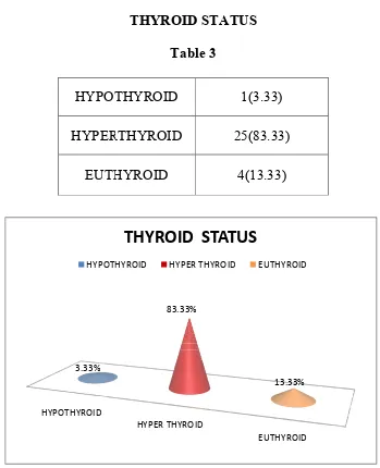
[image:54.612.135.484.71.501.2]THYROID STATUS
Table 3
HYPOTHYROID 1(3.33)
HYPERTHYROID 25(83.33)
EUTHYROID 4(13.33)
In our study the disease process was most common in 83.33%
3.33% in Hypothyroid patient and 13.33% in
and these parameters were comparable with the study
conducted by Prummel et al.40Another study conducted by Bartley et al
showed that Graves ophthalmology was more frequent in Hyperthyroidism
(90%),3% in Hypothyroidism and 6% in Euthyroid which was also
orrelating with our study.44
ocess was most common in 83.33% in
3.33% in Hypothyroid patient and 13.33% in
and these parameters were comparable with the study
Another study conducted by Bartley et al
showed that Graves ophthalmology was more frequent in Hyperthyroidism
SYMMETRICAL B/L
21(70%)
70% patients had bilateral and symmetrical involvement whereas
30% presented asymmetrically. The study was comparable with the study
conducted by Bartley et al
ASYMMETRICAL 30%
[image:55.612.136.496.78.505.2]LATERALITY
Table 4
SYMMETRICAL B/L ASYMMETRICAL B/L
21(70%) 9(30%)
70% patients had bilateral and symmetrical involvement whereas
30% presented asymmetrically. The study was comparable with the study
et al13.
SYMMETRICAL B/L 70% ASYMMETRICAL 30%
LATERALITY
ASYMMETRICAL B/L70% patients had bilateral and symmetrical involvement whereas
CLINICAL MANIFESTATION
Table 5
CLINICAL MANIFESTATION NO OF
PATIENTS PERCENTAGE
LID SIGNS 30 100%
SOFT TISSUE INFLAMMATION WITH CAS >4
OCULAR MOVEMENT
RESTRICTION
15
15
50%
50%
DIPLOPIA 3 10%
ACTIVE STAGE MILD MODERATE SEVERE 15 9 6 50% 30% 20%
OPTIC NERVE COMPRESSION - -
DIFFERENTIAL IOP >4mm 6 20%
PROPTOSIS >23mm 6 20%
IMAGING-CT ORBIT
EOM ENLARGEMENT 12 40%
FAT HYPERTROPHY 3 10%
RISK FACTORS
SMOKING 9 30%
30
15 15
3
6 6
9
2
100% 50% 50% 10% 20% 20% 30% 6.66%
CLINICAL MANIFESTATION
NO OF CASES PERCENTAGE
In our study proptosis with lid retraction was most common initial
presentation .Subjective symptoms like Oppressive Retro orbital feeling
and pain on eye movement was present in all our patients. (mouritus et
al1989,bartley et al1996b)40.
Objective signs like Conjunctival congestion was present in 73.33%
and eyelid swelling in 33.33%.Severe proptosis >23mm was present in
20%.Extraocular movement restriction was present in 50% but 10%
presented with intermittent diplopia, the differential IOP elevation >4mm
Bartley et al 1996b which states that 50% of orbitopathy presents with
motility restriction .13
Soft tissue features were present in 50% of our patients which was
compared to the study by kendler et al 1993 and Bartley et al 1996b13,
which showed association of soft tissue features in 34-75%.
In our study, 83.33% of patients presented with 6/6 Vision with
correction, 6/18-6/12 in 13.33% which was due to cataractous changes
and 3.33% with vision <1/60 which was due to associated Retinitis
pigmentosa and cataract .No patient presented with vision loss due to optic
nerve compression or corneal involvement in our study.
Patients were categorized as mild by insidious onset,lid lag,lid
retraction,minimal proptosis,moderately active based on lid signs,soft
tissue changes,moderate proptosis with intermittent myopathy,imaging
showing disproportionate proptosis with mild extraocular
enlargement.severe stage is characterized by rapid onset with predominant
inflammatory and mass effects,progressive myopathy and compressive
optic neuropathy, imaging showing evidence of extraocular muscle
CLINICAL ACTIVITY
Patients presenting with clinical activity score >4 was categorized as
mild stage in 50% of patients, moderately active in 30% and severe disease
in 20% of patients. Our study was correlating with the study conducted by
Bartley et al13.
50%
30% 20%
MILD STAGE
MODERATE ACTIVE STAGE
SEVERE ACTIVITY
LID EDEMA PERIOBITAL PUFFINESS
CONJUNCTIVAL CONGESTION GOITER
EXTRAOCULAR MOVEMENTS RESTRICTION
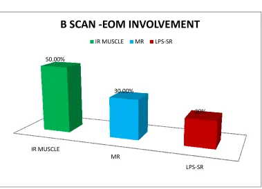
IR MUSCLE 50.00%
B SCAN
EOM THICKENING
IR MUSCLE
MR MUSCLE
LPS-SR
MR
LPS-SR 30.00%
20%
B SCAN -EOM INVOLVEMENT
IR MUSCLE MR LPS-SR
[image:64.612.127.506.390.664.2]B SCAN
Table 6
NO OF PATIENTS PERCENTAGE
15 50%
9 30%
6 20.00%
PERCENTAGE
The B scan was done in longitudinal mode and the extraocular
muscle thickness was compared with normative data given by Byrene et al.
Inferior rectus was most frequently involved about 50% followed by
medial rectus 30% and LPS-SR in 20% of our patients. Our study was
comparable to the study by EVnagi et al which showed that the inferior
rectus (93%) were the most frequently enlarged ,Medial, lateral and
superior rectuses were enlarged in 59%, 37% and 34% of the orbits
B SCAN
TENDON SPARING MR MUSCLE ENLARGEMENT
TENDON SPARING MUSCLE ENLARGEMENT
MEDIAL RECTUS
40% 10%
CT SCAN FINDING
EOM ENLARGEMENT FAT HYPERTOPHY
CT ORBIT AXIAL AND CORONAL VIEW
CT orbit was done for all the patients. Extraocular muscle
enlargement was present in 40% patients and fat hypertrophy in 10% of
patients. The fat hypertrophy was more frequent in young individual. Our
study correlating with the study, Graves Orbitopathy - Current Imaging
procedures by Berhard Krish et al.26
CT ORBIT AXIAL SCAN
CT CORONAL VIEW
FAT HYPERTROPHY
BIOCHEMICAL ANALYSIS
Biochemical investigation included TFT, IL-6, HS-CRP. The serum
samples of patients in moderate and severe stage were taken and IL-6 and
HS-CRP was done using ELISA technique and the results were compared
with control group without TED.
The results were analyzed using ANOVA followed by TURKEY
HSD test. The P value was significant at 5% level for both IL-6 and
HS-CRP30 in severely active patients and was not significant in moderate
activity group. The mean difference was significant at 0.05 level when
severe group was compared with control group and not significant when
moderate group was compared with control group. There was no
correlation between IL-6 and HS-CRP in the same group. Similar study
conducted by Prummel et al states that proinflammatory cytokines like
IL-1b, IL-6 and IL-10 are elevated in active TED when compared to inactive
stage.38 Another study by Molnar and Balazs found that significantly
increased serum IL-6 was found in Graves Ophthalmopathy and suggested
that IL-6 may be an important factor in the inflammatory events of Graves’
ANOVA FOLLOWED BY TURKEY HSD TEST
IL-6
Activity N Mean Standard
deviation P value
Severe 6 102.5317 150.58581
0.013*
Moderate 9 2.0311 .41093
Control 18 15.4328 26.81282
Total 33 27.6139 72.42253
Note :* denotes significance at 5% level.
POST HOC TEST: TURKEY HSD
(I)group (J)group Mean
difference Standard error P value
Severe
Moderate 100.5006(*) 34.10272 0.016*
control 34.10272 30.50240 0.021*
Moderate control -13.4017 26.41586 0.868
HS-CRP
ANOVA
Activity N Mean Standard
deviation P value
Severe 6 4.9233 4.69085
0.041*
Moderate 9 3.3444 3.22068
Control 18 1.7722 .85530
Total 33 2.7739 2.81818
POST HOC TEST : TURKEY HSD
(I)group (J)group Mean difference
Standard
error P value
Severe
Moderate 1.5789 1.37921 0.495
control 3.1511(*) 1.23360 0.041*
Moderate control -1.5722 1.06833 0.319
*The mean difference is significant at 0.05 level.
CORRELATIONS
Activity Correlation between IL6 and HS
-CRP P Value
Severe -0.399 0.433
Moderate 0.054 0.891
Control 0.150 0.553
PITFALLS
Though IL-6 and HS-CRP was statistically significant in severe
disease, the role of these investigation in moderate disease could not be
assessed because the sample size was small in our study.IL-6 is ELISA
based analysis which is expensive and needs trained personnel to perform
the procedure. So it could not be routinely used to screen the patients for
activity of the disease.
HS-CRP is nonspecific inflammatory marker and again its role in
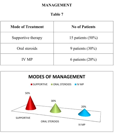
SUPPORTIVE 50%
MODES OF MANAGEMENT
SUPPORTIVE
Mode of Treatment
Supportive therapy
Oral steroids
IV MP
The patients in Mild stage
therapy like topical lubricants and head end elevation.30% of patients
moderately active stage were treated with oral steroids and 20% of them in
severe stage with IV Methyl Prednisolone pulse therapy 1gram in 500ml of
Normal saline for 3 days followed by oral steroids 40
followed weekly and assessed fo
ORAL STEROIDS
IV MP 30%
20%
MODES OF MANAGEMENT
SUPPORTIVE ORAL STEROIDS IV MP
[image:77.612.125.501.73.511.2]MANAGEMENT
Table 7
Treatment No of Patients
Supportive therapy 15 patients (50%)
Oral steroids 9 patients (30%)
6 patients (20%)
he patients in Mild stage (50%) were treated with supportive
therapy like topical lubricants and head end elevation.30% of patients
moderately active stage were treated with oral steroids and 20% of them in
severe stage with IV Methyl Prednisolone pulse therapy 1gram in 500ml of
Normal saline for 3 days followed by oral steroids 40-60mg. Patients were
followed weekly and assessed for disease activity for first 4 weeks.
(50%) were treated with supportive
therapy like topical lubricants and head end elevation.30% of patients in
moderately active stage were treated with oral steroids and 20% of them in
severe stage with IV Methyl Prednisolone pulse therapy 1gram in 500ml of
Patients were
Response to treatment
Remission
Exacerbation
RESPONSE TO TREATMENT
[image:78.612.133.493.97.426.2]FOLLOW UP
Table 8
Response to treatment No of patients
Remission 26 (86.66%)
Exacerbation 4(13.33)
87% 13%
RESPONSE TO TREATMENT
FOLLOW UP
The patients in mild stage who were treated with supportive
management were followed up every 6 months, moderate and severely
active patients who were put on either oral or intravenous steroid
respectively were followed up weekly and biweekly respectively till 4
weeks to assess the response to treatment. Visualacuity, slit lamp
examination, differential IOP, fields, colour vision, Hertels
exophthalmometry, Blood Pressure recording, Random Blood glucose
and systemic side effects of steroid therapy were monitored. Follow up
showed remission in 86.66% patients in mild and moderately active stage
where as 13.33% patients in severe stage treated with IV
Methylprednisolone pulse therapy showed exacerbation after
6months.Those patients were treated with pulse therapy again, followed by
oral steroids.Remission was attained in all our patients with steroid
treatment. The study conducted by Thambe K Bargawa concluded that IV
Methylprednisolone is more effective in moderate and severely active TED
patients.39Eyelid retraction remained the same in all patients and were
symptomatically better, inflammatory signs were reduced and disease
progression was curtailed. The patients were then followed up monthly for
RESULTS
A total of 30 patients in the age group of 20-60years with thyroid
eye disease were studied over the period of two years for age at
presentation, sex incidence, presenting clinical features, thyroid status and
disease activity, associated risk factors. All the patients underwent detailed
clinical evaluation, supportive investigation was done and treatment was
recorded.
1. Of the 30 cases examined, most common age group was between
20-30years (33.33%), females (60%) are most commonly affected
than males.
2. The disease process was most common in Hyperthyroid( 83.33%)
patients as compared to Hypothyroid (3.33%) and Euthyroid status
(13.33%).Risk factors associated with TED in our study was
smoking in 30% and Diabetes Mellitus in 6.66%
3. 70% patients had bilateral and symmetrical involvement whereas
30% presented asymmetrically.
5. Subjective symptoms like oppressive retro orbital feeling and pain
on eye movement was present in all our patients.
6. Objective signs like Conjunctival congestion was present in 73.33%
and Eyelid swelling in 33.33%.Severe proptosis >23mm was
present in 20% and soft tissue features were present in 50% of our
patients.
7. Extraocular movement restriction was found in 50% of patients in
moderate and severely active patient, associated with diplopia only
in 10% patients, and differential IOP of >4mm difference in about
20% 0f them.
8. Visual acuity with Snellen chart showed 6/6 in 83.33%, 13.33%
patients had 6/18-6/12 due to cataractous changes, 3.33% patients
with vision <1/60 which was due to associated Retinitis Pigmentosa
and cataract.
9. Patients presenting with clinical activity score >4 were categorized
as moderately active in 30%, severe disease in 20%, mild stage in
10.CT ORBIT axial and coronal view showed tendon sparing muscle
enlargement in 40% of patients and fat hypertrophy in 10% of
patients. Fat hypertrophy was more common in younger patient.
11.B SCAN- Tendon sparing EOM thickening was found in
longitudinal mode and the maximum muscle belly was measured,
corresponding low eye muscle reflectivity was noted in A scan.
Inferior rectus was most frequently involved (50%) followed by
medial rectus (30%) and LPS –SR complex in (20%).
12.The patients in moderate and severely active stage, IL-6 and
HS-CRP was done which was compared with control group without
TED and the results were analyzed using ANOVA followed by
TURKEY HSD test. The P value was significant at 5% level for
both IL-6(0.013) and HS-CRP(0.041) in severely active patients and
was not significant in moderate activity group. The mean difference
was significant at 0.05 level with the P value of .016 when severe
group was compared with moderate and control group and not
13.50% of the patients presented as mild disease who were treated with
supportive therapy. Moderatly active patients (30%) were treated
with oral steroids and 20% of the patients in severely active phase
were treated with pulse Intravenous Methylprednisolone followed
by oral steroids.
14.Follow up showed remission in 86.66% patients in mild and
moderately active stage where as 4 patients in severe stage treated
with iv methylprednisolone pulse therapy showed exacerbation after
6months.Those patients were treated with pulse therapy again,
followed by oral steroids. Remission was attained in all our patients
with steroid treatment, Eyelid retraction remained the same in all
patients.
CONCLUSION
1. Clinical assessment remains the paramount importance in diagnosing
the activity in Thyroid Eye Disease although controversies do exist in
clinical evaluation and management.
2. A scan with orbital B scan aids in the diagnosis of thyroid eye disease
in active stage, which is very economical with relatively short
examination time and no risk of radiation .Follow up of the patients
can also be performed easily.
3. IL-6 and HS-CRP was statistically significant at 5% in patients with
severe disease when compared to control group. The correlation in
between HS-CRP and IL-6 in all three groups were not significant.
Though the parameters shows significance for severe disease, its role
in moderate disease could not be assessed.
4. Identifying the disease activity early and aggressive management with
systemic steroids in moderately active and severe stage has decreased
BIBLIOGRAPHY
1. The thyroid gland - Endocrinology - NCBI Bookshelf (Nussey S,
Whitehead S.Endocrinology: An Integrated Approach. Oxford: BIOS
Scientific Publishers; 2001.Bookshelf ID: NBK28)
2. Diseases of the Orbit: A Multidisciplinary Approach, 2nd Edition,
Rootman, Jack
3. Garrity, Bahn, Pathogenesis of graves ophthalmopathy: implications for
prediction, prevention, and treatment, Am J Ophthalmol, 142,147-153,
2006
4. Prabhakar BS, Bahn RS, Smith TJ. Current perspective on the
pathogenesis of Graves’ disease and ophthalmopathy. Endocr Rev.
2003;24(6):802-35.
5. Immunopathogenesis of thyroid eye disease: emerging paradigms. Surv
Ophthalmol. 2010;55:215-26
6. The putative role of fibroblasts in the pathogenesis of Graves’ disease:
evidence for the involvement of the insulinlike growth
factor-1receptor in fibroblast activation, Autoimmunity, 36, 409-15, 2003.
7. Controversies in the clinical evaluation of active thyroid-associated
orbitopathy: use of a detailed protocol with comparative photographs
Endocrinology. Sep 2001, Vol. 55, No. 3: 283-303Allen, C., Stetz,
D., Roman, S.H., Podos, S., Som,
8. Davies,T.F. (1985) Prevalence and clinical associations of intraocular
pressure changes in Graves' disease. Journal of Clinical Endocrinology
and Metabolism, 61 183–187.
9. Immunology Of Thyroid Eye Disease:New Treatments On The
Horizon? Raymond S. Douglas, M.D., Ph.D.Jules Stein Eye
Institute/Ucla Los Angeles, Ca
10.Clinical activity score as a guide in the management of patients with
graves orbitopathy by maarten ph,mourits mark f.prummel 1997
11.Rundle F, Wilson C: Bulging of the eyelids with exophthalmos. Clin
Sci 1944; 5:31–45.
12.Bartley GB: The differential diagnosis and classification of eyelid
retraction.Trans Am Ophthalmol Soc 1995; 93:371–387.
13.Bartley GB, Fatourechi V, Kadrmas EF,et al: Clinical features of
Graves’ ophthalmopathy in an incidence cohort.Am J Ophthalmol
1996; 121:284–290.
14.Vardizer Y, Tomkins O, Briscoe D. Clinical assessment of thyroid
related orbitopathy: a review. Pediatr Endocrinol Rev. 2010;7 Suppl
15.An evidence-based approach to the treatment of Graves'
Ophthalmopathy Wilmar M. Wiersinga, Mark F.
Prummel.Endocrinology and Metabolism Clinics of North America.
Jun 2000, Vol. 29, No. 2: 297-319
16.Thomas HM Jr, Woods AC: Progressive exophthalmos following
thyroidectomy.Bulletin of The Johns Hopkins Hospital 1936; 59:99
17.Hosojima H, Uchida K: Availability of an anti-platelet aggregation
inhibitor,ticlopidine, in the treatment of Graves’ophthalmopathy. J
Drug Dev Clin Pract 1996:129–133.
18.Kahaly G, Pitz S, Muller-Forell W,Hommel G: Randomized trial of
intravenous immunoglobulins versus prednisolone inGraves’
ophthalmopathy. Clin Exp Immunol1996; 106:197–202.
19.Prummel MF, Mouritis MP, Berghout A, et al. Prednisolone and
cyclosporine in the treatment of severe Graves’ ophthalmopathy.N Engl
J Med. 1989; 321(20):1353-9
20.Hiromatsu Y. Steroid therapy for Graves’ ophthalmopathy.
NipponRinsho. 2006;64(12):2279-85 Antonelli A, Saracino A, Alberti
B, et al:
21.High-dose intravenous immunoglobulin treatment in Graves’
22.Prummel MF, Mourits MP, Blank L, et al: Randomized double-blind
trial of prednisone versus radiotherapy in Graves’ophthalmopathy.
Lancet 1993; 342:949–954.
23.Kahaly G, Roesler HP, Pitz S, et al. Low-versus high dose radiotherapy
for Graves’ ophthalmopathy: A randomized single blind trial. J Clin
Endocrinol Metab. 2000;85(1):102-8.
24.The eye and thyroid disease Ajay E Kuriyan, Richard P Phipps, Steven
E Feldon Current Opinion in Ophthalmology. Nov 2008, Vol. 19, No.
6: 499-506.
25.A mode ultrasound to assess disease activity in graves ophthalmopathy
by pummel et al(1993b ) found positive predictive value of 73% in
predicting the outcome of prednisolone or radiotherapy treatment.
26.Graves orbitopathy –current imaging procedure by Berhard Krish.
27.Investigation of ocular changes,extraocular muscle enlargement and eye
movements in Graves ophthalmopathy(medicina 2006).
28.Ultrasonic measurement of ocular recti muscle thickness in patients
with graves ophthalmopathy Medicina (Kaunas) 2010 ;46(7):472-6 .
29.Interleukin - 6 stimulates thyrotropin expression in human orbital
preadipocyte fibroblasts from patients with graves ophthalmopathy
30.Testing serum and plasma for thyroid function and the presence of
markers of autoimmunity including TSI,TRAB, c-reactive protein, and
fibrocyte index as objective markers for disease activity, severity and
progression.Laban-Guceva, Bogoev, Antova,
31.Serum concentrations of interleukin (IL-) 1alpha, 1beta, 6 and tumor
necrosis factor (TNF-) alpha in patients with thyroid eye disease (TED),
Med Arh, 61, 203-6, 200712. Smith.
32. Molnar I, Balazs C. High circulating IL-6 level in Graves’
ophthalmopathy. Autoimmunity 1997; 25:91-6.
33.Mourits MP, van Kempen-Harteveld ML,Garcia MB, et al:
Radiotherapy for Graves’ orbitopathy: randomised placebocontrolled
study. Lancet 2000;
34.Bartalena L, Marcocci C, Manetti L, et al. Orbital radiotherapy for
Graves’ ophthalmopathy. Thyroid. 1998;8(5):439-41.
35.Pinchera A, Bartalena L, Chiovato L, et al. Radiotherapy of Graves’
ophthalmopathy. In: Gorman CA, Waller RR, Dyer JA,editors. The eye
and the orbit in thyroid disease. New York: RavenPress; 1984:301-16
36.Department of Eye diseases,laboratory of ophthalmology,Institute for
biomedical research,Kaunas university of Medicine ,Lithuania.
37.Graves ophthalmopathy-Eye muscle involvement in patients with
38.TSH-R expression and cytokine profile in orbital tissue of active VS
inactive Graves ophthalmopathy patients . Clin Endocrinol (Oxf). 2003
Mar;58(3):280-7.
39.Role of IV Methyl prednisolonein the management of active thyroid
eye disease. Journal Orbit 29(5)227-231,2010 .
40.Mourits et al., 1989; Wiersinga, 1992; Kendler et al., 1993; Prummel
et al., 1993b; Kahaly et al., 1995; Bartalena et al., 2000.
41.Wiersinga WM, Bartalena L 2002 Epidemiology and prevention of
Graves’ ophthalmopathy. Thyroid 12:855–860.
42.Bartalena L, Marcocci C, Pinchera A 2002 Graves’ ophthalmopathy: a
preventable disease? Eur J Endocrinol 146:457–461.
43.Hagg E, Asplund K 1987 Is endocrine ophthalmopathy related to
smoking? Br Med J 295:634–635.
44.Clinical features of Graves' ophthalmopathy in an incidence cohort.
Bartley GB, Fatourechi V, Kadrmas EF, Jacobsen SJ, Ilstrup DM,
PROFORMA
Name:
Age: Sex: IP No: DOA: DOD:
Address:
Complaints: Onset, duration, rate of progression
Pain
Redness and edema of lids and conjunctiva
Photophobia
Diplopia
Forward protrusion of eyes
Defective vision
Past history:
Duration of thyroid dysfunction:
Symptoms pertaining to thyroid disorder:
Hypothyroidism--Weight gain/ Cold intolerance/ Fatigue/ constipation/
Hyperthyroidism—weight loss inspite of increased apetite /heat
intolerance/ restlessness/ diarrhea/ amnorrhoea/ irritability/ tremors and
palpitation Presence of swelling in the neck.
Treatment history:
Personal history: Smoking / diabetic/ Hypertention
General Examination: Nutrition Pallor
Cyanosis Clubbing
Tremors Icterus
Pulse rate Blood Pressure:
C.V.S
R.S
C.N.S
RE
Ocular examination Head posture Lid Lid signs Position Redness Swelling lagophthalmos Orbit Proptosis-axial/eccentric Hertels Exophthalmometry Pulsation Compressibility/reducibilityMass/resistance to retropulsion
Valsalva’s maneuver
Visual acuity by snellens chart
basal
value
RE
LE
Facial symmetry
RE LE
axial/eccentric
Hertels Exophthalmometry
Pulsation
Compressibility/reducibility
retropulsion
Slit lamp examination of anterior segment
Extraocular movements
Conjunctiva-congestion/chemosis/cauncle edema
Corne