0022-538X/97/$04.0010
Copyright © 1997, American Society for Microbiology
Isolation and Characterization of Human Papillomavirus Type
6-Specific T Cells Infiltrating Genital Warts
KENNETH HONG, CATHERINE E. GREER, NZEERA KETTER, GARY VAN NEST,
ANDXAVIER PALIARD*
Department of Virology and Vaccine Development, Chiron Corporation, Emeryville, California 94608
Received 18 February 1997/Accepted 2 June 1997
The potential role of T cells in the control of human papillomavirus type 6 (HPV-6) infections is an appealing premise, but their actual role has been sparsely investigated. Since HPV-6 infections are confined to the epithelium, such an investigation should focus on the T cells present at the site of infection (i.e., the warts). Therefore, we isolated wart-infiltrating lymphocytes (WIL) from patients with clinically diagnosed anogenital warts. These WIL were characterized by their phenotype and their specificity for E7 and L1 proteins of HPV-6. The phenotype of WIL varied drastically from patient to patient, as determined by their expression of CD4,
CD8, T-cell receptor alpha/beta chain (TCRab), and TCRgd. Despite this heterogeneity in phenotype, HPV-6
E7 and/or L1-specific WIL, as determined by lymphoproliferation, could be isolated from more than 75% of the patients studied. Among all L1 peptides recognized by WIL, peptides 311-330 and 411-430 were the most consistently detected, with seven of nine patients for whom L1 peptide reactivity was observed responding to at least one of them. Moreover, the HPV-6 epitopic peptides recognized by WIL differed to some extent from those recognized by peripheral T cells.
Human papillomaviruses (HPV) are small double-stranded DNA viruses that comprise more than 75 genotypes with con-siderable sequence variability. The incidence of HPV infection has risen eightfold in the last 30 years, with a reported 47% of U.S. college women carrying the virus (4). HPV infect mucosal or keratinized epithelium, and different genotypes have spe-cific tropism for different anatomical sites. For example, HPV type 16 (HPV-16) and HPV-18, -31, -33, and -45 are most commonly associated with cervical intraepithelial neoplasia, carcinoma, and invasive cervical cancer, whereas HPV-6, -11, -40, and -42 are the types most frequently found in anogenital warts (condylomata acuminata), with HPV-6 being the most prevalent (reviewed in reference 27). Anogenital warts caused by HPV infection do not exhibit a propensity for malignant transformation but cause great psychosocial morbidity.
Infection with HPV-6 probably occurs as a result of exposure of the basal cells to virus particles after minor trauma to the epithelium, probably occurring during sexual intercourse. The virus stimulates the proliferation of basal cells, resulting in the formation of a wart. Although all wart cells contain the viral genome, expression of the various viral genes is linked to the state of differentiation of the cells, with genes encoding for virus particles and capsid antigens expressed only in the most superficial layers of the epithelium (30).
The immunology of genital condylomata is poorly under-stood, due in part to the facts that the virus cannot be easily cultured and no suitable animal model exists. Recent reports indicate that only some subjects with genital warts mount a significant humoral response against virus-like particles (7, 11, 14). Even though this response in subjects that seroconvert is type specific, it does not predict the clinical course of the disease (14, 35, 36). In contrast, some clinical and histopatho-logical observations suggest that T-cell-mediated immunity
may play a role in determining the course of HPV infection. These include a correlation between spontaneous regression of warts and infiltration of mononuclear cells into the dermis and epidermis (9, 33) and an increased incidence of HPV lesions in immunocompromised patients (10, 17, 21, 23, 24). Some his-tological evidence also suggests a role for cellular immune responses in wart regression, since in regressing plane warts caused by HPV-3, an infiltration of T cells and monocytes was observed in proximity to the damaged keratinocytes (16). Fi-nally, a delayed-type hypersensitivity response to an HPV-16 L1 fusion protein was observed in 5 of 7 patients with cervical intraepithelial neoplasia attributed to HPV-16 or -18 infection but in none of 10 uninfected patients (15).
To develop immunologically based strategies to treat or pre-vent HPV-6 infections, viral targets that can be recognized by the immune system must be identified. Since HPV-6 infection is confined to the epithelium, an analysis of the potential in-volvement of T cells should focus on T cells isolated from the site of the disease (i.e., the wart). The study reported here is the first in which wart-infiltrating lymphocytes (WIL) were isolated and phenotypically characterized and the viral epi-topes that they recognized were identified.
MATERIALS AND METHODS
Isolation of PBMC.Peripheral blood mononuclear cells (PBMC) from
pa-tients with anogenital warts were isolated by centrifugation over a Ficoll-Hypaque gradient. PBMC were either used directly or cryopreserved in liquid nitrogen.
HLA typing.HLA typing of patients was performed by the HLA and
Immu-nogenetics Laboratory, Los Angeles, Calif., using serological methods.
HPV typing by PCR.The L1 consensus PCR system (primers MY09 and
MY11) was used to determine the HPV types in each excised wart. This method has been previously described (3).
HPV-6 L1 serology.Sera from the patients were tested by enzyme-linked
immunosorbent assay for the presence of anti-HPV-6 L1 virus-like particle antibodies (10a).
Isolation and culture of wart-infiltrating T cells.After local anesthesia, genital
warts were shaved off as close to the base as possible, using a sterile scalpel blade. These wart biopsies (if multiple warts from a single patient were available, they were treated separately) were cut into small pieces (#1mm2) and placed in
AIM-V medium (Gibco, Grand Island, N.Y.) supplemented with 1% human AB
* Corresponding author. Mailing address: Chiron Corporation, De-partment of Virology and Vaccine Development, 4560 Horton St., Emeryville, CA 94608. Phone: (510) 923-3545. Fax: (510) 923-4265. E-mail: [email protected].
6427
on November 9, 2019 by guest
http://jvi.asm.org/
serum (Gemini, Calabasas, Calif.) (the lot of human serum used did not contain antibodies to HPV-6 L1 virus-like particles [10b]), 1% penicillin-streptomycin, 200 U of interleukin-2 (IL-2; Chiron) per ml, and 5% T-STIM without phyto-hemagglutinin (Collaborative Biomedical Products, Bedford, Mass.). WIL started to migrate out of the tissue by days 5 to 10. WIL were expanded in the absence of any stimuli (i.e., antigen, mitogen, or mitogenic antibody) unless specified.
Fluorescence-activated cell sorting analysis.Cells were incubated for 15 min
at room temperature in phosphate-buffered saline supplemented with 10% hu-man serum, 10 mg of huhu-man immunoglobulin G (Sigma, St. Louis, Mo.) per ml, and 0.1% NaN3to block potential Fc receptors. After two washes in
phosphate-buffered saline–NaN3, cells were incubated with fluorescein isothiocyanate- or
phycoerythrin-labeled anti-CD4, anti-CD8, anti-T-cell receptor alpha/beta chain (TCRab), or anti-TCRgdmonoclonal antibodies (all from Pharmingen, San Diego, Calif.) for 30 min at 4°C. After three additional washes, cells were analyzed on a Coulter EPICS cell sorter.
Proliferation assays.WIL isolated from the biopsies were tested for the ability
to proliferate in a standard lymphoproliferation assay. Briefly, WIL were plated in triplicates at 53104
cells per well in 96-well round-bottom plates. Cells were cultured in medium alone (AIM-V, 1% human AB serum, 1% antibiotics) or in the presence of the autologous Epstein-Barr virus-transformed B-cell line (53 104
cells per well) irradiated at 4,000 rads with or without peptide/protein. RecombinantEscherichia colipurified HPV-6b E7 protein (referred to as HPV-6 E7), recombinant yeast purified HPV-6 L1 virus-like particles (both kindly pro-vided by B. Gervase), and HPV-6 E7/L1 peptides spanning the entire lengths of HPV-6 E7 and HPV-6 L1 proteins (20-mers overlapping by 10 amino acids; Chiron Mimotopes) were used at 10mg/ml. After 72 h, plates were pulsed with 1mCi of [3
H]thymidine per well, and cells were harvested 6 to 8 h later. The PBMC proliferative response to HPV-6 proteins was assessed as follows. PBMC were plated at 23105
cells per well, in 24-plicates, in 96-well round-bottom plates in the presence or absence of E7 and L1 proteins. Plates were pulsed with 1mCi of [3
H]thymidine per well after 120 h for 6 to 8 h. For peptide identification, frozen PBMC were thawed, restimulated at 106
per ml with HPV-6 E7 or L1, and cultured for 10 to 15 days. Cells were then plated in triplicates at 53104
cells per well in 96-well round-bottom plates in the presence of 53104
autologous Epstein-Barr virus-transformed B-cells irradiated at 4,000 rads with or without peptide (all at 10mg/ml). Plates were pulsed with 1mCi of [3
H]thymidine per well 72 h later for 6 to 8 h, and cells were harvested with a Tomtec cell harvester.
Results are presented as a stimulation index (SI) calculated as mean experi-mental counts per minute/mean counts per minute in the absence of antigen. WIL were scored positive for an SI of$2.0 andDcpm of$3,000, and PBMC were scored positive for an SI of$3.0 andDcpm of$3,000.Dcpm was calculated as mean experimental counts per minute2mean counts per minute in the absence of antigen. Different SIs were chosen as a cutoff for positivity in PBMC and WIL, since a higher background was observed for PBMC than for WIL.
RESULTS
Patient characteristics.The characteristics of the patients
from whom WIL were isolated are presented in Table 1. Pa-tients were recruited from three different clinics and were part of a study designed in part to evaluate the natural immune response in patients with genital warts. For this particular study (of cells infiltrating warts), 24 patients were entered consecutively on the basis of diagnosis (2 to 20 measurable genital warts and a total wart area of#10 cm2), availability of
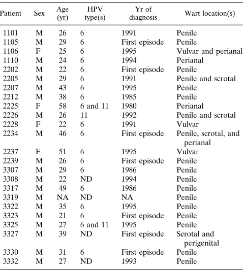
wart biopsy, and availability of peripheral blood. None of the patients had undergone any wart-specific therapy in the 10 weeks prior to wart and blood sample collection. Four of these patients were female, 20 were male, and all had clinically defined anogenital warts. In 17 of 24 (71%) of these patients, only HPV-6 was detected by PCR in the anogenital warts. In 2 of 24 (8%) (patients 2225 and 3325), both HPV-6 and HPV-11 were detected, and in 1 of 24 (4%) (patient 2226), only HPV-11 was present. HPV typing of 4 of 24 patients (17%) could not be determined due to insufficient DNA (Table 1). These data confirmed that HPV-6 is the most prevalent HPV type associated with anogenital warts.
Phenotypes of WIL. The phenotypes of WIL (originating
from a single wart) from these 24 subjects were characterized by immunofluorescence. The data presented in Fig. 1 indicate that the lymphocyte populations present in most warts differed from those present in the periphery, since in the peripheral blood lymphocytes of adults, CD41 cells, CD81 cells,
TCRab1cells, and TCRgd1cells represent about 42% (range, 38 to 46%), 35% (range, 31 to 40%), 69% (range, 63 to 73%), and 3% (range, 1 to 10%), respectively (6, 13). Also, the phenotype of WIL differed significantly from patient to pa-tient. CD41T cells represented more than 80% of the infil-trating lymphocytes in 9 of 24 patients (38%), whereas in 5 of 24 patients (21%), the majority ($60%) of WIL were of the CD8 phenotype (Fig. 1A). When the TCR phenotype of these cells was analyzed, a similar variability between patients was observed (Fig. 1B). In 13 of 24 patients (54%), WIL were mostly ($80%) TCRab1. On the other hand, WIL from 8 of 24 patients (33%) consisted of more than 20% TCRgd1 T cells, with TCRgd1T cells making up over 50% of the total WIL in 3 of these patients (Fig. 1B). Figure 1B also indicates that all but two WIL cultures contained mostly T cells. WIL from patients 1110 and 3322 had, 33 and 59%, respectively, TCRab2TCRgd2cells. These cells were unlikely to be mac-rophages/monocytes, since their side scatter-versus-forward scatter profile was similar to that of TCRab1 and TCRgd1 cells (data not shown).
Two distinct wart biopsies were obtained from one patient (1106). Notably, the WIL from both warts were 99% CD41 and 99% TCRab1(data not shown). Unfortunately, the lim-ited availability of PBMC from this patient did not allow us to conduct any further characterization of these cells.
[image:2.612.317.556.80.347.2]WIL and PBMC proliferative responses to HPV-6 E7 and L1.HPV-6 E7 and L1 antigens were tested for the ability to stimulate WIL and PBMC isolated from 13 of these patients in an initial proliferation assay. The lymphoproliferative response to HPV-6 antigens could not be tested for the PBMC of two
TABLE 1. Donor characteristicsa
Patient Sex Age (yr)
HPV type(s)
Yr of
diagnosis Wart location(s)
1101 M 26 6 1991 Penile 1105 M 29 6 First episode Penile
1106 F 25 6 1995 Vulvar and perianal 1110 M 24 6 1994 Perianal
2202 M 22 6 First episode Penile
2205 M 29 6 1991 Penile and scrotal 2207 M 43 6 1995 Penile
2212 M 38 6 1985 Penile 2225 F 58 6 and 11 1980 Perianal 2226 M 26 11 1992 Penile and scrotal 2228 F 22 6 1991 Vulvar
2234 M 46 6 First episode Penile, scrotal, and perianal 2237 F 51 6 1995 Vulvar 2239 M 26 6 First episode Penile 3307 M 29 6 1986 Penile 3308 M 22 ND 1994 Penile 3317 M 49 6 1986 Penile 3319 M NA ND NA Penile 3322 M 35 6 1995 Penile 3323 M 21 6 First episode Penile 3325 M 27 6 and 11 1995 Penile 3327 M 39 ND First episode Scrotal and
perigenital 3330 M 31 6 First episode Penile 3332 M 27 ND 1993 Penile
aPatients were monitored at Solano Dermatology Clinic, Vallejo, Calif.
(pa-tients 11XX); University of Texas—Medical Branch Center for Clinical Studies, Galveston, Tex. (patients 22XX); and Harbor View Medical Center, Seattle, Wash. (patients 33XX). All patients had clinically diagnosed anogenital warts but were otherwise healthy. None of the patients had undergone any therapy for their warts at least 10 weeks prior to sampling. M, male; F, female; ND, not
determined; NA, not available.
on November 9, 2019 by guest
http://jvi.asm.org/
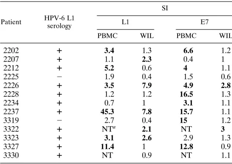
donors (3322 and 3330) due to sample unavailability. Both E7 and L1 antigens were capable of eliciting a significant prolif-erative response in a number of cases. WIL isolated from 39% (5 of 13) of the patients tested responded to HPV-6 L1 protein, and WIL from 15% (2 of 13) of the patients responded to HPV-6 E7 protein. Also, PBMC from 55% (6 of 11) of the patients responded to L1, and PBMC from 73% (8 of 11) of the patients responded to E7 (Table 2).
T-cell epitopes recognized by WIL.To determine which
re-gions of the HPV-6 E7 and L1 proteins were recognized by WIL, peptides (20-mers overlapping by 10 amino acids) span-ning the entire sequences of these two proteins were synthe-sized. The resulting 58 peptides (9 for E7 and 49 for L1) were tested independently for the ability to stimulate WIL. As shown in Table 3, E7 epitopic peptides could be identified in only 2 of 13 patients (15%): WIL from patient 2226 recognized E7 peptide 1-20, and WIL from patient 3322 were induced to proliferate in response to three E7 peptides (11-30, 41-60, and 51-70).
In contrast, L1 epitopic peptides recognized by WIL could be identified in 9 of 13 patients (69%) (Table 3). Most of these peptides were located in the second half of the HPV-6 L1 protein. In most cases, only a few L1 epitopes were recognized by WIL isolated from a single wart, inasmuch as only 1 or 2 peptides of the 49 representing the entire L1 protein stimu-lated WIL isostimu-lated from seven of nine patients from whom L1 epitopic peptides could be identified (Table 3). However, one patient (2225) recognized three contiguous L1 peptides (201-220, 221-240, and 241-260), thereby defining a discrete, highly immunogenic region in the L1 protein for this particular pa-tient. A response to multiple, nonadjacent L1 peptides was observed only for the WIL of a single patient (2237) that recognized seven L1 peptides (51-70, 111-130, 241-260, 411-430, 421-440, 431-450, and 471-490) (Table 3). The results in Table 3 also indicate that the majority (seven of nine, or 78%) of WIL responding to L1 peptides reacted to at least one of the L1 peptides 331-330 and 411-430, suggesting that these were dominant T-cell epitopes in most patients. Table 3 also indi-cates that, for at least the small number of patients tested, there did not seem to be an association between the HLA type of the patient and his or her ability to mount a T-cell response to E7/L1 peptides.
In summary, these data indicated that WIL isolated from the
warts of 10 of 13 patients (77%) responded to L1 protein and/or peptides. In addition, WIL isolated from 2 of 13 pa-tients (15%) responded to HPV-6 E7 protein and/or peptide, whereas WIL isolated from only 3 of 13 patients (23%) showed no response to any of the proteins and/or peptides tested.
Comparison of the T-cell epitopes recognized by peripheral
blood and wart-infiltrating T cells. To find out whether
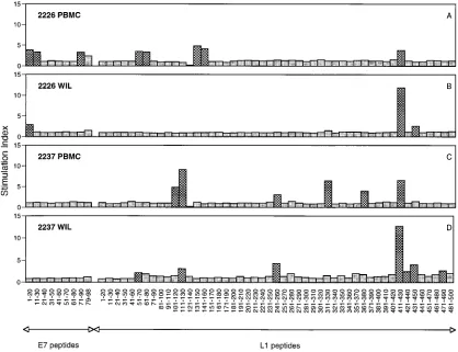
[image:3.612.127.485.67.239.2]pe-ripheral and wart-infiltrating T cells recognized the same pep-tides, we compared the responses of PBMC and WIL to the full set of 58 peptides corresponding to HPV-6 E7 and L1 in four patients. The results obtained from two of these patients are presented in Fig. 2. These data indicated that some iden-tical HPV-6 E7 and L1 epitopic peptides were recognized by both WIL and PBMC. The T cells isolated from both the wart and the periphery of patient 2237 recognized L1 peptides 111-130, 241-260, and 411-430 (Fig. 2C and D). Similarly, WIL and PBMC of patient 2226 recognized E7 peptide 1-20 and L1 peptide 411-430 (Fig. 2A and B). However, a number of dis-parities were present. The WIL but not the PBMC of patient 2237, for example, responded to HPV-6 L1 peptides 51-70,
TABLE 2. Comparison of lymphoproliferative responses of WIL and PBMC to HPV-6 E7 and L1 proteins
Patient HPV-6 L1 serology
SI
L1 E7
PBMC WIL PBMC WIL
2202 1 3.4 1.3 6.6 1.2
2207 1 1.1 2.3 0.4 1
2212 1 5.2 0.6 4 1.1
2225 2 1.9 0.4 1.5 0.6
2226 1 3.5 7.9 4.9 2.8
2228 1 1.2 1.2 16.5 1.3
2234 1 0.7 1 3.1 1.1
2237 1 45.3 7.8 15.7 1.1
3319 2 2.7 0.4 15 1.2
3322 1 NTa 2.1 NT 3
3323 1 3.1 2.6 2.9 1.3
3327 1 11.4 1 12.8 0.9
3330 1 NT 0.9 NT 1.1
aNT, not tested due to lack of PBMC.
FIG. 1. Phenotypic analysis of WIL. WIL from 24 patients were analyzed for expression of CD4, CD8, TCRab, and TCRgd. Each filled dot represents the percentage of WIL from a single donor (and a single wart) expressing CD4 and CD8 (A) or TCRaband TCRgd(B). Each open dot represents the usual percentage of peripheral blood lymphocytes expressing CD4 and CD8 (A) or TCRaband TCRgd(B).
on November 9, 2019 by guest
http://jvi.asm.org/
[image:3.612.316.555.549.718.2]421-440, 431-450, and 471-490. Conversely, the PBMC but not the WIL of this patient responded to HPV-6 L1 peptides 101-120, 311-330, and 361-380 (Fig. 2C and D).
DISCUSSION
The cell-mediated immune response to HPV-6 infections is poorly understood. Identification of HPV-6 determinants rec-ognized by WIL is crucial not only for a better understanding
of natural immunity to HPV-6 but also for the development of prophylactic and therapeutic immunological strategies. Some HPV determinants recognized by T cells have been described for particular HPV types; one report describes epitopes for the HPV-11 E7 proteins (34), and others describe T-cell epitopes for HPV-16, -18, -2, and -1 (1, 2, 8, 18, 20, 25, 28, 29, 32). All of these studies were conducted with peripheral blood lympho-cytes isolated from infected or asymptomatic individuals. How-ever, HPV infections are localized to the epithelium. There-TABLE 3. T-cell epitopes recognized by wart-infiltrating T cells
Patient HLA type Peptide(s)
a
E7 L1
2202 A2,3 B57,60 Cw3,w7 DR4,7 DQ2,7 None None
2207 A3,2B7,2Cw7,2DR14,15 DQ1,2 None 411
2212 A1,68 B7,8 Cw7,2DR15,17 DQ1,2 None 311
2225 A24,69 B7,35 Cw4,w7 DR8,11 DQ4,7 None 201, 221, 241
2226 A2,3 B35,62 Cw3,w4 DR1,8 DQ1,4 1 411, 431
2228 A2,30 B15,18 Cw3,w5 DR4,18 DQ4,8 None None
2234 A3,2B7,14 Cw7,w8 DR1,2DQ1,2 None 311
2237 A1,31 B8,44 Cw2,w7 DR1,11 DQ1,7 None 51, 111, 241, 411, 421, 431, 471
3319 A1,11 B7,57 Cw6,w7 DR6,15 DQ1,2 None None
3322 A24,2B35,2Cw1,w4 DR4,2DQ8,2 11, 41, 51 None
3323 A23,26 B57,2Cw4,2DR8,11 DQ7,2 None 91
3327 A2,3 B53,60 Cw2,w3 DR8,13 DQ1,7 None 311, 411
3330 A2,3 B13,35 Cw4,2DR1,15 DQ1,2 None 311
aEach number refers to the first amino acid of a 20-mer peptide. The peptides were identified by using a lymphoproliferation assay as described in Materials and
[image:4.612.59.559.82.235.2]Methods.
FIG. 2. Comparison of the HPV-6 E7 and L1 epitopes recognized by WIL and PBMC. Positivity (dotted columns) was defined according to the criteria described in Materials and Methods.
on November 9, 2019 by guest
http://jvi.asm.org/
[image:4.612.98.516.390.711.2]fore, the potential role of T cells in modulating HPV infections seems more rigorously addressed by studying T cells present at the site of disease. Thus, in these experiments, WIL were isolated from patients with anogenital warts, characterized by phenotype and by the HPV-6 E7 and L1 epitopes that they recognized. Since we wanted to explore both the specificity of WIL and their potential involvement in controlling HPV in-fection, WIL were expanded in an IL-2 enriched medium in the absence of any antigenic or mitogenic stimulus. In this environment, the T cells most likely to expand were those expressing the IL-2 receptor; these T cells are likely to repre-sent the specific immune response to HPV, since they were presumably activated in vivo.
Staining of WIL with anti-CD4, -CD8, -TCRab, and -TCRgd monoclonal antibodies indicated that proportional representation of these T-cell subsets varied from patient to patient (Fig. 1). Infiltration of the warts by mononuclear cells has been previously observed in studies using immunohisto-chemical techniques (5, 9). Bishop et al. (5) found no differ-ence in the T-cell populations infiltrating regressing warts and nonregressing warts. Coleman et al. (9), however, found an increased infiltration of activated CD41 T cells and macro-phages in regressing warts compared to nonregressing warts. In this context, WIL from 38% of our patients were mostly (.80%) CD41(Fig. 1), and some were HPV-6 specific (Tables 2 and 3).
The immunological significance of TCRgd1T cells, which represent about 3% of peripheral T cells (6, 12), remains poorly understood. TCRgd1 T cells can recognize antigens derived from various microbial taxa (19), including herpes sim-plex virus (26) and Mycobacterium leprae (22), which, like HPV, infect the epithelium. Therefore, it is intriguing that they represented more than 20% of the WIL in 8 of 24 patients (33%) and more than 50% in three of these patients (Fig. 1B). However, whether these TCRgd1WIL are HPV-6 specific and play a role in the control of HPV infection remains to be determined.
The specificity of WIL was investigated by using a proliferation assay. For both E7 and L1, a significant lympho-proliferative response was observed in most (10 of 13) patients. Only WIL isolated from 3 of 13 patients showed no response to HPV-6 E7/L1 proteins or peptides (Tables 2 and 3). Ten of 13 patients (77%) for whom the presence of HPV-6-specific T cells was investigated were infected by HPV-6; one of the patients (2225) was infected with both HPV-6 and -11 (Table 1). One patient (2226) who responded to both HPV-6 E7 and L1 had HPV-11-infected warts (Tables 1 to 3). This observa-tion can be explained by the fact that the E7 and L1 proteins from HPV-6 and HPV-11 are 85.6 and 92.2%, respectively, identical. Alternatively, patient 2226 may have had no detect-able HPV-6 DNA at the sites assayed but could have had undetected sites of HPV-6 infection or previous exposure to HPV-6.
A number of different E7 and L1 epitopes recognized by WIL were identified in 15 and 69%, respectively, of the pa-tients (Table 3). Since E7 is expressed in all layers of infected epithelium but L1 is expressed only in terminally differentiated cells (30), it appears that the T-cell responses within the warts are directed mostly against terminally differentiated cells. WIL isolated from 78% of patients responding to L1 recognized at least one of the two HPV-6 L1 peptides (311-330 and 411-430) (Table 3). Response to these peptides (especially peptide 411-430) led often to a higher SI compared to all other peptides (Fig. 2 and data not shown). Taken together, these data sug-gest that these two L1 peptides represent dominant T-cell epitopes.
The epitopes recognized by peripheral and wart-infiltrating T cells differed to some extent since some epitopic peptides for a given patient were recognized only by WIL and not by PBMC. Conversely, some HPV-6 E7 and L1 peptides were recognized by peripheral T cells but not by WIL (Fig. 2). Thus, there appeared to be a preferential sequestration of some HPV-specific T-cell clonotypes in anogenital warts. Alterna-tively, the PBMC reactivity could reflect a memory response, while the WIL reactivity that is not shared by PBMC might indicate more recent responses that are not yet demonstrable in peripheral T-cell populations.
For the limited number of patients studied, no correlation was apparent between a T-cell response to HPV-6 proteins/ peptides and a given HLA allele, the presence of antibodies against HPV-6 L1, or the duration of disease (Tables 1 to 3). Moreover, since the studied warts were surgically removed, whether these warts would have spontaneously regressed can-not be known. Thus, whether HPV-6-specific WIL provide the right kind of help for clearing HPV infections or preventing recurrences awaits further studies, including some longitudinal studies. Also, we were unable to detect any WIL cytotoxic T-lymphocyte (CTL) activity against HPV-6 E7 and L1 unless cultures were restimulated with HPV-6 antigens multiple times (data not shown). This observation indicated that if HPV-6-specific CTL do infiltrate anogenital warts, their frequency is very low or they have some activity directed against HPV-6 proteins other than E7 and L1. Nevertheless, the absence of detectable CTL activity might be why some individuals appear unable to prevent the development of anogenital warts and that recurrence in these subjects is high (35 to 60% within 3 months) (31). In this context, it is worth noting that an in-creased incidence of warts has been found in immunocompro-mised patients (10, 17, 21, 23, 24). Conversely, cross-sectional epidemiological studies of normal adults have shown that the number of patients harboring HPV-6 or -11 is far greater than the number of patients with overt genital warts. This observa-tion suggests that in some individuals, the immune system can prevent development of lesions or of long-lasting lesions. Whether such subjects, in contrast to patients with recurrent or long-lasting lesions as in this study, are able to locally mount a strong CTL response against HPV-6 remains to be deter-mined. Nevertheless, one can postulate that immunological strategies inducing a strong CTL response against HPV-6 could be beneficial.
ACKNOWLEDGMENTS
We acknowledge Barbara Gervase for providing purified HPV-6 E7 and L1 proteins, Marty Giedlin for help with flow cytometry analyses, Kit Boggio for coordinating the study, D. B. Tyan for HLA typing of the patients, Stewart Cooper for critical review of the manuscript, and S. Tyring, K. Beutner, and H. Hansfield for providing the clinical samples.
REFERENCES
1.Altmann, A., I. Jochmus-Kudielka, R. Frank, H. Gausepohl, U. Moebius, L.
Gissmann, and S. C. Meuer.1992. Definition of immunogenic determinants
of the human papillomavirus type 16 nucleoprotein E7. Eur. J. Cancer
28:326–333.
2.Bartholomew, J. S., S. N. Stacey, B. Coles, D. J. Burt, J. R. Arrand, and P. L.
Stern.1994. Identification of a naturally processed HLA A0201-restricted
viral peptide from cells expressing human papillomavirus type 16 E6 onco-protein. Eur. J. Immunol.24:3175–3179.
3.Bauer, H. M., C. E. Greer, and M. M. Manos.1992. Detection of genital
human papillomavirus using PCR, p. 131–152.InC. S. Herrington and J. O. McGee (ed.), Diagnostic molecular pathology: a practical approach. Oxford University Press, Oxford, England.
4.Bauer, H. M., Y. Ting, C. E. Greer, J. C. Chambers, C. J. Tashiro, J.
Chimera, A. Reingold, and M. M. Manos.1991. Genital human
papilloma-virus infection in female university students as determined by a PCR-based method. JAMA265:472–477.
on November 9, 2019 by guest
http://jvi.asm.org/
5.Bishop, P. E., A. McMillan, and S. Flesher.1990. An immunohistological study of spontaneous regression in condylomata acuminata. Genitourin. Med.66:79–81.
6.Brenner, M. B., J. L. Strominger, and M. S. Krangel.1988. ThegdT cell
receptor. Adv. Immunol.43:133–192.
7.Carter, J. J., G. C. Wipf, M. E. Hagensee, B. McNight, L. A. Habel, S. Lee,
J. Kuypers, N. Kiviat, J. R. Daline, L. A. Koutsky, D. H. Watts, K. K.
Holmes, and D. Galloway.1995. Use of human papillomavirus type 6 capsids
to detect antibodies in people with genital warts. J. Infect. Dis.172:11–18.
8.Charleston, F. C., N. Norval, E. C. Benton, and J. A. A. Hunter.1992.
Lymphoproliferative responses to human papillomaviruses in patients with cutaneous warts. Br. J. Dermatol.127:551–559.
9.Coleman, N., H. D. L. Birley, A. M. Renton, N. F. Hanna, B. K. Ryait, M.
Byrne, D. Taylor-Robinson, and M. A. Stanley.1994. Immunological events
in regressing warts. Am. J. Clin. Pathol.102:768–774.
10. Gassenmaier, A. P., G. Fuchs, H. Schell, and H. Pfister.1986. Papillomavirus
DNA in warts of immunosuppressed renal allograft recipients. Arch. Der-matol. Res.278:219–223.
10a.Greer, C. E., et al.Unpublished data.
10b.Greer, C. E., and X. Paliard.Unpublished data.
11. Greer, C. E., C. M. Wheeler, M. B. Ladner, K. Butner, M. Y. Coyle, H. Liang,
A. Langenberg, T. S. B. Yen, and R. Ralston.1995. Human papillomavirus
type distribution and serological response to HPV type 6 virus-like particles in subjects with genital warts. J. Clin. Microbiol.33:2058–2063.
12. Groh, V., S. Porcelli, M. Fabbi, L. L. Lanier, L. J. Picker, T. Anderson, R. A.
Warnke, A. K. Bhan, J. L. Strominger, and M. B. Brenner.1989. Human
lymphocytes bearing T cell receptor gamma/delta are phenotypically diverse and evenly distributed throughout the lymphoid system. J. Exp. Med.169:
1277–1294.
13. Hannet, I., F. Erkeller-Yuksel, P. Lydyard, V. Deneys, and M. DeBruyere.
1992. Developmental and maturational changes in human blood lymphocyte subpopulations. Immunol. Today13:215–218.
14. Heim, K., N. D. Christensen, R. Hoepfl, B. Wartush, G. Pinzger, P.
Baum-gartner, J. W. Kreider, and O. Dapunt.1995. Serum IgG, IgM and IgA
reactivity to human papillomavirus types 11 and 6 virus-like particles in different gynecological subjects groups. J. Infect. Dis.172:395–402.
15. Ho¨pfl, R., M. Sandbichler, N. Sepp, K. Heim, E. Muller-Holzne, B.
War-tusch, O. Dapunt, I. Jochmus-Kudielka, J. ter Meulen, L. Gissmann, and P.
Fritsch.1991. Skin test for HPV type 16 proteins in cervical intraepithelial
neoplasia. Lancet337:373–374.
16. Iwatsuki, K., H. Tagami, M. Takigawa, and M. Yamada.1986. Plane warts
under spontaneous regression. Immunopathologic study on cellular constit-uents leading to the inflammatory reaction. Arch. Dermatol.122:655–659.
17. Johnson, J. C., A. F. Burnett, G. D. Willet, M. A. Young, and J. Doniger.
1992. High frequency of latent and clinical human papillomavirus cervical infections in immunocompromised human immunodeficiency-virus-infected women. Obstet. Gynaecol.79:321–327.
18. Kadish, A. S., S. L. Romney, R. Ledwidge, R. Tindle, G. J. P. Fernando, S. Y.
Zee, M. A. Van Ranst, and R. D. Burk.1994. Cell-mediated immune
re-sponses to E7 peptides of human papillomavirus (HPV) type 16 are depen-dent on the HPV type infecting the cervix whereas serological reactivity is not type-specific. J. Gen. Virol.75:2277–2284.
19. Kaufmann, S. H. E.1996.g/dand other unconventional T lymphocytes: what
do they see and what do they do? Proc. Natl. Acad. Sci. USA93:2272–2279.
20. Luxton, J. C., A. J. Rowe, J. C. Cridland, T. Coletart, P. Wilson, and P. S.
Shepherd.1996. Proliferative T cell responses to the human papillomavirus
type 16 E7 protein in women with cervical displasia and cervical carcinoma and in healthy individuals. J. Gen. Virol.77:1585–1593.
21. Melbye, M., J. Palefsky, J. Gonzales, L. P. Ryder, H. Nielsen, O. Bergmann,
J. Pindborg, and R. J. Biggar.1990. Immune status as a determinant of
human papillomavirus detection and its association with anal epithelial ab-normalities. Int. J. Cancer46:203–206.
22. Modlin, R. L., C. Pirmez, F. M. Hofman, V. Torigian, K. Uyemura, T. H. Rea,
B. R. Bloom, and M. B. Brenner.1989. Lymphocytes bearing antigen-specific
gdT cell receptors accumulate in human infectious disease lesions. Nature
339:544–548.
23. Orth, G.1987. Epidermodysplasia verruciformis, p. 199–235.InN. P.
Salz-man and P. M. Howley (ed.), The Papovaviridae. Planum Press, New York, N.Y.
24. Porreco, R., I. Penn, W. Droegemueller, B. Greer, and E. Makowski.1975.
Gynaecological malignancies in immunosuppressed organ homograft recip-ients. Obstet. Gynaecol.45:359–364.
25. Sadovnikova, E., and H. J. Stauss.1994. T cell epitopes in human papilloma
virus proteins. Behring Inst. Mitt.94:87–93.
26. Sciammas, R., R. M. Johnson, A. I. Sperling, W. Brady, P. S. Linsley, P. G.
Spear, F. W. Fitch, and J. A. Bluestone.1994. Unique antigen recognition by
a herpesvirus-specific TCRgdcell. J. Immunol.152:5392–5397.
27. Shah, K. V., and P. M. Howley.1996. Papillomaviruses, p. 2077–2109.In
B. N. Fields, D. M. Knipe, and P. M. Howley (ed.), Fields Virology, 3rd ed. Lippincott-Raven Publishers, Philadelphia, Pa.
28. Shepherd, P. S., A. J. Rowe, J. C. Cridland, T. Coletart, P. Wilson, and J. C.
Luxton.1996. Proliferative T cell responses to human papillomavirus type 16
L1 peptides in patients with cervical dysplasia. J. Gen. Virol.77:593–602.
29. Steele, J. C., T. Stankovic, and P. H. Gallimore.1993. Production and
characterization of human proliferative T-cell clones specific for human papillomavirus type 1 E4 protein. J. Virol.76:2799–2806.
30. Stoler, M. H., S. M. Wolinsky, A. Whitbeck, T. R. Broker, and L. T. Chow.
1989. Differentiation-linked human papillomavirus types 6 and 11 transcrip-tion in genital condylomata revealed by in situ hybridizatranscrip-tion with message-specific RNA probes. Virology172:331–340.
31. Stone, K. M.1995. Human papillomavirus infection and genital warts:
up-date on epidemiology and treatment. Clin. Infect. Dis.20(Suppl. 1):S91–S97.
32. Strang, G., J. K. Hickling, G. A. J. McIndoe, K. Howland, D. Wilkinson, H.
Ikeda, and J. B. Rothbard.1990. Human T cell responses to human
papil-lomavirus type 16 L1 and E6 synthetic peptides: identification of T cell determinants, HLA-DR restriction and virus type specificity. J. Gen. Virol.
71:423–431.
33. Tagami, R. W., M. Oguchi, and S. Ofuji.1993. Immunological aspects of
wart regression with special reference to regression phenomenon of numer-ous flat warts. An experiment on tumor immunity in man by nature. J. Dermatol.10:1–12.
34. Tarpey, I., S. Stacey, J. Hickling, H. D. Birley, A. Renton, A. McIndoe, and
D. H. Davies.1994. Human cytotoxic T lymphocytes stimulated by
endog-enously processed human papillomavirus type 11 E7 recognize a peptide containing a HLA-A2 (A*0201) motif. Immunology81:222–227.
35. Wilkstro¨m, A., G. J. J. van Doornum, R. Kirnbauer, W. G. V. Quint, and J.
Diller.1995. Prospective study on the development of antibodies against
human papillomavirus type 6 among subjects with condyloma acuminata or new asymptomatic infection. J. Med. Virol.46:368–374.
36. Wilkstro¨m, A., G. J. J. van Doornum, W. G. V. Quint, J. T. Schiller, and J.
Diller.1995. Identification of human papillomavirus seroconversions. J. Gen.
Virol.76:529–539.