0022-538X/97/$04.00
1
0
Copyright © 1997, American Society for Microbiology
Effects of Nucleocapsid Mutations on Human
Immunodeficiency Virus Assembly and RNA Encapsidation
YAQIANG ZHANG
ANDERIC BARKLIS*
Vollum Institute for Advanced Biomedical Research and Department of Molecular Microbiology and Immunology,
Oregon Health Sciences University, Portland, Oregon 97201-3098
Received 3 December 1996/Accepted 20 May 1997
The human immunodeficiency virus (HIV) Pr55
Gagprecursor proteins direct virus particle assembly. While
Gag-Gag protein interactions which affect HIV assembly occur in the capsid (CA) domain of Pr55
Gag, the
nucleocapsid (NC) domain, which functions in viral RNA encapsidation, also appears to participate in virus
assembly. In order to dissect the roles of the NC domain and the p6 domain, the C-terminal Gag protein
domain, we examined the effects of NC and p6 mutations on virus assembly and RNA encapsidation. In our
experimental system, the p6 domain did not appear to affect virus release efficiency but p6 deletions and
truncations reduced the specificity of genomic HIV-1 RNA encapsidation. Mutations in the nucleocapsid region
reduced particle release, especially when the p2 interdomain peptide or the amino-terminal portion of the NC
region was mutated, and NC mutations also reduced both the specificity and the efficiency of HIV-1 RNA
encapsidation. These results implicated a linkage between RNA encapsidation and virus particle assembly or
release. However, we found that the mutant ApoMTRB, in which the nucleocapsid and p6 domains of HIV-1
Pr55
Gagwere replaced with the
Bacillus subtilis
MtrB protein domain, released particles efficiently but
pack-aged no detectable RNA. These results suggest that, for the purposes of virus-like particle assembly and
release, NC can be replaced by a protein that does not appear to encapsidate RNA.
The human immunodeficiency virus type 1 (HIV-1) proviral
DNA encodes three major genes,
gag
,
pol
, and
env
, as well as
a number of accessory genes. Expression from the proviral long
terminal repeat promoters ultimately leads to the transcription
of spliced and full-length viral RNAs. The viral Gag protein is
translated from the full-length messenger RNA and is
synthe-sized initially as the polyprotein precursor Pr55
Gag(9, 55, 65).
Cellular expression of Pr55
Gaghas been shown to result in the
formation of virus-like particles (18, 22, 35, 44, 58, 59). During
or shortly after budding, Pr55
Gagproteins are cleaved by the
protease (PR) encoded by the
pol
open reading frame (ORF),
which yields a processing intermediate (p41) and the mature
Gag proteins matrix (MA), capsid (CA), nucleocapsid (NC),
and p6 (26, 39, 46, 51, 55). The interdomain peptides p2
(be-tween CA and NC) and p1 (be(be-tween NC and p6) also are
generated via this event. Coincident with processing, HIV
par-ticles take on new morphologies, acquiring an electron-dense
conical or cylindrical core, and become more sensitive to
dis-ruption by nonionic detergents.
Several studies have indicated that protein-protein
interac-tions required for wild-type (wt) levels of Pr55
Gagself-assem-bly into virus particles occur in the CA region (53, 60, 62, 66,
68), while numerous studies have implicated HIV NC in the
incorporation of HIV RNA into virus particles (1, 3, 11, 16, 19,
31, 45, 71). This is not surprising, since the nucleocapsid
do-main consists of an N-terminal, positively charged region, a
proximal Cys-His finger motif, an interfinger region, a distal
Cys-His finger motif, and a C-terminal section (28, 47, 50, 64).
Nevertheless, while experiments have shown that NC
contrib-utes to the specificity of RNA encapsidation (7, 71), the
influ-ence of other Gag domains on this process remains unclear.
Also unclear is the relationship between RNA encapsidation
and HIV particle assembly. Recent findings have indicated that
NC and the p2 and p1 interdomain regions define important
assembly determinants (8, 10, 38). These regions may act
di-rectly by mediating critical Gag-Gag contacts or Gag protein
interactions with cellular components. Alternatively, NC might
exert its effect on assembly via its association with the capsid
domain or with RNA, as has been observed in vitro (8).
To study the effects of the p2, NC, p1, and p6 domains of
HIV-1 on virus assembly and RNA encapsidation, we have
examined a series of C-terminal
gag
mutations in
protease-minus (PR
2) and wt contexts. In our experimental system, we
found that PR
2constructs were released more efficiently than
the wt and that p1 and p6 mutations were neutral with regard
to particle release. In contrast, other amino-terminal
muta-tions, especially those in p2 or near the N terminus of NC,
produced Pr
Gagproteins that were deficient in virus particle
release. Additionally, two types of effects on encapsidation
were observed: p2 and NC mutant particles packaged low
levels of RNA and p1 and p6 mutants incorporated abnormally
high levels of spliced viral RNAs, indicating a loss of
encapsi-dation specificity. We also characterized a mutant, ApoMTRB,
in which the HIV p1, NC, p2, and p6 domains were replaced
with the tryptophan leader RNA binding protein encoded by
the
Bacillus subtilis mtrB
gene (49). While ApoMTRB proteins
efficiently assembled virus-like particles, the particles
con-tained no detectable incorporation of spliced or unspliced viral
RNA. Assuming that spliced RNA accurately assesses
nonspe-cific RNA encapsidation, our results suggest that the assembly
function of HIV-1 NC can be replaced by a protein that does
not encapsidate RNA.
MATERIALS AND METHODS
Recombinant constructs.All the NC and p6 mutants were created from the parental wt construct HIVgpt (52, 66, 67), which is based on the HIV HXB2 strain (17), and the locations of all the mutations in this paper are numbered according to the numbering system of the proviral DNA sequence of HIV HXB2. HIVgpt uses the simian virus 40 origin of replication and early promoter to express the drug resistance guanosine phosphoribosyltransferase (gpt) (48) gene in place of theenvcoding sequence, while other viral genes remain intact.
* Corresponding author. Phone: (503) 8098. Fax: (503)
494-6862. E-mail: [email protected].
6765
on November 9, 2019 by guest
http://jvi.asm.org/
Expression of HIVgpt in Cos7 cells results in the production of noninfectious (env-minus) but otherwise normal virus particles. The construct 2498T has an HpaI linker insertion at theHindIII site at nucleotide (nt) 2498 of the HIV HXB2 proviral DNA sequence, stopping all three ORFs; it expresses wt Gag but produces immature virions, sincepolgene products are not expressed. HIVgpt A15 (1, 66, 67, 71) is a construct in which the first two cysteine residues of the C-terminal NC Cys-His motif were mutated to tyrosine (1). Two other mutants, ApaI andBglII, possess linker insertions at nt 2010 and 2096, respectively, and have been described previously (66). The newly created ApoTE, MunTE, TARK, and TAM constructs cause thegagORF to terminate at different posi-tions in p2 or NC and eliminatepolORF expression. The mutant sequences are as follows, where the 59and 39proviral nucleotide numbers are provided, wt HIV sequences are in normal type, oligonucleotides are in boldface type, and the termination codons are underlined: ApoTE, 59ACAAAT TGA CCC GGG TCA ATT C 39(nt 1899 to 1906); TAM, 59ACAAAT TCA GCT ACC ATA ATG TGA CTG GAA TTC 39(nt 1899 to 1906); TARK, 59ACAAAT TCT GCT ACC ATC ATG ATG CAG AGA GGC AAT TTT AGG AAC CAA AGA AAG TAGAAT TC 39(nt 1899 to 1906); and MunTE, 59TGT TTCAAT TGA CCC GGG TCA ATT G 39(nt 1962 to 1972). Another mutant, ApoMTRB, is one in which the HIV-1 NC and p6 domains have been replaced by theB. subtilis trpleader RNA binding protein encoded by the methyltryptophan resistance (mtrB) locus (49). The ApoMTRB DNA junction sequence starting at HIV-1 proviral nt 1899 is 59ACA
AAT TCC GGG CTG CAG GAA TTA ATTCAA AAG CAT TCA39, where the HIV sequence is in normal type, the linker sequence is in boldface type, and the mtrBsequence, which starts atmtrBcodon 3, is in italic type. In addition to the above-named mutations, three internal deletions were created in the HIV-1 NC coding region.DMun deletes NC sequences from theMunI site at nt 1968 to the RsaI site at nt 2067, yielding the junction sequences of nt 1962 to 2069, 59TGT TTCAAT TCC TGC AGC CCG GGG GAT CCG CGG GGTACT 39, where the linker sequence is in boldface type and the HIV sequence is in normal type. Similarly,DNC deletes from theApoI site at nt 1902 to theRsaI site at nt 2067 (59ACAAAT TCC TGC AGC CCG GGG GAT CCG CGG GGTACT 39[nt 1899 to 2069]), andDp7bf deletes from nt 1932 to 2094 (59ATG CAG AGA GGC
GGG GAT CGA TCC CAT CAG ATC TGG CCT 39[nt 1920 2105]). Of these deletions,DMun andDNC retainpolgene functions, butDp7bf, which disrupts thepolframeshift signal, does not.
Three p6 mutations also were investigated. The p6 termination mutations p6T1 and p6T2 have linker insertions at the HIV-1BglII site at nt 2096, resulting in the terminations of thegagORF before the p6 coding region while thegag-pol ORF is not terminated. The junction region for p6T1 is nt 2094 to 2105, 59AAG ATC TGA TAT CAT CGA TGA ATT CGA GCT CGG TAC CCG GGG ATC TGG CCT 39, while the junction region for p6T2 is nt 2094 to 2105, 59AAG ATC CCC GGG TAC CGA GCT CGA ATT CAT CGA TGA TAA CAG ATC TGG CCT 39(termination codons are underlined). The internal deletionDp6 deletes nucleotides from the HIV-1BglII site (nt 2096) to the compatibleBclI site at 2429. With this mutation, thepolframe is retained; however, the protease-coding region in thepolframe is partially deleted such that the mutant is protease minus.
Cell culture.Cos7 cells were maintained in Dulbecco’s modified Eagle’s me-dium (DMEM) supplemented with 10% heat-inactivated fetal calf serum and penicillin plus streptomycin. For calcium phosphate transfections, 20- to 30%-confluent Cos7 cells on 10-cm-diameter plates were transfected as described previously (21, 66, 67, 68). Ordinarily, medium supernatants and cells were collected at 72 h posttransfection, but for time course experiments, at 48 h posttransfection, plates were mock treated or cycloheximide was added to each plate to a final concentration of 100mg/ml. After a 15- to 20-min incubation, the medium was aspirated, cells were washed three times with 5 ml of DMEM plus fetal calf serum, and each plate was refed with 10 ml of medium6100mg of cycloheximide per ml. At subsequent time points after refeeding, cells and medium supernatants were collected and processed for protein gels. For immu-nofluorescence experiments, confluent Cos7 cells from 10-cm-diameter plates were split 1:40 onto coverslips 24 h before being transfected with wt or mutant plasmids and transfected cells were processed for immunofluorescence 2 days later.
Gag protein analysis.At 72 h posttransfection, medium supernatants were collected and centrifuged at 4°C for 10 min at 1,0003gto remove cell debris. For virus release assays, the cell-free supernatants were then centrifuged through 2 ml of 20% sucrose cushions in TSE (10 mM Tris hydrochloride, 100 mM NaCl, 1 mM EDTA, 0.1 mM phenylmethylsulfonyl fluoride [PMSF]) at 4°C for 45 min at 274,000 3g (SW41 rotor at 40,000 rpm; 2 ml of cushion for 10 ml of supernatants from each plate). The pellets were resuspended in 100ml of IPB (20 mM Tris hydrochloride [pH 7.5], 150 mM NaCl, 1 mM EDTA, 0.1% sodium dodecyl sulfate [SDS], 0.5% sodium deoxycholate, 1% Triton X-100, 0.002% sodium azide) plus 0.1 mM PMSF. Cells were washed twice with 10 ml of ice-cold phosphate-buffered saline (PBS) and then collected into 2 ml of PBS in a 15-ml falcon tube for each 10-cm-diameter plate. Cells were then pelleted at 4°C for 10 min at 1,0003g. The cell pellets were lysed in 1 ml of IPB plus 0.1 mM PMSF, followed by 10 min of microcentrifugation at 13,7003gto remove debris. Cell lysate samples of 100ml were aliquoted for virus release assays, while the rest was saved. Equal volumes of 23sample buffer (12.5 mM Tris hydrochloride [pH 6.8], 2% SDS, 20% glycerol, 0.25% bromophenol blue) and 1/10 volume ofb -mer-captoethanol were then added to the 100ml of IPB suspensions of the virus and
cell samples. After 5 min of boiling, samples were fractionated by SDS-polyacryl-amide gel electrophoresis (PAGE) along with an internal control recombinant HIV CA standard for Gag protein quantitation purposes. After SDS-PAGE and electroblotting onto nitrocellulose filters, Gag proteins were immunodetected with mouse anti-HIV CA monoclonal antibody from hybridoma cell line Hy183 (made by Bruce Chesebro and obtained from the AIDS Research and Reference Reagent Program, Division of AIDS, National Institute of Allergy and Infectious Diseases [NIAID], National Institutes of Health [NIH]) as the primary antibody and an alkaline phosphatase-conjugated goat anti-mouse immunoglobulin G as the secondary antibody. HIV Gag proteins immunodetected on the nitrocellu-lose membranes were quantitated with DeskScan II, version 2.0 alias, and NIH image 1.59/fat software, and levels were normalized with the internal control recombinant HIV CA. For analysis of proteolytic processing, viral Gag precursor and intermediate- and final-processing product levels were calculated as percent-ages of the total amount of Gag present in a sample. For limiting antibody dilution experiments, virus pellets of the wt and 2498T were resuspended and aliquoted for protein gels. The virus samples were electroblotted in parallel onto a nitrocellulose membrane and detected with dilutions of the primary antibody. For sucrose density gradient fractionations (23, 32, 66), 72 h posttransfection, supernatants were collected from three 10-cm-diameter plates of transfected Cos7 cells and centrifuged at 4°C for 10 min at 1,0003gto remove cell debris. Cell-free supernatant material was pelleted by centrifugation (4°C, 2 h at 83,0003g, SW28 rotor) through 4 ml of 20% sucrose cushions, resuspended in 200ml of PBS, mixed with internal control Moloney murine leukemia virus (M-MuLV), and layered onto linear 20 to 60% sucrose gradients in TSE buffer in SW50.1 rotor polyallomer tubes. The gradients were centrifuged at 4°C for 24 h at 240,000 3g (equilibrium for particles of size 3S or greater). After centrifugation, 400-ml fractions were collected from the top to the bottom of the gradients. Each fraction was aliquoted for measurement of density and levels of HIV and M-MuLV Gag proteins.
To assay the subcellular localization of wt and mutant Gag proteins by immu-nofluorescence, 48 h after transfection of cells on coverslips, cells were fixed, permeabilized, and processed for indirect immunofluorescence by following standard methods (32, 66). The primary antibody was a tissue culture superna-tant of hybridoma cell line Hy183 used undiluted, and the secondary antibody was rhodamine-conjugated anti-mouse immunoglobulin G antibody used at a 1:300 dilution. After the final washes with DMEM plus 10% heat-inactivated calf serum, penicillin, streptomycin, and 10 mM HEPES, pH 7.4, coverslips were washed three times for 5 min in PBS and mounted on slides in 50% glycerol in PBS. Cells were viewed and photographed with a Leitz Dialux 22/22 EB immu-nofluorescence microscope equipped with a standard rhodamine filter.
RNA analysis.For RNA isolation from cell-free-medium supernatants, parti-cles were pelleted through 4 ml of 20% sucrose cushions at 4°C for 2 h at 83,0003g(SW28 rotor). The virus pellets were resuspended in 600ml of IPB, and a 100-ml aliquot was taken for protein analysis. To the rest of each of the suspensions (500ml), 30mg of carrierSaccharomyces cerevisiaetRNA was added, and samples were phenol-chloroform extracted twice, chloroform extracted twice, ethanol precipitated, and resuspended in 100ml of TE buffer (10 mM Tris [pH 7.4], 1 mM EDTA). For cellular RNA preparation, cells from three trans-fection plates were washed twice with ice-cold PBS, collected into 2 ml of PBS, and pelleted at 4°C at 1,0003gfor 10 min. The cell pellets were lysed with 3 ml of GTC buffer (6 M guanidium thiocyanate, 25 mM sodium citrate [pH 7.0], 0.5% Sarkosyl, 100 mMb-mercaptoethanol), loaded on a 2-ml cushion of 6.2 M cesium chloride–100 mM EDTA (pH 7.0), and then centrifuged at 15°C for 18 h at 115,0003g(SW 50.1 rotor). The pellets were washed with 70% ethanol, air dried, quantitated spectrophotometrically, and stored at280°C.
An antisense probe for RNase protection assays was prepared from Blue HX 680-831 by in vitro transcription with T3 polymerase according to standard methods (67). The32
P-labeled probe is 183 bases in length, including 59and 39 non-HIV sequences derived from the pBluescribe (Stratagene) vector. HIV-1 spliced and unspliced genomic RNAs were expected to yield protected fragments of 64 and 150 bases, respectively. For riboprobe hybridizations, 10% of the viral RNA samples or 40mg of the cellular RNAs was mixed with 10mg of yeast tRNA, ethanol precipitated, dried, and resuspended for use. Hybridizations, RNase digestions, electrophoresis, and detection of protected RNA bands have been described previously (67). Protected bands on X-ray films and Gag protein signals from corresponding Western blots were processed by DeskScan II, ver-sion 2.0 alias, and NIH image 1.59/fat software for quantitation by following previously outlined methods (71).
RESULTS
Assembly of HIV-1 p2, NC, p1, and p6 mutants.
Previous
experiments have shown that the NC domain of HIV-1 is an
important determinant for the efficiency of RNA packaging (1,
3, 11, 16, 19, 31, 42). In vitro and in vivo evidence also suggests
that HIV-1 NC exerts some effect on the specificity of viral
RNA encapsidation (6, 7, 12, 14, 33, 41, 57, 71). With regard to
virus assembly, previous work has shown that the HIV-1 p2,
NC, p1, and p6 regions influence particle assembly or release
6766
ZHANG AND BARKLIS
J. V
IROL.
on November 9, 2019 by guest
http://jvi.asm.org/
(10, 38) and in some systems, RNA appears to have an impact
on assembly or release processes (8). To further elucidate the
function of the C-terminal region of HIV Gag, we decided to
examine the effects of mutations in the p2, NC, p1, and p6
domains of HIV-1 on particle release and RNA encapsidation.
The constructs used are illustrated in Fig. 1 and were based on
a parental wt construct, HIVgpt, in which the HIV
env
gene
was replaced by a
gpt
gene driven by the simian virus 40 origin
of replication and early promoter (52, 66, 67); when the
con-struct is transfected into Cos7 cells, Env-minus but otherwise
wt virus particles can be produced from this construct. In
addition to wt HIVgpt, we also have used a
pol
ORF truncation
mutant, 2498T (Fig. 1), which produces nonprocessed
imma-ture virus particles (43). Within the p6 coding region, we
cre-ated the premature Gag termination mutants p6T1 and p6T2,
which retain PR function, and
D
p6, which is PR deficient. Also
available were
Bgl
II, a linker insertion mutation in p1, and two
directed mutations in NC, namely,
Apa
I, a linker insertion
between the Cys-His finger motifs, and A15, which possesses
site-directed mutations of cysteines in the second zinc finger
motif (1). Major NC mutations included internal deletions
(
D
Mun,
D
p7bf, and
D
NC) and premature terminations
(MunTE, TARK, TAM, and ApoTE). Of the internal deletion
constructs,
D
Mun removed the two Cys-His fingers but
re-tained p2, p1, the amino- and carboxy-terminal segments of
NC, and the
pol
frame. The
D
p7bf and
D
NC mutations
simi-larly deleted almost all of the NC domain, but
D
p7bf excised
part of p1 and was PR
2while
D
NC excised part of p2 and was
PR
1. The truncation mutations also removed large portions of
NC, and all of these were PR
2. They differ in the placement of
translation terminations: for MunTE, the
gag
ORF terminates
at the start of the first Cys-His motif, 17 codons into NC; for
TARK, the
gag
ORF terminates 6 codons sooner; for TAM,
translation stops exactly at the end of p2; and for ApoTE,
translation terminates halfway through p2. The final mutation,
ApoMTRB, was based on ApoTE but consisted of the
replace-ment of the HIV-1 Gag NC, p1, and p6 domains with the
B.
subtilis
RNA binding protein MtrB (49).
The abilities of the above-described mutant constructs to
direct virus particle formation were studied in transiently
transfected Cos7 cells. At 72 h after transfection of DNAs into
Cos7 cells, medium supernatant and cell lysate samples were
prepared, fractionated by SDS-PAGE, and electroblotted onto
nitrocellulose membranes. The HIV Gag proteins then were
immunodetected with an anti-HIV-CA antibody as detailed in
Materials and Methods. A rough indication of the release
efficiencies of wt and mutant constructs could be obtained by
comparison of Gag protein levels in medium versus cell
sam-ples, and examples of immunoblot results are shown in Fig. 2.
As illustrated in Fig. 2A, lanes A and B, wt HIVgpt Gag
proteins appeared to release particles efficiently, as evidenced
by the relatively high levels of Gag proteins present in the
medium (lane A) versus the cell (lane B). Similarly, the PR
2but otherwise wt construct, 2498T, also directed the efficient
release of Gag proteins from transfected cells (Fig. 2B, lanes A
and B). Compared with HIVgpt or 2498T, it was apparent that
certain mutants, notably
D
NC (Fig. 2A, lanes C and D),
ApoTE (Fig. 2B, lanes C and D), and TAM (Fig. 2B, lanes G
and H), released Gag proteins inefficiently.
In the process of quantitating Gag protein release values, a
disparity was observed for the release of proteolytically
pro-cessed versus unpropro-cessed Gag proteins. In particular, when
release levels of wt and 2498T were compared, it appeared that
release from the PR
2construct (2498T) was more efficient
than that of the wt (compare Fig. 2, lanes A and B). There are
several potential reasons for the apparent difference in PR
2and PR
1virus assembly and release. Released, unprocessed
virus particles may be more stable than processed ones.
Alter-natively, the antibody used in detection might react better to
the precursor Gag than the cleavage product CA, leading to an
underestimation of the Gag proteins of the processed virus
particles in the medium. Or, in transfected cells, if Gag
pro-teins from the PR
2construct are less stable than Gag proteins
from the wt construct, the construct may show an artificially
high ratio of Gag protein levels outside versus inside cells.
Finally, the Gag protein release rate for our transfection
sys-tem might be higher for PR
2than for PR
1constructs. We
performed several experiments to distinguish between these
possibilities (Fig. 3). The results shown in Fig. 3E showed that
the extracellular stabilities of Gag proteins from PR
1and PR
2constructs were similar, and antibody dilution experiments
(Fig. 3F) suggested that our antibody reacts equally well to
precursor, partially processed, and mature forms of Gag.
These results implied that the observed differences in levels of
release derived from differences in the cellular handling of the
proteins. To examine cellular processes, medium supernatant
and cell samples were collected at various time points from
Cos7 cells transfected with wt (PR
1) and 2498T (PR
2)
con-structs (Fig. 3A and B). Although intracellular Gag protein
levels remained roughly steady during the time course, the
relative amount of virus release for the PR
2construct
ex-ceeded that for the PR
1construct by 3- to 10-fold, suggesting
more efficient release for the PR
2construct. As shown in Fig.
3C and D, experiments were performed similarly, except that
transfected cells were treated with cycloheximide prior to and
during time course collections to assess intracellular Gag
pro-tein stabilities. As shown, cycloheximide greatly reduced Gag
release from both PR
1and PR
2construct-transfected Cos7
cells, suggesting that active synthesis might be required for
efficient particle assembly and release. However, results also
showed that under cycloheximide treatment, Gag protein
sta-bilities were approximately 165 min for the wt and 280 min for
the PR
2construct. These results indicate that the observed
apparent higher level of release for PR
2Gag is not due to a
low intracellular PR
2Gag stability. Rather, wt virus particles
seem to be released from the transfected cells less quickly and
the nonreleased Gag proteins appear to be degraded more
quickly. These observations will be discussed (see below) in the
context of previous studies (5, 29, 34, 54).
The results shown in Fig. 3 indicated that PR
2virus particles
were released more efficiently than wt virus particles in our
system. Consequently, for evaluation of assembly efficiencies, it
was necessary to compare PR
1mutants with the wt and PR
2mutants with 2498T (PR
2). The results of such comparisons
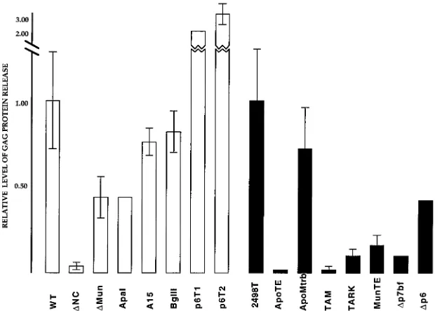
are shown in Fig. 4, in which the relative levels of assembly
efficiency of the PR
1and PR
2constructs are shown. With the
exception of the p6T1 and p6T2 constructs, all mutants
ap-peared to assemble less efficiently than their wt counterparts.
We believe the release levels for p6T1 and p6T2 appear
anom-alously high because they are processed less well than wt
HIVgpt (see below). Insofar as other mutants were concerned,
there was considerable variation in levels of Gag protein
re-lease. Some mutations, such as the linker insertions (
Apa
I and
Bgl
II), the site-directed Cys-His motif mutation (A15), and the
PR
2p6 deletion (
D
p6), reduced the efficiency of virus
assem-bly about twofold or less. However, major deletions and
trun-cations of the NC region significantly reduced the levels of
assembly. Furthermore, it appeared that p2 and the amino
terminus of NC were important to Gag protein assembly and
release, as evidenced by the extremely low release ratios of
D
NC, ApoTE, and TAM constructs, in which deletions
ex-tended to the N terminus of NC or into p2. However, it is not
on November 9, 2019 by guest
http://jvi.asm.org/
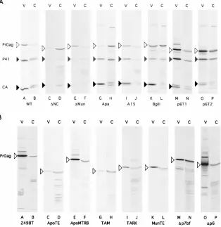
FIG. 1. Mutant HIV Gag constructs. The parental construct HIVgpt (55, 66, 67) is based on HIV HXB2 (17) and is diagrammed to show the C-terminal portion of thegaggene and the beginning of thepolgene. Only the C termini of CA, p2, NC, p1, and p6 of thegagORF are shown in the diagram; they are labeled above their respective segments of the diagram. Cys-His motifs in the NC region are indicated as diamonds, and HIV-1 proviral nucleotide numbers are designated. An arrow indicates when thepolframe is intact. The construct 2498T has a terminator oligonucleotide insertion at theHindII site at nt 2498, stopping all three ORFs; it expresses wt Gag but notpolgene products (43). The p6 terminator mutations (p6T-1 and p6T-2) have linker insertions at theBglII site at nt 2096, resulting in the termination of Gag ORFs before the p6 coding sequences, while thegag-polORF is not terminated. InDp6, the deleted HIV sequence from theBglII site at nt 2096 to theBclI site at nt 2429 is indicated by the thin lines in the diagram; thegag-polframe is retained, although the PR coding region is disrupted, so the mutant is PR2.BglII is a linker insertion mutant with a linker coding for 4 amino acid residues inserted at theBglII site at nt 2096 in p1, between the NC and p6 domains.ApaI is a mutant with a linker inserted at theApaI site at nt 2010, which adds six codons between the two Cys-His motifs of the NC domain (66). In A15, the first two cysteine residues of the C-terminal NC Cys-His motif were mutated to tyrosine (1). Of the NC deletions,DMun andDNC retained thepolframe whileDp7bf waspolminus. Specific deletions were as follows:DMun (from theMunI site [nt 1968] to theRsaI site [nt 2067] removed the two zinc fingers of NC;DNC deleted the region from theApoI site (nt 1900) in p2 to theRsaI site at nt 2067; andDp7bf deleted nt 1928 to 2096, leaving only four amino-terminal and four carboxy-terminal codons of the nucleocapsid domain. The terminator mutants MunTE, TARK, TAM, and ApoTE have oligonucleotide insertions which cause the Gag ORF to stop at different positions in p2 or NC, and neither p6 of thegaggene nor thepolgene is expressed. MunTE has an oligonucleotide at theMunI site (nt 1968) and terminates translation 17 codons after the beginning of NC, before the two zinc fingers. TARK has an oligonucleotide inserted at theApoI site at nt 1900 and should terminate translation 11 residues into NC. TAM and ApoTE also have sequences inserted at nt 1900, and TAM terminates precisely at the end of p2, while ApoTE causes thegagORF to end in p2, five codons before the beginning of NC. Instead of terminating thegagORF at theApoI site at nt 1900 as in ApoTE, in ApoMTRB, the NC and p6 domains of HIV are swapped for a bacterial RNA binding protein, MtrB (49), which is indicated by a shaded bar. MtrB (8 kDa) is encoded by the methyltryptophan resistance (mtr) locus ofB. subtilis, which is a two-gene operon consisting ofmtrAandmtrB. MtrB has been shown to bind specifically totrpleader RNA in a tryptophan-dependent manner. Constructs with activepolframes have them indicated by arrows, and the precise sequences of the junctions of these constructs are provided in Materials and Methods.
6768
ZHANG AND BARKLIS
J. V
IROL.
on November 9, 2019 by guest
http://jvi.asm.org/
clear how p2 and NC impact particle assembly: their
contribu-tions to this process may or may not be sequence specific. An
argument against a sequence-specific requirement is that
ApoMTRB, in which p2, NC, p1, and p6 are replaced by an
unrelated sequence, directed the efficient release of chimeric
Gag proteins.
As illustrated above (Fig. 2 and 4), some of our mutant
proteins were not assembled and released efficiently, even
though Gag proteins were readily detected within cells. It is
possible that these mutant Gag proteins were blocked during
transport to the cell surface, as has been observed previously
for some mutant Gag proteins (23, 32, 66, 68). Alternatively,
plasma membrane-localized proteins might have been
defec-tive in the processes of assembly or release. To find where Gag
proteins resided in cells, we examined the subcellular
localiza-tions of wt and release-impaired truncation mutant Gag
pro-teins (ApoTE, TAM, TARK, and MunTE) by
immunofluores-cence light microscopy. Our results indicated that wt Gag
proteins stained as a perinuclear ring plus a heterogeneous
pattern through the cell periphery, while ApoTE, TAM,
TARK, and MunTE proteins showed heterogeneous
non-nuclear staining, with staining along cell edges (data not
shown). These results did not support the notion that ApoTE,
TAM, TARK, and MunTE proteins were trapped at
intracel-lular membranes but suggested that they were delivered to the
cell surface, as has been reported for other NC mutant
retro-viruses (38).
Characterization of wt and mutant virus particles.
In our
virus assembly and release assays, collection of cell-free
me-dium supernatant samples involved pelleting through 2 ml of
20% sucrose cushions for 45 min at 274,000
3
g
. Based on
centrifugation clearing rates, the minimum particle size to
pel-let is 165S, and over 90% of our medium HIVgpt Gag protein
was recoverable by this method in control experiments (data
not shown).
These data suggest that wt and mutant Gag proteins were
released from cells in particle forms. Although a complete
analysis of each mutant virus awaits electron microscope
anal-ysis, biochemical characterization of the particles was also of
interest. For Gag proteins produced from PR
1constructs, one
avenue of analysis was to examine whether released Gag
pro-teins were processed by the viral protease. Consequently, CA,
p41, and Pr
Gaglevels in pelleted medium supernatant samples
[image:5.612.151.465.72.395.2]for PR
1constructs were determined (Table 1). Not
surpris-ingly the site-directed and linker insertion mutants (A15,
Apa
I,
and
Bgl
II) were processed at approximately wt levels. The
p6T1 and p6T2 proteins also were processed, albeit at lower
efficiency than the wt. More dramatic were results for the
FIG. 2. Gag protein levels in cell lysate and medium samples. Medium supernatant (V) and cell (C) samples were collected 72 h after transfection of Cos7 cells with the indicated constructs. Particles were pelleted from medium samples and resuspended; half of each pelleted medium preparation was used for loading on SDS-polyacrylamide gels. Cell pellets were lysed and centrifuged to remove debris, and 1/20 of each cell lysate sample was prepared for SDS-PAGE. Gag proteins in samples were separated by SDS-PAGE, electroblotted onto nitrocellulose filters, and immunodetected with a mouse anti-p24 monoclonal antibody from hybridoma cell line Hy183 as the primary antibody. Precursor (open triangles), partially processed (shaded triangles), and mature (filled triangles) Gag proteins were identified by antibody reactivity and comparison of gel migration mobilities to known standards. For each of the two panels, medium samples are in lanes A, C, E, G, I, K, M, and O and cell samples are in lanes B, D, F, H, J, L, N, and P. (A) PR1constructs are the wt,DNC,DMun,ApaI, A15,BglII, p6T1, and p6T2. (B) PR2constructs are 2498T, ApoTE, ApoMTRB, TAM, TARK, MunTE,Dp7bf, andDp6.
on November 9, 2019 by guest
http://jvi.asm.org/
mutants which had major deletions in NC (
D
NC and
D
Mun).
Seventy percent or more of the Gag proteins synthesized by
these two constructs remained unprocessed, suggesting that
mutant Gag-Pol proteins do not assemble into virions or are
impaired for PR activity or that Gag proteins are packed in
such a way that they are not accessible for processing.
[image:6.612.138.477.67.550.2]Another method for analysis of released particles is by
den-sity gradient fractionation. Retrovirus particles have densities
of 1.140 to 1.180 g/ml, and while evidence has suggested that
retroviral densities are not grossly dependent on RNA
encap-sidation levels, aberrant densities might be an indication of
different arrangements of packing the Gag proteins within
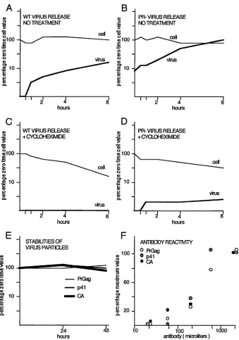
FIG. 3. Stability and release of Gag proteins. (A and B) At 48 h after transfection of Cos7 cells with wt HIVgpt (A) or 2498T (B), cells were washed and refed. At time points thereafter, cell (thin line) and medium supernatant (thick line) samples were collected and processed for immunodetection. Gag protein signals were quantitated for each time point and are plotted as percentages of the zero time cellular Gag value versus collection time. (C and D) Experiments were performed and results were plotted as in panels A and B, except that 100mg (final concentration) of cycloheximide per ml was present in the wash, during a 20-min preincubation step, and throughout each time course. (E) Medium supernatants from wt- and 2498T-transfected Cos7 cells were collected and incubated 0, 24, or 48 h at 37°C, after which virus particles were pelleted and processed for immunodetection of Gag proteins. Results are plotted as percentages of zero time Gag protein levels versus collection times and are given for PrGagproteins from 2498T particles and p41 and CA proteins from wt particles. (F) Equal aliquots of wt and 2498T particles were electrophoresed, electroblotted, and immunodetected in parallel with various dilutions of the mouse Hy183 anti-Ca monoclonal antibody. Gag protein signals were quantitated and are plotted as percentages of the maximum values of PrGag, p41, or CA versus antibody dilutions. The p41 and CA results derive from wt particles, while PrGagresults derive from 2498T particles.
6770
ZHANG AND BARKLIS
J. V
IROL.
on November 9, 2019 by guest
http://jvi.asm.org/
virus particles (5). To measure the densities of wt and mutant
particles, virus resuspensions recovered from the transfected
Cos7 media were mixed with an internal control mouse
retro-virus stock (Sup1 M-MuLV [56]) and fractionated by sucrose
density gradient centrifugation. Collected fractions were
as-sayed for density and Gag protein content (HIV and
M-MuLV), and results are shown in Fig. 5. The fractionation
profiles of the PR
1constructs are shown in the left two
col-umns for comparison with wt HIVgpt, while the profiles of
PR
2constructs are shown in the right two columns for
com-parison with 2498T, since we found that processed wt HIVgpt
particles came to equilibrium at a density of 1.148 g/ml (equal
to that of M-MuLV) while the PR
22498T particles came to a
density equilibrium of 1.168 g/ml (greater than that of
M-MuLV). Relative to that of wt HIVgpt, the p6 terminators
(p6T1 and p6T2) showed higher densities, but since the p6T1
and p6T2 proteins were incompletely processed (Table 1), it
was not surprising that they had densities similar to that of
2498T.
Apa
I, A15,
Bgl
II, TARK, MunTE,
D
Mun, and
D
p6 had
densities similar to those of their wt counterparts, while
D
NC,
ApoTE, TAM, and
D
p7bf showed densities lower than those of
their wt counterparts. These results suggest that p2 and the
amino-terminal portion of NC are necessary for packing Gag
proteins into wt-like virus: critical residues apparently map
between the fourth (
D
p7bf) and the eleventh (TARK) residues
of NC, although the results with the
D
Mun construct are not in
strict agreement with this assessment. Interestingly, while
ApoMTRB particles assemble and are released at reasonable
levels of efficiency, the particles are of low density, at least
compared with the PR
22498T particles. This result is
remi-niscent of some MuLV Gag fusion proteins (32) and suggests
that high-density packing mediated by NC domains is not
es-sential to efficient Gag protein release from cells.
Encapsidation of retroviral RNA.
Efficient and selective
[image:7.612.154.469.74.296.2]en-capsidation of viral genomic RNA into virus particles requires
incompletely defined interactions between virus core proteins
and the viral encapsidation signal, which appears to be
local-ized near the 5
9
portion of the HIV-1 RNA (1, 11, 25, 40). To
examine viral RNA packaging into wt and mutant particles,
Cos7 cells were transiently transfected with wt or mutant
con-structs and cellular and viral RNAs were isolated 72 h
post-transfection, as described in Materials and Methods. Aliquots
also were taken from virus preparations prior to RNA isolation
for Gag protein quantitation. A quantitative RNase protection
assay (67, 71) was employed to measure the viral RNAs
present in cells and particles, using an antisense riboprobe
designed to span the major splice donor site, which allowed
[image:7.612.59.298.545.654.2]FIG. 4. Levels of Gag protein release from transfected cells. Gag proteins in matched cell and medium supernatant samples were detected as described in the legend to Fig. 2 and quantitated with the programs DeskScan II (version 2.0 alias) and NIH image 1.59/fat. After normalization of Gag protein levels with a bacterially expressed HIV CA protein standard run on each gel, ratios of the total Gag protein levels in the media versus in cells were calculated. The ratios of the PR1constructs (open bars) were normalized to that of wt HIVgpt, and the ratios of the PR2constructs (filled bars) were normalized to that of 2498T. Thus, the higher the release ratio, the greater the level of Gag protein release. Note that the release ratio of 2498T was 2.6 times that of HIVgpt and that standard deviations are shown, when available.
TABLE 1. Proteolytic processing of HIV-1 Gag proteins
Construct % of total viral Gag a
PrGag p41 CA
wt
20.8
6
7.6
20.9
6
8.6
58.3
6
12.9
D
NC
69.9
6
17.8
15.9
6
14.9
14.2
6
15.8
D
Mun
92.2
6
0.8
6.1
6
1.1
1.7
6
1.8
Apa
20.2
6
10.6
23.6
6
9.0
56.2
6
14.6
A15
14.9
6
7.4
31.4
6
7.5
53.7
6
11.1
Bgl
II
37.8
6
8.6
9.7
6
3.7
52.5
6
11.5
P6T1
33.5
6
1.0
26.2
6
1.2
40.3
6
0.6
P6T2
47.2
6
6.8
32.4
6
6.2
20.4
6
11.9
aMedium supernatant pellets from Cos7 cells transfected with the indicated
constructs were fractionated by SDS-PAGE, electroblotted, and immunode-tected as described in the legend to Fig. 2. The particle-associated PrGag, partially processed p41, and CA protein signals were quantitated and are expressed in the table as percentages of the total Gag protein signals of the respective samples. Multiple experiments were carried out to obtain the averages and the standard deviations; the numbers of experiments averaged to obtain values were as fol-lows: 30 for the wt, 5 forDNC, 2 forDMun, 8 forApaI, 13 for A15, 3 forBglI, 3 for p6T1, and 6 for p6T2.
on November 9, 2019 by guest
http://jvi.asm.org/
FIG. 5. Sucrose density gradient fractionation of wt and mutant HIV-1 particles. Virus pellets prepared from 30 ml of cell-free supernatants from transfected Cos7 cells were resuspended in 200ml of PBS, mixed with mouse M-MuLV suspensions, and layered on top of the linear 20 to 60% sucrose gradients. Gradients were centrifuged for 24 h at 240,0003g so that particles with a sedimentation coefficient of 3S or greater would come to equilibrium. After centrifugation, 400ml-fractions were collected from the top to the bottom and each fraction was monitored for density and for HIV-1 and M-MuLV Gag protein content. Gag protein bands were quantitated with the DeskScan II (version 2.0 alias) and NIH image 1.59/fat programs, and HIV or M-MuLV Gag protein levels in fractions are expressed as percentages of the respective peak fraction values. The x axes indicate the density gradient fractions from top (left, low numbers) to bottom (right, high numbers). The peak densities of each gradient were 1.152 g/ml for the wt, 1.137 g/ml forDNC, 1.131 g/ml forDMun, 1.150 g/ml for ApaI, 1.148 g/ml for A15, 1.161 g/ml for BglII, 1.164 g/ml for p6T1, 1.177 g/ml for p6T2, 1.168 g/ml for 2498T, 1.155 g/ml for ApoTE, 1.155 g/ml for ApoMTRB, 1.140 g/ml for TAM, 1.180 g/ml for TARK, 1.160 g/ml for MunTE, 1.146 g/ml forDp7bf, and 1.182 g/ml forDp6.
6772
ZHANG AND BARKLIS
J. V
IROL.
on November 9, 2019 by guest
http://jvi.asm.org/
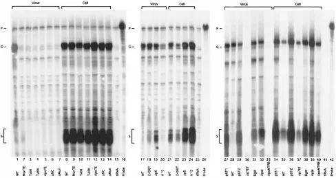
differentiation of full-length viral genomic RNA and spliced
viral transcripts. Examples of the protection gels are shown in
Fig. 6. The probe of 183 bases long is shown in lanes 16, 26, and
42, and little background protection was detected with the
control yeast tRNA samples (lanes 15, 25, and 41). As
ex-pected, wt HIVgpt was expressed well in the transfected Cos7
cells (lanes 8, 21, and 35), showing bands corresponding to
viral full-length (150-nt) and spliced (64-nt) RNAs. Full-length
viral RNA was detected in particles released from wt
HIVgpt-transfected cells, but little spliced viral RNA could be detected
in the particles (lanes 1, 17, and 28), indicating that full-length
genomic RNA was efficiently and specifically packaged into wt
virus particles. Similar results were obtained with the PR
2construct, 2498T (lanes 18 and 22). By comparison, all mutants
showed high levels of full-length genomic and spliced RNAs in
cellular RNA samples (lanes 8 to 14, 21 to 24, and 34 to 40).
However, the constructs with major NC deletions, truncations,
or replacements showed no RNA in the particle samples (lanes
2 to 7, 30, and 33). As seen previously (71), A15, in which
cysteines of the second Cys-His motif of NC were mutated to
tyrosines, showed a reduced level of HIV RNA in the virus
particles and a lower ratio of genomic versus spliced RNA,
suggesting that the zinc finger affects both packaging efficiency
and specificity. The other constructs,
Apa
I (lanes 32 and 39),
Bgl
II (lanes 31 and 38), and the three p6 mutants (lanes 19 and
23, 27 and 34, and 29 and 36) all appeared to show efficient
genomic RNA encapsidation, but reduced specificity, as
evi-denced by high levels of spliced RNA in virus particles.
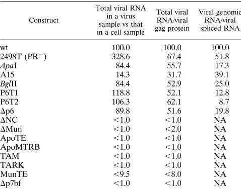
To evaluate encapsidation results, cell and virus protection
band signals and their corresponding Gag protein signals from
Western blots were quantitated with DeskScan II, version 2.0
alias, and NIH image 1.59/fat software programs. From these
data, ratios were calculated for virus RNA versus cell RNA,
virus RNA versus virus Gag protein, and virus genomic RNA
versus virus spliced RNA. Values, normalized to wt HIVgpt
results, are given in Table 2. As shown, the PR
22498T
con-struct gave no clear-cut indication of a loss of encapsidation
efficiency but appeared to show a somewhat increased
ten-dency to package spliced transcripts. However, less subtle
re-sults were obtained for other mutants. Notably, all deletions or
truncations that removed the HIV-1 NC Cys-His fingers failed
to package viral RNA. This result also applied to ApoMTRB,
in which the NC, p1, and p6 domains are replaced with the
unrelated
trp
leader RNA binding protein encoded by the
B.
subtilis
methyltryptophan resistance (
mtrB
) gene. Results for
A15, the construct with the site-directed Cys-His finger
muta-tion, showed reduced packaging efficiency and specificity, as
observed previously (71). The situation was slightly different
for the linker insertion between the two NC fingers (
Apa
I), the
insertion in p1 (
Bgl
II), and the p6 deletions and truncations.
All of these appeared to package RNA efficiently, with
virus-to-cell RNA ratios of at least 80% of wt levels, and
particle-RNA-to-Gag ratios at least half that of the wt. However, the
encapsidation specificities for these mutants were clearly
re-duced, as indicated by particle-associated
unspliced-to-spliced-RNA ratios 8 to 25% of that of the wt. These results indicate
that both the NC and p6 domains contribute to the specificity
of HIV RNA encapsidation, although p6 may act indirectly, by
influencing NC folding.
DISCUSSION
It has been shown that mutations affecting retrovirus NC
Cys-His motifs can reduce levels of genomic RNAs packaged
FIG. 6. Genomic and spliced RNA levels in cells and virus particles. RNA samples were prepared from transfected cells and virus pellets, as described in Materials and Methods. RNAs (10% of viral samples or 40mg of the cellular samples) were mixed with 10mg of yeast tRNA, ethanol precipitated, dried, and processed for RNase protection assays with an antisense probe (P) of 183 nt (lanes 16, 26, and 42) capable of detecting both spliced viral transcripts (S) at a fragment size of 63 to 64 nt and unspliced genomic RNAs (G) at a protected fragment size of 150 nt. In each panel, results from mock reactions with yeast tRNA samples were also used (lanes 15, 25, and 41). Lane contents (experimental virus [listed first] and cell [listed second] samples) are as labeled on the figure. Note that viral RNA signals were normalized for total Gag protein content to yield encapsidation efficiencies as described in Materials and Methods.
on November 9, 2019 by guest
http://jvi.asm.org/
into retrovirus particles (1, 3, 11, 16, 19, 31, 45), although the
extent to which NC contributes to encapsidation specificity has
not been elucidated completely (6, 7, 12, 33, 41, 57, 71). In
addition to its function in RNA encapsidation, NC and the
C-terminal portions of retroviral Gag proteins appear to
pos-sess a region involved in increasing the efficiency of virus
par-ticle assembly; this region has been referred to as an assembly
domain (5), although its exact contribution to virus assembly is
not clear (10, 15, 18, 20, 27, 38). Our study focused on the
evaluation of the effects of the p2, NC, p1, and p6 domains of
HIV-1 on virus assembly and RNA encapsidation.
In accord with previous observations (33, 37), major
dele-tions, truncadele-tions, or replacements of the NC domain of HIV-1
gag
were found to eliminate the encapsidation of HIV genomic
or spliced RNAs into virus particles. Although they are
theo-retically possible, we do not believe these results are due to a
defect of mutant construct
cis
-active encapsidation (Psi)
sig-nals, since the mutations occurred away from the known Psi
signals (4, 24, 25, 42, 57). We have not measured the content of
nonviral RNAs in these mutant particles, but if packaging of
spliced viral RNA is an indication of nonspecific RNA
encap-sidation, we would expect little RNA of any kind in these virus
particles, although this assumption has yet to be proven. While
major NC mutations apparently eliminated encapsidation,
other mutants showed more subtle effects. Linker insertion
between the two NC zinc fingers in the mutant
Apa
I reduced
the specificity of encapsidation, while the total amount of RNA
packaged was normal. This observation supports the previously
described model that NC contributes to the specificity of RNA
encapsidation (7, 71). Perhaps more surprisingly, mutants of p1
or p6 maintained wt levels of encapsidated RNA, but their
specificities of RNA encapsidation were reduced (Fig. 6; Table
2). This strongly suggests that Pr
Gagdetermines packaging
specificity and implicates the entire C-terminal region of
HIV-1 Gag in the process.
The PR
2construct, 2498T, seemed to release particles more
efficiently than wt HIVgpt in our experimental system. This
result differs somewhat from results with avian (63) and
mu-rine (13, 36) retroviruses in which protease activity does not
appear to affect release levels and contrasts with results of one
study of HIV, in which PR
2particles were released less
effi-ciently than PR
1particles (34). It is unclear how the PR
1phenotype might impair transport of Gag protein or
establish-ment of interprotein contacts required for assembly and
bud-ding, although a number of studies have shown that
perturba-tion of Gag protein-to-PR activity ratios can alter virus release
efficiencies (29, 34, 44, 53, 54). Because of the differences
observed with wt HIVgpt and 2498T, we compared mutant
particle release efficiencies with that of either wt HIVgpt or
2498T, depending on the PR phenotype of the mutant
con-struct (Fig. 4). In performing such comparisons, we observed
that mutants without p6 were released at reasonable
efficien-cies, which is consistent with the notion that the effects of p6 on
virus release may be affected by PR or may be cell type specific
(29, 34, 44, 61). However, constructs with mutations in p2 and
the amino-terminal portion of NC were released much less
well than the wt, supporting the notion that this region defines
an assembly domain which appears to function after Gag
pro-tein delivery to the plasma membrane (Fig. 3) (10, 38, 69, 70).
Still, it is not evident how this region contributes to the
effi-ciency of HIV-1 assembly, since replacement of this region
with the unrelated sequence in ApoMTRB permitted efficient
release of virus-like particles. This result is reminiscent of the
ability of some M-MuLV Gag fusion proteins to direct particle
assembly (23, 32), and the ability of ApoMTRB proteins to
release particles at reasonable efficiency might be interpreted
in several ways. One possibility is that the Mtrb domain may
prevent the C terminus of CA from folding into an
assembly-incompatible conformation. Alternatively, the Mtrb domain
may substitute for an active assembly function of p2 or NC.
One might favor the first of these alternatives, since in addition
to the assembly and release of HIV Gag-Mtrb particles, we
have observed that MuLV Gag fusions to
b
-galactosidase also
form virus-like particles. However, both Mtrb and
b
-galacto-sidase proteins form higher-order oligomers (2, 30), so it may
be that the essential assembly function provided by NC is a
nonspecific ability to make interprotein contacts. Although the
Mtrb domain was fused to HIV-1
gag
because it potentially acts
as an RNA binding protein (49), no evidence of specific or
nonspecific RNA incorporation into ApoMTRB particles was
observed in our experiments with either HIV RNA (Fig. 6) or
Mtrb target RNA (data not shown). Thus, it appears that the
NC assembly function can be replaced by that of a protein that
does not encapsidate detectable levels of RNA in our system.
We believe that further analysis of the mechanism(s) by which
the NC assembly domain acts will be of basic and practical
interest.
ACKNOWLEDGMENTS
[image:10.612.58.297.81.271.2]We thank Marylene Mougel, Jason McDermott, Sonya Karanjia,
Zachary Love, Chin-tien Wang, and Mark Hansen for help and advice
throughout the course of this work. The anti–M-MuLV–CA
monoclo-nal antibody was a gift from Bruce Chesebro, who also made the
anti-HIV-CA Hy183 hybridoma cell line that was obtained from the
AIDS Research and Reference Reagent Program, Division of AIDS,
NIAID, NIH. A molecular clone encoding the Mtrb protein was kindly
provided by Paul Gollnick, and the A15 clone originally was from
Anna Aldovini.
TABLE 2. RNA encapsidation into virus particles
aConstruct
Total viral RNA in a virus sample vs that in a cell sample
Total viral RNA/viral gag protein Viral genomic RNA/viral spliced RNA
wt
100.0
100.0
100.0
2498T (PR
2)
328.6
67.4
51.8
Apa
I
84.4
55.7
17.3
A15
14.3
31.7
39.1
Bgl
II
84.4
52.9
25.0
P6T1
118.8
52.1
12.8
P6T2
106.3
62.1
8.7
D
p6
89.8
51.6
19.8
D
NC
,
1.0
,
1.0
NA
D
Mun
,
1.0
,
2.0
NA
ApoTE
,
1.0
,
1.0
NA
ApoMTRB
,
1.0
,
1.0
NA
TAM
,
1.0
,
1.0
NA
TARK
,
1.0
,
1.0
NA
MunTE
,
9.5
,
8.0
NA
D
p7bf
,
1.0
,
1.0
NA
aGenomic and spliced RNA signals from cell and virus samples listed in Fig.
6 and corresponding Gag protein levels from virus particles detected by Western blots were quantitated with DeskScan II (version 2.0 alias) and NIH image 1.59/fat software. From these data, the following ratios were calculated for each indicated construct; the total level of viral spliced and unspliced RNAs in a particle sample divided by that of a cell sample, the total particle-associated viral RNA signal divided by the particle Gag protein signal, and the particle genomic RNA signal divided by the particle spliced viral RNA signal. Calculated ratios were normalized to wt HIVgpt ratios and are listed as percentages of wt levels (wt5100%). For mutants which showed no particle-associated viral RNA signal above background, values in the second and third columns are given as less than a given level, while values for the right-most column were not applicable (NA). Note that although some mutants released particles very inefficiently, experi-ments were scaled up as necessary to obtain the Gag-normalized levels in the third column of the table.
6774
ZHANG AND BARKLIS
J. V
IROL.
on November 9, 2019 by guest
http://jvi.asm.org/
This work was supported by grant 2RO1CA47088-07 from the
Na-tional Cancer Institute.
REFERENCES
1.Aldovini, A., and R. A. Young.1990. Mutations of RNA and protein se-quences involved in human immunodeficiency virus type 1 packaging result in production of noninfectious virus. J. Virol.64:1920–1926.
2.Antson, A. A., J. Otridge, A. M. Brzozowski, E. J. Dodson, G. G. Dodson, K. S. Wilson, T. M. Smith, M. Yang, T. Kurecki, and P. Gollnick.1995. The structure of trp RNA-binding attenuation protein. Nature374:693–700. 3.Aronoff, R., A. M. Hajjar, and M. L. Linial.1993. Avian retroviral RNA
encapsidation: reexamination of functional 59RNA sequences and the role of nucleocapsid Cys-His motifs. J. Virol.67:178–188.
4.Baudin, F., R. Marquet, C. Isel, J.-L. Darlix, B. Ehresmann, and C. Ehres-mann.1993. Functional sites in the 59region of human immunodeficiency virus type 1 RNA form defined structural domains. J. Mol. Biol.229:382– 397.
5.Bennett, R. P., T. D. Nelle, and J. W. Wills.1993. Functional chimeras of the Rous sarcoma virus and human immunodeficiency virus Gag proteins. J. Vi-rol.67:6487–6498.
6.Berkowitz, R. D., J. Luban, and S. P. Goff.1993. Specific binding of human immunodeficiency virus type 1 gag polyprotein and nucleocapsid to viral RNAs detected by RNA mobility shift assays. J. Virol.67:7190–7200. 7.Berkowitz, R. D., A. Ohagen, S. Hoglung, and S. P. Goff.1995. Retroviral
nucleocapsid domains mediate the specific recognition of genomic viral RNAs by chimeric Gag polyproteins during RNA packaging in vivo. J. Virol.
69:6445–6456.
8.Campbell, S., and V. Vogt.1995. Self-assembly in vitro of purified CA-NC proteins from Rous sarcoma virus and human immunodeficiency virus type 1. J. Virol.69:6487–6497.
9.Cann, A. J., and J. Karn.1989. Molecular biology of HIV-1: new insights into the virus life cycle. AIDS3(Suppl. 1):S19–S34.
10. Carriere, C., B. Gay, N. Chazal, N. Morin, and P. Boulanger.1995. Sequence requirements for encapsidation of deletion mutants and chimeras of human immunodeficiency virus type 1 Gag precursor into retrovirus-like particles. J. Virol.69:2366–2377.
11. Clavel, F., and J. M. Orenstein.1990. A mutant of human immunodeficiency virus with reduced RNA packaging and abnormal particle morphology. J. Vi-rol.64:5230–5234.
12. Clever, J., C. Sassetti, and T. G. Parslow.1995. RNA secondary structure and binding sites forgaggene products in the 59packaging signal of human immunodeficiency virus type 1. J. Virol.69:2101–2109.
13. Crawford, S., and S. Goff.1985. A deletion mutation in the 59part of thepol gene of Moloney murine leukemia virus blocks proteolytic processing of the gagandpolpolyproteins. J. Virol.53:899–907.
14. Dannull, J., A. Surovoy, G. Jung, and K. Moelling.1994. Specific binding of HIV-1 nucleocapsid protein to PSI RNA in vitro requires N-terminal zinc finger and flanking basic amino acid residues. EMBO J.13:1525–1533. 15. Dorfman, T., J. Luban, S. P. Goff, W. A. Haseltine, and H. G. Gottlinger.
1993. Mapping of functionally important residues of a cysteine-histidine box in the human immunodeficiency virus type 1 nucleocapsid protein. J. Virol.
67:6159–6169.
16. Dupraz, P., S. Oertle, C. Meric, P. Danay, and P.-F. Spahr.1990. Point mutations in the proximal Cys-His box of Rous sarcoma virus nucleocapsid protein. J. Virol.64:4978–4987.
17. Fisher, A. G., M. B. Feinberg, S. R. Josephs, M. E. Harper, L. M. Marselle, G. Reyes, F. A. Gonda, A. Aldovini, C. Debouk, R. C. Gallo, and F. Wong-Staal.1986. The transactivator gene of HTLV-III is essential for virus rep-lication. Nature320:367–371.
18. Gheysen, D., E. Jacobs, F. de Foresta, D. Thiriart, M. Francotte, D. Thines, and M. De Wilde.1989. Assembly and release of HIV-1 precursor pr55gag virus-like particles from recombinant baculovirus-infected cells. Cell59:103– 112.
19. Gorelick, R. J., S. M. Nigida, Jr., J. R. Bess, Jr., L. O. Arthur, L. E. Henderson, and A. Rein.1990. Noninfectious human immunodeficiency vi-rus type 1 mutants deficient in genomic RNA. J. Virol.64:3207–3211. 20. Gottlinger, H. G., J. G. Sodroski, and W. A. Haseltine.1989. Role of capsid
precursor processing and myristylation in morphogenesis and infectivity of human immunodeficiency virus type 1. Proc. Natl. Acad. Sci. USA86:5781– 5785.
21. Graham, R., and A. van der Eb.1973. A new technique for the assay of infectivity of human adenovirus 5 DNA. Virology52:456–467.
22. Haffar, O., J. Garrigues, B. Travis, P. Moran, J. Zarling, and S.-L. Hu.1990. Human immunodeficiency virus-like, nonreplicatinggag-envparticles assem-ble in a recombinant vaccinia virus expression system. J. Virol.64:2653–2659. 23. Hansen, M., L. Jelinek, S. Whiting, and E. Barklis.1990. Transport and assembly ofgagproteins into Moloney murine leukemia virus. J. Virol.
64:5306–5316.
24. Harrison, G. P., and A. M. L. Lever.1992. The human immunodeficiency virus type 1 packaging signal and major splice donor region have a conserved stable secondary structure. J. Virol.66:4144–4153.
25. Hayashi, T., T. Shioda, Y. Iwakura, and H. Shibuta.1992. RNA packaging
signal of human immunodeficiency virus type 1. Virology188:590–599. 26. Henderson, L. E., M. A. Bowers, R. C. Sowder II, S. A. Serabyn, D. G.
Johnson, J. W. Bess, Jr., L. O. Arthur, D. K. Bryant, and C. Fenselau.1992. Gag proteins of the highly replicative MN strain of human immunodeficiency virus type 1: posttranslational modifications, proteolytic processing, and complete amino acid sequences. J. Virol.66:1856–1865.
27. Hong, S. S., and P. Boulanger.1993. Assembly-defective point mutants of the human immunodeficiency virus type 1 Gag precursor phenotypically expressed in recombinant baculovirus-infected cells. J. Virol.67:2787–2798. 28. Housset, V., H. De Rocquigny, B. Roques, and J.-L. Darlix.1993. Basic amino acids flanking the zinc finger of Moloney murine leukemia virus nucleocapsid protein NCp10 are critical for virus infectivity. J. Virol.67:
2537–2545.
29. Huang, M., J. M. Orenstein, M. A. Martin, and E. O. Freed.1995. p6Gagis required for particle production from full-length human immunodeficiency virus type 1 molecular clones expressing protease. J. Virol.69:6810–6818. 30. Jacobson, R. H., and B. W. Matthews.1992. Crystallization of
beta-galacto-sidase from Escherichia coli. J. Mol. Biol.223:1177–1182.
31. Jentoft, J. E., L. M. Smith, X. Fu, M. Johnson, and J. Leis.1988. Conserved cysteine and histidine residues of the avian myeloblastosis virus nucleocapsid protein are essential for viral replication but not “zinc-binding fingers.” Proc. Natl. Acad. Sci. USA85:7094–7098.
32. Jones, T., G. Blaug, M. Hansen, and E. Barklis.1990. Assembly of Gag–b -galactosidase proteins into retrovirus particles. J. Virol.64:2265–2279. 33. Jowett, J. M. B., D. J. Hockley, M. V. Nermut, and I. M. Jones.1992. Distinct
signals in human immunodeficiency virus type 1 Pr55 necessary for RNA binding and particle formation. J. Gen. Virol.73:3079–3086.
34. Kaplan, A. H., M. Manchester, and R. Swanstrom.1994. The activity of the protease of human immunodeficiency virus type 1 is initiated at the mem-brane of infected cells before the release of viral proteins and is required for release to occur with maximum efficiency. J. Virol.68:6782–6786. 35. Karacostas, V., K. Nagashima, M. A. Gonda, and B. Moss.1989. Human
immunodeficiency virus-like particles produced by a vaccinia virus expres-sion vector. Proc. Natl. Acad. Sci. USA86:8964–8967.
36. Katoh, I., Y. Yoshinaka, A. Rein, M. Shibuya, T. Odaka, and S. Oroszlan.
1985. Murine leukemia virus maturation: protease region required for con-version from “immature” to “mature” core form and for virus infectivity. Virology145:280–292.
37. Kaye, J. F., and A. M. Lever.1996.trans-acting proteins involved in RNA encapsidation and viral assembly in human immunodeficiency virus type 1. J. Virol.70:880–886.
38. Krausslich, H. G., M. Facke, A. M. Heuser, J. Konvalinka, and H. Zentgraf.
1995. The spacer peptide between human immunodeficiency virus capsid and nucleocapsid proteins is essential for ordered assembly and viral infectivity. J. Virol.69:3407–3419.
39. Leis, J., D. Baltimore, J. B. Bishop, J. Coffin, E. Fleissner, S. P. Goff, S. Oroszlan, H. Robinson, A. M. Skalka, H. M. Temin, and V. Vogt.1988. Standardized and simplified nomenclature for proteins common to all ret-roviruses. J. Virol.62:1808–1809.
40. Lever, A., H. Gottlinger, W. Haseltine, and J. Sodroski.1989. Identification of a sequence required for efficient packaging of human immunodeficiency virus type 1 RNA into virions. J. Virol.63:4085–4087.
41. Luban, J., and S. P. Goff.1991. Binding of human immunodeficiency virus type 1 (HIV-1) RNA to recombinant HIV-1gagpolyprotein. J. Virol.65:
3203–3212.
42. McBride, M. S., and A. T. Panganiban.1996. The human immunodeficiency virus type 1 encapsidation site is a multipartite RNA element composed of functional hairpin structures. J. Virol.70:2963–2973.
43. McDermott, J., L. Farrell, R. Ross, and E. Barklis.1996. Structural analysis of human immunodeficiency virus type 1 Gag protein interactions, using cysteine-specific reagents. J. Virol.70:5106–5114.
44. Mergener, K., M. Facke, R. Welker, V. Brinkmann, H. R. Gelderblom, and H. G. Krausslich.1992. Analysis of HIV particle formation using transient expression of subviral constructs in mammalian cells. Virology186:25–39. 45. Meric, C., and S. P. Goff.1989. Characterization of Moloney murine
leuke-mia virus mutants with single-amino-acid substitutions in the Cys-His box of the nucleocapsid protein. J. Virol.63:1558–1568.
46. Mervis, R. J., N. Ahmad, E. P. Lillehoj, M. G. Raum, F. H. R. Salazar, H. W. Chan, and S. Venkatesan.1988. Thegaggene products of human immuno-deficiency virus type 1: alignment within thegagopen reading frame, iden-tification of posttranslational modifications, and evidence for alternativegag precursors. J. Virol.62:3993–4002.
47. Morellet, N., H. de Rocquigny, Y. Mely, N. Jullian, H. Demene, M. Ottmann, D. Gerard, J.-L. Darlix, M. C. Fournie-Zaluski, and B. P. Roques.1994. Conformational behaviour of the active and inactive forms of the nucleo-capsid NCp7 of HIV-1 studied by1HNMR. J. Mol. Biol.235:287–301. 48. Mulligan, R. C., and P. Berg.1981. Selection for animal cells that express the
Escherichia coli gene coding for xanthine-guanine phosphoribosyltrans-ferase. Proc. Natl. Acad. Sci. USA78:2072–2076.
49. Otridge, J., and P. Gollnick.1993. MtrB from Bacillus subtilis binds specif-ically to trp leader RNA in a tryptophan-dependent manner. Proc. Natl. Acad. Sci. USA90:128–132.