EFFECTIVENESS OF CHEST PHYSIOTHERAPY AND
INCENTIVE SPIROMETRY ON POSTOPERATIVE
RESPIRATORY STATUS AND RESPIRATORY
COMPLICATIONS AMONG CHILDREN UNDERGOING
ABDOMINAL SURGERY
A DISSERTATION SUBMITTED TO THE TAMILNADU DR.
M.G.R MEDICAL UNIVERSITY, CHENNAI, IN PARTIAL
FULFILLMENT OF THE REQUIREMENTS FOR THE DEGREE
OF MASTER OF SCIENCE IN NURSING.
CERTIFICATE
This is the bonafide work of S. BEULA ANGEL M.Sc., Nursing II year student from Sacred Heart Nursing College, Ultra Trust, Madurai, submitted in partial
fulfillment for the Degree of Master of Science in Nursing, under The Tamilnadu Dr. M.G.R. Medical Univeristy, Chennai.
Dr. Nalini Jeyavanth Santha, M.Sc., (N), Ph. D.,
Principal
Sacred Heart Nursing College
Ultra Trust
ACKNOWLEDGEMENT
“I will praise you, O Jesus, with my whole heart, I will tell all your marvelous works,
I will be glad and rejoice in you;
I will sing and praise your name, oh! Most high”
Drops of water make an ocean. Although bricks give the structure of house, proper foundation makes it strong. A thesis, however, insignificant it is, can seldom be claimed as the work of an individual. These have been persons who stood by me all my efforts to successfully complete my study. In the absence of staunch support of those people, all the toil would have been in vain.
I praise and thank God Almighty for his abundant grace, kindness, care and blessings showered upon me through out the study. I, the investigator of this study, owe my sincere thanks and gratitude to all those who have contributed towards the successful completion of the endeavor.
I, the investigator, also extend some thoughtful words of gratitude and feelings of praise for those brilliant minds and helping hands that lent me in my hour of need and helped in a way cherished forever’s. I greatly appreciate the wisdom with which their efforts have enriched the study.
I express my sincere thanks to Prof. K.R Arumugam, M. Pharm., the correspondent, Ultra Trust, Madurai for his support and for providing the required facilities for the successful completion of the study.
with never ending willingness and patience to provide expert guidance and suggestions in moulding this study.
I express my gratitude to my medical guide Dr. Durai raj, M.D(Pediatrics), D.M., (Neuro), madurai-20 for his comment and guidance throughout the study.
I am extremely thankful to Mrs. Chandrakala, M.Sc(N)., Ph. D., Vice – Principal, Sacred Heart Nursing College, Ultra Trust, Madurai for her encouragement.
I extend my thanks to Mrs. Deva Kirubai, M.Sc(N)., Ph.D., for her suggestion and guidance.
I express my sincere thanks to Mrs. Sarojini, M.Sc(N)., and Mrs. Jothilakshmi, M.Sc (N)., for their guidance and suggestion.
I express my special thanks to Mr. Senthil Kumar, M.Sc., M.Phil., for his excellent guidance in the statistical analysis through out the study.
I am thankful to Mr. Thiunavukarasu, M.Lib., Senior Librarian, Sacred Heart Nursing College.
I extend my sincere thanks to Mr. Rajesh Kumar, M.A., M.Phil, B.Ed., Ph.d., for editing this manuscript.
My affectionate thanks to my dear lovable father Mr.M.Solomon Raja, my lovable mother Mrs. S. Jansi rani and my sisters and my brother for their blessings, prayers, encouragements and support throughout the study which cannot be expressed in words.
I express my thanks to my dear friends and all my classmates for their help and support for the completion of the study.
ABSTRACT
TABLE OF CONTENTS
Chapter Contents Page No.
I INTRODUCTION 1-15
Background of the study 1
Significance and need for the study 5
Statement of problem 7
Objectives 8
Hypotheses 8
Operational definition 9
Assumptions 12
Delimitations 13
Projected outcomes 13
Conceptual framework 13
II REVIEW OF LITERATURE 16-30
Literature related to overview of postoperative pulmonary 16 complication
Literature related to chest physiotherapy and incentive 25 spirometry
III METHODOLOGY 31-44
Research approach 31
Research design 31
Setting of the study 32
Population 32
Sample 32
Chapter Contents Page No
Sample size 33
Sampling technique 33
Criteria for sample selection 33
Description of the tool 34
Testing of the study 35
Description of the intervention 37
Pilot study 41
Procedure for data collection 41 Protection of human rights 43
IV ANALYSIS AND INTERPRETATION OF DATA 45-72
V DISCUSSION 73-85
VI SUMMARY, CONCLUSIONS, IMPLICATIONS AND 86-96
RECOMMENDATIONS
Summary of the study 86
Major findings of the study 88
Conclusions 92
Implications 92
Limitations 95
Recommendations 95
REFERENCES 97-103
LIST OF TABLES
Table No Title Page 1.
Frequency and percentage distribution of the samples
47
according to their age and sex.
2.
Frequency and percentage distribution of samples
49
according to the type of surgery.
3.
Frequency and percentage distribution of samples based
50
on respiratory status preoperatively.
4.
Frequency and percentage distribution of samples based
51
on respiratory status on the first postoperative day.
5.
Frequency and percentage distribution of samples based
53
on respiratory status on the fourth postoperative day.
6.
Comparison of mean respiratory status of the experimental 55
group between first and second postoperative day.
7.
Comparison of mean respiratory status of the experimental 56
group between first and third postoperative day.
8.
Comparison of mean respiratory status of the experimental 57
group between first and fourth postoperative day.
9.
Comparison of mean posttest respiratory status of the
59
experimental group and control group on second
Table No Title Page
10.
Comparison of mean posttest respiratory status of the
61
experimental group and control group on third
postoperative day
11.
Comparison of mean posttest respiratory status of the
63
experimental group and control group on fourth
postoperative day.
12.
Comparison of frequency and percentage distribution of
65
respiratory complications between the experimental group
and control group on fifth postoperative day.
13.
Comparison of mean respiratory complications of the
67
experimental group and control group.
14.
Association of posttest respiratory status of experimental
69
group and selected variables.
15.
Association of posttest respiratory complications of
71
experimental group and selected variables
LIST OF FIGURES
Figure No Title Page No
1 Conceptual framework based on J.W. Kenny’s Open System Model
15
2 Distribution of samples according to age. 48 3 Distribution of samples according to sex. 48 4 Comparison of mean respiratory status of the experimental
group between first and fourth postoperative day.
58
5 Comparison of mean posttest respiratory status of the experimental group and control group on second postoperative day.
60
6 Comparison of mean posttest respiratory status of the experimental group and control group on third postoperative day
.
62
7 Comparison of mean posttest respiratory status of the experimental group and control group on fourth postoperative day.
64
8 Comparison of frequency and percentage distribution of respiratory complications between the experimental group and control group on fifth postoperative day.
66
9 Comparison of mean respiratory complications of the experimental group and control group.
68
LIST OF APPENDICES
Appendix Title Page No
I Copy of letter seeking permission to conduct study in Government Rajaji Hospital, Madurai
104
II Copy of letter seeking experts opinion for tool and content validity
105
III List of Experts consulted for the research tool and content validity
106
IV Demographic Profile 107
CHAPTER- I
INTRODUCTION
“I brought children into this dark world because It needed the light that only a child can bring" -Liz armbruster
BACKGROUND OF THE STUDY
“Children are the brightest treasures we bring forth into this world”
The wealth and strength of the nation depends on the children because they are the future leaders. But they become the victim of health problems during their developmental period. The process of invasive and surgical procedures is extremely hard on children. This stress period includes venipuncture, separation from parents at the time of transport to the operating room and anesthesia induction.
Children experiencing surgical procedures require both psychological and physical preparation. Even though children are asleep for the actual surgical intervention, they are subjected to numerous preoperative and postoperative procedures. Although most preoperative care procedures are routine, nurse should keep in mind that they could be anxiety provoking for children and parents.(Hockenberry, 2009).
laryngospasm) and decreasing long-term behavioural effects of surgery. (Romino et al., 2005).
Inspite of the scientifical and technological development, squeal after the diseases are still problematic and requires immediate attention. Various psychological and physical interventions and observations are required to prevent or minimize possible untoward effects from anesthesia and the surgical procedure. Managing pain is a major nursing responsibility after surgery. The nurse should assess pain frequently and administer analgesics to provide comfort and facilitate cooperation with postoperative care such as ambulation and deep breathing.(Warner, 2005).
Postoperative complications are defined as the unexpected disease that occurs either early or late that is connected with the exacerbation of a pre-existing illness. Postanesthesia complications such as airway obstruction, postextubation croup, laryngospasm and bronchospasm make maintaining a patent airway and maximum ventilation is crucial.(LuizJoia et al., 2005).
Various studies have demonstrated that, upper abdominal surgery correlates more strongly with postoperative pulmonary complications. LuizJoiaNeto, et al., (2005) estimated that diaphragm dysfunction, postoperative pain and alveolar collapse result in a 50% to 60% decrease in vital capacity and a 30% decrease in residual functional capacity after abdominal surgeries. They also found that the most frequent postoperative pulmonary complications were pneumonia and the incidence was higher in the 0-11 age group.
Wong and Whaley (2008) suggest that the nurse plays a significant role in preparing the child for surgery, maintaining surveillance of the child during surgery, preventing complications and facilitating recovery. When preparing the child for surgery, it is important to realize that, no matter how mature the child may act, he or she still needs to be treated differently than adults. Some children show eagerness to discover, exactly what will happen to them in detail. Others do not want much detail. So the nurse has to implement her teaching or intervention before surgery in such a way keep in mind that the particular level of a child’s development will determine the specific concerns.
Breathing is an automatic function, but when a person has respiratory difficulties, breathing becomes more difficult. Breathing exercises helps the people to breathe easier. There are many benefits to breathing exercise. These exercises help to get more lung expansion and improve lung capacity. The diaphragm is also strengthened by breathing exercises. All types of exercise strengthen the muscles of diaphragm and chest capacity (Arruda et al., 2004).
Thoren reported that chest physiotherapy reduced the incidence of pulmonary complications after cholecystectomy, there has been an acceptance that patients undergoing abdominal surgery require some form of prophylactic physiotherapy. The nature of chest physiotherapy has varied according to prevailing attitudes about the pathophysiology of atelectasis. Attention has lately been directed towards lung expansion techniques such as incentive spirometry that promote frequent maximum inspiratory effort (Hall et al., 1991).
SIGNIFICANCE AND NEED FOR THE STUDY
“To keep the body in good health is a duty, otherwise we shall not be able to Keep our mind strong and clear”
-Buddha. Postoperative pulmonary complications contribute to surgical morbidity and mortality. Causes of complications are multifactorial and include patient related factors , surgery and anesthesia type. Pulmonary function testing has a limited role in preoperative assessment and chest x-ray and arterial blood gas measurement are rarely required.
It is estimated that at a minimum 2.5% of patients undergoing surgery may experience postoperative complication. This translates to as many as a million postoperative pulmonary complications of the 40 million patients undergoing non-thoracic surgery annually in North America. This figure may be as high as 25% to 30% among high risk patients undergoing major surgery (Peter rock 2006).
MC Alister(2005) said that the prediction ofpostoperative pulmonary complications is still in an under investigated field. He conducted a prospective cohort study, of 1055 consecutive patients 28(2.7%) suffered a postoperative pulmonary complications within 7 days of surgery: 13 patients developed respiratory failure requiring ventilator support, ten pneumonia, five atelectasis requiring bronchoscopic intervention and one pnemothorax, requiring intervention. Mean lengths of stay were substantially prolonged for these patients who developed pulmonary complications within seven days of surgery; 27.9 days versus 4.5days, p=0.006.
According to “National Hospital Discharge Survey” during 1989-2004, the estimated abdominal operations and postoperative pulmonary complications and the estimated number of major abdominal operations performed were 12,897,800. Postoperative pulmonary complications occurred after surgery in 388,400 men (7.8%) and in 452,800 women (5.7%).
High risk children should be examined for perioperative respiratory adverse events at the preoperative period, and the information gained there could be used to optimize anesthetic care. They might also benefit from specifically targeted anesthesia management (Becky McCall et al., 2010).
Beverly D. Delacruz et al., (2009) has done a study to develop guidelines and proposed a scoring systemfor pediatric risk stratification for postoperative pulmonarycomplications and to determine the accuracy of this simple and rapid pre-operative scoring system for postpre-operative pulmonary complicationsin pediatric patients undergoing elective surgery. The total samples included in this study were 506 children. Hospital medical records of children aged 6 to 19 years old,who underwent elective surgery were analyzed. Among the total samples, 330 (65.2%) developedpost-operative complications while 176 (34.8%) with none. Atelectasis(25.6%) was the top complications.
Wallis and Prasad (1999) explained that physiotherapy is often prescribed after abdominal or cardiac surgery in an attempt to counter the negative pathophysiological changes that occur in the postoperative period. Adult patients undergoing upper abdominal surgery and stay in the hospital for upto 2 weeks, a degree of atelectasis is almost invariable. Children are even more predisposed to postoperative respiratory failure because of poorly developed intercostal muscles and a compliant chest wall but less compliant lungs and poorly established collateral alveolar collapse. Specific respiratory therapy techniques might have different effects on oxygen saturation and hemodynamic stability in different age groups and careful assessment should ensure that the intervention is beneficial and effective rather than hazardous.
Breathing exercise using incentive spirometer had been found to be effective in strengthening the respiratory muscles during postoperative period. Teaching patients about incentive spirometry exercise can maintain the respiratory status and can prevent respiratory complication due to abdominal surgery (Griffin et al., 2007).
Patient education and patient counseling are independent functions of a nurse. In addition, not so many studies are conducted on these aspects in south India. With this background and knowledge the researcher intended to study the effect of chest physiotherapy and incentive spirometry among children who had undergone abdominal surgery.
STATEMENT OF THE PROBLEM
OBJECTIVES
To determine the respiratory status of children preoperatively.
To find out the postoperative respiratory status of children who had abdominal surgery after receiving chest physiotherapy and incentive spirometry.
To evaluate the effectiveness of chest physiotherapy and incentive spirometry. on postoperative respiratory status of children who had abdominal surgery. To find out the postoperative respiratory complications of children who had abdominal surgery after receiving chest physiotherapy and incentive spirometry.
To evaluate the effectiveness of chest physiotherapy and incentive spirometry on postoperative respiratory complications of children who had abdominal surgery.
To find out the association between posttest respiratory status of the children in
Experimental group and their selected variables. (age, sex, type of surgery). To find out the association between posttest respiratory complication of the children in experimental group and their selected variables. (age, sex, type of surgery).
HYPOTHESES
All the hypotheses were tested at 0.05 level of significance:
H2-The mean post-test respiratory status of the experimental group who had chest physiotherapy and incentive spirometry will be significantly higher than the mean posttest respiratory status of the control group. H3-The postoperative respiratory complications of the experimental group who had chest physiotherapy and incentive spirometry will be significantly lower than the postoperative respiratory complications of control group. H4-There will be significant association between postoperative respiratory status of the children who received chest physiotherapy and incentive spirometry& their selected variables. (age, sex, type of surgery).
H5-There will be significant association between postoperative respiratory complication of the children who received chest physiotherapy & their selected variables. ( age, sex, type of surgery).
OPERATIONAL DEFINITION
Effectiveness:
It means that the way in which it produces the intended result. In this study, it is the statistical measure of the difference in the respiratory status and respiratory complications score obtained between the experimental group and control group after the administration of chest physiotherapy and incentive spirometry which is measured by observational checklist for respiratory status and respiratory complications.
Respiratory status:
Respiratory status is defined as the movement of air in and out of the lungs and exchange of carbon dioxide and oxygen at the alveolar level.
-Nursing outcome classification
include chest x-ray results, arterial blood gas values, pulse oximetry, and pulmonary function test results.
In this study, Respiratory status denotes the condition of the respiratory system as assessed by an observational checklist. It includes respiratory rate, pulse rate, temperature, chest retractions, unilateral expansion of chest, nasal flaring, air entry, shape of the chest, breath sounds, cough, wheezing, use of accessory muscles, dyspnea and discoloration of finger and lips.
Respiratory complications
:
It is defined as an identifiable respiratory disease or dysfunction that is clinically relevant and adversely affects the clinical course.
In this study, Respiratory complications refer to those respiratory passage problems & lung problems that will decrease air entry due to effects of surgery. It includes atelectasis, pneumonia, pleural effusion, emphysema.
Atelectasis:
It is defined as a collapsed or airless state of the lung, which may be acute or chronic and may involve all or part of the lung.
In this study it refers to the characteristics such as fever, diminished respiratory movements, and diminished breath sounds, tracheal displacement toward affected side and X –ray features such as mediastinal shift or tracheal deviation.
Pneumonia:
It is defined as the inflammation of the lung with consolidation and exudation. In this study it refers to the characteristics such as fever, tachypnea, cough, decreased breath sounds, intercostals retractions and x-ray features such as lobar consolidation or interstitial infiltrates.
It is characterized by inflammation and exudation of serous fluid into the pleural cavity.
In this study it refers to the characteristics such as dyspnea on exertion, chest expansion reduced & limited diaphragm movement, frothy white or pink mucoid sputum and diminished breath sounds.
Emphysema:
It is defined as the abnormal presence of air in tissues or cavities of the body. In this study it refers to the characteristics such as low grade fever, tachypnea, and pleuritic chest pain, dullness to percussion over effusion, diminished or absent breath sounds.
Chest physiotherapy:
Chest physiotherapy (CPT) is an airway clearance technique that combines manual percussion of the chest wall by the caregiver, strategic positioning of the patient for mucus drainage with cough and breathing techniques.
In this study chest physiotherapy comprises of postural drainage and various manipulative procedures like chest percussion, cough stimulation and deep breathing exercise through pursed lips and assisting with tactile sense.
Incentive Spirometry:
Incentive spirometryis a method of deep breathing that provides visual feedback to help the patient inhale slowly and deeply to maximize lung inflation.
Abdominal surgery:
Abdominal surgery is defined as surgery pertaining to the contents of the abdominal cavity, its walls & orifices. It includes the following planned surgeries like Appendicectomy, Herniotomy, Colostomy, Ureterolithotomy, Cholecystectomy, Laproscopic surgery, Herniorrhaphy.
Appendicectomy:
It refers to the surgical excision of vermiform appendix. Herniotomy:
It is defined as an operation to remove a hernial sac. Colostomy:
An artificial opening (stoma) in the large intestine brought to the surface of the abdomen for the purpose of evacuating the bowel.
Ureterolithotomy:
Surgical removal of a calculus lodged in a ureter. Cholecystectomy:
It refers to the excision of gall bladder. Laparoscopic surgery:
It is defined as the type of surgery viewing the abdominal cavity by passing an endoscope through the abdominal wall by making one or more incisions.
Herniorrhaphy:
It is defined as the removal of a hernia sac and repair of the abdominal wall.
ASSUMPTIONS
Chest physiotherapy and incentive spirometry is a part of nursing activity in the post-operative period.
Chest physiotherapy and incentive spirometry increases pulmonary volume, improves gas exchange and ventilation distribution.
Chest physiotherapy and incentive spirometry helps to get more air into the lungs and improves lung capacity.
DELIMITATION
The following delimitations are set for the study,Children between the age group of 3-12 years, who are admitted in GovernmentRajajiHospital in Madurai.
Children undergoing abdominal surgery during the period of data collection. Study is confirmed to Tamil/English speaking children.
PROJECTED OUTCOME
The study findings will determine the effectiveness of chest physiotherapy and incentive spirometry in reducing postoperative respiratory complications. The study findings will help the nursing personnel to select the breathing intervention in reducing postoperative respiratory complications.
It will help the health personnel to conduct further research studies in chest physiotherapy and incentive spirometry to prevent respiratory complications.
CONCEPTUAL FRAMEWORK
The conceptual framework of this study is based on, “J.W.Kenny’s open system model”. The model consists of 3 components.
Input Process Output
All the systems are open; there is a continued exchange of matter, energy & information.Open system receives varying degrees of interaction with the environment, from which the system receives input & gives feedback, output in the form of matter, energy & information for survival, systems must receive varying types of matter, energy & information.
Input:
It refers to energy or information that enters into the system through its boundary.
Process:
It refers to transformation, which enables the input to be transformed in such a way that it can be used readily by the system.
Output:
It refers to the energy in formats or matter that is transformed to the environment.
In this study, input is chest physiotherapy and incentive spirometry to children undergoing abdominal surgeries only for experimental group.
FEED BACK
FIGURE 1: CONCEPTUAL FRAME WORK BASED ON J.W. KENNY’S OPEN SYSTEM MODEL
Assessment of samples for respiratory status who are undergoing abdominal surgery
Selecting and allotting
samples who are undergoing abdominal surgery in both control and experimental group group
INPUT
Administration of chest physiotherapy and incentive spirometry to the experimental groupNo treatment to the control group
THROUGHPUT
Rapid recoveryin regaining the respiratory
function, and aids in lung expansion and airway clearance, thereby preventing complication.
Delayed recovery in regaining the respiratory function and poor lung
expansion and airway clearance.
P
O
S
T
T
E
S
T
Improvement in the respiratory status of children and reduction of postoperative respiratory complication. No improvement in the respiratory status of children and more
complication.
OUTPUT
1
CHAPTER II
REVIEW OF LITERATURE
According to Polit and Hungler (1999) the task of reviewing research literature involves the task of reviewing research literature involves the identification, selection, critical analysis and written description of existing information on the topic. Related literature which was reviewed is discussed under the following headings.
1. Studies related to overview of the postoperative pulmonary complications. 2. Studies related to chest physiotherapy and incentive spirometry.
Studies related to overview of postoperative pulmonary complications Soledad Chumillas (1998) posited that pulmonary function is commonly altered after surgery, particularly in patients who have had chest or upper abdominal surgery. The physiological changes observed are directly related to anaesthesia (general or regional) and to the type of incision and surgical technique employed, and are reflected by decreases in total pulmonary capacity and pulmonary volumes and by a parallel decrease in Pa02.
effects of anesthesia and postoperative analgesics, inhibit cough, impair mucociliary clearance, and contribute to the risk of postoperative pulmonary complications.
David Warner (2005) described that many factors responsible for PPCs are related to disruption of the normal activity of the respiratory muscles, disruption that begins with the induction of anaesthesia and that may continue into the postoperative period. The effects of anaesthesia can persist into the postoperative period, though via different mechanisms, as the effects of surgical trauma come into play. These are most pronounced following thoracic and abdominal surgery, and arise from at least three mechanisms. First, functional disruption of respiratory muscles by incisions, even after surgical repair, may impair their effectiveness. Postoperative pain may cause voluntary limitation of respiratory function. Finally, stimulation of the viscera, such as provided by mechanical traction on the gallbladder or esophageal dilation, markedly decreases phrenic motor neurone output and changes the activation of other respiratory muscles, in general acting to minimize diaphragmatic descent. Other factors that may contribute to PPCs include:
1. Reflex stimulation during surgery, and release of inflammatory mediators by drug administration, increasing airway resistance and limiting expiratory gas flow from the lung; if severe this can produce hyperinflation with risk of barotrauma and gas exchange abnormalities.
2. Impairment of normal mucociliary transport by anaesthetic gasses and endotracheal intubation which may delay clearance of pathogens and promote retained secretions
4. Impaired upper airway reflexes postoperatively, with may increase the risk of aspiration, and
5. Incomplete reversal of neuromuscular blockade.
Rochelle Wynne and Mari Botti (2004) postulated that the pathogenesis of postoperative pulmonary dysfunction is associated with anomalies in gas exchange, alterations in lung mechanics, or both. Abnormalities in gas exchange are evidenced by a widening of the alveolar-arterial oxygen gradient, increased micro vascular permeability in the lung, increased pulmonary vascular resistance, increased pulmonary shunt fraction, and intrapulmonary aggregation of leukocytes and platelets. Variations in the mechanical properties of the lung lead to reductions in vital capacity, functional residual capacity, and static and dynamic lung compliance.
Woerlee (2009) listed certain performance criteria for the respiratory system of a surgical patient. They are:
• The lungs must have sufficient oxygen to oxygenate the blood.
• The pulmonary circulation must eliminate carbon dioxide from the body to
prevent carbon dioxide accumulation.
• The client must be able to generate a productive cough; otherwise mucus
accumulation will occur resulting in atelectasis and/or lung infection or pneumonia.
• The client must be able to significantly increase their respiratory minute
Postoperative pulmonary complications account for a substantial portion of the risks related to surgery and anaesthesia and are a source of postoperative morbidity, mortality and longer hospital stays. The current basis for our understanding of the nature of Postoperative pulmonary complications is weak; only a small number of high quality studies are available, a uniform definition has not emerged, and studies have focused on specific patients and kinds of surgeries. Current evidence suggests that risk factors for Postoperative pulmonary complications are related to the patient’s health status and the particular anaesthetic and surgical procedures chosen. Age, pre-existing respiratory and cardiac diseases, the use of general anaesthesia and overall surgical insult are the most significant factors associated with complications. Election of anaesthetic technique, postoperative analgesia and chest physiotherapy seem to be the preventive measures that are best supported by evidence. (J.Canet, V.Mazo, 2010).
Postoperative pulmonary complications contribute significantly to the overall perioperative morbidity and mortality. Pulmonary complications occur significantly more often in patients undergoing elective surgery of the thorax and abdomen. These include atelectasis, infections including bronchitis and pneumonia, respiratory failure and bronchospasm. Sharma (2000).
The study findings of Brooks-Brunn (1995) revealed that atelectasis and infectious complications account for the majority of reported pulmonary complications. Risk factors were thought to exaggerate pulmonary function deterioration, which occurred both during and after surgical procedures. 18 risk factors were reviewed regarding their Pathophysiology, impact on preoperative, intra operative and postoperative pulmonary function in this study. Identification of risk factor and prediction of postoperative pulmonary complications are important. Preoperative assessment and identification of patients at risk for postoperative pulmonary complications can guide our respiratory care to prevent or minimize these complications.
demonstrated that postoperative pulmonary complications are frequently observed in paediatric recipients undergoing orthotopic liver transplantation. (Toshihide et.al., 1994).
Kanat et al., (2007) studied the risk factors for postoperative pulmonary complications in upper abdominal surgery. They concluded that pulmonary complications are the most frequent causes of postoperative morbidity and mortality in upper abdominal surgery. A prospective study on 60 consecutive patients was conducted who underwent elective upper abdominal surgery in general surgical unit. Each patient’s preoperative pulmonary status was assessed by an experienced chest physician using clinical examination, chest radiographs, spirometry, blood analysis, anaesthetical risks, surgical indications, operation time, incision type, duration of nasogastric catheter and mobilization time. Complications were observed in 35 patients (58.3%). The most complications were pneumonia followed by pneumonitis, atelectasis, bronchitis, pulmonary emboli and acute respiratory failure. They recommend a detailed pulmonary examinations and spirometry in patients who will undergo upper abdominal surgery by chest physicians to identify the patients at high risk for postoperative pulmonary complications, to manage respiratory problems of the patients before surgery and also to help surgeons to take early measures in such patients before a most likely postoperative pulmonary complications occurrence.
consecutive children were included and 75 (28.2%) developed postoperative pulmonary complications. Pulmonary complications are frequent among children undergoing abdominal surgery and lead to increased length of hospital stay and death rate.
Kilpadi, et al., (1999) in a prospective study of respiratory complications, conducted a study for a period of six months with total samples of 584 patients, who underwent elective or emergency surgery. He found that 81 of them had 13.9% of respiratory complications, 68% had pneumonia and others included pleural effusion, empyema and exacerbation of asthma.
Felardo et al., (2002) investigated the postoperative pulmonary complications after upper abdominal surgery. Two hundred and eighty three patients were followed from pre to postoperative period. A protocol including a questionnaire, physical examination, thoracic radiogram and spirometry was used during preoperative period. Sixty nine (24.4%) patients had pulmonary complications in 87 events registered. Pneumonia was the most frequent event 34% (30/87) followed by atelectasis 24% (21/87), broncho constriction 17% (15/87), acute respiratory failure 13% (11/87), prolonged mechanical ventilation 9% (8/87) and bronchial infection 2% (2/87).
against the risks it imposes. When possible, stabilization of respiratory status is advisable before surgery. (Muhammed Aslam, Syed Hussain, 2005).
Decline in pulmonary function after major abdominal surgery is thought to be identified in daily assessment by observation of breathing and pain intensity. Measurement of pulmonary function is usually not included in the assessment of the patient in postoperative period. The aim of this study was to investigate the relationship between clinical observation of breathing and decline in pulmonary function and pain. Eighty nine patients admitted for elective major, mild and upper abdominal surgery, participated in the study. Clinical observation of breathing covered the following parameters like abdominal expansion, side expansion, high thoracic expansion, paradoxical breathing, symmetry of thorax expansion, ability to huff and signs of mucus retention. Pain intensity was assessed at rest and during breathing exercises and during coughing using a visual analogue scale. Peak expiratory flow rate were performed on the preoperative day and for seven postoperative days. A poor correlation is found between clinical observation of breathing and pulmonary function after abdominal surgery. (Johannes vandeleor et al., 2003).
36 obese children had minor complications versus 12 controls (P = 0.000). Obese children had significantly more upper airway obstruction (19 vs. 4, P = 0.0003), particularly during the immediate postoperative period. The mean hospital stay was significantly longer for the obese group (18 vs. 8 hours, P = 0.000, mean difference of 10 hours). He concluded that Obesity in children significantly increases the risk of respiratory complications following surgery for sleep-disordered breathing.
Sixty patients were studied to determine the incidence of postoperative pulmonary complications and the value of preoperative spirometry in producing pulmonary complications after upper abdominal surgery. On the day before the operation and for 15 days after the operation, each patient’s respiratory status was assessed by clinical examinations, chest x-ray, spirometry and blood gas analysis. A chest physician and surgeon monitored patients for pulmonary complications independently. In this study postoperative pulmonary complications developed in 21(35%) patients (pneumonia in 10 patient, bronchitis in 9 patients, atelectasis in 1 patient, pulmonary embolism in 1 patient) of 31 patients with abnormal preoperative spirometry, 14 patients showed normal preoperative spirometry, 7 patients showed complications. It was concluded that postoperative pulmonary complications was still a serious cause of postoperative morbidity. (Kocabas et al., 1996).
associated with presence of gastric bacteria during operation and transmission of gastric bacteria to the pulmonary tree after surgery.
Studies related to chest physiotherapy and incentive spirometry
Chest physiotherapy is an important therapy in the treatment of respiratory illness. It is very important to carry out this procedure in children for the purpose of loosening secretions from the lungs.Morran, et al., (1993) has done a randomized controlled trial on physiotherapy for postoperative pulmonary complications. A sample size of 102 patients undergoing cholecystectomy was assigned to control group and study group. The patients in the control group did not receive chest physiotherapy, while patients in the study group received chest physiotherapy. The study proved that without chest physiotherapy 21 patients developed atelectasis and 19 patients developed chest infections whereas with chest physiotherapy 15 patients developed atelectasis and 7 developed chest infection and 40 patients developed no complication. The author concluded that routine prophylactic chest physiotherapy significantly decreased frequency of chest infection (p<0.02).
able to reduce the development of postoperative pneumonia. He recommends chest physiotherapy as prophylactic treatment after abdominal surgery.
Manzano et al., (2008) conducted randomized clinical trial on chest physiotherapy during immediate postoperative period among patients undergoing upper abdominal surgery in the post-anesthesia care unit of a public university hospital. 31 patients were randomly assigned to control (n=16) and chest physiotherapy (n=15) groups. The chest physiotherapy group received treatment at the post-anaesthesia care unit, while the control did not. The control groups presented decreased spirometry values in contrast the chest physiotherapy group presented improved oxygen-hemoglobin saturation during the immediate postoperative period (p<0.03). He concluded that chest physiotherapy was very effective in improving the 0xy-hemoglobin saturation during the immediate postoperative period. Breathing exercises could be adopted at post-anaesthesia care units with benefits for patients.
expiratory flow rate or forced vital capacity. Postoperative pulmonary complications occurred in 6 per cent of patients in the treatment group and in 27 per cent of controls (P<0·001). In high-risk patients the numbers with pulmonary complications were six of 40 and 20 of 39 respectively. Pulmonary complications were particularly common in patients with morbid obesity. This study showed that, preoperative chest physiotherapy reduced the incidence of postoperative pulmonary complications and improved mobilization and oxygen saturation after major abdominal surgery.
Warren and Grim Wood (1996) conducted a study on postoperative pulmonary complications in patients who underwent cholecystectomy. In this study 194 patients were included. Postoperative pulmonary complications such as atelectasis and pneumonia developed in 42 patients. Patients receiving physiotherapy had substantially fewer pulmonary complications complications. So from this study, we can conclude that, patients receiving chest physiotherapy are prone to develop fewer pulmonary complications.
The effectiveness of incentive spirometry versus chest physiotherapy for prevention of postoperative pulmonary complications was investigated after abdominal surgery. The researcher entered 876 patients into a clinical trial aimed at preventing pulmonary complications after abdominal surgery. Patients either received conventional chest physiotherapy or were encouraged to perform maximal inspiratory manoeuvres for 5 min during each hour while awake, using an incentive spirometer. The incidence of pulmonary complications did not differ significantly between the groups: incentive spirometry 68 of 431 (15·8%, 95% Cl 14·0-17·6%), and chest physiotherapy 68 of 445 (15·3%, Cl 13·6-17·0%). Nor was there a difference between the groups in the incidence of positive clinical signs, pyrexia,
Hg), or length of stay in hospital. They concluded that prophylactic incentive spirometry and chest physiotherapy are of equivalent clinical efficacy in the general management of patients undergoing abdominal surgery. (Hall et al., 1996).
Spirometry is one of the most common pulmonary lung function tests and it also a lung expansion technique. It is used to test for a variety of lung problems and to determine the effectiveness of treatment. The patient takes a deep breath and breaths out with force into a spirometer to the best of their ability. The spirometer measures both the amount of air expelled and how quickly the air was expelled from the lungs.
An ineffective cough leads on to retention of secretions, decreased lung compliance and atelectasis. The collapsed regions of lungs that are not reinflated can get infected. Incentive spirometry is a commonly used, effective and inexpensive bronchial hygiene tool for lung expansion. A lung expansion technique is one that increases lung volume above that of usual unassisted inspiration. Lung expansion techniques are indicated to prevent atelectasis or pneumonia in patient who cannot or will not perform periodic hyperinflation, like postoperative patients with thoracic or abdominal surgery, patients with obstructive pulmonary disease, neuromuscular or chest wall disorders. (Naveen Malhotra et al., 2007).
Prevention of respiratory complications after abdominal surgery was investigated by conducting a randomised clinical trial by a comparison of a global policy of incentive spirometry with a regimen consisting of deep breathing exercises
for low risk patients and incentive spirometry plus physiotherapy for high risk
patients. Stratified randomised trial was undertaken in a general surgical service of an
urban teaching hospital.c456 patients undergoing abdominal surgery were included.
The researcher defined respiratory complications as clinical features consistent with
collapse or consolidation, a temperature above 38°C, plus either confirmatory chest
radiology or positive results on sputum microbiology. He also recorded the time that
staff devoted to prophylactic respiratory therapy. There was good baseline
equivalence between the groups. The incidence of respiratory complications was 15%
(35/231) for patients in the incentive spirometry group and 12% (28/225) for patients
in the mixed therapy group (P=0.40; 95% confidence interval -3.6% to 9.0%). It
required similar amounts of staff time to provide incentive spirometry and deep
breathing exercises for low risk patients. The inclusion of chest physiotherapy for
time per patient. He concluded that when the use of resources is taken into account,
the most efficient regimen of prophylaxis against respiratory complications after
abdominal surgery is deep breathing exercises for low risk patients and incentive
spirometry for high risk patients. (Hall et al., 1996).
Pankaj Kundra et al., (2010) conducted a study that was designed to compare
the effects of preoperative and postoperative incentive spirometry on lung functions after laproscopic cholecystectomy in 50 patients. Patients were randomized into a control group (n=25) and a study group (n=25). Patients in study group were instructed to carry out incentive spirometry before the surgery 15 times, every fourth hourly, for 1 week whereas in control group, incentive spirometry was carried out during the postoperative period. Lung functions were recorded at the time of preanesthetic assessment, on the day before the surgery, postoperatively at 6, 24 and 48 hours, and at discharge. Significant improvement in the lung functions was seen after preoperative incentive spirometry (study group, p<0.05). The lung functions were significantly reduced till discharge in both the groups. However, lung functions were better preserved in study group at all times when compared with control group p<0.05. He concluded that, lung functions are better preserved with preoperative than postoperative incentive spirometry.
CHAPTER-III
RESEARCH METHODOLOGY
Research methodology provides a brief description of the method adopted by the investigator in the study. Research methodology includes research approaches, research design, the setting, the population, sample, sample size, sampling technique & criteria for sample selection description of the tools validity, reliability, pilot study & data gathering process, plan for data analysis & the protection of human subjects.
RESEARCH APPROACH
Quasi-experimental approach was used for the present study. Quasi experiments involve the manipulation of an independent variable that is, the institution of a treatment. Quasi-experiments however either lack the randomization or control group feature that characterizes true experiment. The study aimed at evaluating the effectiveness of intervention package on reduction of respiratory complications. Random selection & randomization is not possible since it is impossible to have the entire listing of surgical clients, hence the researcher choose Quasi-experimental design.
RESEARCH DESIGN
Diagrammatic representation of the design is given below
Experimental group : X O1 XY O2 XY O3 XY O4 XY O5 Control group : O1 O2 O3 O4 O5 01 - Observation of respiratory status on first postoperative day.
X -Manipulation of independent variable (incentive spirometry) Y -Manipulation of independent variable (chest physiotherapy) O2- O4 -Observation of respiratory status post-operatively for 3
consecutive days.
O5 -Observation of postoperative respiratory complications.
RESEARCH VARIABLES
Dependent variables: Postoperative respiratory status and respiratory complications. Independent variables: Chest physiotherapy and incentive spirometry.
SETTING OF THE STUDY
The study was conducted in Government Rajaji Hospital, Madurai which is situated at Goripalayam. Data was collected from pediatric surgical ward. 75 beds are allotted for surgical unit. As an average 30 patients are admitted every week.
STUDY POPULATION
The study population were the children who have undergone for major abdominal surgery in Government Rajaji hospital at Madurai.
SAMPLE
SAMPLING SIZE
The total sample size was 60, out of which 30 samples for experimental group & 30 samples for control group.
SAMPLING TECHNIQUE
Convenient sampling technique was followed for sample selection. The sampling units were selected simply because they were available – they were in the right place at the right time – that was convenient for the researcher.
CRITERIA FOR SELECTION OF SAMPLE
Inclusion criteria:
The samples were selected based on the following criteria.
Children who underwent abdominal surgery from government Rajaji hospital, Madurai during the data collection period.
Both male & female between the age group of 3-12 years. Children who spoke & understood Tamil or English. Children who were undergoing elective surgery.
Children who were having normal respiratory status preoperatively.
Exclusion criteria:
Children who were unconscious. Children who were critically ill.
RESEARCH TOOL AND TECHNIQUE
The tools used for this study consists of demographic data and observational checklist regarding the assessment of postoperative respiratory status and respiratory complications. The tool was prepared after going through related literature and with the guidance of experts in the field.
DESCRIPTION OF THE TOOL
The data collection tool consists of:Part I : Demographic data (Appendix IV)
Part II : Observational checklist for assessment of respiratory status (Appendix V)
Part III : Observational checklist for respiratory complications. (Appendix VI)
Part I:
Part I consists of personal data of the client, which includes name, age, sex, hospital number, ward, diagnosis, type of surgery and date of surgery.
Part II:
Scoring interpretation: Score
1-14 : Normal respiratory status 15-28 : Altered respiratory status
Part III:
The observational checklist consists of respiratory characteristics like Fever, diminished respiratory movements, diminished breath sounds, tracheal displacement toward affected side, tachypnea, cough , intercostals retractions, dyspnea on exertion, chest expansion reduced & diaphragm movement is limited, frothy white or pink mucoid sputum, pleuritic chest pain, dullness to percussion over effusion and x-ray features for assessing respiratory complications. The checklist consists of abnormal respiratory characteristics. A score of one (1) was allotted for presence of each finding & a score of zero (0) for absent findings. Higher the score, higher the respiratory complications.
Scoring interpretation: Score
0 – No complication
1-4 – complications present.
TESTING OF THE TOOL
Validity
Reliability
The reliability of measuring the tool was a major criterion in assessing the accuracy. The reliability of the check list to assess respiratory status and respiratory complications is established by inter rater reliability and the R value is 0.912 and 0.926 respectively.
Testing of the physiological tools
Evaluation of physiological measures requires a slightly different perspective from that of behavioral measures. Gift and Soe ken in 1998 as cited by burns 2001 identified the following terms as critical to the evaluation of physiological measures, accuracy, selectivity, precision and error.
Precision
Precision is the degree of consistency or reproductability of measurement made with physiological instrument. It is comparable to reliability.
To find out the precision of the thermometer, temperature was taken for seven patients. The reading of each patient was measured by researcher and her colleague simultaneously for the same patients immediately. Obtained values of the researcher and colleague were correlated. It was found to be correlating with each other and ‘r’ value is 0.96.
Error
Selectivity
It is the ability to identify correctly the signal under and to distinguish from other signal (Gift and Soeken, 1998 as cited by Burns, 2001).
The instrument used in the present study has selectivity for the dimensions which are to be studied. The volume spirometry was selected for forced inspiratory volume, vital capacity etc…
INTERVENTION:
CHEST PHYSIOTHERAPY
Definition:
Chest physiotherapy (CPT) is a method used to mobilize or loose secretions in the lungs and respiratory tract.
It usually refers to the use of postural drainage in combination with adjunctive techniques that are thought to enhance the clearance of mucus from the airway.
Along with the postural drainage the adjunctive techniques used in this study, consists of various manipulative procedures like chest percussion, cough stimulation and deep breathing exercise through pursed lips and assisting with tactile sense.
Purpose:
To mobilize secretions and facilitate effective airway clearance.
Equipments:
pillows for patient comfort stethoscope
towel gloves
Preliminary assessment for chest physiotherapy:
The following are the assessment criteria: Monitor vital signs.
Collect the child’s medication history, particularly diuretics and antihypertensive cause fluid and hemodynamic changes. These decrease child’s tolerance to positional changes and postural drainage.
Collect the child’s medical history; certain conditions with increased ICP and spinal cord injuries, contra indicate the positional change to postural drainage. Know the child’s cognitive level of functioning.
Beware of child’s exercise tolerance.
Procedure:
1. If the child and family are not familiar with chest physiotherapy, explain the procedure to them using developmentally appropriate language.
2. To provide a basis for determining response to the treatment, assess the child’s baseline respiratory status before beginning the procedure. Before the treatment, ask the child to cough or suction the trachea to remove secretions that may have accumulated in the trachea.
3. The child should be dressed in lightweight shirt or gown covering the chest. 4. Place the child in a postural drainage position.
5. Percussion: Gently but firmly clap the chest wall with cupped hands. A “popping”, hollow sound should be the result, not a slapping sound.
6. Cough stimulation: Child can be asked to cough. The child should be advised to cough out while the hand of the operator reinforces anticipated cough by synchronously compressing the lower half of the chest.
8. Deep breathing: Encourage the child to take deep breathe through pursed lips. Inhale through the nose while counting to 3, then purse the lips as like blowing a whistle and then asked to breathe out gently through pursed lips for 6 counts.
9. Assisting with tactile sense: Place the hand over the child’s chest where the muscular movement is desired and the child is encouraged to concentrate on expanding that part of chest under the placed hand.
10.Expiration after these deep breaths will often stimulate coughing. Assist with removal of secretions if needed.
11.Reposition the child as needed to complete the procedure, maintaining each position for approximately 5 to 10 minutes.
INCENTIVE
SPIROMETRY EXERCISE
Incentive spirometry:
Incentive devices use the principle of sustained maximal inspiration (SMI) and incentive spirometry is a technique used to encourage the patient to take a maximal inspiration and to measure a flow or volume. A maximal inspiration sustained over three seconds may increase the transpulmonary pressure thereby improving inspiratory volumes and inspiratory muscle performance. When the procedure is repeated on a regular basis,
airway patency may be maintained and lung atelectasis prevented and reversed.
Incentive spirometry approximately resembles to natural sighing or yawning thereby encourages the patient to take long, slow, deep breaths. It is fulfilled by the use of incentive spirometer which helps the children to visualize and gives
Definition:
An incentive spirometer is a device which measures deep breaths that expand lungs & helps to prevent pulmonary complications after surgery.
Procedure:
1. Assemble Incentive spirometer unit:
• Remove all parts from plastic bag.
• Attach mouthpiece to one end of wide-boring tubing and attach other end of tubing to large bore fittings on spirometer.
2. Explain the procedure to the child according to the level of understanding. 3. Sit on the edge of the bed if possible, or sit up as far as the child can in bed. 4. With the unit in an upright position, exhale normally & then place the lips
tightly around the mouthpiece.
5. Inhale as deeply and slowly as possible from the mouthpiece. 6. Continue to hold for three seconds.
7. For a low flow rate: Inhale at a sufficient rate, raise only the ball in the first chamber while the second chamber ball remains in the rest position.
8. For a high flow rate: Inhale at a rate sufficient to raise the first & second chamber balls while the third chamber ball remains in rest position.
9. Relax, remove mouthpiece and let air out into the room. 10.Rest for a few seconds and repeat exercise.
11.Each treatment should consist of at least ten deep inhalations, followed by three to five normal breathing cycles.
13.Have child rest as needed.
PILOT STUDY
In order to test the relevance & practicability of the study a pilot study was conducted among six subjects in the same manner of the original study in government Rajaji hospital, Madurai. Data was analyzed to find out suitability of statistics & feasibility.
DATA COLLECTION PROCEDURE
stomach is preferred. The observational checklist regarding respiratory status and respiratory complications were used to assess the respiratory condition of the patient.
The posttest respiratory status was assessed for both experimental group and control group on second, third and fourth postoperative days. The posttest respiratory complications were assessed for both experimental group and control group on fifth postoperative day. The respiratory status and respiratory complications were again counter checked by the pediatrician for each patient.
FLOW CHART
Selection of samples by assessing the preoperative respiratory status
Number of samples in Experimental Number of samples in control
group = 30. group =30
Preoperatively, incentive spirometry No intervention given. exercises were started.
Postoperative respiratory status assessment & Postoperative respiratory
administration of chest physiotherapy Days 1-4 status assessment only and incentive spirometry & no intervention given.
Assessment of postoperative respiratory Day 5 Assessment of
postoperative complication. respiratory complication.
DATA ANALYSIS
Data analysis was done according to the objectives of the study. Both descriptive and inferential statistics were used, to analyze the data.
PROTECTION OF HUMAN RIGHTS
Permission was obtained from pediatric surgical ward of government Rajaji hospital, Madurai.
Oral consent of each subject will be obtained before starting the data collection.
SUMMARY
This chapter dealt with research approach, research design, setting population, and sample, sampling technique, development of data collection tool and testing of tool.
CHAPTER - IV
DATA ANALYSIS AND INTERPRETATION
This chapter deals with the description of sample analysis and interpretation of the data collected to evaluate the achievement of the objectives of the study. The data collected is tabulated and described as follows:
SECTION I: Demographic profile of the samples.
• Frequency and percentage distribution of the samples according to their age & sex.
• Frequency and percentage distribution of samples according to the type of surgery.
SECTION II:
• Frequency and percentage distribution of samples based on respiratory Status preoperatively.
SECTION III:
• Frequency and percentage distribution of samples based on respiratory status on first postoperative day.
SECTION IV:
• Frequency and percentage distribution of samples based on respiratory status on fourth postoperative day.
SECTION V:
• Comparison of mean respiratory status of the experimental group between first and third postoperative day.
• Comparison of mean respiratory status of the experimental group between first and fourth postoperative day.
SECTION VI:
• Comparison of mean post test respiratory status of experimental & control group on second postoperative day.
• Comparison of mean post test respiratory status of experimental group & control group on third postoperative day.
• Comparison of mean posttest respiratory status of experimental & control group on fourth postoperative day.
SECTION VII:
• Comparison of frequency and percentage distribution of respiratory complications
between experimental group & control group.
• Comparison of mean respiratory complications of experimental & control group on fifth postoperative day.
SECTION VIII:
• Association of post test respiratory status of experimental group & selected demographic variables.
SECTION - I
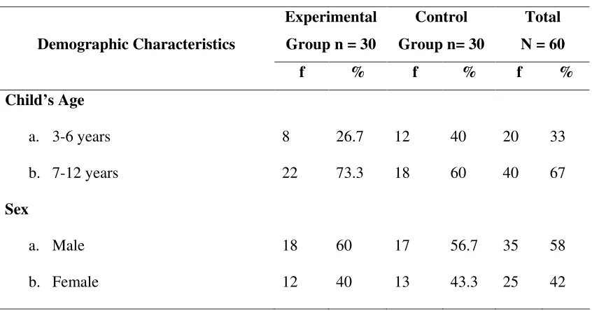
[image:59.612.124.548.233.456.2]DEMOGRAPHIC PROFILE OF THE SAMPLES
TABLE 1
Frequency and percentage distribution of the samples according to their
age &
sex.N = 60 Experimental
Group n = 30
Control Group n= 30
Total N = 60 Demographic Characteristics
f % f % f %
Child’s Age a. 3-6 years b. 7-12 years Sex a. Male b. Female 8 22 18 12 26.7 73.3 60 40 12 18 17 13 40 60 56.7 43.3 20 40 35 25 33 67 58 42
[image:59.612.124.547.234.456.2]Figure 2 : Distribution of samples according to age
TABLE 2:
Frequency and percentage distribution of samples according to the type of
surgery
N=60
Experimental Group n = 30
Control Group n= 30
Total N = 60 Demographic Characteristics
f % f % f %
Appendicectomy Colostomy Ureterolithotomy Cholecystectomy Laproscopic surgery Herniorrhaphy Herniotomy 8 4 5 4 3 4 2 26.7 13.3 16.7 13.3 10 13.3 6.7 7 2 4 6 3 5 3 23.3 6.7 13.3 20 10 16.7 10 15 6 9 10 6 9 5 25 10 15 17 10 15 8
As for the type of abdominal surgery in the experimental group out of 30 children, 8 (26.7%) underwent appendicectomy, 4 (13.3%) colostomy,5(16.7%) ureterolithotomy, 4 (13.3%) cholecystectomy, 3 (10%) laproscopic surgery, 4 (13.3%) herniorrhaphy, 2 (6.7%) herniotomy.
SECTION - II
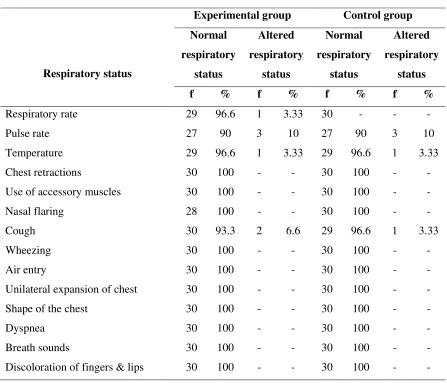
TABLE 3:
Distribution of samples based on respiratory status pre operatively.
N = 60
Experimental group Control group Normal respiratory status Altered respiratory status Normal respiratory status Altered respiratory status Respiratory status
f % f % f % f %
Respiratory rate Pulse rate Temperature Chest retractions
Use of accessory muscles Nasal flaring
Cough Wheezing Air entry
Unilateral expansion of chest Shape of the chest
Dyspnea Breath sounds
Discoloration of fingers & lips
29 27 29 30 30 28 30 30 30 30 30 30 30 30 96.6 90 96.6 100 100 100 93.3 100 100 100 100 100 100 100 1 3 1 - - - 2 - - - - - - - 3.33 10 3.33 - - - 6.6 - - - - - - - 30 27 29 30 30 30 29 30 30 30 30 30 30 30 - 90 96.6 100 100 100 96.6 100 100 100 100 100 100 100 - 3 1 - - - 1 - - - - - - - - 10 3.33 - - - 3.33 - - - - - - -
SECTION - III
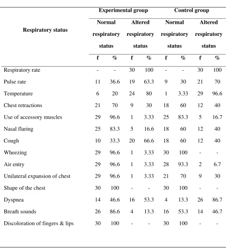
TABLE 4:
Distribution of samples based on respiratory status on the first post
operative day.
N = 60
Experimental group Control group Normal respiratory status Altered respiratory status Normal respiratory status Altered respiratory status Respiratory status
f % f % f % f %
Respiratory rate Pulse rate Temperature Chest retractions
Use of accessory muscles Nasal flaring
Cough Wheezing Air entry
Unilateral expansion of chest Shape of the chest
Dyspnea Breath sounds
Discoloration of fingers & lips
Table 4 represents the distribution of samples based on respiratory status in the experimental group and control group on the first postoperative day.
On the first postoperative day in experimental group, 30 (100%) children had altered respiratory rate, 19 (63.3%) had altered pulse rate, 24 (80%) had altered temperature, 9 (30%) had chest retractions, one (3.33%) child used accessory muscle for breathing, 5 (16.6%) had nasal flaring, 20 (66.6%) had cough, wheezing, altered air entry and unilateral expansion of chest were present each in one (3.33%) child, 16 (53.3%) had dyspnea, 4 (13.3%) had