CERTIFICATE
This is to certify that this dissertation titled “A study and analysis of cutaneous small vessel vasculitis” submitted by DR.O.R.JAYANTHI to the TamilNadu Dr. M.G.R Medical University, Chennai in partial fulfillment of the requirement for the award of MD degree branch – XII A, is a bonafide research work carried out by her under direct supervision and guidance.
DR.S.Krishnan The Dean,
Professor and Head, Madurai Medical College, Department of Dermatology, Madurai.
DECLARATION
I , Dr.O.R.JAYANTHI, solemnly declare that the dissertation titled “A study and analysis of cutaneous small vessel vasculitis” has been prepared by me. This is submitted to The Tamil Nadu Dr. M.G.R Medical University, Chennai, in partial fulfillment of the regulations for the award of MD degree branch – XII A Dermato Venereo Leprology.
Govt. Rajaji Hospital,
ACKNOWLEDGEMENT
At the outset, I thank our Dean Dr.S.M.SIVAKUMAR for permitting me to use the facilities of Madurai Medical College and Government Rajaji Hospital to conduct this study.
I wish to express my respect and sincere gratitude to my beloved teacher and Head of the Department of Dermatology, Dr.S.KRISHNAN for his valuable guidance and encouragement throughout the study and also during my post graduate course. I owe my sincere thanks to him.
I express my thanks and deep sense of gratitude to my teachers Dr.A.S.Krishnaram and Dr.G.Geetharani for their valuable guidance.
I profoundly thank the Head of the Department of Venereology i/c Dr.D.Amalraja for his valuable advice and support.
I sincerely thank the Head of the Department of Pathology Dr.Usha Ravikumar for her valuable guidance.
I also wish to thank all my Assistant Professors of the Department of Dermatology and Venereology, and all my colleagues, who have stood by me and helped me to complete my work.
CONTENTS
S.NO. CONTENTS PAGE NO.
1. INTRODUCTION 1
2. REVIEW OF LITERATURE 3
3. AIM OF THE STUDY 23
4. MATERIALS AND METHODS 24
5. OBSERVATIONS AND RESULTS 26
6. DISCUSSION 39
7. SUMMARY 48
8. CONCLUSION 51
9. APPENDIX
(i) BIBLIOGRAPHY (ii) PHOTOGRAPHS (iii) PROFORMA (iv) MASTER CHART
INTRODUCTION
Vasculitis is inflammatory process affecting the vessel walls and leading
to its compromise or destruction and subsequent hemorrhagic and ischemic
events. Vessels of any organ can be affected that results in a wide variety of
signs and symptoms1,2 The unique feature of this group is multiorgan
involvement. Because of the rich vasculature, the skin is prone to be frequently
affected in vasculitis.
Cutaneous involvement in vasculitides may be primary or reflector of a
fatal systemic disease or evidence of association with some other systemic
disorder. Cutaneous vasculitic lesions offer a window to diagnosis and a ready
source of accessible tissue for histopathologic examination.
Small vessel vasculitis is defined as one which affects mostly vessels
smaller than arteries such as arterioles, capillaries and venules3. These
heterogeneous clinical manifestations, combined with the etiologic non
specificity of the histologic lesions, complicate the diagnosis of specific form of
vasculitis.The gold standard for a diagnosis of vasculitis is histologic
confirmation on biopsy, as few forms of vasculitis have a pathognomonic
laboratory or imaging finding. As a clinicopathological process, vasculitis
occurs both as a primary process or idiopathic vasculitis and as a secondary
malignancy and adverse drugs reaction. Many times the initial presentation of
vasculitis is on the skin and it is the dermatologist who must diagnose and treat
REVIEW OF LITERATURE
Classification
In 1952, Zeek- based upon vessel size and histopathology4, 5, 6
Gilliam and Smiley in 19767- by subdividing Zeek's existing categories
In 1990, American College of Rheumatology (ACR) - based upon clinical,
laboratory, and histologic criteria, and those of the Chapel Hill Consensus
Conferences - based mainly upon histologic criteria8, 9.
An attempt to present a working classification has been previously presented by
Jorizzo in 1993 and others 10, 11.
Proposed working classification of vasculitis [updated version of Gilliam’s
1976 scheme] 12.
Small vessel vasculitis
Cutaneous small vessel vasculitis – not further classified
Henoch – Schonlein purpura
Essential mixed cryoglobulinemia
Waldenstroms hypergammaglobulinaemic purpura
Urticarial vasculitis
Erythema elevatum diutinum
Eosinophilic vasculitis
Rheumatoid nodules
Reactive leprosy
Septic vasculitis
Etiology
Bacterial antigens may be noted in the wall of the small vessels, as may
hepatitis B antigen13, 14. Still, as in all forms of vasculitis, most cases of small
vessel vasculitis are idiopathic (45-54%), due to medications (10-45%),
infections (10-36%), including hepatitis B (5%)13-16. Medications most
commonly associated with small vessel vasculitis are antibiotics and NSAIDS
(especially beta-lactams) and diuretics. Upper respiratory tract infections were
the most common (20%) infectious cause of small vessel vasculitis in one series
from Spain14.
Autoimmune diseases associated with secondary small vessel vasculitis
include rheumatoid arthritis and systemic lupus erythematosus13-16 .Examples of
implicated drugs induce vasculitis are aspirin, penicillin, thiazides, and
necrotizing venulitis are hepatitis B, streptococcal agents, and mycobacterium
tuberculosis. Diseases associated with immune-complex formation include
malignancies, connective tissue diseases, inflammatory bowel disease, and
chronic active hepatitis13, 17.
Pathogenesis 18, 11, 19
Antigenic triggers of immunological responses targeted at components of
blood vessel walls elicit most vasculitides. The deposition of immune
complexes in blood vessel walls is the best characterized mechanism for the
vascular injury associated with vasculitis 18. Potential antigens of relevance
include bacteria, viruses, drugs and other chemicals. The circulating immune
complexes interact with the complement system to generate C3a, C5a,
anaphylotoxins which stimulate the production of chemotactic factors and
subsequent chemotaxis , the release of vasoactive amines (histamine) and the
release of proinflammatory cytokines which induce expression of adhesion
molecules subsequently, immune complex with a sedimentation coefficient
greater than 19S are deposited in vessel walls.
Neutrophils get attracted to this site and these release lysosomal enzymes
in an attempt to engulf the immune complexes. This causes degranulation and
destruction of the neutrophils (leukocytoclasis) with release of collagenase and
elastase, generation of reactive oxygen species, ultimately resulting in
It is of practical importance to appreciate that neutrophils degrade
immune complexes within 24 – 48 hours they are deposited20 usually within 24
hours. Hence direct immunofluorescence of vasculitis lesions older than 3 – 12
hours will generally yield negative results11, 21.
Phenotypic manifestations of cutaneous lesions due to small vessel vasculitis
• The signs and symptoms of small-vessel vasculitis are extremely varied,
and many are shared by all vessel vasculitis Presentation:
o usually:
erythematous macules,
palpable purpura – hall mark feature, initial lesion non
palpable becomes pustule or sometimes ulcerate.
o may also be:
nodules22
hemorrhagic vesicles and bullae
crusted ulcers23,24
gangrene,infarct
less commonly:
pustules32
annular lesion26,29
o lesions:
1mm to several cm diameter
sometimes painful30/ itching
may be:
single crop that subsides spontaneously after few
weeks
crops at different stages of evolution that recur
intermittently31
• Location:
o predilection for dependent parts:
particularly lower legs32,33
o rarely in previously traumatized skin:
Koebner phenomenon34
• Extracutaneous manifestations:
o ≈20% of affected individuals
o include:
arthralgia (synovial) (polyarthritis)
myositis
low-grade fever
o less commonly: 28,11,36,37,38
renal - glomeruli (proteinuria or hematuria)
gastrointestinal (abdominal pain or gastrointestinal bleeding)
pulmonary
neurological32,39 - focal or diffuse, central, or peripheral
neurologic involvement.
o not predicted by severity of histopathological changes40,35
Laboratory findings
These are both to confirm the diagnosis and to determine the extent of
systemic vasculitis, or the existence of underlying associated diseases.
Histopathology
A definitive diagnosis of vasculitis requires histological confirmation in
almost all cases. The hallmark histopathologic pattern of small vessel vasculitis
is leukocytoclastic vasculitis.
• Dynamic:
o not all features necessarily present at a particular stage30
o lesion of 18–24 hours’ duration shows most diagnostic features41
• Usually in small venules (postcapillary venules) in dermis:
Features
Neutrophil infiltration of vessel walls and extending into and beyond
perivascular zone, neutrophils degenerate (leukocytoclasis) with formation of
nuclear dust.
Walls thickened by:
Exudate of inflammatory cells and due to edema fluid and due to
exudation of fibrin (‘fibrinoid necrosis') which often extends into adjacent
perivascular connective tissue.
Endothelial cells:
Usually swollen, some degenerate.
Sometimes thrombosis can occur.
Variable:
o edema
o extravasation of red blood cells
Sometimes eosinophils and lymphocytes:
perivascular
Other Changes
• Macrophages scattered in interstitium:
o even in early stages
o time-dependent increase42
• Mixed inflammatory cell infiltrate of eosinophils, lymphocytes and
occasional neutrophils:
o common in vasculitis due to drugs43
Resolving Lesions33
• Usually only mild perivascular infiltrate of lymphocytes and some
eosinophils
• May be rare plasma cell
• Late resolving lesions:
o hypercellular (busy) dermis striking feature with increased number
of:
interstitial fibroblasts
histiocytes
• Sometimes mild increase in acid mucopolysaccharides:
o gives vague ‘necrobiotic’ appearance
• Uncommonly, pronounced subepidermal edema results in vesiculobullous
lesions
o usually involving only epidermis and upper third of dermis
may follow thrombosis of affected vessels
A lymphocytic form (in which lymphocytes predominate) has also been
described. There is still not enough evidence, however, to prove that the
lymphocyte pattern is truly etiologically or clinically relevant. Old lesions of
small vessel vasculitis may no longer demonstrate leukocytoclastic vasculitis
and may contain mainly lymphocytes around blood vessels44-48. This latter
consideration stresses the importance of timing when taking a biopsy in a
dynamic process such as the vasculitic one.
Cutaneous lymphocytic vasculitis
This form of vasculitis is characterized by lymphomonocytic cell
infiltration as a primary response to various antigens such as medications or
antigens found in connective tissue diseases such as lupus erythematosus,
primary Sjögren's syndrome. These inflammatory cells are present in the
involved vessel wall, while fibrinoid necrosis and leukocytoclasis is absent.
Activated lymphocytes elaborate cytokines, thereby damaging the vessel wall,
either by direct action of the cytokine or promotion of apoptosis. This form of
Drug-induced vasculitis
Drug-Induced vasculitis should be considered in any patient with
small-vessel vasculitis and will be substantiated most often in patients with vasculitis
confined to the skin. Drug causes approximately 10 percent of vasculitic skin
lesions. Drug-Induced vasculitis usually develops within 7 to 21 days after
treatment begins 82, 32. Most common drugs include Beta – lactams, Penicillin,
Sulfadrugs , Nonsteroidal anti- inflammatory drugs53. Identification of tissue
eosinophilia in these biopsies is a clue to a drug etiology54.
Henoch Schonlein purpura55-60
It comprises about 10 % of all cases of cutaneous vasculitis and the most
Common vasculitis in children ( 90 % of all cases)55,56.Upper respiratory tract
infection precedes by 50% , in pediatric patients and 40% in adult patients.
Other factors include drugs, malignancy, foods57, 58. The classic findings are
palpable purpura, arthralgia and abdominal pain58, 59. Furthermore urticaria,
vesicles, bullae and necrotic ulcers may develop.
Henoch Schonlein purpura typically involving the extensor aspect of the
limbs and buttocks in a symmetrical fashion. Henoch Schonlein purpura may
also affect the trunk and face60. Renal involvement occurring in approximately
hematuria. Gastrointestinal bleeding being demonstrated in 50 – 75 % of
patients.
USG Abdomen is indicated to exclude intussusceptions, bowel wall
edema, thickening , or perforation. Arthritis is seen in about 75 % of patients,
most frequently affecting the knees and ankles. ASO titre may be done to rule
out streptococcal infection59. HSP is a clinical diagnosis, with conformation by
direct immune fluorescence and routine histology. Histopathology shows
leukocytoclastic vasculitis. Perivascular IgA deposits are characteristic of HSP.
Essential Mixed Cryoglobulinemia
Cryoglobulins are immunoglobulins that precipitate in the cold and
dissolve on reworming. Type II or essential mixed cryoglobulinemia, in which
the cryoglobulin contains both a polyclonal IgG (Antigen)and a monoclonal
IgM Rheumatoid factor directed against the IgG. Most cases are due to chronic
infection with hepatitis C virus 61-65.
Although infection with hepatitis B virus and Epstein –Barr virus have
been implicated in some patients61-67. Type III , in which there is also mixed
cryoglobulin , but both the IgG and the rheumatoid factor IgM are
polyclonal.this condition is often seen in chronic inflammatory and autoimmune
disorders ( such as lupus and leukocytoclastic vasculitis), lymphoproliferative
The median age at diagnosis of cryoglobulinemia is early to middle sixth
decade, with a female to male ratio of 2: 1 69.
Recurrent showers of dependent palpable purpura associated with arthritis
or arthralgia is the common presentation. Sometime cutaneous infarction may
be the presenting feature. Histopathology shows the presence of hyaline thrombi
in vessel wall with evidence of vessel wall damage. Conformation of
cryoglobulins requires cryoglobulin test followed by serum protein
electrophoresis69.
Waldenstroms Hypergammaglobulinaemic Purpura
Its a polyclonal disorder most commonly linked with sarcoidosis, lupus
erythematosus, sjogren syndrome and other auto immune conditions 71, 77. The
majority of patients have positive anti nuclear antibody and anti- SSA (Ro) or
anti –SSB (La) antibodies73-77. Other features associated include arthropathy,
renal tubular acidosis, chest infections, lymphopenia and immune
hypersensitivity pneumonitis. Most patients are female 78.
Clinically the pattern of purpura usually consisting of crops of small
erythematous macular or palpable purpuric spots on the lower leg70-77.
Prolonged walking, standing or sitting with the legs dependent, or other increase
in venous pressure, may be an obvious provocative factor76,77. Histology may
walls75,79, 80. There may be an abnormal ratio of IgG subclasses, with low
IgG1/IgG2 ratio81.
Vasculitis Associated With Collagen Vascular Diseases
Around 12% of cutaneous vasculitis cases will be associated with a
connective tissue disorders55, 56. Secondary vasculitis due to connective tissue
disease should be considered in patients presenting with biopsy – proven
cutaneous vasculitis who have signs and symtoms of dry eyes, drymouth,
arthritis, sclerosis, photosensitivity, or serologic evidence of ANA, RF,
antiphospholipid antibodies, or anti- DNA, Ro, or La antibodies. Cutaneous
vasculitis occurs frequently in SLE, RA and Sjogren syndrome, less commonly
in dermatomyositis, scleroderma, and polychondritis55, 56, 82.
Increased or enhanced expression of vascular adhesion molecules
attracting and activating neutrophils is suspected to play a role in the
pathogenesis of connective tissue vasculitis83. In general, CTD vasculitis show
more widespread organ involvement and the calibre of vessels affected shows
more variation compared with conventional CLA. For example, lupus vasculitis
patients represent a heterogenous population of which nearly 60% of cases do
not fulfil the categories and definitions adopted by the CHCC84.
Small vessel vasculitis in LV patients is predominantly cutaneous84. LV
patients are more likely to have livedo racemosa, anemia, elevated ESR, anti-La
Arterioles and post – capillary venules are the vessels most commonly affected
by vasculitis, manifesting as purpura, vesiculobullous lesions , urticaria , and
splinter haemorrhages.
Skin biopsy will reveal a mixed, mostly small and less commonly
muscular vessel neutrophilic vasculitis with lesions that can resemble either
typical CLA or PAN. Extravascular histologies can provide clues to the
diagnosis of CTD vasculitis such as the presence of interface dermatitis with
dermal mucin deposition (found in SLE and Dermatomyositis), dermal and / or
subcutaneous sclerosis in scleroderma, palisaded neutrophilic and
granulomarous dermatitis in RA or SLE and tissue neutrophilia in SLE and
Sjogren syndrome 85.
Urticarial Vasculitis
Of patients with urticarial lesions, roughly 5-10% have urticarial
vasculitius86, 87. It is a chronic disease, which presents as urticarial lesions that
most often occur on the trunk or proximal limbs, frequently with associated
angioedema88. Lesions differ from those of simple urticaria in that individual
lesions persist for greater than 24hours, often demonstrate purpura and post
inflammatory pigmentation, and cause symptoms of burning.
Two types described 1) UV associated with hypocomplementaemia 2)
normocomplementaemic UV. Most UV patients are women (female to male
connective tissue diseases (sjogren syndrome, SLE86, physical urticarias,
hepatitis B or C, IgM or IgA gammopathies, serum sickness, colon cancer and
drug ingestion. UV is thought to represent a type III hypersensitivity reaction, as
circulating immune complexes may be demonstrated in upto 75% of patients89.
Lesions of UV demonstrate leukocytoclastic vasculitis.
Erythema Elevatum Diutinum
EED is a rare chronic cutaneous eruption that is most commonly seen in
adults. Though the exact etiology is unknown it has been associated with
autoimmune diseases such as Rheumatoid arthritis, celiac disease, inflammatory
bowel disease and type I diapetes mellitu, infections (streptococcus, hepatitis,
syphilis, HIV 90-100. In addition it has been associated with
hypergammaglobulinemia and IgA monoclonal gammopathies, myelodysplasia,
pyoderma gangrenosum and relapsing polychondritis. The association with
haematological disorders, such as multiple myeloma, is strong; however EED
may precede the haematological disease by several years101.
Histopathology of acute lesions show leukocytoclastic vasculitis.
Chronic lesions demonstrate fibrosis, capillary proliferations and infiltration of
macrophages, plasma cellsand lymphocytes. Lesions of EED most commonly
appear chronically in a symmetrical fashion over the dorsa of the hands, the
knees, buttocks and Achilles tendons. They are red violaceous, red brown or
atrophic scars. It may last from 5 to 35 years, with crops of new lesions
developing every few weeks to months.
Eosinophilic Vasculitis
Rare vasculitis, cause is unknown. Eosinophil cytokines such as IL-5, and
toxic eosinophil granule proteins such as major basic protein, have been
demonstrated in serum and tissues, respectively, and presumably play a part in
the tissue damage. Neutrophil elastase is predominantly around vessels, and
mast cell degranulation occurs. Eosinophilic vasculitis has also been reported in
a patient with the hypereosinophilic syndrome. In this patient CD40 (a
glycoprotein of the TNF receptor family) was considered to be important in
pathogenesis 102.
Recurrent pruritic papules and urticarial lesions occur at any site,
especially the head and neck, with angioedema of the face and extremities.
Either sex and any age group may be affected. The course is long and recurrent
but fever, arthralgia and visceral involvement are absent. Raynaud’s
phenomenon and digital gangrene were reported in a patient with cutaneous
eosinophilic vasculitis associated with the hypereosinophilic syndrome 102
Histopathology shows fibrinoid deposition and necrosis of small dermal vessels
with an infiltrate of eosinophils and absent or minimal leukocytoclasis.
Small vesicles containing eosinophils may be present. Immunoglobulin
from other vasculitides such as CSS, in which medium to large vessels are
affected.
Rheumatoid Nodule
Palpable subcutaneous nodules occur in approximately 20% of patients
with RA. The most common site is on ulnar border of the forearm.Less
commonly ,they occur on the dorsa of the hands, on the knees, on the ears, over
the scapulae. They vary in size upto several centimetres in diameter, are firm in
cocsistency and tend to ulcerate with trauma. Nodules are almost invariably
asocited with more severe forms of the disease, and rheumatoid factor and
antinuclear factor are frequently found in the serum.
Nodules were characterized by granuloma formation secondary to
leukocytoclastic vasculitis103. Histologically, rheumatoid nodules consists of
fibrous tissue in which foci of fibrinoid necrosis are scattered, surrounded by a
palisade of cells, mainly fibroblasts and histiocytes. A peripheral zone of
lymphocytes and plasma cells occurs. Within the necrotic area are thin
reticulum fibres, amorphous material and some nuclear debris are seen.
Reactive Leprosy
Erythema nonosum leprosum (ENL) or type II reaction is an immune
complex syndrome seen in multibacillary leprosy 104. ENL usually occurs after
observed in patients who have not been treated. ENL presents with
inflammatory skin nodules and involvement of multiple organs, often running a
protracted course. Over one half of lepromatous patients and one quarter of
borderline lepromatous patients will experience type 2 reaction105.
The clinical manifestation is of evanescent crops of tender erythematous
papules or nodules located on interlesional skin. They are all dome shaped with
ill defined margins. The papules may turn into pustules or may simply ulcerate.
They can also occur as subcutaneous plaques. The lesions are most common on
the face and extensor surface of limbs and less commonly on the trunk,
accompanied by fever, malaise, arthralgia and leukocytosis.
Lesions tend to recur at the same sites and if they do not resolve
completely, a chronic painful panniculitis develops which may persist for
months or years.
Histopathology: In ENL, the lesions are foci of acute inflammation
superimposed on chronic multibacillary leprosy106. It is characterized by edema
of papillary dermis and a mixed dermal infiltrate of neutrophils and
lymphocytes superimposed on collection of macrophages. There may be
involvement of subcutis, with the development of mixed lobular and septal
panniculitis. However, in majority of case involvement of the dermis is the
primary and predominant finding. Macrophage in dermis contain fragmented
dermal abscess with ulceration. Whereas foamy macrophages containing
fragmented bacilli are usual, in some patients no bacilli remain and
macrophages have a granular pink hue on Wade-Fite staining, indicating
mycobacterial debris.
Lesions exhibit necrotizing vasculitis involving capillaries, venules, and
small-to-medium arteries and veins. In the superficial dermis, affected venules
and capillaries showed endothelial cell enlargement and focal necrosis
associated with perivascular infiltrates of lymphocytes.
In the deep dermis and subcutaneous tissue, affected venules, arterioles
and arteries exhibited endothelial cell necrosis and matted fibrin in the vessel
walls associated with perivascular infiltrates of neutrophils107. Throughout the
dermis, mononuclear phagocytes with vacuoles containing numerous
fragmented organisms were observed. These patients may have superficial
ulceration106.
Lucio phenomenon108,109,110
Lucio phenomenon is a vasculitis clinically described in 1852 and
microscopically documented in 1948 in patients with diffuse lepromatous
leprosy. Usually occurs in patients who have received either no treatment or
inadequate treatment. In contrast to ENL, fever, tenderness and leukocytosis are
marginated, irregular plaques. They develop into crusted lesions and,
particularly on the legs, into ulcers.
Histopathologically, Lucio phenomenon is a distinctive type of
granulomatous and necrotizing panvasculitis108 of small vessels in the upper and
mid dermis that results in ulceration of epidermis. Occasionally the process
extends deep into the subcutaneous fat with small vessel vasculitis in the fat
lobule109.
Septic Vasculitis
About 22% of all cases of cutaneous vasculitis are associated with
infection, and all types of infectious agents (viruses, bacteria, fungi, protozoa
and helminthes) have been associated with the development of vasculitis55, 56.
The cutaneous pathology most often found in these cases is a small-vessel
neutrophilic vasculitis affecting superficial dermal vessels.
LCV histology found in infection-related vasculitis is likely A common
morphologic endpoint of several pathways: Immune complex formation,
alternate pathway of complement activation, and endotoxin-mediated
expression of vascular adhesion molecules108, 109. In the case of Immune
Complex formations, infection-triggered LCV is suspected to show a greater
frequency of subcorneal, intraepidermal, and subepidermal neutrophilic
deposits, and to have relatively fewer eosinophils and lymphocytes than
idiopathic CLA and drug-related cutaneous LCV 110.
Septic vasculitis is a variant of small-vessel neutrophilic vasculitis that is
typically immune-complex negative and caused by infective endocarditis or
septicemia from gonococci, meningococci, pseudomonads, staphylococci,
streptococci, certain rickettsial infections, and other microorganisms55.
The clinical lesions of septic vasculitis are characterized by purpura
(petechiae and ecchymoses), vesiculo-pustules (often with gray roofs signifying
necrosis), hemorrhagic bullae, and, rarely ulceration. Patients with chronic
gonococcemia and chronic meningococcemia have a triad of intermittent fever,
arthralgia, and fewer clinical lesions, distributed over extremities, particularly
acral surfaces, which are mostly petechiae surrounded by a rim of erythema
vesiculo-pustules with a gray necrotic roof, and rarely hemorrhagic bullae.
Biopsy reveals mixed neutrophilic small and muscular-vessel vasculitis
with deep dermal and subcutaneous vessel involvement associated with scant
perivascular fibrin or fibrin thrombi, and no or little nuclear debris. These
AIM OF THE STUDY
1) To study the epidemiological spectrum of cutaneous small vessel vasculitides.
2) To determine the clinicopathological correlation.
MATERIALS AND METHODS
A study was conducted during the period from May 2008 – May
2010 in the department of dermatology, Govt Rajaji Hospital, Madurai
Medical college, Madurai, among the patients with cutaneous small
vessel vasculitis attending the dermatology department as well as those
referred from other departments mainly Medicine, Rheumatology and
paediatrics.
Inclusion criteria:
1. All patients with clinical features suggestive of small vessel vasculitis
i.e palpable purpura , infiltrated erythema , hemorrhagic vesicles and
bulla, ulcers, infarct, digital gangrene, erythematous plaques and
nodules, urticaria, livedoreticularis which was subsequently supported
by histopathological examination.
Exclusioncriteria:
1. Patients who were unwilling for the study
2. Patients with abnormal bleeding parameters.
History:
A detailed history was taken which includes, symptoms (itching , burning
sensation, pain) duration of skin lesions, occupational history, systemic
symptoms, history of sore throat in the recent past, history of drug intake,
history suggestive of malignancy and collagen vascular disorders.
Clinical examination:
Detailed general and systemic examinations were done. Detailed
examination of skin lesion which includes morphology of skin lesions,
distribution of lesions, symmetry, tenderness, diascopy were done.
Investigations:
1) Baseline laboratory investigations included are complete hemogram,
serum urea, serum creatinine, liver function tests , chest X ray, urine
(routine and microscopy), Mantoux test, test for stool occult blood,
ASO titre, blood culture and skin smears for acid fast bacilli, USG
Abdomen and pelvis .
2) Screening for HIV, Hepatitis B, C and syphilis were also done for
high risk patients with history of sexual exposure or occupational
exposure to blood and blood products.
3) Tests to rule out cryoglobulinemia, (cryoglobulin test , serum protein
electrophoresis, complement ) malignancy and collagen vascular
4) Incisional elliptical skin biopsies were done from the early tender skin
lesions with a caution not to include the resolving lesion and they
were sent to pathologist for histopahology Special stains like AFB
were done when required.
5) Other tests to rule out bacterial infections include gram stain and
blood culture.
Classification of patients with features of cutaneous small vessel vasculitis in
our study was based on Proposed working classification of vasculitis
[updated version of Gilliam’s 1976 scheme 12 .
Analysis :
A descriptive analysis of the clinical characteristics, laboratory
parameters and histopathological features of various cutaneous small vessel
vasculitis in our study was done.The data was analysed and compared with
OBSERVATION AND RESULTS
Fifty one patients with clinical features of cutaneous small vessel vasculitis
were seen during the study period from May 2008 to May 2010. Of these, 11
cases were excluded from the study because of the patients denial for the study
(4), not willing for biopsy (7) .40 patients were diagnosed as cutaneous small
[image:33.612.131.484.323.710.2]vessel vasculitis were included in our study.
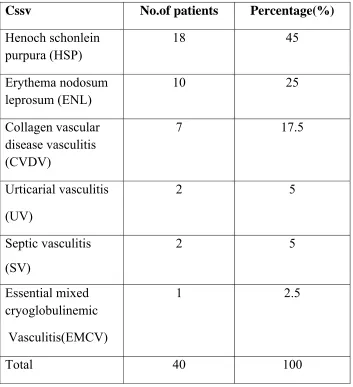
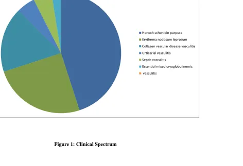
Table -1
Clinical spectrum of cutaneous small vessel vasculitis
Cssv No.of patients Percentage(%)
Henoch schonlein purpura (HSP) 18 45 Erythema nodosum leprosum (ENL) 10 25 Collagen vascular disease vasculitis (CVDV) 7 17.5 Urticarial vasculitis (UV) 2 5 Septic vasculitis (SV) 2 5 Essential mixed cryoglobulinemic Vasculitis(EMCV) 1 2.5
Key
Cssv = Cutaneous small vessel vasculitis
The common types were Henoch schonlein purpura (18) , Erythema
nodosum leprosum(10), small vessel vasculitis associated with collagen
vascular disease (7). The less common types were urticarial vasculitis (2),
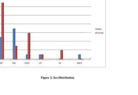
Table – 2
Sex distribution
Disease Male Female
No.of cases
% No.of
cases
%
HSP 5 28 13 72
ENL 7 70 3 30
CVDV 1 14 6 86
UV 1 50 1 50
SV - - 2 100
EMCV 1 100 - -
Total 15 38 25 62
Key
HSP = Henoch Schonlein purpura
ENL = Erythema nodosum leprosum
CVDV = Collagen vascular disease vasculitis
UV = Urticaril vasculitis
SV = Septic vasculitis
EMCV = Essential mixed cryoglobulinemic vasculitis
There were 15 (38%) male and 25 (62%) female patients .
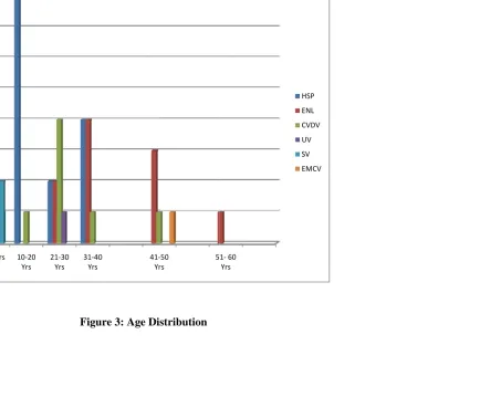
Table- 3
Age distribution Cssv <10
Yrs 10-20 Yrs 21-30 Yrs 31-40 Yrs 41-50 Yrs 51- 60 Yrs >60 Yrs Total
HSP 4 8 2 4 18
ENL 2 4 3 1 10
CVDV
1 4 1 1 7
UV
1 1 2
SV
2 2
EMCV
1 1
TOTAL
7 9 9 9 5 1 40
Key – Cssv = Cutaneous small vessel vasculitis
HSP = Henoch Schonlein purpura
ENL = Erythema nodosum leprosum
SV = Septic vasculitis Yrs = Years.
EMCV = Essential mixed cryoglobulinemic vasculitis
Majority of patients were between the ages of 11 – 40 years.
The mean age of our study group was 31 years for males and 24 years for
[image:38.612.90.526.298.566.2]females.
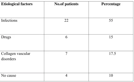
Table -4
Etiological factors
Etiological factors No.of patients Percentage
Infections 22 55
Drugs 6 15
Collagen vascular disorders
7 17.5
No cause 4 10
Approximately twenty two ( 55%) of the patients had infections, and seven
( 17.5%) had positive connective tissue disease workup without any overt
manifestations, six (15%) were attributed to drugs ( These included NSAIDS
(2.5%) patient had cryoglobulinemia. No cause was found in four (10%)
[image:39.612.88.525.138.457.2]cases.
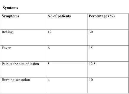
Table -5
Symtoms
Itching was the commonest presenting symptom in twelve (30%) patients. six
patients complained of fever (15%), while burning sensation and pain at the site
of the lesion were encountered in four and five patients respectively.
Symptoms No.of patients Percentage (%)
Itching 12 30
Fever 6 15
Pain at the site of lesion 5 12.5
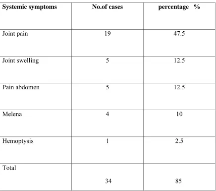
Table – 6
Systemic symptoms
Systemic symptoms were encountered in 34 (85%) patients. Associated joint
pains were the commonest systemic presentation in 19 (47.5%) patients with
knee joint being the most commonly involved joint (11). Other joints involved
were ankle joint and small joints of feet and wrists. Joint swelling was observed
in 5 patients. There was history of abdominal pain in 5 patients, melena in 4
Systemic symptoms No.of cases percentage %
Joint pain 19 47.5
Joint swelling 5 12.5
Pain abdomen 5 12.5
Melena 4 10
Hemoptysis 1 2.5
Total
34
patients and hemoptysis in 1 patient .
Table – 7
Signs
Signs No.of patients Percentage (%)
Palpable purpura 21 52.5
Nodule 7 17.5
Plaque 3 7.5
Ulcers (crusted & necrotic) 3 7.5
Urticarial lesions 2 5
Bulla 1 2.5
Ecchymoses 1 2.5
Pustule 1 2.5
Palpable purpura was the commonest cutaneous presentation noticed in 21
patients (16 females and 5 males). The other cutaneous lesions seen in 19
patients were in the form of nodules, plaques, ulcers, bullae, vesicles, gangrene
of toes, urticarial lesions and Koebner phenomenon. The time since onset of
[image:42.612.92.524.368.566.2]lesions varied from 1 day to 9 months.
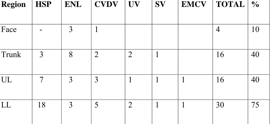
Table- 8
Distribution
Region HSP ENL CVDV UV SV EMCV TOTAL %
Face - 3 1 4 10
Trunk 3 8 2 2 1 16 40
UL 7 3 3 1 1 1 16 40
Key
HSP = Henoch Schonlein purpura
ENL = Erythema nodosum leprosum
CVDV = Collagen vascular disease vasculitis
UV = Urticaril vasculitis
SV = Septic vasculitis
EMCV = Essential mixed cryoglobulinemic vasculitis
LL = Lower limb UL = Upper limb
The commonest sites were lower limb (75 %) mainly the legs and anklle
Table – 9
Laboratory findings
Laboratory parameters No.of patients Percentage (%)
Anaemia 16 40
Leukocytosis 12 30
Raised ESR 27 67.5
Elevated urea 6 15
Elevated serum creatinine 6 15
Albuminuria 7 17.5
Urine examination
RBC 7 17.5
Pus cells - -
Bacilli - -
Stool for occult blood 8 20
Abnormal chest x ray 1 2.5
Anti nuclear antibody 5 12.5
Rheumatoid factor 2 5
ASO titre 10 25
Mantoux test 1 2.5
Hepatitis B Virus - -
Hepatitis C Virus - -
HIV - -
Cryoglobulin Test 1 2.5
USG abdomen – abnormality
KEY
ESR = Erythrocyte sedimentation rate
ASO titre = Anti streptolysin O titre
RBC = Red blood cells
USG abdomen = Ultrasonography abdomen
HIV = Human immunodeficiency virus.
The hematological and biochemistry workup revealed anemia in sixteen
(40%)patients, leukocytosis in twelve(30%), elevated ESR in twenty
seven(67.5%), raised serum-urea in six and raised creatinine levels in six
patients. Routine urine examination showed albuminuria in seven patients,
while urine microscopy demonstrated blood cells in seven patients. The stool
for occult blood was positive in eight patients. Chest x ray showing cavity in
one patient with history of hemoptysis. Smear from pustular lesion in one
patient revealed gonococci. Blood culture from one patient with ecchymoses
showed growth of Pseudomonas aeruginosa. Anti-nuclear antibody and
rheumatoid factor were positive in five and two patients, serum cryoglobulins
were positive in one patient . ASO titer was also raised in ten patients, while
Mantoux was positive in one patient. USG abdomen showed bowel wall edema
Table - 10 Histopathology Clinical diagnosis No .of cases % Histopathological diagnosis No. Of cases % NEV %
HSP 18 45 LCV 15 37.5 3 7.5
ENL 10 25 LCV , mixed
panniculitis
8 20 2 5
CVDV
LE 6 15 LCV 5 12.5 1 2.5
RA 1 2.5 LCV 1 2.5
Urticarial
Vasculitis
2 5 LCV 2 5
Septic vasculitis 2 5 LCV 2 5
Essential mixed cryoglobulinemic vasculitis
1 2.5 LCV with hyaline
thrombi
1 2.5
Total
Key – LCV = Leukocytoclastic vasculitis LE =Lupus erythematosus
NEV = No evidence of vasculitis RA = Rheumatoid arthritis
HSP = Henoch Schonlein purpura
ENL = Erythema nodosum leprosum
CVDV = Collagen vascular disease vasculitis
Based on histopathological findings, 34 (85%) patients were given a diagnosis
of leukocytoclastic vasculitis, while 6 (15%) patients showed perivascular
lymphocytic infiltrates with no evidence of vasculitis. The skin biopsy showed
typical features of endothelial swelling, fibrinoid necrosis, RBC extravasation
and leukocytoclasis. Additional findings include subepidermal bulla , Hyaline
DISCUSSION
Cutaneous small vessel vasculitis is a poorly understood entity due to its
protean clinical manifestation and its overlap with various infections, collagen
vascular disorders and malignancies. In our study, we analyzed cases of
cutaneous small vessel vasculitis diagnosed on the basis of history, clinical
features, and various laboratory tests. The clinical diagnosis was supported by
skin biopsy. Our study confirmed various established facts regarding cutaneous
small vessel vasculitis and throws light on some new aspects.
One of the published study on cutaneous vasculitis was by Gupta et al112
between 2004 -2006 Postgraduate Institute of Medical Education and Research,
Chandigarh, India. They included 50 patients. The results of the study by Gupta
et al 112 indicated that there were 20 male and 30 female patients. The mean age
of the study group was 41.1 years for males and 35.9 years for females. In our
study, 15(32%) were male and 25(62%) females. Females were more commonly
affected than males in our study consistent with the study by Gupta et al.112 .
The mean age of our study group was 31 years for males and 24 years for
Etiology
A possible etiological association was suspected in 90% of our patients
which was comparable to 67.2% seen by Sais et al 111. Most common cause
found in our study was infections 55%, mainly upper respiratory tract infections
and this is consistent with study by Martinez-Taboada et al14 collagen vascular
disorders (17.5%) and drugs (15%) were other causes of cutaneous small vessel
vasculitis in our study. Lupus erythematosus was the common collagen vascular
disease found in our study. NSAIDS were the common drugs followed by
antibiotics. This was comparable with the results of the study by Sams et al 13,
Callen et al15, Hautmann et al16.
Symptoms
The common symtoms observed in cutaneous lesions of our study were itching
(30%), pain (12.5%) and burning sensation (10%). This was comparable with
earlier studies by Sais et al111 where 41.3% patients complained of itching and
30% had painful lesions. Fever was seen in 15% of our patients while it was
31.6% in the study by Sais et al 111.
Signs
The most common clinical presentation in our study was crops of
nonthrombogenic palpable purpura, primarily involving dependent areas such as
with earlier studies by Sais et al111 , Ekenstam et al113,Gupta et al112 and Lopez
Maturana et al116 in 89.2%, 62%, 86% and 62% of cases, respectively.
Koebner phenomenon was observed in two (5%) patients as reported in
standard literature34 . The second most common type of lesion in our study was
cutaneous nodules which comprised 17.5% of patients as against 2% observed
by Sais et al111.This may be due to high percentage of reactive leprosy in our
study and high prevalence of multibacillary leprosy patients in India.
Systemic involvement
Systemic involvement was observed in 87.5% of patients with the joint
pain being the commonest presenting manifestation (52.5%). This was again in
consonance with the systemic involvement observed by Ekenstam et al113 in
51% of patients where the musculoskeletal system was most commonly
involved (43%). However Sais et al 111 observed systemic involvement only in
20% of the cases with joint involvement in 36.7% of all cases.
Gastrointestinal involvement mainly in the form of abdominal pain,
melena, occult blood in stools and bowel edema in Ultrasonography abdomen,
was observed in 13(32.5%) patients which was consistent with 9.5% cases
reported by Saiset al111.
Winkelmann and Ditto114 found renal involvement in 61% of their
patients, while in our study, renal involvement was seen in only 11(27.5%) of
disorders in the form of elevated serum urea and creatinine, proteinuria and
microscopic hematuria. In patients of Henoch Schonlein purpura with renal
involvement, three had extensive cutaneous lesions all over the body while
seven patients had musculoskeletal symptoms.This is consistent with study by
Gupta et al112 where 16 % of cases had renal involvement, in that 3 had
extensive cutaneous lesions, while 3 had musculoskeletal symptoms.
Pulmonary involvement was seen in only one case in the form of positive
mantoux test and cavities (tuberculosis) in a patient of Henoch Schonlein
purpura vasculitis. Tuberculosis was seen in (2.5 %) of our patients in contrast
to no case detection in study by Sais et al 111 and Ekenstam et al113.This was
probably due to the high prevalence of tuberculosis in India.
Laboratory findings
The laboratory parameters reflecting systemic inflammatory responses
were elevated ESR in 27 (67.5%) patients, anemia in 16 (40%) and leukocytosis
in 12(30%) patients. It was consistent with study by Sais et al111 where elevated
ESR (52.4%), anemia (37%) and leukocytosis ( 18%) was observed.
The renal functions were altered in 6 (15%) patients in the form of
elevated urea and creatinine and abnormal urine examination in 14 (35%)
patients. Saiset al 111 found these parameters to be 26 and 21.1%, respectively.
Liver function tests were within normal limits in all patients while Sais et al111
The collagen vascular disease workup revealed positive Anti nuclear
antibodies in 12.5% and rheumatoid factor in 5% patients, which was in
consistent with study results reported by Gupta et al 112 . In addition serum
cryoglobulins were found positive in 1 (2.5%) patient consistent with study by
Gupta et al in which 1(2%) patient had serum cryoglobulin. ASO titer was
raised in 10 (25%) patients, while Mantoux test was positive in one (2.5%) of
our patients. Gupta et al 112 reported ASO titre was found raised only one
(2%) patient.
Histopathology
Histopathology showed features of leukocytoclastic vasculitis in 34
patients. In the remaining 6, perivascular lymphocytic infiltrate with no
evidence of vasculitis was observed which was consistent with study by Gupta
et al111 where out of 42 patients, 5 patients did not show evidence of vasculitis.
Hence the diagnosis of cutaneous small vessel vasculitis was considered on the
basis of high index of clinical suspicion. Moreover, histopathology was
noncontributory in these cases, probably due to the biopsy of the lesion at a late
stage in the disease evolution as mentioned in the literatue44-48. We found 25 %
of patients with Erythema nodosum leprosum showing features of
leukocytoclastic vasculitis in addition to mixed panniculitis as reported by Giam
SUMMARY
40 patients, diagnosed with cutaneous small vessel vasculitides, based on
history, clinical features, laboratory findings, histopathology were included in
the study. This study was done for a period of 24 months ( May2008 to May
2010) .
Sex
15 were male (38%) and 25 were female (62%). The male : female ratio
was 1:1.7.
Age
Majority of patients were between the ages of 11 – 40 years. The mean
age of our study group was 31 years for males and 24 years for females.
.
Clinical spectrum
The common types of cutaneous small vessel vasculitis included
Henoch Schonlein purpura (45%), Erythema nodosum leprosum (25%),
connective tissue disorders with vasculitis (17.5%). The less common types
were urticarial vasculitis (5%), septic vasculitis (5%) and Essential mixed
cryoglobulinemic vasculitis(2.5%).
Aetiology
Infections(55%) mainly the upper respiratory tract infections were the
commonest underlying cause encountered in our study followed by collagen
vascular disorder (17.5%) and drugs (15%).
Symptoms associated with skin lesions
Itching was the predominant symptom noted in 30 % of patients followed
by fever and burning sensation.
Systemic symptoms
Joint pain was the most common systemic symptom observed in 47.5%
of patients followed by joint swelling. In patients of HSP with renal
involvement, three had extensive cutaneous lesions all over the body while
seven patients had musculoskeletal symptoms.
Morphology & distribution
The morphology of lesions and their distribution were concordant with
those described in the literature . Clinically most of the patients presented with
palpable purpura in the dependent parts especially the legs. Legs involvement
and palpable purpura were noted in 100% of cases of Henoch Schonlein
Laboratory findings
Common laboratory findings observed were elevated ESR(67.5%), and
anemia (40%) .
Histopathology
Leukoocytoclastic vasculitis was the most common histopathological
pattern observed in 34 cases.We found a significant proportion(25%) of patients
with ENL presenting as leukocytoclastic vasculitis in addition to mixed
panniculitis.
CONCLUSION
1. Henoch Schonlein purpura was the most common cutaneous small vessel
vasculitis followed by Erythema nodosum leprosum.
2. Cutaneous small vessel vasculitis was more common in females.
3. Majority of the patients were between 11 – 40 years of age.
4. Upper respiratory tract infection was the commonest etiologic factor in
cutaneous small vessel vasculitis.
5. Joint pain was the common systemic symptom found in majority of
patients.
6. Nonthrombogenic palpable purpura represents clinical hallmark in
cutaneous small vessel vasculitis.
7. Widespread cutaneous manifestations may be the cutaneous marker of
serious systemic involvement.
8. Elevated ESR and anemia were the common laboratory abnormalities.
9. Leukocytoclastic vasculitis was the common histopathological pattern in
cutaneous small vessel vasculitis.
10.Significant proportion of patients with Erythema nodosum Leprosum
presenting as leukocytoclastic vasculitis .
11.Early biopsy and Clinicopathological correlation is necessary for the
KEY TO MASTER CHART
Morphology
1 = Palpable purpura 2 = Nodules
3 = Plaques
4 = Nodules and plaques 5 = Ulcer
6 = Urticaria 7 = Gangrene 8 = Pustules
9 = Bulla with / without ulcer 10 = Ecchymoses
DSL = Duration of skin lesions Symptoms
1 = Burning sensation 2 = Itching
3 = pain 4 = Fever Systemic symptoms
2 = Joint swelling 3 = Melena
4 = Abdominal pain 5 = Diarrhoea 6 = Hemoptysis Drugs
1 = NSAIDS (Non steroidal anti inflammatory drugs) 2 = Antibiotics
3 = Unknown Region
1 = Lower limb
2 = Lower limb and trunk
3 = lower limb, trunk and upper limb 4 = Lower limb and upper limb ASO titre = Anti Streptolysin O titre Urine routine
1 = Hematuria 2 = Proteinuria
RFT = Renal function test
1 = Elevated urea and creatinine LFT = Liver function test
RF = Rheumatoid factor ANA = Anti nuclear antibody CRGT = Cryoglobulin test CXR = Chest X ray
Cavt = Cavity MX = Mantoux test
USG Abdomen = Ultrasonography of Abdomen B.E = Bowel wall edema
SSS = Slit skin smear CD = Clinical diagnosis
HSP = Henoch Schonlein purpura
LEV = Lupus erythematosus with vasculitis RAV = Rheumatoid arthritis with vasculitis ENL = Erythema nodosum leprosum UV = Urticarial vasculitis
SV = Septic vasculitis
HP = Histopathology
LCV = Leukocytoclastic vasculitis MP = Mixed panniculitis
NEV = No evidence of vasculitis P = Positive
Ag e / Se x Morplolog y DSL Sy mt oms Sy st emic sy m to ms Sore T hroat Drug s Regi ons AS O ti tre Stool Occult Blood Ur in e R out in e
RFT LFT RF AN
A
CRGT CXR MX
V irual Markers USG A bdomen Le uk o Cy to si s Anem ia Elev
ated ESR SSS CD HP
1 Deepika 12y/F 1 2w 2 1 P _ 3 P P 3 1 NRL N N N NRL N N NRL P P P _ HSP LCV
2 Arul pondi 19y/M 1 3M 2 4 _ _ 3 P P 3 1 NRL N N N NRL N N B.E P _ P _ HSP LCV
3 Sumathy 28y/F 2 2M _ 1 _ _ 1 _ _ _ NRL NRL N N N NRL N N NRL _ P P P ENL LCV, MP
4 Arumugam 50y/M 7 15D _ _ _ _ 4 _ _ _ NRL NRL P N P NRL N N NRL _ _ P _ EMCV LCV
5 Baby of Gayathiri 15D /F 8 5D 4 2 _ _ 4 _ _ _ NRL NRL N N N NRL N N NRL P _ P _ SV LCV
6 Abdul Majeet 45y/M 2 6M _ _ _ _ 3 _ _ _ NRL NRL N N N NRL N N NRL _ _ P P ENL LCV, MP
7 Dinesh Kumar 9y/M 1 1D 4 1 P _ 1 P P _ NRL NRL N N N NRL N N NRL P P _ _ HSP NEV
8 Deepa 18y/F 1 1M 4 1 _ _ 1 _ _ 2 1 NRL N P N NRL N N NRL _ P P _ LEV LCV
9 Bhuvana 7 y/F 1 1D _ 1 P _ 1 P _ 1 NRL NRL N N N NRL N N NRL P _ P _ HSP LCV
10 Perumal 50y/M 2 6M _ 0 _ _ 3 _ _ _ NRL NRL N N N NRL N N NRL P _ _ P ENL LCV, MP
11 Alagu 48y/M 5 1M 3 1 _ _ 3 _ _ 2 1 NRL N P N NRL N N NRL _ _ P _ LEV LCV
12 Priya 3y/F 10 2D _ _ _ _ 1 _ _ _ NRL NRL N N N NRL N N NRL P _ P _ SV LCV
13 Praveen 7 y/M 6 15D 1 4 _ _ 4 _ _ _ NRL NRL N N N NRL N N NRL _ _ P _ UV LCV
14 Kavitha 6 y/F 1 7D 2 2 P _ 2 P P _ NRL NRL N N N NRL N N NRL P _ _ _ HSP NEV
15 Angayarkanni 28y/M 5 6M 3 1 _ _ 1 _ _ _ NRL NRL N N N NRL N N NRL _ P P _ LEV LCV
16 Eswar 40y/M 4 9M 3 _ _ _ 1 _ _ _ NRL NRL N N N NRL N N NRL P P _ P ENL NEV
17 Pondiammal 40y/F 9 9M _ 2 _ _ 1 _ _ _ NRL NRL P P N NRL N N NRL P P _ _ RAV LCV
18 Angammal 35 y/F 1 6M 2 6 _ 3 2 _ _ 1 NRL NRL N N N Cavt P N NRL _ _ _ _ HSP LCV
19 Karthick 40y/M 1 3D 2 3 _ 2 1 _ P _ NRL NRL N N N NRL N N NRL _ _ P P HSP NEV
20 Panchavarnam 23y/F
Name
Henoch schonlein purpura
Erythema nodosum leprosum
Collagen vascular disease vasculitis
Urticarial vasculitis
Septic vasculitis
Essential mixed cryoglobulinemic
vasculitis
[image:66.612.140.686.94.417.2]
0 2 4 6 8 10 12 14
[image:67.612.143.656.90.413.2]HSP ENL CVDV
Figure 2:
UV
Sex Distribu
SV
ution
EMCV
Mal
Fem le
0 1 2 3 4 5 6 7 8
<<10 Yrs 10‐20
Yrs
Figu
21‐30
Yrs
31‐40 Yrs
ure 3: Age Dis
0 41‐
Yr
stribution
‐50
rs
51‐60
[image:68.612.178.617.88.447.2]PROFORMA
• Name • Age • Sex • Hosp.No • Occupation• Presenting complaint
• Duration of skin lesions
• Precipitating factors
Drugs Infection(sore throat) Trauma
• Symptoms Fever Pain Burning sensation Itching Symmetrical Site, Description
• Systemic symptoms Joint pain Joint swelling Melena Abdominal pain Diarrhoea Hemoptysis
• General Examination
Weight Temperature
Pallor Lymphadenopathy
• Type of skin lesion Purpura
Urticaria Gangrene Pustule Bulla
Ecchymoses
• Systemic examination
CVS RS CNS GIT • Investigations ASO titre
Stool occult blood Urine examination Complete hemogram
Hb (gm%)
TC
DC
ESR
Platelet count
Diascopy LFT RFT ANA RF Cryoglobulin test Chest x ray Mantoux test Viral markers (Hep B , C and HIV) USG Abdomen
Slit skin smear for AFB Skin Biopsy Special stain
• Histopathological findings
BIBLIOGRAPHY
1. Asad s, Smith AG, cutaneous vasculitis : a retrospective study. J Am Acad Dermatology 2004; 50 (3)(Suppl) : 113.
2. Fiorentino DF. Cutaneous vasculitis. J Am Acad Dermatol 2003; 48(3):311-40.
3. Jennette JC. Vasculitis affecting the skin. Arch Derm 1994; 130:899. 4. Zeek PM, Smith CC, Weeter JC. Studies on periarteritis nodosa III. The
differentiation between the vascular lesions of periarteritis nodosa and of hypersensitivity. Am J Pathol 1984; 24: 889-917.
5. Zeek PM, Periarteritis nodosa: a critical review. Am J Clin Pathol 1952;22:777-790.
6. Zeek PM. Periarteritis nodosa and other forms of necrotizing angiitis. New England J Med 1953; 248:764-772.
7. Gilliam, J.N. and Smiley, J.D. (1976) Cutaneous necrotizing vasculitis and related disorders. Annals of Allergy 1976;37: 328-339.
8. Matteson. Historical perspective on the classification of vasculitis. Arthritis Care Res 2000;13:122-127.
9. Cantillo Turbay J, Iglesias A, Restrepo JF. Análisis crítico de las clasificaciones de las vasculitis. Rev Col de Reumatol 2006;13:48-64.
11. Lotti T, Ghersetich I, Comacchi C, Jorozzo JL. Cutaneous small vessel vasculitis. J Am Acad Dermatol 1998;39:667-87.
12. Barham K.L. , Jorizzo J.L , Grattan B & Cox N.H. Vasculitis and Neutrophilic Vascular Reactions: Burns T, Seventh edition , Text Book of Dermatology. Vol. III, 7th Edition, Blackwell Scientific Publications Ltd., Oxford, London, Edinburgh and Melbourne, 1979, 49.1 -32.
13. Sams HH, Sams WM Jr. Cutaneous leukocytoclastic vasculitis in vasculitis. Ball GV, Bridges SL Jr, eds. Oxford University Press 2002;pp.467-475.
14. Martinez-Taboada VM, Blanco R, Garcia-Guente M, Rodriguez-Valverde V. Clinical features and outcome of 95 patients with hypersensitivity vasculitis. Am J Medicine 1997; 102: 186-191.
15. Callen JP, Chandra JJ, Voorhees JJ. Cutaneous angiitis (vasculitis). Int Dermatol 1978;17:105-108.
16. Hautmann G, Campanile G, Lotti TM. The many faces of cutaneous vasculitis. Clin Dermatol 1999;51:31-37.
18. Dixon FJ , Cochrane CG. The pathogenicity of antigen –antibody complexes. Pathol Annu 1970; 5:355-79.
19. Klippel JH , Dieppe PA . Rheumatology, 2nd edn . London: Mosby, 1998: 7.19.1-8.
20. Cochrane CG , Weigle WO ,. Dixon FJ The role of polymorphonuclear leukocytes in the initiation and cessation of arthus vasculitis. J Exp Med 1959; 110: 481- 94.
21. Yancey KB , Lawley TJ , Circulating immune complexes: their immunochemistry , biology and detection in selected dermatologic and systemic diseases. J Am Acad Dermatol 198; 10: 711-31.
22. Gougerot H, Duperrat B. The nodular allergides of Gougerot.B r J Dermatol 1954;66:283–286.
23. Ratnam KV, Boon YH, Pang BK. Idiopathic hypersensitivity vasculitis: clinicopathologic correlation of 61 cases. Int J Dermatol 1995; 34 : 786–789.
24. Hafeez ZH. Unusual presentation of cutaneous vasculitis. Int J Dermatol. 1998 ; 37: 687–690.
26. Branford WA, Farr PM, Porter DI. Annular vasculitis of the head and neck in a patient with sarcoidosis. Br J Dermatol . 1982; 106 : 713–716.
27. Kelly FI, Cook MG, Marsden RA. Annular vasculitis associated with pregnancy. B r J Dermatol. 1993; 129 : 599–601.
28. Cribier B, Cuny JF, Schubert B, et al. Recurrent annular erythema with purpura: a new variant of leucocytoclastic vasculitis responsive to dapsone. Br J Dermatol. 1996; 135 : 972–975.
29. Nousari HC, Kimyai-Asadi A, Stone JH. Annular leukocytoclastic vasculitis associated with monoclonal gammopathy of unknown significance.J Am Acad Dermatol. 2000; 43 : 955–957.
30. Sams WM. Immunologic aspects of cutaneous vasculitis.Semin Dermatol. 1988;7:140–148.
31. Fauci AS. The spectrum of vasculitis. Clinical, pathologic, immunologic, and therapeutic considerations. Ann Intern Med. 1978;89:660–676.
32. Ekenstam EA, Callen JP. Cutaneous leukocytoclastic vasculitis. Clinical and laboratory features of 82 patients seen in private practice. Arch Dermatol. 1984;120 : 484–489.
34. Chan LS, Cooper KD, Rasmussen JE. Koebnerization as a cutaneous manifestation of immune complex-mediated vasculitis. J Am Acad Dermatol . 1990;22:775–781.
35. Sais G, Vidaller A, JucglB A, et al.. Prognostic factors in leukocytoclastic vasculitis. A clinicopathologic study of 160 patients. Arch Dermatol . 1998;134:309–315.
36. Ghersetich I, Comachic, Jorizzo JL, Katsambas A, Lotti TM. Proposal for a working classification of cutaneous vasculitis. Clinics in Dermatology 1999; 17: 499-503.
37. Lotti T, Comacchi C, Ghersetich I. Cutaneous necrotizing vasculitis. Int J Dermatol 1996; 35: 457-474.
38. Jorizzo JL, Solomon AR, Zanolli MD, et al. Neutrophilic vascular. Arch Dermatol 1976; 112: 219-216.
39. Ramsay C, Fry L. Allergic vasculitis: clinical and histological features and incidence of renal involvement.Br J Dermatol. 1969;81:96–102.
42. Bielsa I, Carrascosa JM, Hausmann G, Ferr C. An immunohistopathologic study in cutaneous necrotizing vasculitis. J Cutan Pathol. 2000;27:130–135.
43. Mullick FG, McAllister HA, Wagner BM, Fenoglio JJ. Drug related vasculitis. Clinicopathologic correlations in 30 patients. Hum Pathol. 1979;10:313–325.
44. Soter A, Mihm MC Jr, Gigli L, et al. Two distinct cellular patterns in cutaneous necrotizing angiitis. J Invest Dermatol 1976;66:344-350.
45. Soter NA. Cutaneous necrotizing venulitis. Fitzpatrik TB, Eisen AZ, Wolff K, eds. Dermatology in General Medicine Vol. 1. New York, McGraw Hill, 1993;1501-1510.
46. Alexander EE, Moyer C, Travlos GS, Roth JB, Murphy ED. Two histopathologic types of Inflammatory vascular disease in MRL/MP autoimmune mice. Model for human vasculitis in connective tissue disease. Arthritis Rheum 1985;28:1146-1155.
47. Massa MG, Su wpd: Lymphocytic vasculitis is it a specific clinicopathologic entity? J Cutaneous Pathol 1984;11:132-139.
49. Neil-Crowson A, Mihm Jr MC, Magro CM. Cutaneous vasculitis: a review. J Cutan Pathol 2003;30:161-173.
50. David FF. Cutaneous vasculitis. J Am Acad Dermatol 2003;48:311-343.
51. Jorizzo JL. Classification of vasculitis. J Invest Dermatol 1993;100(suppl):106S.
52. Jennette JC, Falk RJ, Andrassy K, Bacon PA, Churg J, Gross WL, et al. Nomenclatura of systemic vasculitides: proposal of an internacional consensus conference. Arthritis Rheum 1994;37:187-192.
53. Russel JP, Gibson EL. Primary cutaneous small vessel vasculitis: approach to diagnosis and treatment.Int J Dermatol 2006;45:3-13.
54. Watts RA, Scott DG. Classification and epidemiology of the vasculitides.Baillieres Clin Rheumatol 1997;11(2):430-8.
55. Carlson JA , Ng BT , Chen KR. Cutaneous vasculitis update : diagnostic criteria , classification , epidemiology , etiology , pathogenesis ,evaluation and prognosis. Am J Dermatopathol 2005 Dec ; 27(6): 504 – 28.
57. Helander SD , De Castro FR , Gibson LE. Henoch schonlein purpura: Clinicopathological correlation of cutaneous vascular IgA Deposits and the relationship to leukocytoclastic vasculitis.Acta Derm Venereol.Mar 1995 ;75(2):125-9
58. Gunasekaran TS . . Henoch schonlein purpura: What does the “rash” look like in the GI mucosa: J PGN31(3):324-325,SEP 2000.
59. Sheyyb MA , Shanti HE , Ajlouni S , Batieha A , Daoud AS . Henoch schonlein purpura: Clinical experience and contemplations on a streptococcal association. Oxford J Tropical paediatrics , vol .42; p200-203.
60. Barham K.L. , Jorizzo J.L , Grattan B & Cox N.H. Vasculitis and Neutrophilic Vascular Reactions: Burns T, Seventh edition , Text Book of Dermatology. Vol. III, 7th Edition, Blackwell Scientific Publications Ltd., Oxford, London, Edinburgh and Melbourne, 1979, 49.1 -32
61. Lunel , F , Musset, L , Cacoub , P, et al.Cryoglobulinemia in chronic liver diseases: Role of hepatitis c virus and liver damage.gastroenterology 1994;1061291.
63. Pozzato , G, Mazzaro, C , Crrovatto ,M , et al. Low grade malignant lymphoma , hepatitis C virus infection , and mixed cryoglobulinemia.Blood 1994; 84:3047.
64. Misiani , R, Bellavita, P, Fenili D ,et al. Hepatitis C virus infection in patients with essential mixed cryoglobulinemia. Ann Intern Med 1992;117:573.
65. Roccatello , D , Fornasieri, A , Giachino , O , et al. Multicenter study on hepatitis C virus – related cryoglobulinemic glomerulonephritis.Am J Kidney Dis 2007; 49:69.
66. Kawakami ,T , Ooka , S , Mizokuchi M , et al. Remission of hepatitis B related cryoglobulinemic vasculitis. Ann Intern Med 2008;149:911.
67. Enomoto , M , Nakamishi , T, Ishii , M , et al. Entecavir to traet hepatitis B associated cryoglobulinemic vasculitis. Ann Intern Med 2008;149:912
68. Monti , G , Galli M , Invernizzi , F , et al.cryoglobulinemias : a multi centre study of early clinical and laboratory manifestations of primary and secondary diseases.GISC.Italian group for the study of cryoglobulinemias. QJM 1995 ; 88:115.