0022-538X/95/$04.0010
Copyrightq1995, American Society for Microbiology
The Enhancer in the Long Control Region of Human
Papillomavirus Type 16 is Up-Regulated by
PEF-1 and Down-Regulated by Oct-1
GARY J. SIBBET,* SCOTT CUTHILL,†ANDM. SAVERIA CAMPO
Beatson Institute for Cancer Research, CRC Beatson Laboratories, Glasgow G61 1BD, Scotland
Received 2 December 1994/Accepted 27 March 1995
The minimal enhancer in the long control region of human papillomavirus type 16 regulates cell type and constitutive expression from the promoter P97. This region contains at least four DNase I footprints (fp4e, fp5e, fp6e, and fp7e). We have shown that fp5e is crucial to enhancer function and have described an apparently novel factor (PEF-1) binding fp5e (S. Cuthill, G. J. Sibbet, and M. S. Campo, Mol. Carcinog. 8:96–104, 1993). Further analyses reveal that Oct-1 or an Oct-related factor binds fp5e at a site overlapping that of PEF-1. The binding of Oct-1 to fp5e has been demonstrated by electrophoretic mobility shift assays, by oligonucleotide competition studies, and by using an Oct-1-specific anti-POU serum. The location of the Oct-1 site has been confirmed by a panel of mutants across fp5e. Mutations that block PEF-1 binding to fp5e also block enhancer/promoter activity of the long control region, whereas mutations that block Oct-1 binding significantly increase enhancer/promoter activity. Thus, although both PEF-1 and Oct-1 interact with fp5e, they appear to regulate human papillomavirus expression in opposite ways.
Human papillomaviruses (HPVs) are epitheliotropic tumor viruses with a circular double-stranded DNA genome. Over 80 HPVs have been described, and a subset of these are respon-sible for initiating anogenital neoplasia (17, 34, 35). HPV type 16 (HPV16) in particular is frequently associated with the initiation of cervical intraepithelial neoplasia and its progres-sion to squamous cell carcinoma. In vivo, the virally encod-ed oncogenes E6 and E7 are expressencod-ed at very low levels in cervical intraepithelial neoplasia but are much more transcrip-tionally active in squamous cell carcinoma (3, 19, 33). There-fore, increased expression of these transforming genes corre-lates with tumorigenesis. Indeed, E6 and E7 are together capable of immortalizing human primary keratinocytes in vitro, and their continued expression is required to maintain the transformed phenotype of cervical carcinoma cell lines (4). Thus, HPV16-induced carcinogenesis is critically dependent on the regulatory mechanisms governing papillomavirus E6 and E7 expression.
Transcription of the transforming genes in HPV16 is initi-ated from the promoter P97 and is reguliniti-ated by an enhancer in the long control region (LCR). We and other laboratories have previously mapped DNase I footprints of nuclear factors bind-ing within the enhancer (E, F, G, and H [28] or, respectively, fp4e, fp5e, fp6e, and fp7e [13]). These footprints have been shown to bind a variety of transcription factors (6–8, 20) and
we have recently described an apparently novel ;110-kDa
factor, PEF-1 (papillomavirus enhancer binding factor 1), which binds footprint fp5e (10). We showed that this footprint was important to enhancer function and described a mutation that disrupted both PEF-1 binding and enhancer activity (10). However the mutation in fp5e also down-regulated the
en-hancer in cells in which PEF-1 was almost absent, indicating that additional factors may bind this site (our unpublished observations). Furthermore, the close proximity of factors binding along the enhancer suggests that they interact syner-gistically to control the viral enhancer (7). These observations led us to search for other factors that could regulated P97 through footprint fp5e.
We show here that in addition to PEF-1, footprint fp5e binds Oct-1, and that while mutating fp5e in the LCR of HPV16 to block PEF-1 binding causes enhancer/promoter activity to be dramatically reduced, mutations that block Oct-1 binding sig-nificantly increase the activity of the enhancer/promoter. Thus, PEF-1 binding and Oct-1 binding to fp5e appear to have op-posite effects, PEF-1 up-regulating and Oct-1 down-regulating HPV expression.
MATERIALS AND METHODS
Cell culture.HaCaT cells were grown in Joklik modified minimal essential medium (MEM; Ca21free; Gibco) supplemented with 10% fetal calf serum
(Gibco), 10 mM glutamine, 100mg of kanamycin per ml, 50mg of streptomycin per ml, and 37mg of penicillin per ml (Gibco). HeLa, SiHa, and MRC-5 (human fibroblasts immortalized with simian virus 40) cell lines were grown in Special Liquid Medium (Gibco) supplemented as described above.
Oligonucleotides.All oligonucleotides were synthesized on an Applied Bio-systems model 381A DNA synthesizer. Oligonucleotides for electrophoretic mo-bility shift assays (EMSAs) were annealed by using standard methods. Double-stranded oligonucleotides were32P labelled by using T4 polynucleotide kinase (Gibco) as recommended by the manufacturer and were purified by preparative 6% polyacrylamide gel electrophoresis. The HPV16 LCR (2568 to18 relative to the P97 promoter) was amplified for cloning by PCR with oligonucleotide primers Bam-568 (59 -ctaaagggaatGGATCCCCATTTTGTAGCTTCAACCG-39) and Hind18 (59-ctatagggcAAGCTTTGCAGTTCTCTTTTGGTGCAT-39) (inserted BamHI and HindIII restriction sites are underlined, and nonhomolo-gous flanking sequences are in lowercase). The Oct oligonucleotide sequence was from reference 11.
Nuclear extract and plasmids.Nuclear extracts were prepared as previously described (28). The protein concentration of these extracts was typically 3 mg/ml, as determined by the method of Bradford (5). The pGL2-P-based luciferase plasmids andb-galactosidase plasmid were described by Cuthill et al. (10). The pGL2-LCR-based luciferase plasmids were constructed by replacing the simian virus 40 promoter of pGL2-P (Promega) with a PCR-amplified 576-bp fragment corresponding to the enhancer and promoter within the LCR of HPV16 (nucle-otides [nt]2568 to18 relative to the P97 promoter). The upstream and down-* Corresponding author. Mailing address: Papillomavirus Group,
Beatson Institute for Cancer Research, CRC Beatson Laboratories, Garscube Estate, Switchback Road, Glasgow G61 1BD, Scotland. Phone: (141) 942 9361. Fax: (141) 942 6521.
† Present address: Comprehensive Cancer Centre, University of Wisconsin, Madison, WI 53792.
4006
on November 9, 2019 by guest
http://jvi.asm.org/
stream PCR primers contained BamHI and HindIII restriction sites, respectively, which enabled directional cloning of the PCR product into the BglII and HindIII sites of pGL2-P. Mutant LCR constructs were produced by two-stage PCR using the mutant fp5e or fp5eL oligonucleotides as primers along with primers Bam-568 and Hind18. All wild-type and mutated plasmid constructs were purified by two rounds of CsCl centrifugation and were sequenced directly on both DNA strands.
EMSAs.The conditions used for EMSAs were as follows. Typically 1mg of nuclear extract was added to 1 to 2mg poly(dI-dC) and preincubated on ice in a final volume of 50ml in 20 mM NaCl–8 mM N-2-hydroxyethylpiperazine-N9 -2-ethanesulfonic acid (HEPES; pH 7.9)–2 mM MgCl2–0.1 mM EDTA–1 mM dithiothreitol–10% glycerol. After 10 min of preincubation;5 fmol of32 P-labelled oligonucleotide probe was added, and the mixture was incubated for a further 10 min at room temperature before being loaded onto a 6% polyacryl-amide gel. Competition gel retardation experiments were carried out under the same conditions except that a 20- to 100-fold molar excess of unlabelled oligo-nucleotide was included in the incubation. EMSAs with an Oct-1-specific anti-POU (a-POU) serum were performed as described above except that the nuclear extract was preincubated for 30 min on ice with 2ml of 10-fold-diluteda-POU serum. Autoradiographs of EMSA gels were scanned on a Molecular Dynamics densitometer and displayed with P.D.I. image analysis software.
Transfection protocols.HaCaT cells were transfected with Polybrene and then subjected to a glycerol shock in a procedure similar to that described by Staedel et al. (29). Typically, transfections were performed in triplicate with 2.53105 HaCaT cells plated in 3 ml of medium per 3-cm-diameter dish. Medium on the cells was replaced with 5mg of pGL2-LCR-based luciferase plasmid per dish in 1.5 ml of Joklik MEM containing 30mg of Polybrene per ml. After 4 h of transfection, the Polybrene medium was removed, cells were washed with phos-phate-buffered saline, and the medium was replaced with 2 ml of serum-free Joklik MEM and 15% glycerol per dish. Following a 4-min glycerol shock, the medium was replaced with 3 ml of standard Joklik MEM. Additional transfec-tions of SiHa and MRC-5 cells were performed in parallel by lipofection with DOTAP (Boehringer, Mannheim, Germany) as recommended by the manufac-turer. Typically 106cells were plated in 5 ml per 6-cm-diameter dish. Transfec-tions were performed with 15mg of pGL2-based luciferase plasmids and 5mg of b-galactosidase plasmid with 160ml of DOTAP in 1 ml of Special Liquid Me-dium per set of four dishes. Cells were harvested and lysed after 40 h and luciferase assays were performed on an LKB1251 luminometer as described by the manufacturer of the assay reagents (Promega).
RESULTS
Factors other than PEF-1 bind fp5e. Copper-orthophenan-throline footprinting on fp5e had previously shown that the PEF-1 binding site was centered over the CCAAC motif and that mutating this sequence to CCTTC in the fp5e oligonucle-otide (fp5e-mt1) completely disrupted PEF-1 binding in EMSA (10). Furthermore, in transfection experiments, the mt1 mutation strongly reduced the activity of the minimal enhancer in primary keratinocytes and SiHa cells (10). How-ever, the mt1 mutation also reduced enhancer activity in cells that contained only very low levels of PEF-1, as assayed by EMSA (data not shown). This finding suggested that addi-tional regulatory transcription factors might bind fp5e but had previously remained undetected by EMSA.
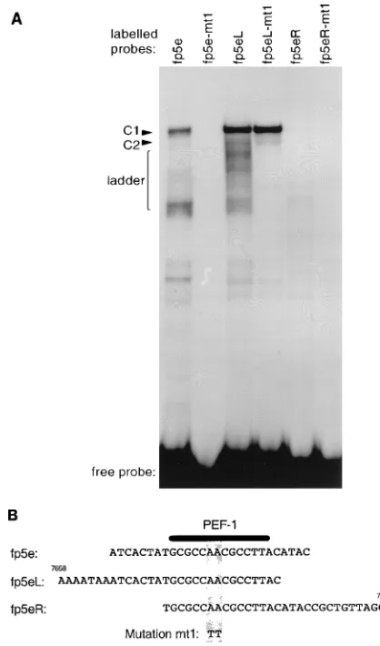
As the length or configuration of the original fp5e oligonu-cleotide may have selected against the binding of other factors, we performed an EMSA with oligonucleotides that contained the PEF-1 binding site but extended either upstream (fp5eL, nt 7658 to 7687) or downstream (fp5eR, nt 7672 to 7701) of the original fp5e oligonucleotide (nt 7665 to 7691). In addition, mutation mt1, which abrogates PEF-1 binding to fp5e, was also introduced into these oligonucleotides (Fig. 1). The EMSA pattern of complexes formed with oligonucleotide fp5eL was superficially similar to that with fp5e, whereas the ability of oligonucleotide fp5eR to form complexes was dramatically re-duced (Fig. 1A). Both oligonucleotide fp5e and oligonucleo-tide fp5eL formed a main complex, C1. While the C1 complex formed with fp5e contained PEF-1 as previously demonstrated and was sensitive to the mt1 mutation, the C1 complex formed with fp5eL was unaffected by the mt1 mutation, suggesting that fp5eL bound a factor distinct from PEF-1 (Fig. 1A). Oligonu-cleotide fp5eL also formed a faster-migrating ladder of com-plexes, which were disrupted by mt1. A very faint band formed
with fp5eR corresponding to the C1 complex, seen on longer exposures of the EMSA autoradiographs, was disrupted by mt1, suggesting that there was a very weak interaction between fp5eR and PEF-1 (Fig. 1A). The sequences, location of the mt1 mutation, and overlap between oligonucleotides fp5e, fp5eL, and fp5eR are shown in Fig. 1B.
Fp5eL binds regulatory factors other than PEF-1.To con-firm that the factor forming C1 with fp5eL was distinct from PEF-1, we performed EMSAs with a set of unlabelled com-petitor oligonucleotides at 20-fold molar excess. Figure 2A shows the control EMSA without competitors. Figures 2B to D show results of EMSAs in which competitor oligonucleotides fp5e, fp5eL, and fp5eL-mt1, respectively in (Fig. 1B), were used.
[image:2.612.320.529.71.426.2]Competition with unlabelled fp5e blocked the formation of the PEF-1/fp5e C1 complex but not the formation by fp5eL of either the C1 complex or the faster-migrating ladder (Fig. 2B). Conversely, competition with fp5eL did not affect the PEF-1/ fp5e C1 complex but abolished all other complexes formed with either fp5e or fp5eL (Fig. 2C). The fp5eL-mt1 competitor inhibited the formation of C1 and C2 by fp5eL but not the complexes formed by fp5e (Fig. 2D). Therefore, both direct
FIG. 1. Comparison of the nuclear factors binding oligonucleotides fp5e, fp5eL, and fp5eR. (A) EMSA with the indicated oligonucleotides containing either the wild-type or mt1 mutated sequence and HeLa nuclear extract. C1, C2, and ladder refer to retarded complexes. (B) Alignment of oligonucleotides fp5e, fp5eL, and fp5eR and their locations in nucleotide map units or relative to the P97 promoter. The A-to-T transversion (mutation mt1) in these oligonucleotides is shown along with the PEF-1 binding site (10).
on November 9, 2019 by guest
http://jvi.asm.org/
binding EMSA and competition EMSA confirm that the C1 complex formed with fp5eL is insensitive to the mt1 mutation and thus does not contain PEF-1.
Oligonucleotide fp7e was also chosen as a competitor oligo-nucleotide since sequence analysis of fp5e or fp5eL reveals a weak homology to fp7e (Fig. 2G). Chong et al. had previously shown that fp7e binds Oct-1 (7), which suggested to us that fp5eL might likewise bind Oct-1. Competition with oligonucle-otide fp7e abolished only the C1 complex formed by fp5eL and had no effect on C2 or the other, faster-migrating complexes
(Fig. 2E). Furthermore, an oligonucleotide (Oct) containing a high-affinity Oct-1 binding site from the murine immunoglob-ulin heavy-chain (IgH) enhancer also specifically abolished the fp5eL C1 complex but not the faster-migrating ladder of bands (Fig. 2F). These results indicate that the C1 complex of fp5eL contains an Oct factor rather than PEF-1.
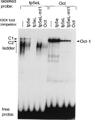
Fp5eL binds an Oct factor. To directly compare the C1 complex formed by fp5eL with the Oct-1 complex formed by the murine IgH enhancer Oct oligonucleotide, we labelled these oligonucleotides and performed competition EMSAs with 100-fold-molar-excess unlabelled oligonucleotides (Fig. 3). These experiments showed that while the fp5eL C1 com-plex and the Oct oligonucleotide comcom-plex comigrated, the Oct oligonucleotide competed for the fp5eL C1 complex with a much higher efficiency than oligonucleotide fp5eL competed for the murine IgH oligonucleotide Oct-1 complex. Thus, an Oct factor binds to fp5eL but with significantly lower affinity than to the Oct-1 binding site of the murine IgH enhancer. Furthermore, the mt1 mutation had no effect on the affinity of the Oct factor for fp5eL, since the fp5eL and fp5eL-mt1 oli-gonucleotides competed for Oct-1 binding to the murine IgH enhancer oligonucleotide to equivalent extents, and both com-pletely abolished formation of C1 with fp5eL (Fig. 3).
The Oct factor binding site on fp5eL overlaps the PEF-1 site on fp5e.The location of the Oct factor binding site in fp5eL, partly overlapping the PEF-1 binding site, is suggested by se-quence comparison with oligonucleotide fp7e (Fig. 2G). The putative site has good homology to the consensus Oct motif in
its 59half but is GC rich and has poor homology in the 39half
overlapping the PEF-1 site. To confirm the location by direct EMSAs, a series of mutations was introduced into both fp5e and fp5eL oligonucleotides within and flanking the putative Oct site (Fig. 4C). Mutation mt1 was described above. Muta-tion mt2 is a C-to-G transversion within the GC-rich region of the site and would be expected to have little effect on Oct binding. In contrast, mutation mt3, which is within the region of best homology, would be expected to abolish Oct factor binding. Mutation mt4 maintained the similarity of the
[image:3.612.84.264.70.521.2]puta-FIG. 2. Competition EMSA of complexes formed with oligonucleotides fp5e and fp5eL and the mt1 mutated oligonucleotides. (A to F) EMSA performed as for Fig. 1 except that nuclear extracts were preincubated with 20-fold-excess unlabelled competitor oligonucleotides. Only the retarded complexes are pre-sented. C1, C2, and ladder are as in Fig. 1. (G) Sequences of the oligonucleotides aligned according to the putative Oct binding site and its location relative to the PEF-1 site in fp5e. The locations of the sequences with the HPV16 genome are shown in map units. The sequence of the Oct-1 binding site in the murine IgH enhancer is from reference 11.
FIG. 3. Direct comparison of the nuclear factors binding oligonucleotide fp5eL and Oct-1 binding the murine IgH enhancer oligonucleotide Oct. The retarded complexes formed with both labelled oligonucleotides fp5eL and Oct were competed for with 100-fold-excess oligonucleotides. C1, C2, and ladder are as in Fig. 1.
on November 9, 2019 by guest
http://jvi.asm.org/
[image:3.612.360.513.74.275.2]tive site to the consensus and introduced a C-to-A transversion into the GC-rich region of the site. An additional pair of mutations, mt5 and mt6, were also used to examine the role of flanking sequences on Oct factor binding (Fig. 4C).
As expected for oligonucleotide fp5e, mutations mt2, mt3, and mt4 had little effect on the C1 complex containing PEF-1, although mutation mt2 strongly enhanced the ladder of faster-migrating complexes (Fig. 4A). The identical mutations in oli-gonucleotide fp5eL did, however, modulate the Oct factor C1 complex. Mutation mt3 completely abolished the fp5eL C1 complex but had no effect on the faster-migrating ladder of bands. Conversely, mutations mt2 and mt4 in fp5eL both main-tained C1 formation, or Oct factor binding, but with reduced affinity (Fig. 4B). However, these mutations had opposite ef-fects on the ladder of faster-migrating complexes, since mt2 in fp5eL strongly enhanced their formation, as it did with oligo-nucleotide fp5e, whereas mt4 specifically disrupted the ladder of complexes.
Additional EMSA competition studies showed that Oct-fac-tor binding to fp5eL was specifically blocked by mutation mt5 but unaffected by mutation mt6. PEF-1 binding to fp5e was reduced by mutation mt6 but not completely blocked (data not shown). Thus, by EMSA, mutations mt3 and mt5 behaved alike, as did mutations mt1 and mt6, and the locations of the
mutations modulating the fp5eL C1 complex are consistent with the presence of an Oct factor binding site adjacent or overlapping that of PEF-1.
Fp5eL binds Oct-1.To further distinguish between and iden-tify the factors binding fp5e and fp5eL, the wild-type and mutated oligonucleotides were subjected to EMSA in the
pres-ence of an Oct-1-specifica-POU serum under conditions that
would specifically disrupt Oct-1 binding. As predicted, the
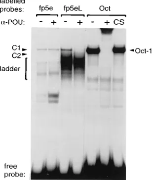
a-POU serum had no effect on any of the complexes formed
with fp5e but specifically abrogated formation of the C1 com-plex with the fp5eL oligonucleotide (Fig. 5). None of the mu-tants in oligonucleotide fp5e formed complexes that were
dis-rupted by thea-POU serum, whereas mutants mt1, mt2, and
mt4 of oligonucleotide fp5eL all formed C1 complexes
suscep-tible to disruption by thea-POU serum (data not shown). This
finding confirmed that Oct-1 binds fp5eL and forms the C1 complex.
Oct-1 binding negatively regulates the fp5e PEF-1 site.To correlate the binding of PEF-1 and Oct-1 to the functional role of fp5e in the HPV16 enhancer, the mutations were introduced into the LCR of HPV16. The wild-type and mutated LCRs were inserted into the pGL2-P luciferase vector. The plasmids were transfected into the keratinocyte cell line HaCaT, and luciferase activity was assayed, comparing two independent clones of wild-type LCR with the mutant LCRs. HaCaT cells were chosen since they contain both PEF-1 and Oct-1, are free of papillomaviral DNA, and retain the capacity to differentiate, therefore providing a cellular environment more like normal keratinocytes.
[image:4.612.83.267.64.407.2]Mutation mt1, which abrogates PEF-1 binding without af-fecting Oct-1, dramatically reduced enhancer/promoter func-tion when introduced into the LCR. In contrast, mutafunc-tion mt3, which specifically blocks Oct-1 binding, significantly enhanced LCR enhancer/promoter expression (Fig. 6). Likewise, muta-tion mt6, which reduced PEF-1 binding, also reduced enhanc-er/promoter activity, while mutation mt5, which blocked Oct-1 binding, markedly increased activity (Fig. 6). Mutations mt2 and mt4, which only marginally affected PEF-1 or Oct-1 bind-ing (C1 complexes), also had only a limited effect on enhancer/ promoter function of the LCR (Fig. 6), despite the dramati-cally altered levels of the faster-migrating ladder complexes
FIG. 4. Mapping of the nuclear factor binding sites along oligonucleotides fp5e and fp5eL by mutations mt1 to mt6. (A) Direct EMSA comparison of the wild-type (wt) fp5e oligonucleotide with mutants mt1, mt2, mt3, and mt4. (B) As in panel A, but with fp5eL-based oligonucleotides. C1, C2, and ladder are as in Fig. 1. (C) Mutations mt1 to mt6 are shown relative to oligonucleotides fp5e and fp5eL. The locations of the PEF-1 site and the Oct-1 binding homology site are also shown.
FIG. 5. Identification of the nuclear factor binding fp5eL to form complex C1. Lanes1, preincubation of HeLa nuclear extract with Oct-1-specifica-POU serum prior to EMSA with labelled oligonucleotides fp5e, fp5eL, and Oct. CS, control serum. C1, C2, and ladder are as in Fig. 1.
on November 9, 2019 by guest
http://jvi.asm.org/
[image:4.612.356.512.75.257.2](Fig. 4A and B). Thus, the overall contribution to enhancer function of the factors forming the faster-migrating ladder is small in comparison with that of PEF-1 and Oct-1. These functional assays in conjunction with the EMSA data are therefore consistent with PEF-1 acting as an up-regulatory factor and Oct-1 acting as a down-regulatory factor.
DISCUSSION
The HPV16 enhancer is nucleosome free and densely bound by a wide variety of transcription factors (2, 7, 9, 13, 14, 16, 20, 25, 26, 28, 31). Our previous studies of the factors binding the enhancer of HPV16 have revealed that footprint fp5e is critical to enhancer activity and is bound by an apparently novel factor, PEF-1 (10, 28).
However, PEF-1 is not the only factor to bind fp5e. For example, Gloss et al. had previously described the very weak binding of NF-I/CTF to this footprint (14). More recently, List et al. discovered a novel methylation-sensitive transcription factor, MSPF, that binds fp5e (21). We have not looked at the methylation sensitivity of PEF-1 binding to fp5e, but compar-ative EMSAs with the unrelated oligonucleotide tat-GRU-D (glucocorticoid response unit of the tat gene) which also binds MSPF (21), should enable a direct analysis of both factors. While the relationship, if any, between PEF-1 and MSPF re-mains to be determined, we have found that the transcription factor Oct-1 also binds to fp5e, at a site adjacent to or over-lapping that of PEF-1. The Oct-1 binding activity was
charac-terized by cross-competition EMSA, use of ana-POU serum,
and analysis of a panel of mutations (mt1 to mt6) across fp5e. Binding of Oct factor to the site is revealed only with
oligo-nucleotide fp5eL, which extends 59 of the original fp5e and
presumably provides a larger target sequence. Given the ap-parent overlap between the two sites, we might expect the
binding of PEF-1 and Oct-1 to be mutually exclusive. We have not observed any complexes containing both factors, but until this is tested directly by using a larger oligonucleotide encom-passing both fp5e and fp5eL, we cannot formally exclude the possibility that PEF-1 and Oct-1 bind fp5e together. Neverthe-less, in functional studies, the two factors have opposite effects on LCR activity, which is consistent with PEF-1 and Oct-1 competing for fp5e.
It has to be stressed that the effect of PEF-1 and Oct-1 binding to their respective sites has been monitored in the setting of a 576-bp LCR fragment containing the enhancer and the P97 promoter of HPV16, and therefore in the context of all other cellular transcription factors known to regulate enhancer function and with physiological levels of PEF-1 and Oct-1. Furthermore, the analysis has been conducted in HaCaT cells, which do not contain HPV DNA, and thus in the absence of overlaying effects of virally encoded transcription fac-tors. The binding of PEF-1 or Oct-1 can be selectively abol-ished by appropriate mutations. The abrogation of PEF-1 binding results in down-regulation of LCR activity (mutations mt1 and mt6), whereas abolition of Oct-1 binding leads to an increase in activity (mutations mt3 and mt5). PEF-1 and Oct-1 are therefore antagonists, and the balance between the two may determine the overall level of LCR activity in epithelial cells.
The interplay between PEF-1 and Oct-1 resembles the situ-ation reported for simian virus 40 enhancer repeats, in which case the activating SphI motifs are sterically hindered by Oct-1 binding (30). Similarly, KRF-1, thought to be a novel tissue-specific activating factor which regulates the HPV18 enhancer, appears to be blocked by Oct-1 binding at an overlapping site (22). Overexpression of Oct-1 has also been shown to down-regulate transcription through a region of the HPV18 en-hancer which includes the overlapping Oct site and KRF-1 site, although this may be due to squelching rather than steric hindrance (18).
A role for Oct factors in cell type and cell cycle regulation of papillomaviral and cytokeratin gene expression has recently emerged (1, 12, 15, 23). The human cell line HeLa has been shown to contain an Oct factor which is absent in BHK cells. This Oct factor causes up-regulated expression of a reporter plasmid through fp7e of HPV16, while Oct-1 binding causes down-regulation (23). This role for Oct-1 binding fp7e is, how-ever disputed, since it has been recently demonstrated that mutating the site to reduce Oct-1 binding dramatically reduces enhancer function (24). Another Oct-1-related factor, p92, has been found in HeLa cells (27). Nuclear factor p92 binds the enhancer of HPV18 at sites including the KRF-1/Oct-1 site and is cell cycle regulated by an inhibitor I-92 (32). It would be interesting to know whether p92 binds fp5eL and confers cell cycle regulation upon HPV16 expression. Other novel cell-type-specific Oct factors such as Skn-1a (Oct-11) and Oct-6 have been shown to regulate cytokeratin gene expression, and thus they may play a similar role in regulating HPV expression (1, 12).
ACKNOWLEDGMENTS
We thank P. van der Vliet and P. Verrijzer for their very generous gift of Oct-1-specifica-POU serum. We are grateful to W. Herr for suggesting the mutants mt5 and mt6 and F. Hoppe-Seyler, L. Laimins, G. Klock, L. Turek, and D. Apt for helpful advice.
[image:5.612.102.251.67.299.2]We also thank the Cancer Research Campaign for supporting this work.
FIG. 6. Functional assays of the pGL2-LCR-based luciferase reporter plas-mids transfected into HaCaT keratinocytes. Mutations mt1 to mt6 (shown in Fig. 4C) were introduced into the LCR (nt2568 to18 relative to the P97 promoter) of pGL2-LCR. The activities of the mutated LCR luciferase reporter plasmids are shown relative to the activities of two independent wild-type (pGL2-LCR WT) clones. The means and standard errors of six duplicate transient transfec-tions are shown.
on November 9, 2019 by guest
http://jvi.asm.org/
REFERENCES
1. Andersen, B., M. D. Schonemann, S. E. Flynn, R. V. I. Pearse, H. Singh, and
M. G. Rosenfeld.1993. Skn-1a and Skn-1i: two functionally distinct Oct-2-related factors expressed in epidermis. Science 260:78–82.
2. Apt, D., T. Chong, Y. Liu, and H.-U. Bernard. 1993. Nuclear factor I and epithelial cell-specific transcription of human papillomavirus type 16. J. Virol. 67:4455–4463.
3. Bohm, S., S. P. Wilczynski, H. Pfister, and T. Iftner. 1993. The predominant mRNA class in HPV16-infected genital neoplasias does not encode the E6 or the E7 protein. Int. J. Cancer 55:791–798.
4. Bosch, F. X., E. Schwarz, P. Boukamp, N. E. Fusenig, D. Bartsch, and H. zur
Hausen.1990. Suppression in vivo of human papillomavirus type 18 E6-E7 gene expression in nontumorigenic HeLa3fibroblast hybrid cells. J. Virol.
64:4743–4754.
5. Bradford, M. M. 1976. A rapid and sensitive method for the quantitation of microgram quantities of protein utilizing the principle of protein-dye bind-ing. Anal. Biochem. 72:248–254.
6. Chan, W. K., T. Chong, H.-U. Bernard, and G. Klock. 1990. Transcription of the transforming genes of the oncogenic human papillomavirus-16 is stimu-lated by tumor promoters through AP1 binding sites. Nucleic Acids Res.
18:763–769.
7. Chong, T., D. Apt, B. Gloss, M. Isa, and H.-U. Bernard. 1991. The enhancer of human papillomavirus type 16: binding sites for the ubiquitous transcrip-tion factors oct-1, NFA, TEF-2, NF1, and AP-1 participate in epithelial cell-specific transcription. J. Virol. 65:5933–5943.
8. Chong, T., W. K. Chan, and H.-U. Bernard. 1990. Transcriptional activation of human papillomavirus 16 by nuclear factor I, AP1, steroid receptors and a possibly novel transcription factor, PVF: a model for the composition of genital papillomavirus enhancers. Nucleic Acids Res. 18:465–470. 9. Cripe, T. P., A. Alderborn, R. D. Anderson, S. Parkkinen, P. Bergman, T. H.
Haugen, U. Pettersson, and L. P. Turek.1990. Transcriptional activation of the human papillomavirus-16 P97 promoter by an 88-nucleotide enhancer containing distinct cell-dependent and AP-1-responsive modules. New Biol.
2:450–463.
10. Cuthill, S., G. J. Sibbet, and M. S. Campo. 1993. Characterization of a nuclear factor, papilloma enhancer binding factor-1, that binds the long control region of human papillomavirus type 16 and contributes to enhancer activity. Mol. Carcinog. 8:96–104.
11. Ephrussi, A., G. M. Church, S. Tonegawa, and W. Gilbert. 1985. B lineage-specific interactions of an immunoglobulin enhancer with cellular factors in vivo. Science 227:134–140.
12. Faus, I., H. J. Hsu, and E. Fuchs. 1994. Oct-6: a regulator of keratinocyte gene expression in stratified squamous epithelia. Mol. Cell. Biol. 14:3263–3275. 13. Gloss, B., T. Chong, and H.-U. Bernard. 1989. Numerous nuclear proteins
bind to the long control region of human papillomavirus type 16: a subset of 6 of 23 DNase I-protected segments coincides with the location of the cell-type-specific enhancer. J. Virol. 63:1142–1152.
14. Gloss, B., M. Yeo Gloss, M. Meisterenst, L. Rogge, E. L. Winnacker, and
H.-U. Bernard.1989. Clusters of nuclear factor I binding sites identify en-hancers of several papillomaviruses but alone are not sufficient for enhancer function. Nucleic Acids Res. 17:3519–3533.
15. Goldsborough, A. S., L. E. Healy, N. G. Copeland, D. J. Gilbert, N. A.
Jenkins, K. R. Willison, and A. Ashworth.1993. Cloning, chromosomal localization and expression pattern of the POU domain gene Oct-11. Nucleic Acids Res. 21:127–134.
16. Haugen, T. H., T. P. Cripe, G. D. Ginder, M. Karin, and L. P. Turek. 1987. Trans-activation of an upstream early gene promoter of bovine papilloma virus-1 by a product of the viral E2 gene. EMBO J. 6:145–152.
17. Hoppe-Seyler, F., and K. Butz. 1994. Cellular control of human papilloma-virus oncogene transcription. Mol. Carcinog. 10:134–141.
18. Hoppe-Seyler, F., K. Butz, and H. zur Hausen. 1991. Repression of the
human papillomavirus type 18 enhancer by the cellular transcription factor Oct-1. J. Virol. 65:5613–5618.
19. Iftner, T., M. Oft, S. Bohm, S. P. Wilczynski, and H. Pfister. 1992. Tran-scription of the E6 and E7 genes of human papillomavirus type 6 in ano-genital condylomata is restricted to undifferentiated cell layers of the epi-thelium. J. Virol. 66:4639–4646.
20. Ishiji, T., M. J. Lace, S. Parkkinen, R. D. Anderson, T. H. Haugen, T. P.
Cripe, J. H. Xiao, I. Davidson, P. Chambon, and L. P. Turek.1992. Tran-scriptional enhancer factor (TEF)-1 and its cell-specific co-activator activate human papillomavirus-16 E6 and E7 oncogene transcription in keratinocytes and cervical carcinoma cells. EMBO J. 11:2271–2281.
21. List, H., V. Patzel, U. Zeidler, A. Schopen, G. Ruhl, J. Stollwerk, and G.
Klock.1994. Methylation sensitivity of the enhancer from the human pap-illomavirus type 16. J. Biol. Chem. 269:11902–11911.
22. Mack, D. H., and L. A. Laimins. 1991. A keratinocyte-specific transcription factor, KRF-1, interacts with AP-1 to activate expression of human papillo-mavirus type 18 in squamous epithelial cells. Proc. Natl. Acad. Sci. USA
88:9102–9106.
23. Morris, P. J., C. L. Dent, C. J. Ring, and D. S. Latchman. 1993. The octamer binding site in the HPV16 regulatory region produces opposite effects on gene expression in cervical and non-cervical cells. Nucleic Acids Res. 21: 1019–1023.
24. O’Conner, M., and H.-U. Bernard. 1995. Oct-1 activates the epithelial-specific enhancer of human papillomavirus type 16 via a synergistic interac-tion with NFI at a conserved composite regulatory element. Virology 207: 77–88.
25. Offord, E. A., and P. Beard. 1990. A member of the activator protein 1 family found in keratinocytes but not in fibroblasts required for transcription from a human papillomavirus type 18 promoter. J. Virol. 64:4792–4798. 26. Offord, E. A., P. O. Chappuis, and P. Beard. 1993. Different stability of AP1
proteins in human keratinocyte and fibroblast cells: possible role in the cell-type specific expression of human papillomavirus type 18 genes. Carci-nogenesis 14:2447–2455.
27. Royer, H. D., M. P. Freyaldenhoven, I. Napierski, D. D. Spitkovsky, T. D.
Bauknecht, and N. Nathan.1991. Delineation of human papillomavirus type 18 enhancer binding proteins: the intracellular distribution of a novel oc-tamer binding protein p92 is cell cycle regulated. Nucleic Acids Res. 19: 2363–2371.
28. Sibbet, G. J., and M. S. Campo. 1990. Multiple interactions between cellular factors and the non-coding region of human papillomavirus type 16. J. Gen. Virol. 71:2699–2707.
29. Staedel, C., J. S. Remy, Z. Hua, T. R. Broker, L. T. Chow, and J. P. Behr. 1994. High-efficiency transfection of primary human keratinocytes with pos-itively charged lipopolyamine:DNA complexes. J. Invest. Dermatol. 102:768– 772.
30. Tanaka, M., and W. Herr. 1990. Differential transcriptional activation by Oct-1 and Oct-2: interdependent activation domains induce Oct-2 phosphor-ylation. Cell 60:375–386.
31. Thierry, F., G. Spyrou, M. Yaniv, and P. Howley. 1992. Two AP1 sites binding JunB are essential for human papillomavirus type 18 transcription in keratinocytes. J. Virol. 66:3740–3748.
32. Weitz, J., M. Kopun, M. Stoehr, I. Napierski, and H. D. Royer. 1991. A novel nuclear inhibitor I-92 regulates DNA binding activity of octamer binding protein p92 during the cell cycle. Nucleic Acids Res. 19:5725–5730. 33. Zeltner, R., L. A. Borenstein, F. O. Wettstein, and T. Iftner. 1994. Changes
in RNA expression pattern during the malignant progression of cottontail rabbit papillomavirus-induced tumors in rabbits. J. Virol. 68:3620–3630. 34. zur Hausen, H. 1991. Human papillomaviruses in the pathogenesis of
ano-genital cancer. Virology 184:9–13.
35. zur Hausen, H. 1991. Viruses in human cancers. Science 254:1167–1173.