Copyright © 1998, American Society for Microbiology
Herpes Simplex Virus Type 1 Latency-Associated Transcripts
Suppress Viral Replication and Reduce Immediate-Early
Gene mRNA Levels in a Neuronal Cell Line
NURITH MADOR,
1DANIEL GOLDENBERG,
1OREN COHEN,
1AMOS PANET,
2AND
ISRAEL STEINER
1*
Laboratory of Neurovirology, Department of Neurology, Hadassah University Hospital,
1and Department
of Virology, The Hebrew University-Hadassah Medical School,
2Jerusalem, Israel
Received 16 October 1997/Accepted 12 March 1998
During herpes simplex virus type 1 (HSV-1) latent infection in human dorsal root ganglia, limited viral
transcription, which has been linked to HSV-1 reactivation ability, takes place. To study the involvement of this
transcription in HSV-1 replication in neuronal cells and consequently in viral latency, we constructed stably
transfected neuronal cell lines containing (i) the entire HSV-1 latency transcriptionally active DNA fragment,
(ii) the same DNA sequence with deletions of the latency-associated transcript (LAT) promoters, or (iii) the
DNA coding sequence of the LAT domain. Replication of HSV-1 or a LAT-negative mutant was markedly
repressed in the LAT-expressing cells, a phenomenon mediated by the LATs. To study the mechanism
responsible for this effect, we examined LAT influence upon expression of HSV-1 immediate-early (IE) genes
ICP0, ICP4, and ICP27, by Northern blot analysis. Following infection of a LAT-expressing neuronal cell line
with a LAT-negative mutant, the steady-state levels of all three IE mRNAs were reduced compared to those for
control cells. Transient transfections into a neuronal cell line indicated that the LAT suppressive effect upon
ICP0 mRNA was mediated directly and was not due to the LAT effect upon the ICP0 promoter. We therefore
propose that the LATs may repress viral replication in neuronal cells by reducing IE gene mRNA levels and
thus facilitate the establishment of HSV-1 latency in nervous tissue.
Herpes simplex virus type 1 (HSV-1) colonizes and
estab-lishes latent infection in human dorsal root ganglia (DRG) to
produce periodic reactivations (for reviews, see references 59
and 63). No mature virions are detected in latently infected
human nervous tissue (13, 62), and restricted gene expression
from the repeat segments of the viral genome (Fig. 1A) is the
only transcriptional activity present throughout latency. This
latency-associated gene expression consists of the more
abun-dant RNAs, the latency-associated transcripts (LATs) (2.0 and
1.5 kb) (49, 56, 64), and the 8.3-kb minor hybridizing RNA
(mLAT) (41, 70). Whether the latency-associated gene(s)
codes for proteins is yet unclear (summarized in references 17
and 59), but we have recently demonstrated that the LATs are
associated with polyribosomes in latently infected trigeminal
ganglia (TG) of mice (22).
The function of the LATs was studied mainly with HSV-1
mutants harboring genomic changes that modify LAT
tran-scription. It is clear that HSV-1 latency-associated gene
expres-sion is not required for viral lytic replication in vivo (29, 33, 61),
nor does a lack of the latency-associated gene(s) prevent viral
transport from the periphery to the DRG. The possibility that
these RNAs take part in the establishment of latency was
suggested, since LAT-negative mutants established latent
in-fection in fewer neuronal cells than did the parental virus (38,
52, 53, 66). It seems also that the LATs are not required for the
maintenance of latency (5, 28, 29, 33, 61). The most constant
and common phenotypic behavior of HSV-1 mutants that are
unable to express the latency-associated gene(s) is a prolonged
and asynchronous explant reactivation kinetics (29, 33, 61) and
reduced or absent in vivo reactivation from DRG (26, 67).
The latency-associated gene(s) may act through a protein
and/or as a specialized RNA. Its molecular action
mecha-nism(s) is yet unknown, but two main possibilities have been
proposed. (i) The LATs may act during reactivation. Neurons
possess means to inhibit the transactivation of the
immediate-early (IE) genes of HSV-1, a mechanism that renders the virus
incapable of replication in neuronal cells at low multiplicities
of infection (MOIs) (31, 34). This hypothesis assumes that the
LATs, the only RNAs that are expressed during latency (56,
62, 64), may enable the viral replication cycle to bypass this
inhibition at the initial stages of reactivation. (ii) Alternately,
the LATs may act during the establishment of latent infection,
by suppressing HSV-1 IE gene expression. This assumption is
based on the fact that, since HSV-1 is a lytic virus, one of the
early requirements for establishment of latent infection in
neu-ronal cells is prevention of viral replication, before or at the
stage of IE gene expression (50, 58).
The aim of the present study was therefore to examine the
effect of the latency-associated gene(s) of HSV-1 on viral
rep-lication in neuronal cell lines, to map the functional region,
and to investigate whether any effect is associated with
expres-sion of HSV-1 IE genes ICP0, ICP4, and ICP27. Our findings
indicate that the LATs suppress HSV-1 replication in neuronal
cell lines and reduce ICP0 and also ICP4 and ICP27
steady-state mRNA levels in these cells.
MATERIALS AND METHODS
Construction of plasmids.The construction and characteristics of pLAT/ICP0
(Fig. 1C), previously termed pNM3 (22, 37), have been reported before (37). Briefly, the entire HSV-1 DNA fragment that is transcriptionally active during latent infection was inserted into a vector under the control of the cytomegalo-virus (CMV) IE promoter. This DNA insert also contains the ICP0 (46),g34.5 (12), open reading frame (ORF) P (32), and ORF O (48) genes.
* Corresponding author. Mailing address: Department of
Neurol-ogy, Hadassah University Hospital, P.O. Box 12 000, Jerusalem 91120,
Israel. Phone: 972/2/6776941. Fax: 972/2/6437782. E-mail: isteiner
@md2.huji.ac.il
5067
on November 9, 2019 by guest
http://jvi.asm.org/
pDprLAT (Fig. 1D) was derived from pLAT/ICP0 by two deletions that re-moved the CMV IE promoter and the latency-associated promoter 2 (LAP2 [21]) and therefore is incapable of expressing HSV-1 LATs or mLAT but con-tains the ICP0,g34.5, ORF P, and ORF O genes. pLAT/ICP0 was cleaved with HindIII and NdeI enzymes to delete the CMV promoter sequence (500 bp). The 14.4-kb vector with a deletion of the promoter was subjected to Klenow fragment (Promega) activity for a filling-in reaction, followed by ligation by T4 ligase (Promega). This vector was then cleaved with BstEII enzyme at nucleotides 119194 and 120091 to delete the LAP2 promoter (897 bp). The 13.5-kb DNA fragment was subjected to ligation reaction to obtain the pDprLAT vector.
pLAT (Fig. 1E) was derived from pLAT/ICP0 by deletion of the sequences downstream from the 39end of the LAT gene; therefore, it is capable of ex-pressing only the LATs. pLAT/ICP0 was cleaved with restriction enzyme MluI at nucleotide 121651 and with BamHI at nucleotides 123459 and 129259. This yielded three DNA fragments, 1.8, 5.8, and 7.3 kb. The 7.3-kb fragment, which contains only the LAT coding sequence, was subjected to Klenow fragment filling-in reaction, followed by ligation by T4 ligase.
Cell lines and viruses.NA cells (39), a subclone of neuro-2a, a clonal line of
C-1300 mouse neuroblastoma cells (1), were kindly provided by A. McMorris, The Wistar Institute, Philadelphia, Pa. Cells were maintained in minimal essen-tial Eagle medium (MEEM) with 10% heat-inactivated fetal calf serum (FCS). CV-1 cells were maintained in Dulbecco’s modified Eagle’s medium with 10% FCS. HSV-1 strain F was obtained from B. Roizman, University of Chicago, Chicago, Ill. FS1001K, a KOS strain-derived LAT-negative mutant (15), was kindly provided by N. Fraser, The Wistar Institute. The viruses were propagated and their titers were determined on CV-1 cells, and results are represented as PFU per milliliter.
Cell transfections.Transient transfections were performed according to the
method of Cullen (14) and as detailed before (37). Cells were harvested for RNA extraction 48 h posttransfection. Stable transfections were performed as de-scribed by us before (22). pSV2neo, a plasmid containing the neomycin resistance gene, was used for clonal selection.
Isolation of RNA and Northern blot analysis.Total RNA was extracted from
cells with Tri reagent (Molecular Research Center, Inc.) according to the man-ufacturer’s instructions. For reverse transcription-PCR (RT-PCR), RNAs were treated with 5 U of RQ1 DNase (Promega), extracted with phenol-chloroform and chloroform, and precipitated in ethanol. Northern blot analysis was per-formed according to the method of Spivack and Fraser (56). RNA markers (281 to 6,583 bases) were purchased from Promega. Computerized image analysis and quantitation were performed by a Bio-Imaging analyzer (Fuji Corp.) (68).
PCR amplification of reverse-transcribed RNA.RT-PCR was performed as
described by us before (37). The following oligonucleotide primers that flank the intron within the 2.0-kb LAT (Fig. 1F) were prepared according to the published sequence of HSV-1 (45): P2, 59-GACTCTGTTACTTACCCGTCCGAC-39
(HSV-1 bases 119612 to 119635), and P3, 59-GAAAGCATCCTGCCACTGGC ATGGA-39(bases 120426 to 120402). RT was performed with primer P3.
For RT-PCR analysis of the HSV-1 ICP0 gene, the following primers that flank the first 59intron within the gene (Fig. 1F) were used: P11, 59-TCTCGA ACAGTTCCGTGTCCGT-39(bases 123130 to 123151), and P10, 59-TCTCCG CATCACCACAGAAG-39(bases 124192 to 124173). RT was performed with primer P11.
For PCR analysis of the ICP27 gene, the following primers were used (Fig. 1B): P12, 59-CCCTTTCTCCAGTGCTACCTGAA-39(bases 114919 to 114941), and P13, 59-GTGCGTGTCTAGGATTTCGATC-39(bases 115170 to 115149).
For RT-PCR analysis of the mouse CuZn superoxide dismutase (CuZn SOD) gene (2), the following primers were used: P14, 59-GAAAGCGGTGTGCGTG CTGAAG-39(SOD exon 1), and P15, 59-GAGTGAGGATTAAAATGAGGTC C-39(SOD exon 3). RT was performed with primer P15.
The primers were chosen according to the following criteria: (i) GC content of the primer of not less than 50%, (ii) absence of significant homology with all rodent and human sequences present in the GenBank database or with other HSV-1 sequences, (iii) size of the PCR product of between 0.2 and 1 kb, and (iv) avoidance of sequences which contain polypurine or polypyrimidine base repetition.
Southern blot analysis.DNA fragments were resolved by 1.5% agarose gel
electrophoresis and transferred onto a GeneScreen Plus membrane (New Re-search Products), by the salt transfer protocol of the manufacturer. Hybridization and washes were also performed according to the manufacturer’s instructions. The filters were autoradiographed with XAR-5 film at270°C with intensifying screens (Du Pont).
Preparation of radioactively labeled DNA probes.The DNA fragments that
served as probes for the LATs and ICP0 and ICP4 mRNAs were cleaved from the vector pLAT/ICP0 and purified from an agarose gel. We used the following DNA fragments (Fig. 1B): a BstEII-BamHI probe, covering the HSV-1 genomic region from bp 121068 to 123463; a BstEII-BstEII probe consisting of two equimolar fragments, 897 and 977 bp, covering the genomic sequence from bp 119194 to 121068; and an AflIII-BamHI probe covering the genomic sequence from bp 128059 to 129259.
The 251-bp probe for the ICP27 gene was prepared by PCR of BamHI DNA fragment B (47) with primers P12 and P13.
A 198-bp DNA probe for the housekeeping CuZn SOD gene (2) was prepared by RT-PCR of RNA from TG of mice with primers P14 and P15.
All probes were labeled with [a-32P]dCTP by random priming with the
Mul-tiprime DNA labeling system of Amersham. Specific activities of the probes were approximately 108cpm/mg of DNA.
For preparation of the single-stranded DNA probe for ICP0 mRNA, the following primers were used (Fig. 1F): P11 (described above) and P9, 59-TGG TGTTGGTGTTACTGCTG-39(bases 122310 to 122330). Primers were 59end labeled with [g-32P]dATP followed by PCR with the Taq DNA polymerase (sequencing grade; Promega) according to the instructions in the manual. The PCR was performed in the presence of dATP, dGTP, and dTTP (Boehringer) (1.6 mM each); 0.7 mM [a-32P]dCTP; 100 ng of the BstEII-BamHI DNA frag-ment; and 20 ng of the primer.
For detection of the 18S rRNA, we used the following 59-end-labeled single-stranded DNA probe: 59-CTGTCAATCCTGTCCGTGTCCG-39, according to the 18S rRNA sequence in GenBank (accession no. X00686, coordinates 1273 to 1294).
Cell infection and virus titration.NA cells or clones derived from them (23
104cells per well) were seeded in triplicate in 24-well plates for 24 h. Cells were infected at MOIs of 0.1, 1, and 10 in 250ml of MEEM without serum. After 2 h, the medium was removed and 1 ml of fresh medium with serum was added to every well. Twenty-four hours later, the plates were stored at270°C.
For HSV-1 titration, the cells and medium of each well were removed and sonicated. CV-1 cells (1.33105per well) were seeded in 24-well dishes in 0.5 ml of Dulbecco’s modified Eagle’s medium including 10% FCS and 48 h later were used for titration as described elsewhere (60), with 0.5 mg of human gamma globulin per ml.
For RNA extraction, cell lines NA-neo and NA-LAT#1, 23106cells each, were infected at an MOI of 1 with LAT-negative mutant in 4 ml of MEEM without serum. After 1 h, the medium was removed and 10 ml of fresh medium with serum was added to every plate. Six hours later, the cells were extracted for RNA isolation as described above.
Cell viability assay (MTT).This method enabled us to estimate the number of
viable cells seeded for infection and to calibrate the required numbers of viral PFU, to reach similar MOIs during infection. The assay was based upon the ability of cellular mitochondria to convert MTT (3-[4,5-dimethylthiazol-2-yl]-2,5-diphenyl-tetrazolium bromide [Sigma]) into blue formazan product and was performed according to the procedure of Miller and McDevitt (40). In parallel with seeding of the different NA clones for infection, an extra plate was seeded for the MTT assay with the same cells. The optical density (492 nm) was mea-sured in an enzyme-linked immunosorbent assay plate reader (Organon Tek-nika) with a reference wavelength of 630 nm.
Chloramphenicol acetyltransferase (CAT) assay.Cell lysates were analyzed
for CAT activity at 48 h after transfection by the technique of Gorman et al. (24). The pIE-CAT construct, containing the CAT gene sequence under the control of the HSV-1 ICP0 promoter (from 2585 to1150 [19]), and the pRSV-CAT construct, which contains the CAT gene under the control of the long terminal repeat promoter of Rous sarcoma virus (23), were kindly provided by D. Latch-man, London, United Kingdom. Cells (0.53106) were transfected with 1.25 and 0.25mg of DNA, respectively, as described before (37). Following transfections, cells were harvested and the protein content was determined by the method of Bradford (7). Samples equalized for protein content were assayed for CAT activity. The vector pRSV-CAT was used as a control plasmid to correct for differences in transfection efficiencies. This was done in duplicate for each ex-periment.
DNA dot blot.Plasmid DNA was extracted from equal amounts of transfected
cells by the Hirt procedure (27), and identical volumes were denatured in 0.4 N NaOH–10 mM EDTA for 10 min at 96°C. The samples were then added to the slot blot apparatus, transferred to a GeneScreen Plus membrane (New Research Products), hybridized, and washed according to the manufacturer’s instructions. Computerized image analysis and quantitation were performed by a Bio-Imaging analyzer as described for RNA.
RESULTS
Establishing neuronal cell lines that stably express the
HSV-1 latency-associated gene(s).
In order to examine the
effect of the gene(s) expressed during HSV-1 latent infection
upon viral replication in neuronal cells and to delineate the
DNA sequences which are responsible for any observed effect,
we have constructed three vectors. (i) pLAT/ICP0 (Fig. 1C
[37]) is a plasmid containing an HSV-1 10.4-kb DNA fragment
which includes the entire sequences that are transcriptionally
active during latency, under the control of the constitutive IE
CMV promoter. This fragment also contains the coding
se-quences of the ICP0,
g
34.5, ORF P, and ORF O genes. (ii)
p
D
prLAT (Fig. 1D) is a plasmid derived from the pLAT/ICP0
vector by deletion of the CMV and LAP2 (21) promoters. This
vector is unable to express the LATs or the mLAT but still
contains the coding sequences of ICP0,
g
34.5, ORF P, and
ORF O. (iii) pLAT (Fig. 1E) is a plasmid derived from pLAT/
on November 9, 2019 by guest
http://jvi.asm.org/
ICP0 and having deletions of all the sequences downstream
from the DNA fragment that transcribes the LATs.
These vectors and pSV2neo were stably transfected into the
NA neuronal cell line to produce three G418-resistant
popu-lations, NA-LAT/ICP0, NA-
D
prLAT, and NA-LAT. Sixteen
cell clones were randomly isolated from each population and
were examined by RT-PCR and Northern blot analysis for
expression of the relevant transcripts. Clones NA-LAT/
ICP0#1 (previously termed NA4 [22]), NA-
D
prLAT#1, and
NA-LAT#1, which express these transcripts, were randomly
chosen. NA-neo, a negative clone harboring only pSV2neo, was
selected as a negative control.
The neuronal cell clones were examined for expression of
the 2.0- and 1.5-kb LATs by Northern blot analysis (Fig. 2A)
and by RT-PCR (Fig. 2B). While the 2.0-kb LAT was present
at amounts high enough to be easily visualized by Northern
blot analysis in NA-LAT#1 and NA-LAT/ICP0#1 cells and
latently infected mouse TG (Fig. 2A, lanes 4, 5, and 2,
respec-tively) and was absent from NA-
D
prLAT#1 and NA-neo cells
(lanes 3 and 6, respectively), the 1.5-kb LAT was produced at
low levels and was observed on Northern blots only as a faint
band and after overexposure of the film (data not shown). This
transcript was clearly identified in RNA from latently infected
mouse TG (lane 2). However, a DNA band with an expected
size of 255 bp, representative of the 1.5-kb LAT (Fig. 1F), was
obtained by RT-PCR analysis of RNA obtained from
NA-LAT#1 and NA-LAT/ICP0#1 clones (Fig. 2B, lanes 6 and 7,
respectively) and of RNA from TG of latently infected mice
(Fig. 2B, lane 5). Appropriate controls without RT enzyme in
the reaction mixture gave no DNA band (Fig. 2B, lanes 1 to 4).
By Northern blot analysis, clone NA-
D
prLAT#1 was found
to produce the ICP0 transcript (Fig. 2A, lane 3). As expected,
both the 2.7-kb ICP0 transcript and the 2.0-kb LAT were
observed in HSV-1-infected CV-1 cells (Fig. 2A, lane 1).
HSV-1 replication is suppressed in neuronal cell clones that
express the LATs.
In order to study the effect of the gene(s)
expressed during HSV-1 latency on viral replication in
neuro-nal cells, we infected the three neuroneuro-nal cell clones, NA-LAT/
ICP0#1, NA-
D
prLAT#1, and NA-LAT#1, with HSV-1 (strain
F). NA-neo cells were used as a control (Table 1). Twenty-four
hours postinfection, plates were stored at
2
70°C. Prior to
titration on CV-1 cells, the infected cells together with the
medium were sonicated. All experiments were performed in
triplicate. Presented are the averaged results of a
representa-tive experiment. To ensure that the MOI would be accurate,
control plates were subjected to cell quantification by the MTT
assay.
At an MOI of 0.1, no HSV-1 replication could be detected in
NA-LAT/ICP0#1 and NA-LAT#1 cells (Table 1). Since
titra-tion was performed in 0.25 ml of a total volume of 1 ml, lack
of plaques on titration assay is represented as
,
4. HSV-1
replication was detectable in NA-neo and NA-
D
prLAT#1 cell
clones. At an MOI of 1, HSV-1 replication was markedly
suppressed in LAT/ICP0#1 and NA-LAT#1 cells compared to
that in NA-neo cells by similar orders of magnitude of about
330- and 220-fold, respectively. The NA-
D
prLAT#1 cells were
FIG. 1. Map of the HSV-1 genome region expressing the LATs, structure of plasmids, and location of probes and primers used in this study. (A) The enlarged
BamHI fragments B and SP within the repeat region of the HSV-1 genome. Arrows indicate the relevant HSV-1 genes and their transcription orientation. Locations
of LAP1 and the LAT polyadenylation site are indicated according to the HSV-1 DNA sequence in GenBank (accession no. X14112). (B) Location of the DNA probes used in this study. (C to E) Structure of plasmids pLAT/ICP0, pDprLAT, and pLAT (see Materials and Methods for details). (F) Location of the primers used for RT-PCR of the 1.5-kb LAT and of ICP0 mRNA and expected sizes of amplified fragments. Also indicated are locations of the primers used for production of the single-stranded probe for ICP0 mRNA. B, BamHI; LAP1, latency-associated promoter 1; Pv, PvuI; M, MluI; Bs, BstEII; Af, AflIII; SV40, simian virus 40.
on November 9, 2019 by guest
http://jvi.asm.org/
unable to suppress HSV-1 replication. At an MOI of 10, the
suppressive effect of the LATs upon HSV-1 replication was
almost completely abolished. Similar results were obtained in
repeated experiments, with the same cell clones, and in
exper-iments with three additional cell clones randomly selected
from the NA-LAT/ICP0, NA-
D
prLAT, and NA-LAT cell
pop-ulations (data not shown).
To validate these results, similar infection experiments were
carried out with the three neuronal cell populations as well.
The parental neuronal NA cells were used as the control. As
expected, HSV-1 replication was repressed in the
LAT-ex-pressing populations (NA-LAT/ICP0 and NA-LAT) compared
to that in the NA-
D
prLAT and NA populations, and the
sup-pressive effect was MOI dependent (data not shown).
Thus, HSV-1 replication was suppressed in NA-LAT/
ICP0#1 cells, which contain the entire DNA sequence
tran-scriptionally active during latency, including the coding
se-quences for the LATs, mLAT, ICP0,
g
34.5, ORF P, and ORF
O. A similar effect was demonstrated with NA-LAT#1 cells,
which are capable of expressing only the LATs, suggesting that
the observed inhibition of HSV-1 replication is mediated by
the LATs and not by the ICP0,
g
34.5, ORF P, or ORF O gene.
Indeed, the repressive effect upon HSV-1 replication was not
present in NA-
D
prLAT#1 cells, which are incapable of
ex-pressing the LATs but express ICP0 and also contain the three
genes
g
34.5, ORF P, and ORF O.
The suppressive effect of the LATs upon HSV-1 replication
was MOI dependent (Table 1). At an MOI of 0.1, no viral
replication could be detected in LAT/ICP0#1 and
NA-LAT#1 cells, compared to that in NA-neo cells, while at an
MOI of 1 the differences were 330- and 220-fold, respectively.
At an MOI of 10, the suppressive effect was almost completely
abolished.
Replication of HSV-1 LAT-negative mutant is suppressed in
a neuronal cell clone that expresses the LATs.
In order to
further substantiate the findings that HSV-1 LATs suppress
viral replication in neuronal cell clones, we repeated these
experiments in parallel and under similar conditions, with a
LAT-negative HSV-1 mutant. We used the FS1001K mutant,
in which a 1.1-kb fragment starting from the 5
9
end is deleted
from the 2.0-kb LAT coding sequence to abolish all LAT
expression (15).
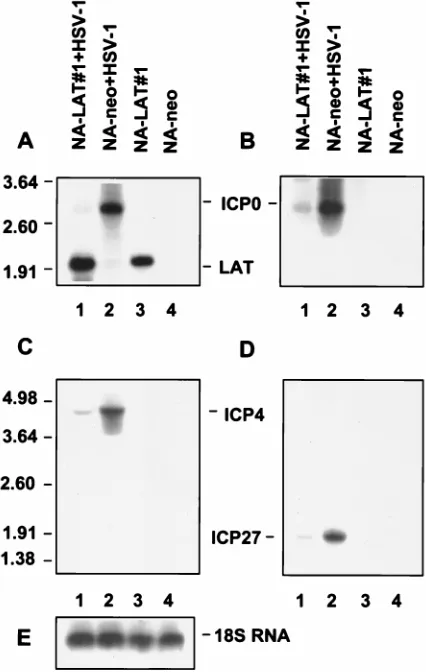
[image:4.612.71.528.67.271.2]Twenty-four hours postinfection, virus yield in NA-LAT#1
and NA-neo cells was determined by titration on CV-1
indi-cator cells. This experiment was performed in triplicate, and
the average results are presented in Table 2. HSV-1
LAT-negative mutant replication was suppressed in NA-LAT#1
cells compared to that in the control NA-neo cells. The effect
FIG. 2. Transcription of HSV-1 LATs and ICP0 in stably transfected neuronal cell clones. (A) Northern blot analysis of total RNAs obtained from NA-DprLAT#1, NA-LAT#1, NA-LAT/ICP0#1, and NA-neo cell clones. Lane 1, RNA from CV-1 cells infected with HSV-1 (MOI of 5) and harvested 5 h postinfection; lane 2, RNA from latently infected TG; lane 3, RNA from NA-DprLAT#1 cells; lane 4, RNA from NA-LAT#1 cells; lane 5, RNA from NA-LAT/ICP0#1 cells; lane 6, RNA from NA-neo cells. Ten micrograms of RNA was loaded in each lane, except lanes 1 and 2, which were loaded with 5mg of RNA. The DNA probe used was BstEII-BamHI. The positions of RNA markers (in kilobases) are indicated on the left. Sizes of the relevant HSV-1 transcripts (in kilobases) are indicated on the right. (B) Southern blot analysis of RT-PCR products from RNAs obtained from NA-LAT#1, NA-LAT/ICP0#1, and NA-neo cells. Lanes 1 and 5, latently infected TG; lanes 2 and 6, NA-LAT#1; lanes 3 and 7, NA-LAT/ICP0#1; lanes 4 and 8, NA-neo. Lanes 1 to 4: control PCR without RT enzyme in the reaction mixture. The 255-bp DNA fragment is representative of the 1.5-kb LATs (Fig. 1F). The DNA probe used was BstEII-BstEII (Fig. 1). RT was performed with primer P3, and PCR was performed with primers P3 and P2.TABLE 1. LAT effect on HSV-1 replication in the stably transfected neuronal clones
aMOI
Value (PFU/ml) for clone: Fold of inhibition for NA-neo vs:
NA-neo NA-DprLAT#1 NA-LAT/ICP0#1 LAT#1NA- NA-LAT#1 NA-LAT/ICP0#1
10
3.8
3
10
42.0
3
10
41.6
3
10
41.4
3
10
42.6
2.4
1
3.3
3
10
33.5
3
10
310
15
220
330
0.1
3.0
3
10
13.0
3
10
1,
4
,
4
aHSV-1, produced by the LAT-expressing NA-LAT/ICP0#1 and NA-LAT#1 clones and by the control NA-DprLAT#1 and NA-neo clones, was quantitated on CV-1 cells. Since titration was performed on 0.25 ml of a total volume of 1 ml, lack of plaques in the titration assay is represented as,4. All experiments were performed in triplicate, and the results represent the average calculations.
on November 9, 2019 by guest
http://jvi.asm.org/
[image:4.612.52.549.630.702.2]was again MOI dependent, i.e., more pronounced at a low
MOI of 0.1, where the inhibition was 160-fold. Similar results
were obtained in repeated experiments. These results
substan-tiate the findings that the LATs suppress HSV-1 replication in
neuronal cells and indicate that the LAT effect has a
trans-acting component.
ICP0 mRNA levels are suppressed in a neuronal cell line
that expresses the LATs.
Since HSV-1 replication is dependent
on the expression of the viral IE genes, we hypothesized that
the LATs may reduce HSV-1 replication in neuronal cells by
down-regulating expression of the IE genes. We chose initially
to examine the influence of the LATs on ICP0 expression for
the following reasons. (i) ICP0 is the first IE gene to be
ex-pressed at very early times postinfection (36). (ii) This IE
protein is a transactivator which enhances HSV-1 replication
during acute infection as well as reactivation from latency (9)
and is capable of activating all three kinetic classes of HSV
genes (10). (iii) The sequences of ICP0 mRNA and of the
LATs overlap at their 3
9
end (and therefore an antisense
mechanism for LAT action has been proposed elsewhere [64]).
(iv) The pLAT/ICP0 vector used in this work (Fig. 1C) includes
the entire DNA sequences required for ICP0 expression (46).
(v) The LATs have been shown elsewhere to inhibit ICP0
protein synthesis (20) and ICP0-mediated transactivation (16).
(i) The levels of ICP0 mRNA produced by an HSV-1
LAT-negative mutant are suppressed in a neuronal cell line that
expresses the LATs.
The LAT effect upon ICP0 expression was
first examined by infecting LAT-expressing NA-LAT#1 and
NA-neo control cells with the LAT-negative mutant at an MOI
of 1. RNA was extracted 6 h postinfection, resolved by gel
electrophoresis, Northern blotted, and hybridized with the
BstEII-BamHI DNA probe specific to both ICP0 mRNA and
LATs (Fig. 1B). ICP0 mRNA was barely expressed in
NA-LAT#1 cells (Fig. 3A, lane 1) and was readily identified in
NA-neo cells (lane 2). Bio-Imager quantification of ICP0
mRNA suggested a 16-fold inhibition.
To confirm that the identified band was indeed ICP0
mRNA, we repeated this experiment with an ICP0-specific
single-stranded DNA probe (Fig. 3B). As expected, a 2.7-kb
RNA band, of the size of ICP0 mRNA, was identified in RNA
obtained from NA-neo cells infected with HSV-1 (lane 2) and
not in control RNA from uninfected cells (lanes 3 and 4). The
ICP0 mRNA was barely visualized in the HSV-1-infected
LAT-expressing NA-LAT#1 cells (lane 1). To validate the
finding that equal amounts of RNA were loaded in each lane,
we also hybridized the blot with a single-stranded DNA probe
specific for the 18S rRNA (Fig. 3E). Measurement of rRNA
levels by Bio-Imager showed a 1.1-fold difference in the
amounts of RNA from NA-LAT#1-infected cells compared to
that from NA-neo-infected control cells (Fig. 3E, lanes 1 and 2,
respectively).
These results indicate that expression of HSV-1 LATs in a
neuronal cell clone reduces the ICP0 mRNA levels. It also
shows that this effect can be mediated in trans.
(ii) LATs reduce ICP0 mRNA levels following transfection
of pLAT/ICP0 into NA cells.
To rule out the possibility that the
LATs interfere with HSV-1 entry into cells, with subsequent
reduction of ICP0 mRNA, and to examine whether the LAT
effect upon ICP0 mRNA is a direct one, we used a second
approach. NA cells were transfected with either pLAT/ICP0,
which contains the LAT and ICP0 genes (Fig. 1C), or with
p
D
prLAT, which has deletions of the CMV promoter and of
LAP2 (Fig. 1D) and is therefore unable to express the LATs
but still expresses ICP0 (Fig. 2).
[image:5.612.321.534.71.407.2]RNAs were extracted from NA cells 48 h posttransfection,
resolved by gel electrophoresis, Northern blotted, and
hybrid-ized with a BstEII-BamHI probe specific to both ICP0 mRNA
and the LATs (Fig. 1B). While ICP0 mRNA was visualized in
NA cells transfected with p
D
prLAT (Fig. 4A, lane 3), no ICP0
FIG. 3. Expression of ICP0, ICP4, and ICP27 genes by an HSV-1 LAT-negative mutant following infection of a LAT-expressing neuronal cell line. Results of Northern blot analysis of total RNAs (10mg) obtained from NA-LAT#1 and NA-neo cells following infection (MOI of 1) with HSV-1 LAT-negative mutant are shown. RNAs were harvested 6 h postinfection. The order of the lanes is identical in all panels. Lane 1, RNA from infected NA-LAT#1 cells; lane 2, RNA from infected NA-neo cells; lane 3, RNA from control NA-LAT#1 cells; lane 4, RNA from control NA-neo cells. Each of the four identical filters was hybridized with a different probe (Fig. 1B and Materials and Methods): BstEII-BamHI probe for the LATs and ICP0 (A), single-stranded DNA probe specific for ICP0 mRNA (B), AflIII-BamHI probe for ICP4 (C), and PCR product specific for ICP27 (D). The positions of RNA markers (in kilo-bases) are indicated on the left. The relevant HSV-1 transcripts are indicated in the center. (E) 18S rRNA hybridized with a specific single-stranded DNA probe (see Materials and Methods).TABLE 2. LAT effect on HSV-1 LAT-negative mutant replication
in the stably transfected neuronal clones
aMOI
Value (PFU/ml) for clone: Fold of
inhibition for NA-neo vs NA-LAT#1
NA-neo LAT#1
NA-10
2.3
3
10
57.0
3
10
43.3
1
3.9
3
10
51.6
3
10
424.4
0.1
4.8
3
10
23.0
160
aFS1001K LAT-negative mutant, produced by the LAT-expressing NA-LAT#1 and the control NA-neo clones, was quantitated on CV-1 cells. All experiments were performed in triplicate, and the results represent the average calculations.
on November 9, 2019 by guest
http://jvi.asm.org/
[image:5.612.50.291.91.164.2]mRNA was observed in pLAT/ICP0-transfected NA cells (lane
2). Bio-Imager quantification suggested a difference of at least
58-fold in ICP0 mRNA levels between the NA cells transfected
with each of these vectors. As described previously,
single-stranded ICP0-specific probe confirmed the identity of the
ICP0 mRNA (Fig. 4B), following rehybridization of the same
blot shown in Fig. 4A. To ensure that equal amounts of RNA
were loaded in each lane, we also hybridized the blot with the
SOD probe (2) (Fig. 4A, bottom). Measurement of SOD
mRNA levels by Bio-Imager showed a 1.4-fold difference in
the amounts of RNA from p
D
prLAT-transfected cells
com-pared to that from pLAT/ICP0-transfected cells (Fig. 4A, lanes
3 and 2, respectively). To quantitate the efficiency of
transfec-tion in the NA cells, a Hirt procedure followed by DNA dot
blot hybridization was performed. It indicated that similar
amounts of p
D
prLAT and pLAT/ICP0 DNAs entered the NA
cells (Fig. 4C).
To investigate whether in the presence of the LATs small
amounts of ICP0 mRNA may still be detected, we have applied
the much more sensitive technique of RT-PCR followed by
Southern blot analysis. The 296-bp DNA fragment
represen-tative of the spliced ICP0 mRNA (Fig. 1F) was present in the
two respective preparations (Fig. 4D, lanes 4 and 5).
Appro-priate controls without RT enzyme in the reaction mixture
identified no DNA band (lanes 1 to 3). This would indicate
that, while the LATs suppress ICP0 mRNA, residual levels
may still be detected.
In order to confirm that the results of the transfection
ex-periments were not due to interference by the strong CMV
promoter in pLAT/ICP0 with the native ICP0 promoter, we
cotransfected 2.5
m
g of pIE-0-CAT (a vector containing the
CAT gene under the control of the HSV-1 ICP0 promoter)
with increasing amounts (0.5 to 2.5
m
g) of the CMV promoter
cloned in the expression vector pCH2N (37). No effect upon
CAT expression from the presence of the CMV promoter was
noted in the cotransfected NA cells (data not shown).
Taken together, these results (i) indicate that HSV-1 LATs
directly reduce ICP0 mRNA levels in neuronal cells and (ii)
rule out the possibility that the findings are the outcome of
entry of different viral DNA amounts into the two neuronal
cell lines or are an experimental in vitro artifact due to the
influence of the CMV promoter upon the ICP0 promoter.
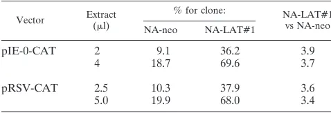
LATs do not reduce ICP0 mRNA levels via repression of the
ICP0 promoter.
Since HSV-1 LATs substantially reduce the
steady-state levels of ICP0 mRNA, we went on to examine
FIG. 4. Transcription of ICP0 and LATs in a transfected neuronal cell line. (A) Northern blot analysis of total RNAs (10 mg) obtained from NA cells following transient transfection with vectors pLAT/ICP0 and pDprLAT. Lane 1, RNA from NA cells infected with HSV-1 (MOI of 5) and harvested 5 h postin-fection; lane 2, RNA from NA cells transfected with pLAT/ICP0 vector; lane 3, RNA from NA cells transfected with pDprLAT vector; lane 4, RNA from NA control cells. The probe used is the BstEII-BamHI DNA fragment (Fig. 1). The positions of RNA markers (in kilobases) are shown on the left. Sizes (in kilo-bases) of the relevant HSV-1 transcripts are indicated on the right. At the bottom is shown the mRNA of the CuZn SOD gene hybridized with an SOD-specific probe (see Materials and Methods). (B) Rehybridization of the same blot as in panel A with a single-stranded DNA probe specific for ICP0 mRNA. (C) Dot blot analysis of plasmid DNA extracted from transfected NA cells by the Hirt procedure. Lane 1, NA cells transfected with pLAT/ICP0 vector; lane 2, NA cells transfected with pDprLAT; lane 3, control NA cells. The amount of the DNA in each row is indicated in microliters. The probe used is the BstEII-BamHI DNA fragment (Fig. 1). (D) Southern blot analysis of RT-PCR products from total RNAs obtained from NA-LAT/ICP0#1, NA-DprLAT#1, and NA-neo cells. Lanes 1 and 4, NA-LAT/ICP0#1; lanes 2 and 5, NA-DprLAT#1; lanes 3 and 6, NA-neo; lanes 1 to 3, control PCR without RT enzyme in the reaction. RT was performed with primer P11, and PCR was performed with P11 and P10. The probe used is the BstEII-BamHI DNA fragment. The 296-bp DNA is represen-tative of the spliced ICP0 mRNA (Fig. 1F).on November 9, 2019 by guest
http://jvi.asm.org/
their direct effect upon the ICP0 promoter. pIE-0-CAT (19)
was transfected into NA-LAT#1 and into NA-neo control
cells. CAT activity was determined in duplicate in crude cell
extracts (Table 3). All experiments included a control plasmid,
pRSV-CAT, in which the Rous sarcoma virus promoter drives
the expression of CAT (23), to correct for differences in
trans-fection efficiency. This control vector was transfected in
dupli-cate in each experiment, and the reported results represent the
average.
While the vector pIE-0-CAT expressed CAT in NA-LAT#1
cells at about 3.8-fold-higher levels relative to that in NA-neo
cells, the control vector, pRSV-CAT, also expressed about
3.5-fold-higher amounts of CAT in NA-LAT#1 cells relative
to that in NA-neo cells. Thus, the difference in the levels of
activity of the ICP0 promoter between these two cell lines
originated from the difference in transfection efficiencies
rather than from any LAT effect on the ICP0 promoter.
The levels of ICP4 and ICP27 mRNA produced by HSV-1
LAT-negative mutant are suppressed in a neuronal cell line
that expresses the LATs.
Following the observation that ICP0
mRNA was reduced by the LATs, we went on to examine LAT
effects upon ICP4 and ICP27 mRNA levels, because these two
IE genes are essential for viral replication. Since it was
pro-posed that the LATs affect ICP0 via an antisense mechanism,
the effect of the LATs on the expression of other IE genes
might shed light upon the LAT action mechanisms.
Measurements of ICP4 and ICP27 mRNA levels were
per-formed in parallel with those of ICP0 mRNA levels, on the
RNA samples from NA-LAT#1 and NA-neo cells following
infection with HSV-1 LAT-negative mutant at an MOI of 1.
RNAs were resolved by gel electrophoresis, Northern blotted,
and hybridized with an AflIII-BamHI DNA probe specific for
ICP4 mRNA (Fig. 1B) or with the PCR product-specific probe
for ICP27 mRNA (see Materials and Methods). Both ICP4
and ICP27 mRNA levels were reduced in the NA-LAT#1 cells
compared to the control NA-neo cells (Fig. 3C, lanes 1 and 2,
and 3D, lanes 1 and 2, respectively). Bio-Imager quantification
indicated a difference of about fourfold in ICP4 mRNA levels
and about threefold in ICP27 mRNA levels in NA-neo cells
compared to that in NA-LAT#1 cells, after correction for
different amounts of RNA loaded from NA-LAT#1 and from
NA-neo cells (1.1-fold).
These results indicate that the LATs also reduce ICP4 and
ICP27 mRNA levels in the neuronal cell line.
DISCUSSION
The LATs suppress viral replication in neuronal cell lines.
Our aim in this study was to examine the role of the
latency-associated gene(s) in HSV-1 replication and to map the viral
genomic region responsible for any observed effect. Any
ap-proach to addressing this question must take into account the
fact that, within the sequence(s) of the HSV-1
latency-associ-ated gene(s), other genes such as
g
34.5, ICP0, ORF P, and
ORF O and transcripts designated L/STs (69) are located.
Therefore, we constructed three different vectors which
en-abled us to separate the expression of LAT from that of the
other genes expressed from this viral genomic region.
Our findings demonstrate that, at an MOI of 0.1, no HSV-1
replication could be detected in the NA-LAT/ICP0#1
neuro-nal cell clone and that, at an MOI of 1, it was reduced up to
330-fold compared to that in the control NA-neo cells (Table
1). However, the other genes located within pLAT/ICP0 may
have also affected HSV-1 replication (8, 10, 44, 48, 55). In
order to delineate the viral locus responsible for replication
inhibition, we used the two other cell clones, NA-LAT#1 and
NA-
D
prLAT#1. Our findings indicate that the suppressive
effect upon HSV-1 replication may be mediated by the LATs
alone and not by the other genes located downstream from the
LAT gene (Table 1). These results were substantiated by a
complementary approach, in which a virus having the LAT
gene deleted was applied. We showed that a LAT-negative
HSV-1 mutant replicated better in the control NA-neo cells
than in NA-LAT#1, the latter cell line complementing in trans
the lack of LATs (Table 2). In this experiment, we conclusively
indicate that the LATs may act via a trans mechanism.
In comparing replication abilities of the LAT-negative
mu-tant and HSV-1 in the various NA cell clones, two observations
should be noted: (i) the LAT-negative mutant replicated better
than HSV-1 (in 10- to 100-fold-higher titers) in NA-neo cells
(Tables 1 and 2), and (ii) the suppressive effect upon viral
replication was less pronounced with the LAT-negative
mu-tant. These findings may suggest that the suppressive effect
upon HSV-1 replication is dependent upon the cellular amounts
of the LATs and that it has also a cis-acting component.
The impact of the LATs upon HSV-1 replication was MOI
dependent, being more pronounced at lower MOIs. This
prob-ably reflects a biologically important mechanism, since the viral
titer that is obtained during acute infection in mice TG
sug-gests a low viral load (reference 61 and our unpublished data).
A similar MOI-dependent effect was demonstrated for the
neuronal transcription factors Oct-2 (35) and N-Oct-3 (25), which
suppressed viral replication in neuronal cells. It seems, therefore,
that cellular and viral factors, which act to reduce HSV-1
repli-cation in neuronal cells, are most effective at low viral loads, which
are indeed present during the in vivo infection.
[image:7.612.49.288.81.163.2]The LATs reduce ICP0, ICP4, and ICP27 mRNA levels in
neuronal cell clones.
To study the mechanism responsible for
viral replication suppression by the LATs, we chose first to
examine the influence of LATs on ICP0 gene expression.
Us-ing an HSV-1 negative mutant for infection of
LAT-expressing neuronal cell lines, we demonstrated that the LATs
reduce ICP0 mRNA steady-state levels at an MOI of 1 (Fig.
3A), an HSV-1 MOI that also showed effective suppression of
viral replication in these cells (Tables 1 and 2). These findings
are in accordance with studies indicating that ICP0-negative
mutants grow poorly at low MOIs (9, 10, 51). A transfection
experiment with a plasmid coding for both ICP0 and the LATs
gave identical results (Fig. 4A), indicating that the LAT effect
upon ICP0 steady-state mRNA levels was most probably
me-diated directly and not through other viral gene products. The
LAT effect upon ICP0 mRNA steady-state levels is not the
result of ICP0 promoter suppression. This conclusion is based
on the observation that the LATs were unable to reduce CAT
expression driven by the ICP0 promoter, following transfection
TABLE 3. LAT effect upon ICP0 promoter
aVector Extract(ml) % for clone: NA-LAT#1vs NA-neo
NA-neo NA-LAT#1
pIE-0-CAT
2
9.1
36.2
3.9
4
18.7
69.6
3.7
pRSV-CAT
2.5
10.3
37.9
3.6
5.0
19.9
68.0
3.4
aQuantification of results was performed with the Bio-Imager analyzer. Val-ues indicate the percentages of chloramphenicol acetylated in lysates prepared from the transfected cells. All transfections included a control plasmid (pRSV-CAT) to correct for differences in transfection efficiency. The control plasmid was transfected in duplicate in each experiment, and the reported results repre-sent the averages.
on November 9, 2019 by guest
http://jvi.asm.org/
of pIE-0-CAT into NA-LAT#1, the LAT-expressing neuronal
cell line (Table 3).
HSV-1 infection experiments have also shown that the LATs
reduce the steady-state mRNA levels of two other IE genes,
ICP4 and ICP27 (Fig. 3C and D). However, while the LATs
suppressed ICP0 mRNA by 16-fold, they reduced ICP4 and
ICP27 mRNA by only 3- to 4-fold.
How do the LATs act upon HSV-1 IE genes? The LAT
sequence overlaps the 3
9
end of the ICP0 transcript and
there-fore may regulate its turnover by an antisense mechanism (16,
64). Nevertheless, an antisense mechanism for ICP0 inhibition
was not supported by a genetic approach that used an HSV
mutant with deletion of the LAT-ICP0 overlapping sequences
(43). Our present finding that the LATs act also upon ICP4
and ICP27 does not resolve this question. It might be that the
LATs act directly and in a similar fashion upon all three IE
genes, making the antisense hypothesis for LAT action
un-likely. Alternatively, the reduced ICP4 and ICP27 mRNA
lev-els in the presence of the LATs might be a consequence of
reduced ICP0 mRNA levels. Indeed, it was shown that, in the
absence of structural viral proteins such as Vmw65 (10) and at
a low MOI, ICP0 can activate the expression of ICP4 and
ICP27 genes (10, 30, 36). Further experiments are in progress
to address the mechanism of action of the LATs upon ICP0,
ICP4, and ICP27.
The possible mechanism(s) of LAT action upon HSV-1
re-activation.
In general, the consistent phenotype of
LAT-neg-ative mutants is defective reactivation (4, 6, 26, 42, 53, 61, 67).
However, no evidence to prove a direct role for LATs in
reactivation has so far been presented. The other possibility is
that this gene(s) functions to promote the ability of HSV-1 to
establish latent infection in neurons, increasing the pool of
latently infected cells and indirectly improving reactivation
ability (3, 53). This is supported by the recent observation that
HSV-1 reactivation efficacy correlated with its ability to
estab-lish latent infection in a high percentage of neurons (66).
Our findings indicate that HSV-1 LATs suppress viral
rep-lication in neuronal cell lines in parallel with reduction of
ICP0, ICP4, and ICP27 mRNA levels. By reducing these IE
gene mRNA levels, the LATs may keep HSV-1 replication
below levels which culminate in neuronal cell destruction and
thus facilitate the establishment of viral latent infection. This
hypothesis is in accordance with the observations derived from
studies with latently infected mouse DRG tissues of increased
numbers (i) of latently infected cells with a LAT-positive virus
compared to those with LAT-negative mutants (38, 52, 53, 66)
and (ii) of neurons expressing ICP4 and ICP27 during
LAT-negative mutant infection compared to wild-type virus (11, 18).
Why would the LATs suppress IE genes and hence
replica-tion during primary infecreplica-tion but not during reactivareplica-tion?
Par-tial explanations may be based on the findings that the cellular
amounts of LATs decrease during reactivation (57) and/or
that, following a reactivation trigger, early genes are expressed
first and prior to IE gene expression, bypassing the LAT
block-ing effect upon IE genes (65).
However, the LATs may not be the only factors involved in
the establishment of latency, as viruses lacking LATs are still
capable of establishing latent infection (28, 29, 54, 61). ORF P
and ORF O together can block the expression of regulatory
proteins essential for viral replication (8, 48), and at least two
neuronal transcription factors may promote the establishment
of latency via blockage of the transactivating action of Vmw65
(25, 35).
Further investigation of the mode of LAT action upon
HSV-1 IE genes may now be performed in the cell lines
con-structed and characterized in this work.
ACKNOWLEDGMENTS
We thank O. Abramsky for continuous interest in our work. We are
grateful to Haya Falk and Itamar Goren for technical assistance and to
A. Honigman and H. Rosen for critical readings of the manuscript.
This work was supported in part by grants from the Chief Scientist,
Ministry of Health, State of Israel; from the Hermann J. Abs Research
Program of the Deutsche Bank; from the German-Israel Foundation
(GIF) for Research; from the Israeli Ministry of Science; and from the
DKFZ, Heidelberg, Germany. D.G. was supported in part by a grant
from the Center of Absorption of Scientists, Ministry of Absorption,
State of Israel.
REFERENCES
1. Augusti-Tocco, G., and G. Sato. 1969. Establishment of functional clonal lines of neurons from mouse neuroblastoma. Proc. Natl. Acad. Sci. USA
64:311–315.
2. Benedetto, M. T., Y. Anzai, and J. W. Gordon. 1991. Isolation and analysis of the mouse genomic sequence encoding Cu21-Zn21superoxide dismutase. Gene 99:191–195.
3. Birmanns, B., I. Reibstein, and I. Steiner. 1993. Characterization of an in vivo reactivation model of herpes simplex virus from mice trigeminal ganglia. J. Gen. Virol. 74:2487–2491.
4. Block, T. M., S. Deshmane, J. Masonis, J. Maggioncalda, T. Valyi-Nagi, and
N. W. Fraser.1993. An HSV LAT null mutant reactivates slowly from latent
infection and makes small plaques on CV-1 monolayers. Virology 192:618–630. 5. Block, T. M., J. G. Spivack, I. Steiner, S. Deshmane, M. T. McIntosh, R. P.
Lirette, and N. W. Fraser. 1990. A herpes simplex virus type 1
latency-associated transcript mutant reactivates with normal kinetics from latent infection. J. Virol. 64:3417–3426. (Erratum, 64:4603.)
6. Bloom, D. C., J. M. Hill, G. Devi-Rao, E. K. Wagner, L. T. Feldman, and J. G.
Stevens.1996. A 348-base-pair region in the latency-associated transcript
facilitates herpes simplex virus type 1 reactivation. J. Virol. 70:2449–2459. 7. Bradford, M. M. 1976. A rapid and sensitive method for the quantitation of
microgram quantities of protein utilizing the principle of protein-dye bind-ing. Anal. Biochem. 72:248–254.
8. Bruni, R., and B. Roizman. 1996. Open reading frame P—a herpes simplex virus gene repressed during productive infection encodes a protein that binds a splicing factor and reduces synthesis of viral proteins made from spliced mRNA. Proc. Natl. Acad. Sci. USA 93:10423–10427.
9. Cai, W., T. L. Astor, L. M. Liptak, C. Cho, D. M. Coen, and P. A. Schaffer. 1993. The herpes simplex virus type 1 regulatory protein ICP0 enhances virus replication during acute infection and reactivation from latency. J. Virol.
67:7501–7512.
10. Cai, W., and P. A. Schaffer. 1992. Herpes simplex virus type 1 ICP0 regulates expression of immediate-early, early, and late genes in productively infected cells. J. Virol. 66:2904–2915.
11. Chen, S.-H., M. F. Kramer, P. A. Schaffer, and D. M. Coen. 1997. A viral function represses accumulation of transcripts from productive-cycle genes in mouse ganglia latently infected with herpes simplex virus. J. Virol. 71: 5878–5884.
12. Chou, J., and B. Roizman. 1990. The herpes simplex virus 1 gene for ICP34.5, which maps in inverted repeats, is conserved in several limited-passage isolates but not in strain 17syn1. J. Virol. 64:1014–1020. 13. Croen, K. D., J. M. Ostrove, L. J. Dragovic, J. E. Smialek, and S. E. Straus.
1987. Latent herpes simplex virus in human trigeminal ganglia. Detection of an immediate early gene “anti-sense” transcript by in situ hybridization. N. Engl. J. Med. 317:1427–1432.
14. Cullen, B. R. 1987. Use of eukaryotic expression technology in the functional analysis of cloned genes. Methods Enzymol. 152:684–704.
15. Fareed, M. U., and J. G. Spivack. 1994. Two open reading frames (ORF1 and ORF2) within the 2.0-kilobase latency-associated transcript of herpes simplex virus type 1 are not essential for reactivation from latency. J. Virol.
68:8071–8081.
16. Farrell, M. J., A. T. Dobson, and L. T. Feldman. 1991. Herpes simplex virus latency-associated transcript is a stable intron. Proc. Natl. Acad. Sci. USA
88:790–794.
17. Fraser, N. W., T. M. Block, and J. G. Spivack. 1992. The latency-associated transcripts of herpes simplex virus: RNA in search of function. Virology
191:1–8.
18. Garber, D. A., P. A. Schaffer, and D. M. Knipe. 1997. A LAT-associated function reduces productive-cycle gene expression during acute infection of murine sensory neurons with herpes simplex virus type 1. J. Virol. 71:5885– 5893.
19. Gelman, I. H., and S. Silverstein. 1987. Herpes simplex virus immediate-early promoters are responsive to virus and cell trans-acting factors. J. Virol.
61:2286–2296.
20. Glorioso, J. C., N. A. Deluca, and D. J. Fink. 1995. Development and application of HSV-1 vectors for human gene therapy. Annu. Rev. Micro-biol. 49:675–710.
21. Goins, W. F., L. R. Sternberg, K. D. Croen, P. R. Krause, R. L. Hendricks,
on November 9, 2019 by guest
http://jvi.asm.org/
D. J. Fink, S. E. Straus, M. Levine, and J. C. Glorioso. 1994. A novel latency-active promoter is contained within the herpes simplex virus type 1 UL flanking repeats. J. Virol. 68:2239–2252.
22. Goldenberg, D., N. Mador, M. J. Ball, A. Panet, and I. Steiner. 1997. The abundant latency-associated transcripts of herpes simplex virus type 1 are bound to polyribosomes in cultured neuronal cells and during latent infec-tion in mouse trigeminal ganglia. J. Virol. 71:2897–2904.
23. Gorman, C. M., G. T. Merlino, M. C. Willingham, I. Pastan, and B. Howard. 1982. The Rous sarcoma virus long terminal repeat is a strong promoter when introduced into a variety of eukaryotic cells by DNA-mediated trans-fection. Proc. Natl. Acad. Sci. USA 79:6777–6781.
24. Gorman, C. M., L. F. Moffat, and B. H. Howard. 1982. Recombinant ge-nomes which express chloramphenicol acetyltransferase in mammalian cells. Mol. Cell. Biol. 2:1044–1051.
25. Hagmann, M., O. Georgiev, W. Schaffner, and P. Douville. 1995. Transcrip-tion factors interacting with herpes simplex virusagene promoters in sen-sory neurons. Nucleic Acids Res. 23:4978–4985.
26. Hill, J. M., F. Sedarati, R. T. Javier, E. K. Wagner, and J. G. Stevens. 1990. Herpes simplex virus latent phase transcription facilitates in vivo reactiva-tion. Virology 174:117–125.
27. Hirt, B. 1967. Selective extraction of polyoma DNA from infected mouse cell cultures. J. Mol. Biol. 26:365–369.
28. Ho, D. Y., and E. S. Mocarski. 1989. Herpes simplex virus latent RNA (LAT) is not required for latent infection in the mouse. Proc. Natl. Acad. Sci. USA
86:7596–7600.
29. Javier, R. T., J. G. Stevens, V. B. Dissette, and E. K. Wagner. 1988. A herpes simplex virus transcript abundant in latently infected neurons is dispensable for establishment of the latent state. Virology 166:254–257.
30. Jordan, R., and P. A. Schaffer. 1997. Activation of gene expression by herpes simplex virus type 1 ICP0 occurs at the level of mRNA synthesis. J. Virol.
71:6850–6862.
31. Kemp, L. M., C. L. Dent, and D. S. Latchman. 1990. Octamer motif mediates transcriptional repression of HSV immediate early genes and octamer-con-taining cellular promoters in neuronal cells. Neuron 4:215–222.
32. Lagunoff, M., and B. Roizman. 1994. Expression of a herpes simplex virus 1 open reading frame antisense to theg134.5 gene and transcribed by an RNA 39coterminal with the unspliced latency-associated transcript. J. Virol. 68: 6021–6028.
33. Leib, D. A., C. L. Bogard, M. Kosz-Vnenchak, K. A. Hicks, D. M. Coen, D. M.
Knipe, and P. A. Schaffer.1989. A deletion mutant of the latency-associated
transcript of herpes simplex virus type 1 reactivates from the latent state with reduced frequency. J. Virol. 63:2893–2900.
34. Lillycrop, K. A., J. K. Estridge, and D. S. Latchman. 1993. The octamer binding protein Oct-2 inhibits transactivation of the herpes simplex virus immediate-early genes by the virion protein Vmw65. Virology 196:888–891. 35. Lillycrop, K. A., M. K. Howard, J. K. Estridge, and D. S. Latchman. 1994. Inhibition of herpes simplex virus infection by ectopic expression of neuronal splice variants of the Oct-2 transcription factor. Nucleic Acids Res. 22:815– 820.
36. Lium, E. K., and S. Silverstein. 1997. Mutational analysis of the herpes simplex virus type 1 ICP0 C3HC4zinc ring finger reveals a requirement for ICP0 in the expression of the essentiala27 gene. J. Virol. 71:8602–8614. 37. Mador, N., A. Panet, D. Latchman, and I. Steiner. 1995. Expression and
splicing of the latency-associated transcripts of herpes simplex virus type 1 in neuronal and non-neuronal cell lines. J. Biochem. 117:1288–1297. 38. Maggioncalda, J., A. Mehta, Y. H. Su, N. W. Fraser, and T. M. Block. 1996.
Correlation between herpes simplex virus type 1 rate of reactivation from latent infection and the number of infected neurons in trigeminal ganglia. Virology 225:72–81.
39. McMorris, F. A., and F. H. Ruddle. 1974. Expression of neuronal phenotypes in neuroblastoma cell hybrids. Dev. Biol. 39:226–246.
40. Miller, R. R., and C. A. McDevitt. 1991. A quantitative microwell assay for chondrocyte cell adhesion. Anal. Biochem. 192:380–383.
41. Mitchell, W. J., R. P. Lirette, and N. W. Fraser. 1990. Mapping of low abundance latency-associated RNA in the trigeminal ganglia of mice latently infected with herpes simplex virus type 1. J. Gen. Virol. 71:125–132. 42. Perng, G.-C., E. C. Dunkel, P. A. Geary, S. M. Slanina, H. Ghiasi, R. Kaiwar,
A. B. Nesburn, and S. L. Wechsler.1994. The latency-associated transcript
gene of herpes simplex virus type 1 (HSV-1) is required for efficient in vivo spontaneous reactivation of HSV-1 from latency. J. Virol. 68:8045–8055. 43. Perng, G.-C., H. Ghiasi, S. M. Slanina, A. B. Nesburn, and S. L. Wechsler.
1996. The spontaneous reactivation function of the herpes simplex virus type 1 LAT gene resides completely within the first 1.5 kilobases of the 8.3-kilobase primary transcript. J. Virol. 70:976–984.
44. Perng, G.-C., R. L. Thompson, N. M. Sawtell, W. E. Taylor, S. M. Slanina,
H. Ghiasi, R. Kaiwar, A. B. Nesburn, and S. L. Wechsler.1995. An avirulent
ICP34.5 deletion mutant of herpes simplex virus type 1 is capable of in vivo spontaneous reactivation. J. Virol. 69:3033–3041.
45. Perry, L. J., and D. J. McGeoch. 1988. The DNA sequences of the long repeat region and adjoining parts of the long unique region in the genome of herpes simplex virus type 1. J. Gen. Virol. 69:2831–2846.
46. Perry, L. J., F. J. Rixon, R. D. Everett, M. G. Frame, and D. J. McGeoch.
1986. Characterization of the IE110 gene of herpes simplex virus type 1. J. Gen. Virol. 67:2365–2380.
47. Post, L. E., A. J. Conley, E. S. Mocarski, and B. Roizman. 1980. Cloning of reiterated and nonreiterated herpes simplex virus 1 sequences as BamHI fragments. Proc. Natl. Acad. Sci. USA 77:4201–4205.
48. Randale, G., M. Lagunoff, and B. Roizman. 1997. The product of ORF O located within the domain of herpes simplex virus 1 genome transcribed during latent infection binds to and inhibits in vitro binding of infected cell protein 4 to its cognate DNA site. Proc. Natl. Acad. Sci. USA 94:10379– 10384.
49. Rock, D. L., A. B. Nesburn, H. Ghiasi, J. Ong, T. L. Lewis, J. R. Lokensgard,
and S. L. Wechsler.1987. Detection of latency-related viral RNAs in
tri-geminal ganglia of rabbits latently infected with herpes simplex virus type 1. J. Virol. 61:3820–3826.
50. Roizman, B., and A. E. Sears. 1987. An inquiry into the mechanisms of herpes simplex virus latency. Annu. Rev. Microbiol. 41:543–571. 51. Sacks, W. R., and P. A. Schaffer. 1987. Deletion mutants in the gene
encod-ing the herpes simplex virus type 1 immediate-early protein ICP0 exhibit impaired growth in cell culture. J. Virol. 61:829–839.
52. Sawtell, N. M. 1997. Comprehensive quantification of herpes simplex virus latency at the single cell level. J. Virol. 71:5423–5431.
53. Sawtell, N. M., and R. L. Thompson. 1992. Herpes simplex virus type 1 latency-associated transcription unit promotes anatomical site-dependent establishment and reactivation from latency. J. Virol. 66:2157–2169. 54. Sedarati, F., K. M. Izumi, E. K. Wagner, and J. G. Stevens. 1989. Herpes
simplex virus type 1 latency-associated transcription plays no role in estab-lishment or maintenance of a latent infection in murine sensory neurons. J. Virol. 63:4455–4458.
55. Spivack, J. G., M. U. Fareed, T. Valyi-Nagy, T. C. Nash, J. S. O’Keefe, R. M.
Gesser, E. A. McKie, A. R. MacLean, N. W. Fraser, and S. M. Brown.1995.
Replication, establishment of latent infection, expression of the latency-associated transcripts and explant reactivation of herpes simplex virus type 1
g34.5 mutants in a mouse eye model. J. Gen. Virol. 76:321–332. 56. Spivack, J. G., and N. W. Fraser. 1987. Detection of herpes simplex virus
type 1 transcripts during latent infection in mice. J. Virol. 61:3841–3847. 57. Spivack, J. G., and N. W. Fraser. 1988. Expression of herpes simplex virus
type 1 latency-associated transcripts in the trigeminal ganglia of mice during acute infection and reactivation of latent infection. J. Virol. 62:1479–1485. 58. Steiner, I., and P. G. E. Kennedy. 1991. Herpes simplex virus latency in the
nervous system—a new model. Neuropathol. Appl. Neurobiol. 17:433–440. 59. Steiner, I., and P. G. E. Kennedy. 1995. Herpes simplex virus latent infection
in the nervous system. J. Neurovirol. 1:19–29.
60. Steiner, I., J. G. Spivack, S. L. Deshmane, C. I. Ace, C. M. Preston, and N. W.
Fraser.1990. A herpes simplex virus type 1 mutant containing a
nontransin-ducing Vmw65 protein establishes latent infection in vivo in the absence of viral replication and reactivates efficiently from explanted trigeminal ganglia. J. Virol. 64:1630–1638.
61. Steiner, I., J. G. Spivack, R. P. Lirette, S. M. Brown, A. R. MacLean, J. H.
Subak-Sharpe, and N. W. Fraser.1989. Herpes simplex virus type 1
latency-associated transcripts are evidently not essential for latent infection. EMBO J. 8:505–511.
62. Steiner, I., J. G. Spivack, D. R. O’Boyle II, E. Lavi, and N. W. Fraser. 1988. Latent herpes simplex virus type 1 transcription in human trigeminal ganglia. J. Virol. 62:3493–3496.
63. Stevens, J. G. 1989. Human herpesviruses: a consideration of the latent state. Microbiol. Rev. 53:318–332.
64. Stevens, J. G., E. K. Wagner, G. B. Devi-Rao, M. L. Cook, and L. T. Feldman. 1987. RNA complementary to a herpesvirusagene mRNA is prominent in latently infected neurons. Science 235:1056–1059.
65. Tal-Singer, R., T. M. Lasner, W. Podrzucki, A. Skokotas, J. J. Leary, S. L.
Berger, and N. W. Fraser.1997. Gene expression during reactivation of
herpes simplex virus type 1 from latency in the peripheral nervous system is different from that during lytic infection of tissue cultures. J. Virol. 71:5268– 5276.
66. Thompson, R. L., and N. M. Sawtell. 1997. The herpes simplex virus type 1 latency-associated transcript gene regulates the establishment of latency. J. Virol. 71:5432–5440.
67. Trousdale, M. D., I. Steiner, J. G. Spivack, S. L. Deshmane, S. M. Brown,
A. R. MacLean, J. H. Subak-Sharpe, and N. W. Fraser.1991. In vivo and in
vitro reactivation impairment of a herpes simplex virus type 1 latency-asso-ciated transcript variant in a rabbit eye model. J. Virol. 65:6989–6993. 68. Tsumoto, H. 1990. Autoradiography of new era replacing traditional x-ray
film. Cell Tech. 9:456–462.
69. Yeh, L., and P. A. Schaffer. 1993. A novel class of transcripts expressed with late kinetics in the absence of ICP4 spans the junction between the long and short segments of the herpes simplex virus type 1 genome. J. Virol. 67:7373– 7382.
70. Zwaagstra, J. C., H. Ghiasi, S. M. Slanina, A. B. Nesburn, S. C. Wheatley, K.
Lillycrop, J. Wood, D. S. Latchman, K. Patel, and S. L. Wechsler.1990.
Activity of herpes simplex virus type 1 latency-associated transcript (LAT) promoter in neuron-derived cells: evidence for neuron specificity and for a large LAT transcript. J. Virol. 64:5019–5028.
on November 9, 2019 by guest
http://jvi.asm.org/