0022-538X/97/$04.0010
Copyrightq1997, American Society for Microbiology
The Human T-Cell Lymphotropic Virus Type 1 Tof Protein Contains a
Bipartite Nuclear Localization Signal That Is Able To Functionally
Replace the Amino-Terminal Domain of Rex
DONNA M. D’AGOSTINO,* VINCENZO CIMINALE, LORENZA ZOTTI, ANTONIO ROSATO,
ANDLUIGI CHIECO-BIANCHI
Department of Oncology and Surgical Sciences, University of Padua, Padua, Italy
Received 6 May 1996/Accepted 17 September 1996
The X region of human T-cell lymphotropic virus type 1 (HTLV-1) encodes two nucleolar/nuclear proteins, the posttranscriptional regulator of mRNA expression Rex and a protein of unknown function named Tof. To gain insight into the possible biological role of Tof, we investigated the mechanism governing its intracellular trafficking and identified its nucleolar/nuclear localization signal (NLS). Mutational analysis of Tof revealed that its NLS was located between amino acids 71 and 98 and contained two arginine-rich domains that functioned in an interdependent manner. Studies of Tof-Rex hybrid proteins showed that the Tof NLS could functionally replace the NLS of Rex at the level of nuclear targeting. As the NLS of Rex is known to mediate its interaction with its RNA target, the Rex-responsive element (RXRE), we tested whether the NLS of Tof could replace that of Rex in mediating activation of a RXRE-containing mRNA. Results showed that the NLS of Tof was indeed able to mediate activation of RXRE-containing mRNAs, suggesting that Tof itself may function as a regulator of RNA expression and utilization. A comparison of their compartmentalization in response to actinomycin D treatment indicated that Tof did not share Rex’s shuttling pathway. Expression of Tof from its natural multiply spliced mRNA required the presence of Rex, suggesting that Tof may regulate viral or cellular mRNA expression during the later stages of viral replication.
The members of the human T-cell lymphotropic virus (HTLV)/bovine leukemia virus group of oncoretroviruses (HTLV type 1 [HTLV-1], HTLV-2, bovine leukemia virus, and simian T-cell leukemia virus type 1) share a complex genome whose expression is regulated by viral factors (for recent re-views, see references 6, 16, and 19). HTLV-1 is the only ret-rovirus that has been demonstrated to play a causative role in a human neoplasia, i.e., adult T-cell leukemia (22, 43), and is also etiologically linked to tropical spastic paraparesis/HTLV-associated myelopathy (18, 40). In addition to the typical ret-roviral genes encoding Gag, Pol, and Env, the HTLVs contain other open reading frames (ORFs) in a portion of the genome
termed the X region, located between the env gene and the 39
long terminal repeat (LTR) (52). Expression of the various ORFs is accessed through differential splicing, producing un-spliced, singly un-spliced, and multiply spliced mRNAs, and by multiple initiation of translation and ribosomal frameshifting (9; reviewed in references 19 and 41). In HTLV-1, the X region is about 1.6 kb in size and contains 10 ORFs (7). To date, mRNAs capable of expressing the x-I, x-II, x-III, and x-IV ORFs have been described. A singly spliced mRNA consisting of the noncoding exon 1 linked to a splice acceptor located at nucleotide (nt) 6383 in the x-I ORF encodes a 99-amino-acid
protein termed p12I(15, 27, 28). The essential regulatory
pro-teins Tax and Rex are coded by overlapping ORFs in a doubly spliced mRNA consisting of exon 1 linked to exons 2 and 3 (26, 36). Exon 2 encodes the N-terminal 20 amino acids of Rex and the initiator methionine of Tax; exon 3 contains the x-III and x-IV ORFs, encoding the remainder of Rex and Tax, respec-tively. Tax increases the rate of transcription from the viral
LTR promoter and is essential for expression of all of the viral mRNAs; in addition, Tax is able to transactivate a number of cellular genes (reviewed in references 6, 16, and 19). Rex activates expression of the unspliced and singly spliced mRNAs encoding gag/pro/pol and env via direct interaction with a cis-acting RNA element termed the Rex-responsive
element (RXRE), present in the 39LTR (reviewed in
refer-ences 19 and 41). A truncated form of Rex termed p21Rexis
expressed from a singly spliced mRNA consisting of exon 1
linked to exon 3 (2, 3, 39). p21Rexlacks functionally important
domains of Rex and does not exhibit Rex-like activity (62). The x-II ORF is expressed from a multiply spliced mRNA containing exons 1 and 2 joined to a splice acceptor at nt 6478, generating exon B (8, 27). These splicing events place the Tax AUG located in exon 2 in frame with the x-II ORF, producing
a 241-amino-acid protein designated Tof or p30II(8, 27). An
87-amino-acid truncated form of Tof, termed p13II, is encoded
in a singly spliced mRNA containing exon 1 joined to a 39
splice site at nt 6875, in the 39portion of the x-II ORF (2, 27).
The 1-2-B mRNA encoding Tof was initially detected by reverse transcription (RT)-PCR and Northern analysis of RNA from HeLa cells transfected with the HTLV-1 molecular clone CS-HTLV-1 and from the chronically infected cell lines MT-2 and C91PL (8). Independent studies based on RT-PCR identified the 1-2-B mRNA in the chronically infected cell line
HTLV-1LAF and in peripheral blood mononuclear cells of a
healthy carrier and a patient with tropical spastic paraparesis/ HTLV-1-associated myelopathy (27). The singly spliced
mRNA encoding p13IIwas detected by RT-PCR of RNA from
HTLV-1LAFcells (27) and from peripheral blood mononuclear
cells of patients with adult T-cell leukemia (2). The coding potential of the x-II ORF in human cells was verified by
trans-ferring the Tof and the p13IIcoding sequences into eukaryotic
expression vectors, followed by transfection into a HeLa cell line and analysis by indirect immunofluorescence or
radioim-* Corresponding author. Mailing address: Department of Oncology and Surgical Sciences, University of Padua, Via Gattamelata 64, 35128 Padua, Italy. Fax: 39-49-807-2854. Phone: 39-49-821-5676. E-mail: [email protected].
75
on November 9, 2019 by guest
http://jvi.asm.org/
munoprecipitation. Results demonstrated that Tof accumu-lates in the nucleoli/nuclei (8, 28) and has an apparent size of
30 kDa (27), while the 13 kDa p13IIprotein has been reported
to accumulate in the nuclear matrix and to be excluded from nucleoli (28).
As noted earlier (8), Tof contains regions rich in serine and threonine that share similarities with serine-rich activation do-mains found in transcriptional activators such as oct-1, oct-2, pit-1, engrailed, and POU-M1, suggesting a possible function as a transcription factor. However, studies aimed at defining
the possible function of Tof and p13IIat either the
transcrip-tional or posttranscriptranscrip-tional level have yielded negative results (8, 48). This report presents an analysis of Tof aimed at pro-viding indications for its possible function. To this end, we identified the signal directing Tof to nucleoli/nuclei and com-pared it to the amino-terminal nuclear localization signal (NLS)/RNA binding domain of Rex. Results indicated a func-tional homology between the NLS of Tof and the NLS/RNA binding domain of Rex. We also evaluated the trafficking prop-erties of Tof and analyzed its expression from the 1-2-B mRNA. Results suggested that Tof may modulate RNA ex-pression at a level distinct from that of Rex and that exex-pression of Tof is dependent on the presence of Rex and may therefore be restricted to later phases of the viral life cycle.
MATERIALS AND METHODS
Cells and transfections.The HeLa-derived cell line HLtat, which constitu-tively expresses the human immunodeficiency virus type 1 (HIV-1) Tat protein (50), was transfected as described previously (20). Plasmids used in transfections were purified by chromatography (Qiagen).
Plasmids.The coding sequences in the Tof and p13IIexpression plasmids were
derived from the infectious HTLV-1 molecular clone CS-HTLV-I (13). Plasmids with the prefix “pLs” were derived from the vector pLdKL3pA, which contains the HIV-1 LTR promoter, a polylinker, and the simian virus 40 polyadenylation signal site (34); plasmids designated “AU” contained a sequence encoding the AU-1 epitope (amino acids DTYRYI) at the 39ends of their coding sequences. pLsTof contained a DNA fragment encoding the Tof protein (nt 4820 to 4831 linked to nt 6478 to 7223). Plasmid pBS1-2-B expressed all noncoding and coding regions of the natural Tof mRNA (the viral 59LTR and exons 1, 2, and B, including the 39LTR), derived from CS-HTLV-I and cloned into pBluescript (Stratagene). The p13IIprotein was expressed from plasmid pLsp13AU, which
contained nt 6919 to 7193 of CS-HTLV-I, followed by a sequence coding for the AU-1 epitope. The Rex protein was expressed from pL3Rex (58) or from the natural tax/rex mRNA, using plasmid BSTR-3, which contained the viral 59LTR and exons 1, 2, and 3 (including the 39 LTR) of CS-HTLV-I, cloned into pBluescript. The Tof coding sequence was altered by site-directed mutagenesis (30) using as a template pTxTof, a pBluescript-derived plasmid containing a DNA fragment (nt 4820 to 4831 linked to nt 6478 to 7223) spanning the Tof ORF; resulting mutated sequences were transferred to LdKL3pA, generating the plasmids specified in the legend to Fig. 3. Plasmid pLsp21Rex contained a DNA fragment (nt 6985 to 7472) encoding p21Rex. Truncated forms of Tof and Rex
(2NTof andDNTof, initiating at amino acids 71 and 99 of Tof, respectively; DNRex, initiating at amino acid 19 of Rex) were generated by PCR using sense oligonucleotide primers encoding an initiator methionine followed by the indi-cated protein sequences; resulting PCR products were cloned into LdKL3pA. Plasmids pLsNRex and pLs2NRex encoded Tof-Rex hybrids in which the amino-terminal 18 amino acids of Rex were replaced by a sequence encoding an initiator methionine followed by the second arginine-rich domain (amino acids 90 to 99) or both arginine-rich domains (amino acids 71 to 99) of Tof, respec-tively; these plasmids also contained a mutation changing the initiator methio-nine of p21Rexto a threonine. In plasmids pLsNp21Rex and pLs2Np21Rex, the
initiator methionine of p21Rexwas replaced by a sequence encoding an initiator
methionine followed by the second or both arginine-rich domains of Tof, respec-tively. Plasmid pLs2NGFP coded for a derivative of Aequorea victoria green fluorescent protein (GFP) containing the two arginine-rich domains of Tof (amino acids 71 to 99) at its amino terminus. PCR amplifications were carried out by using Vent DNA polymerase (New England Biolabs) in a Perkin-Elmer GeneAmp 9600 thermal cycler. Sequences of plasmid inserts were verified by cycle sequencing (fmol DNA Sequencing System; Promega).
Anti-Tof antibodies.As described earlier (8), a rabbit serum recognizing the Tof protein was generated by immunizing a rabbit with a synthetic peptide corresponding to the carboxy terminus of Tof (amino acids 225 to 241); this serum recognized Tof in immunofluorescence but did not function in Western blot assays. For this reason, we generated a mouse serum against Tof by using a denatured glutathione S-transferase (GST)–Tof protein as the antigen. A PCR
fragment encoding amino acids 71 to 177 of Tof was inserted into the GST expression vector pGex2T (56), and the resulting plasmid was expressed in the
Escherichia coli HB101 in the presence of isopropylthiogalactopyranoside
(IPTG). The GST-Tof fusion protein was purified from bacterial lysates by using glutathione-Sepharose (Pharmacia) as described previously (57), separated by sodium dodecyl sulfate (SDS)-polyacrylamide gel electrophoresis (PAGE) through 12% gels, and visualized by staining the gels with 0.1 M KCl (17). Gel fragments containing the GST-Tof fusion protein were pulverized, combined with an equal volume of complete Freund’s adjuvant, and injected into the peritoneal cavity of a 6-week-old BALB/c mouse. Following three boosts with the antigen prepared by using incomplete Freund’s adjuvant, ascites fluid was col-lected and tested for anti-Tof reactivity. Results of Western blot assays showed that the ascites fluid reacted strongly against Tof and 2NTof; it showed weak reactivity againstDNTof (see Fig. 3F), and did not recognize p13II
. The mouse was sacrificed, and its spleen cells were frozen for eventual generation of anti-Tof monoclonal antibodies.
Indirect immunofluorescence and Western blot assays.HLtat cells were trans-fected with the eukaryotic expression plasmids described above; 1 day later, cells were harvested for indirect immunofluorescence (see the legend to Fig. 1) or lysed for SDS-PAGE/Western blot analysis by solubilizing the monolayers in SDS-PAGE sample buffer. Indirect immunofluorescence was carried out with either rabbit anti-Tof serum (8), mouse monoclonal antibody anti-AU-1 (Babco), or rabbit anti-Rex (3), followed by the appropriate fluorescein isothio-cyanate (FITC)-conjugated second antibody (Sigma). Western blot analysis was carried out with mouse anti-Tof, mouse anti-AU-1, or rabbit anti-Rex and an enhanced chemiluminescence (ECL) Western blotting analysis system (Amersham). Chemiluminescent signals were visualized by using Hyperfilm MP (Amersham).
Assay for Rex function.HLtat cells were transfected with the HTLV-1 RXRE reporter plasmid pCgagRXRE (1) and the transfection standard pL3CAT (61) in the absence or presence of pL3Rex, pLsp21Rex, or hybrid Rex proteins contain-ing the arginine-rich domains of Tof. One day later, the cells were lysed by scraping the monolayers into 100 mM Tris (pH 7.5) containing 0.5% Triton X-100 and subjected to SDS-PAGE/Western blot analysis using p24 anti-body (Cellular Products) and the ECL Western blotting analysis system. p24Gag
levels were quantitated by p24 antigen capture assay (Cellular Products). As described earlier, pCgagRXRE contains the HIV-1 LTR and gag gene linked to the HTLV-1 RXRE and produces Gag protein in a Rex-dependent manner (1). Production of chloramphenicol acetyltransferase (CAT) protein was measured by enzyme-linked immunosorbent assay (CAT-ELISA; Boehringer-Mannheim). As reported earlier in cotransfection experiments using various transfection standards in combination with Rex and the related proteins Rev and Tev of HIV-1 (58), we observed that cotransfection of Rex and Rex-related plasmids caused a reduction in CAT values that did not correlate with differences in transfection efficiency; therefore, the p24 antigen capture data reported in the text were not normalized for CAT production.
RESULTS
Identification of the signal directing nucleolar/nuclear
tar-geting of Tof.Selective targeting of proteins into the nucleus is
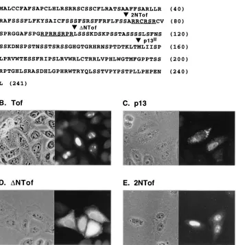
an active process in which the protein initially binds to the nuclear pore complex, followed by opening of the pore and passage of the protein into the nuclear interior (reviewed in reference 53). Recognition by the pore complex requires the presence of an NLS in the protein itself or in a carrier protein; most of the NLSs described thus far consist of regions display-ing a high content of positively charged amino acids (reviewed in reference 14). Figure 1A shows the predicted amino acid sequence of Tof coded by the HTLV-1 molecular clone CS-HTLV-I. Examination of the Tof sequence revealed one re-gion showing a high content of positively charged amino acids, spanning residues 73 to 98. This region contains two arginine-rich segments; the first includes amino acids 73 to 78 and contains four arginines; the second spans amino acids 91 to 98 and contains five arginines. We initially tested the significance of this region to nuclear targeting by comparing the subcellular
localization of Tof with that of p13II, a truncated ORF x-II
isoform translated from a methionine at position 155 of the Tof protein, and of two amino-terminal deletion mutants of
Tof (2NTof and DNTof) initiating either immediately
up-stream or downup-stream of the arginine-rich sequences. Figure 1 shows indirect immunofluorescence assays of HLtat cells
trans-fected with plasmids expressing wild-type Tof (Fig. 1B), p13II
(Fig. 1C), or the deletion mutants (Fig. 1D and E). Fluores-cence assays using a polyclonal rabbit serum recognizing the
76 D’AGOSTINO ET AL. J. VIROL.
on November 9, 2019 by guest
http://jvi.asm.org/
carboxy terminus of the x-II ORF (8) confirmed the nucleolar/ nuclear distribution of Tof (Fig. 1B), as reported earlier (8,
28). The p13II protein showed a mixed distribution, with the
majority of the cells showing a strong punctate signal in the cytoplasm (Fig. 1C), some cells showing a pattern of nuclear fluorescence/nucleolar exclusion as reported earlier (Fig. 1C; also see reference 28), and other cells exhibiting a combination of the cytoplasmic and nuclear signals (not shown). Partial
cytoplasmic localization has also been observed for p13IIcoded
by a molecular clone derived from the HTLV-ILAF cell line
(16a) and suggests that its targeting to the nucleus might be restricted to certain phases of the cell cycle.
Results obtained in assays using the two synthetic truncation mutants of Tof indicated the importance of amino acids 71 to
98 for nucleolar/nuclear targeting. The DNTof deletion
mu-tant, which initiated at amino acid 99 and therefore lacked the arginine-rich sequences, showed a mixed subcellular distribu-tion, accumulating primarily in the cytoplasm of some cells and showing a diffuse pattern in others, but did not exhibit nucle-olar targeting (Fig. 1D). In contrast, 2NTof, which initiated at
amino acid 71 and therefore differed fromDNTof only by the
presence of the two arginine-rich sequences, was efficiently targeted to the nuclei and accumulated in the nucleoli in most of the transfected cells (Fig. 1E). Western blot analysis of cells transfected with the two deletion mutants confirmed their ex-pression (see Fig. 3F).
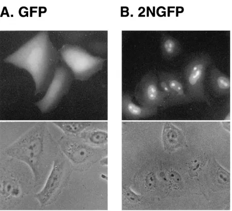
The results obtained with the Tof deletion mutants indicated that the arginine-rich sequences spanning amino acids 71 to 98 represented the NLS of Tof. The ability of the NLS of Tof to change the targeting properties of a nonnuclear protein was confirmed by attaching it to the GFP of the bioluminescent jellyfish A. victoria (44). As shown in Fig. 2A, GFP showed a diffuse pattern of fluorescence in transfected HLtat cells, which was demonstrated by earlier studies to be cytosolic (46). As shown in Fig. 2B, modification of GFP by attachment of both arginine-rich domains of Tof at its amino terminus resulted in efficient targeting of the resulting hybrid to nucleoli/nuclei.
To further study the contribution of the two arginine-rich domains to nucleolar/nuclear targeting, we constructed a series of Tof substitution mutants containing changes in each
do-FIG. 1. Subcellular localization of Tof, p13II
, and Tof deletion mutants. (A) Amino acid sequence of the Tof protein encoded in the HTLV-1 molecular clone CS-HTLV-I. The two arginine-rich sequences are underlined. Sequencing of our CS-HTLV-I plasmid revealed the presence of a glutamine codon at position 222, instead of an arginine codon as reported earlier (8). Triangles indicate the amino termini of synthetic truncation mutants 2NTof andDNTof and the initiator methionine of p13II
. (B to E) HLtat cells that were transfected with plasmid pLsTof, pLsp13AU, pLsDNTof, or pLS2NTof. One day after transfection, the cells were fixed for 20 min with 3.7% formaldehyde–phosphate-buffered saline, permeabilized for 10 min with 0.1% Nonidet P-40–phosphate-buffered saline, and analyzed by indirect immunofluorescence using rabbit anti-Tof serum followed by FITC-conjugated anti-rabbit antibody; shown are representative phase and fluorescence fields.
on November 9, 2019 by guest
http://jvi.asm.org/
[image:3.612.135.479.88.445.2]main, as outlined in Fig. 3. Each mutant was expressed in HLtat cells and detected by indirect immunofluorescence (Fig. 3A to E) and by SDS-PAGE/Western blotting (Fig. 3F). Mu-tants TofP and TofPB contained substitutions in the first argi-nine-rich segment, which contains a total of four arginines. TofP, containing substitutions of the first two arginines (resi-dues 73 and 74), showed a nucleolar/nuclear distribution (Fig. 3B) that was indistinguishable from that of wild-type Tof (Fig. 3A), indicating that these arginines were not necessary for correct targeting. Mutant TofPB, containing substitutions of all four arginines in the first NLS segment (residues 73, 74, 76, and 78), was detected primarily in the nucleus but showed significantly reduced nucleolar accumulation compared to wild-type Tof or TofP (Fig. 3C). This observation indicated that the first arginine-rich domain was necessary for efficient targeting to nucleoli but was not necessary for nuclear import. Mutants TofATK and TofPS were generated to test the role of individual arginines in the second domain in nucleolar/ nuclear targeting. TofATK contained a glycine residue instead of arginine at position 91, a change that is present in the Tof protein coded by the ATK prototype of HTLV-1 (52). TofPS contained the same substitutions found in TofP plus mutations of the arginines at positions 96 and 98 to asparagine and glycine, respectively. Both TofATK and TofPS were targeted to nucleoli/nuclei (Fig. 3D and E), although a slightly greater proportion of the proteins appeared to accumulate in the nu-cleus versus the nucleoli compared to Tof of CS-HTLV-1 (Fig. 3A). These observations suggested that the arginines at posi-tions 91, 96, and 98 were not necessary for nuclear targeting but did promote efficient nucleolar accumulation.
Intracellular targeting of Tof-Rex and Tof-p21Rex
hybrid
proteins.Taken together, the targeting properties of the
sub-stitution mutants indicated that the NLS of Tof functioned as a sum of its parts, with most of the individual arginines appar-ently dispensable for nuclear targeting. As a means of further characterizing the NLS of Tof, we tested its ability to replace the NLS of the HTLV-1 Rex protein and to redirect the
tar-geting of p21Rex. Figure 4 shows results of indirect
[image:4.612.60.299.65.282.2]immuno-FIG. 2. Nucleolar/nuclear targeting of a Tof-GFP hybrid. HLtat cells were transfected with plasmid pLsGFP, which encodes the GFP protein, or with pLs2NGFP, which encodes a derivative of GFP containing the bipartite NLS of Tof at its amino terminus (see Materials and Methods). Thirty-six hours later, the cells were fixed and analyzed for GFP fluorescence.
FIG. 3. Point mutagenesis of the Tof NLS. (A to E) Results of immunoflu-orescence assays of HLtat cells expressing the indicated proteins, from plasmids pLsTof, pLsTofP, pLsTofPBAU, pLsTofATKAU, and pLsTofPSAU, respec-tively. The intact and mutant NLSs in these proteins are represented as boxes containing the two arginine-rich (R) segments. TofP contained arginine-to-as-paragine substitutions at residues 73 and 74. TofPB contained arginine-to-aspar-agine substitutions at residues 73, 74, and 78 and an arginine-to-glycine substi-tution at residue 76. TofATK contained a glycine instead of an arginine at position 91. TofPS contained arginine-to-asparagine substitutions at residues 73 and 74 and arginine-to-glycine substitutions at residues 96 and 98. Immunoflu-orescence assays were carried out with rabbit anti-Tof serum as described in the legend to Fig. 1. Plasmids with the designation “AU” were tagged with a se-quence coding for the AU-1 epitope (see Materials and Methods), the presence of which did not affect subcellular localization of Tof (e.g., compare panel A and Fig. 6). (F) Composite of Western blots carried out with mouse anti-Tof (see Materials and Methods) to detect Tof and the indicated derivatives in lysates of cells transfected with plasmid pLs2NTof, pLsDNTof, pLsTofATKAU, pLsTof PBAU, pLsTofP, pLsTofPSAU, or pLsTof. All of the proteins were electropho-resed through SDS–15% polyacrylamide gels under identical conditions of elec-trophoresis along with prestained protein markers (Broad Range Protein Mark-er; New England Biolabs) as a guide for subsequent alignment of the blots. The weak detection ofDNTof in the Western blot contrasted with its strong reactivity with rabbit anti-Tof in immunofluorescence assays (Fig. 1D); this discrepancy probably reflects the fact thatDNTof lacks the amino-terminal portion of the antigen used to generate mouse anti-Tof.
78 D’AGOSTINO ET AL. J. VIROL.
on November 9, 2019 by guest
http://jvi.asm.org/
[image:4.612.350.516.70.521.2]fluorescence assays of HLtat cells transfected with plasmids
expressing Rex, p21Rex, or hybrid Rex proteins containing Tof
NLS sequences, processed by using a rabbit serum directed against Rex; the apparent size of each protein was confirmed by SDS-PAGE/Western blotting (see Fig. 5A). The amino-terminal NLS of Rex, which consists of the sequence MPK-TRRRPRRSQRKRPPTP (37, 38, 54), directs the protein to the nucleoli/nuclei in a pattern very similar to that of Tof (compare Fig. 4A and 3A). The involvement of this domain in Rex targeting was reflected by the observation that mutant
DNRex, which initiated at amino acid 19 of Rex and therefore
lacked an NLS, was distributed throughout the cell, with some cells showing distinct nucleolar exclusion of the protein (Fig.
4B). p21Rex showed a similar diffuse distribution, although
nucleolar exclusion was not noted (Fig. 4E).
Attachment of the second arginine-rich domain of Tof to
DNRex, resulting in hybrid protein NRex, produced a slightly
more pronounced accumulation of the protein in the nucleus, with nucleolar exclusion no longer evident (Fig. 4C). A more obvious retargeting effect was seen when the entire NLS of Tof
was attached to DNRex, as the resulting hybrid, designated
2NRex, showed clear nuclear targeting (Fig. 4D).
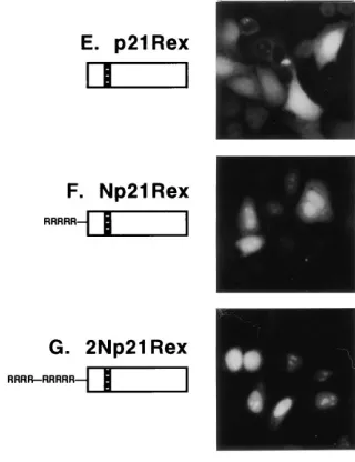
The NLS of Tof functioned more efficiently in the context of
p21Rex, which is translated from a methionine in position 79,
downstream to the start ofDNRex. Np21Rex, which contained
the second arginine-rich domain of Tof, showed a mixed dis-tribution in both the nucleoli/nuclei and cytoplasm (Fig. 4F).
2Np21Rex, carrying the entire NLS of Tof, showed primarily
nucleolar/nuclear targeting, very similar to the pattern of wild-type Rex (Fig. 4G). Therefore, the NLS of Tof directed both nuclear targeting and nucleolar accumulation when attached to
p21Rexbut only nuclear targeting when attached toDNRex.
Functional properties of Tof-Rex and Tof-p21Rex
hybrid
proteins. The amino-terminal NLS of Rex also serves as its
RNA binding domain, mediating association of the protein
with its RNA target, the RXRE (4). Having demonstrated that the NLS of Tof was able to replace that of Rex at the level of nuclear targeting, we were interested in determining whether it could also mediate activation of a Rex/RXRE-dependent mRNA. To test this, HLtat cells were transfected with the Rex-dependent reporter plasmid pCgagRXRE in the absence
or presence of Rex, p21Rex, or the hybrids containing the Tof
NLS. Lysates of the cells were subjected to SDS-PAGE/West-ern blotting to detect the Rex and Gag proteins; results of a
representative experiment are shown in Fig. 5. The anti-p24Gag
Western blot shown in Fig. 5B demonstrated that Gag protein, detected primarily in its uncleaved 55-kDa precursor form, was produced in cells that were cotransfected with Rex, as well as in cells that were cotransfected with NRex or 2NRex (Fig. 5B, lanes 2 and 6 to 9). These results demonstrated that the NLS of Tof was indeed able to functionally replace the amino-terminal domain of Rex at the level of interaction with the RXRE. Results of control transfections showed that the
amount of Rex plasmid used, 0.5mg, produced amounts of Rex
protein that were sufficient to reach a maximal plateau of Gag protein production (data not shown). In contrast, production of Gag protein in the presence of NRex and 2NRex was
aug-mented in transfections using 2.5mg of their expression
plas-mids, indicating that compared to Rex, NRex and 2NRex must be present in higher concentrations within the cell in order to effectively rescue the RXRE-containing mRNA (Fig. 5B, lanes 7 and 9). No induction of Gag protein production was detected
in cells transfected with 2.5mg of plasmids expressingDNRex,
p21Rex, Np21Rex, 2Np21Rex, or Tof itself (Fig. 5, lanes 3 to 5;
data forDNRex and Tof are not shown). Quantitation of Gag
protein by p24Gagantigen capture assay revealed the following
levels of induction of Gag protein production: 383-fold in the
presence of 0.5mg of Rex plasmid, 59-fold in the presence of
2.5mg of NRex plasmid, and 60-fold in the presence of 2.5mg
[image:5.612.315.475.71.279.2]of 2NRex plasmid.
FIG. 4. Tof-Rex and Tof-p21Rexhybrids. HLtat cells were transfected with
pL3Rex (58), pLsDNRex, pLsNRex, pLs2NRex, pLsp21Rex, pLsNp21Rex, or pLs2Np21Rex (see Materials and Methods) and analyzed by indirect immuno-fluorescence using rabbit Rex serum (3) followed by FITC-conjugated anti-rabbit antibody. The following domains of Rex are indicated in the diagrams: box with diagonal lines, the amino-terminal NLS/RNA binding domain (4, 37, 55); black box, multimerization domain (5, 60); black-and-white box, activation do-main (23). The second arginine-rich dodo-main of Tof is represented by RRRRR; the entire Tof NLS is represented by RRRR—RRRRR.
on November 9, 2019 by guest
http://jvi.asm.org/
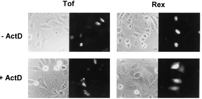
Effect of treatment with actinomycin D on the subcellular
distribution of Tof.Although detected primarily in the nucleoli
and nuclei, Rex is known to shuttle between the nucleus and the cytoplasm (29, 59), presumably reflecting its functional role as a cofactor in the nuclear-cytoplasmic transport of RXRE-containing mRNAs. We were interested in determining if Tof possessed such shuttling properties. One way to analyze the nuclear export leg of the shuttling process and to distinguish between shuttling and nonshuttling proteins is to test their responses to treatment with actinomycin D (actD), an inhibitor of RNA synthesis that is known to disrupt nuclear retention of shuttling proteins, including Rex (29, 59) and Rev of HIV-1 (12, 24, 35, 45). Figure 6 shows immunofluorescence assays of HLtat cells that were transfected with a plasmid expressing Tof or Rex and then incubated for 4 h in the absence or presence
of 10 mg of actD per ml, a concentration sufficient to inhibit
synthesis of both rRNA by RNA polymerase I and mRNA by RNA polymerase II (42). Fluorescence images of the actD-treated cells showed that Rex underwent a partial redistribu-tion of its compartmentalizaredistribu-tion from the nucleoli/nuclei to the cytoplasm, as expected for a shuttling protein that is exported from the nuclear compartment. In contrast, Tof was not re-leased into the cytoplasm in the presence of actD but remained sequestered in the nucleus in numerous small speckles that
might represent remnants of nucleoli. Therefore, although Rex and Tof had similar nucleolar/nuclear import pathways and despite the fact that the NLS of Tof was functionally inter-changeable with the NLS/RNA binding domain of Rex, an involvement of Tof in Rex-like control of mRNA transport appeared to be unlikely.
Rex-dependent expression of Tof from the 1-2-B mRNA.In
previous studies, the Tof protein was detected by transfecting cells with eukaryotic expression plasmids containing the Tof coding sequence and driven by the HIV-1 LTR (8) or human cytomegalovirus promoter (28). We generated a plasmid en-coding the entire multiply spliced 1-2-B Tof mRNA and tested its ability to produce Tof protein and its possible dependence on Rex for efficient expression. This plasmid, designated pBS1-2-B, was transfected into HLtat cells in the absence or pres-ence of the Tax expression plasmid pLCXL (8), needed to promote efficient transcription from the viral LTR, and the Rex expression plasmid pL3Rex (58). Expression of the 1-2-B mRNA was monitored by Northern analysis using a probe specific for tof sequences, and production of Tof protein was detected by Western blotting and indirect immunofluorescence using Tof-specific antibodies. The Northern blot shown in Fig. 7A demonstrated that the Tof mRNA was not detected in cells that were transfected with pBS1-2-B alone, as expected for an LTR-driven gene (Fig. 7A, lane 1). Surprisingly, cells trans-fected with both pBS1-2-B and Tax also failed to express de-tectable levels of the Tof mRNA (Fig. 7A, lane 2); only cells that had been cotransfected with pBS1-2-B and both the Tax and Rex expression plasmids contained the 2.3-kb 1-2-B mRNA (Fig. 7A, lane 3). Immunoblotting assays revealed the presence of the 30-kDa Tof protein band only in lysates of cells that had received all three plasmids (data not shown). Anti-Tof immunofluorescence assays confirmed that cells transfected with pBS1-2-B alone failed to produce detectable levels of Tof protein (i.e., no Tof-positive cells were observed [data not shown]). Immunofluorescence assays of cells transfected with pBS1-2-B and the Tax expression plasmid revealed a few cells that were able to express low levels of Tof. Figure 7B (panel 2) shows an example of a field containing a single cell expressing Tof at low levels; the ability to detect Tof expression in trans-fections that had tested negative by Western blotting and Northern analysis reflected the high sensitivity of the immu-nofluorescence technique to detect proteins that are concen-trated in the nucleoli/nuclei. In contrast, many cells expressing readily detectable levels of Tof protein were observed when pBS1-2-B was cotransfected with both the Tax and Rex expres-sion plasmids, as shown in panel 3 of Fig. 7B. Taken together, these data demonstrated that the Tof mRNA required Rex for efficient expression at both the RNA and protein levels. This property is shared by the unspliced and singly spliced mRNAs encoding the structural proteins of HTLV-1, which are ex-pressed during the later stages of viral replication (21).
DISCUSSION
The present study was aimed at finding clues to the possible functional significance of the Tof protein of HTLV-1 by using as a starting point its accumulation in nucleoli/nuclei, a prop-erty shared with essential retroviral regulatory proteins such as Rex and the Tat and Rev proteins of HIV (11, 49, 54, 55). We mapped the signal directing Tof to nucleoli and demonstrated that the sequence was able to replace the amino-terminal NLS/ RNA binding domain of Rex, both at the level of nuclear targeting and as an RNA binding domain capable of interact-ing with the RXRE and rescuinteract-ing a Rex-dependent mRNA. Although these findings suggested that Tof and Rex had
func-FIG. 5. Functional assay of Tof-Rex and Tof-p21Rex hybrids. (A) Western blot analysis of HLtat cells cotransfected with pCgagRXRE and pL3CAT (as a standard for transfection efficiency [61]) in the presence of pL3Rex (0.5mg), pLsp21Rex (2.5mg), pLsNp21Rex (2.5mg), pLs2Np21Rex (2.5mg), pLsNRex (0.5 or 2.5mg), or pLs2NRex (0.5 or 2.5mg) (lanes 2 to 9). Lane A shows a transfection with pLSDNRex in the absence of pCgagRXRE and pL3CAT. Wild-type and hybrid Rex proteins were electrophoresed through an SDS–15% polyacrylamide gel and subjected to Western blot analysis using rabbit anti-Rex serum (3) and the ECL Western blotting detection system. Indicated in the left margin are positions of prestained protein standards (Rainbow protein molecu-lar weight markers; Amersham). (B) Lanes 2 to 9 show an anti-p24GagWestern
blot of the samples corresponding to lanes 2 to 9 of panel A. Lane 1 contains a lysate of HLtat cells cotransfected with pCgagRXRE and pL3CAT in the ab-sence of Rex. Gag protein production was detected using an anti-p24Gag
mono-clonal antibody (Cellular Products).
80 D’AGOSTINO ET AL. J. VIROL.
on November 9, 2019 by guest
http://jvi.asm.org/
[image:6.612.72.283.70.334.2]tionally analogous NLS/RNA binding domains and that Tof itself may function as a regulator of RNA expression, Tof did not display the nuclear-cytoplasmic shuttling properties of a Rex-like RNA transport chaperone on the basis of its retention in the nucleus in response to treatment with actD. Expression of Tof from the complete 1-2-B mRNA containing all coding and noncoding portions was found to be dependent on both Tax and Rex, indicating a possible role for the protein in the later stages of viral replication.
The NLS of Tof was mapped to amino acids 71 to 98 and consisted of two arginine-rich domains (amino acids 73 to 78 and 91 to 98) separated by a 12-amino-acid spacer (Fig. 1). The targeting activity of this sequence was demonstrated by its efficiency in redirecting GFP from the cytoplasm to the nucle-oli/nuclei (Fig. 2). The interdependence of the individual ar-ginine residues in the two domains for nucleolar/nuclear tar-geting was demonstrated by a series of mutant Tof proteins containing point mutations. The second arginine-rich domain was shown to be sufficient for efficient nuclear targeting (mu-tant TofPB [Fig. 3C]). Arginines 73, 74, 91, 97, and 98 did not appear to be involved in nuclear targeting but promoted nu-cleolar accumulation (Fig. 3B, D, and E). By a process of elimination, arginines 93 and 94 remain as potentially essential for nuclear targeting.
Bipartite NLSs similar to that of Tof have previously been found in other nuclear proteins and are characterized by the presence of two clusters of positively charged amino acids separated by a spacer region of approximately 10 residues (reviewed in reference 14). Characterization of the NLS of the
Xenopus protein nucleoplasmin showed that both clusters are
important for nuclear targeting, while mutations within the spacer region do not affect NLS function (47). Although our data indicate that the nine arginine residues within the Tof NLS work in an interdependent manner determining efficient nucleolar accumulation, the group of five arginines in the sec-ond domain are clearly sufficient for nuclear targeting.
Attachment of the NLS of Tof at the amino terminus of
p21Rexor its substitution for the amino terminal NLS of Rex
conferred different targeting properties to the resulting hy-brids. While attachment of the second arginine-rich domain to
p21Rex(Np21Rex[Fig. 4F]) resulted in partial nuclear
target-ing, the corresponding Rex hybrid (NRex [Fig. 4C]) showed
little change in distribution compared toDNRex, which lacked
the first 18 amino acids of Rex (Fig. 4B). Likewise, the entire bipartite NLS of Tof acted as an efficient nucleolar/nuclear
targeting signal in the context of p21Rex(2Np21Rex[Fig. 4G])
but directed nuclear targeting, but not nucleolar accumulation, of the Rex hybrid 2NRex (Fig. 4D). These observations suggest that the sequences of Rex between amino acids 19 and 79 (the
initiator methionine of p21Rex) influence the subcellular
tar-geting properties of the protein. This region contains a
[image:7.612.132.484.71.244.2]mul-FIG. 6. Effect of actD on the intracellular distribution of Tof and Rex. HLtat cells were transfected with the Tof expression plasmid pLsTofAU or the Tax/Rex expression plasmid pBSTR-3 (see Materials and Methods). Twenty hours later, the cells were incubated for an additional 4 h in the absence or presence of 10mg of actD (Sigma) per ml and then harvested for indirect immunofluorescence using rabbit anti-Tof or rabbit anti-Rex serum.
FIG. 7. Expression of Tof from the 1-2-B mRNA. HLtat cells were trans-fected with plasmid pBS1-2-B in the absence or presence of Tax and Rex expression plasmids. One day later, cells were harvested for Northern analysis or indirect immunofluorescence. (A) Northern analysis of total RNA isolated from cells transfected with the following plasmids: lane 1, pBS1-2-B; lane 2, pBS1-2-B and the Tax expression plasmid pLCXL (8); lane 3, pBS1-2-B, pLCXL, and the Rex expression plasmid pL3Rex. Total RNA was isolated using RNAzol as specified by the manufacturer (Cinna/Biotecx), separated by denaturing electro-phoresis through an agarose gel, transferred to a nylon membrane (Qiabrane; Qiagen), and hybridized to a DNA probe to detect the Tof mRNA. The probe was produced by PCR amplification of HTLV-1 nt 4820 to 4831 joined to nt 6478 to 6948, using plasmid pLsTofAU as a template, and was32P-labeled by the
random priming method (Random Primed DNA Labeling Kit; Boehringer Mannheim). (B) Anti-Tof immunofluorescence assays of cells transfected with pBS1-2-B and pLCXL (panel 2) or with pBS1-2-B, pLCXL and pL3Rex (panel 3). Cells transfected with pBS1-2-B alone did not produce detectable amounts of Tof (not shown).
on November 9, 2019 by guest
http://jvi.asm.org/
timerization domain spanning amino acids 54 to 69 that was identified in studies of Rev-Rex chimeras (60) and in experi-ments based on a two-hybrid system approach (5). Results of
the functional assays of the Tof-p21Rexand Tof-Rex hybrids
confirm the importance of sequences between amino acids 19 and 79 of the Rev protein for activation of the RXRE (Fig.
5B). Although 2Np21Rexexhibited a nucleolar/nuclear
distri-bution very similar to that of Rex, the protein was unable to transactivate the RXRE. In contrast, both NRex and 2NRex were functionally active (Fig. 5B), even though they were not very efficiently retained in the nucleus and did not appear to accumulate in nucleoli. The lack of functional activity of the
p21Rexhybrid probably reflects the absence of the
multimer-ization domain.
The finding that NRex and 2NRex were able to activate a RXRE-containing mRNA suggests that the NLS of Tof may function as an RNA binding domain within the context of Tof and leads to the proposal that the protein plays a functional role in the metabolism of cellular or viral mRNAs. We are currently testing whether Tof interacts with the RXRE or with a cellular RNA. Cotransfection assays performed with Tof expression plasmids indicated that Tof does not possess a Rex-like ability to activate the RXRE (8); however, this result does not exclude the possibility that Tof binds to the RXRE but displays a function that is distinct from that of Rex.
An important question is whether the ability of the Tof NLS to replace that of Rex reflects specific properties of the two arginine-rich segments. Would any positively charged NLS confer functional activity when substituted for the amino-ter-minal domain of Rex? A recent analysis of Rex function tested a Rex hybrid analogous to NRex and 2NRex, in which the amino-terminal 19 amino acids of the protein were replaced by the NLS (P-K-K-K-R-K-V) of the simian virus 40 large T antigen (25). Interestingly, although this hybrid showed nu-clear targeting, it was unable to bind to RXRE in vitro and was functionally inactive in vivo; instead, the hybrid acted as a transdominant inhibitor of Rex function, a property that the investigators mapped to the activation domain of Rex. It is particularly noteworthy that the T-antigen NLS contains five positively charged amino acids, equal to the number of argi-nines present in the second arginine-rich domain of Tof, which was able to efficiently replace the NLS of Rex in hybrid NRex. The failure of the T-antigen signal to direct binding to or activation of the RXRE provides indirect evidence that prop-erties observed for the Tof NLS are indeed specific and rele-vant to the function of the protein.
The ability of the NLS of Tof to functionally replace that of Rex initially suggested a possible involvement of Tof in Rex-like posttranscriptional nuclear-cytoplasmic transport of either cellular or viral mRNAs. However, comparative analyses of the trafficking properties of Tof and Rex carried out in actD-treated cells revealed a clear difference between Tof and Rex in the nuclear export/retention portion of the trafficking path-way, since Tof remained sequestered in the nucleus upon actD treatment, while Rex showed a distinct relocalization to the cytoplasm (Fig. 6). Therefore, while Rex is able to travel back and forth between the nucleus and the cytoplasm, a property that is presumed to be connected to its function in mRNA transport, Tof appears to remain stably associated with nucle-olar/nuclear components. Although these observations suggest that Tof probably is not directly involved in a Rex-like RNA export pathway, we cannot exclude that the protein is involved in some as yet undescribed transport pathway that is not af-fected by actD treatment.
The observation that expression of the 1-2-B mRNA encod-ing Tof is dependent on Rex represents the first instance of a
multiply spliced HTLV-1 mRNA under such posttranscrip-tional control (Fig. 7). An important aspect of this analysis is that we tested expression of an mRNA that was transcribed in its fully spliced form, rather than being derived by splicing of the full-length viral transcript as would be the case during a cycle of viral replication. Therefore, the Rex dependence that we observed must have been exerted at some point after splic-ing, presumably at the level of transport/stabilization, as indi-cated by other studies of Rex and Rev (reviewed in references 6, 31, and 41). One possibility is that the Rex-RXRE interac-tion is necessary to overcome the effects of inhibitory elements within the mRNA, analogous to intragenic inhibitory se-quences that are known to confer Rev dependence to the gag and env mRNAs of HIV (10, 32, 51). Such inhibitory sequences might be located in the proximal portion of exon B, which is absent from the 1-2-3 mRNA; experiments to test this possi-bility are under way.
ACKNOWLEDGMENTS
We thank David Derse, Genoveffa Franchini, Barbara K. Felber, George N. Pavlakis, and Rosario Rizzuto for generously providing plasmids and reagents and Pierantonio Gallo for preparing the artwork. This study was supported by grants from the Istituto Superiore di Sanita`-Progetto AIDS and from the Associazione Italiana per la Ricerca sul Cancro. D.M.D. was supported by sequential fellowships from the National Cancer Institute (National Research Service Award IF32CA60403-01), the Lady Tata Memorial Trust, and the Istituto Superiore di Sanita`-Progetto AIDS.
REFERENCES
1. Benko, D. M., R. Robinson, L. Solomin, M. Mellini, B. K. Felber, and G. N.
Pavlakis.1991. Binding of trans-dominant mutant Rev protein of human immunodeficiency virus type 1 to the cis-acting rev-responsive element does not affect the fate of viral mRNA. New Biol. 2:1111–1122.
2. Berneman, Z. N., R. B. Gartenhaus, M. S. Reitz, W. A. Blattner, A. Manns,
B. Hanchard, O. Ikehara, R. C. Gallo, and M. E. Klotman.1992. Expression of alternatively spliced human T-lymphotropic virus type I pX mRNA in infected cell lines and in primary uncultured cells from patients with adult T-cell leukemia/lymphoma and healthy carriers. Proc. Natl. Acad. Sci. USA
89:3005–3009.
3. Bhat, N. K., Y. Adachi, K. P. Samuel, and D. Derse. 1993. HTLV-I gene expression by defective proviruses in an infected T-cell line. Virology 196:15–24. 4. Bogerd, H. P., G. L. Huckaby, Y. F. Ahmed, S. M. Hanly, and W. C. Greene. 1991. The type I human T-cell leukemia virus (HTLV-I) Rex trans-activator binds directly to the HTLV-I Rex and the type 1 human immunodeficiency virus Rev RNA response elements. Proc. Natl. Acad. Sci. USA 88:5704–5708. 5. Bogerd, H. P., and W. C. Greene. 1993. Dominant negative mutants of human T-cell leukemia virus type I Rex and human immunodeficiency virus type 1 Rev fail to multimerize in vivo. J. Virol. 67:2496–2502.
6. Cann, A. J., and I. S. Y. Chen. 1996. Human T-cell leukemia virus types I and II, p. 1849–1880. In B. N. Fields, D. M. Knipe, P. M. Howley et al. (ed.), Fields virology, 3rd ed. Raven Press, Ltd., New York, N.Y.
7. Caputo, A., and W. A. Haseltine. 1992. Reexamination of the coding poten-tial of the HTLV-I pX region. Virology 188:618–627.
8. Ciminale, V., G. N. Pavlakis, D. Derse, C. P. Cunningham, and B. K. Felber. 1992. Complex splicing in the human T-cell leukemia virus (HTLV) family of retroviruses: novel mRNAs and proteins produced by HTLV-1. J. Virol.
66:1737–1745.
9. Ciminale, V., D. M. D’Agostino, L. Zotti, G. Franchini, B. K. Felber, and L.
Chieco-Bianchi.1995. Expression and characterization of proteins produced by mRNAs spliced into the X region of the human T-cell leukemia/lympho-tropic virus type II. Virology 209:445–456.
10. Cochrane, A. W., S. Jones, S. Beidas, P. J. Dillon, A. M. Skalka, and C. A.
Rosen. 1991. Identification and characterization of intragenic sequences which repress human immunodeficiency virus structural gene expression. J. Virol. 65:5305–5313.
11. Cullen, B. R., J. Hauber, K. Campbell, J. G. Sodroski, W. A. Haseltine, and
C. A. Rosen.1988. Subcellular localization of the human immunodeficiency virus trans-acting art gene product. J. Virol. 62:2498–2501.
12. D’Agostino, D. M., V. Ciminale, G. N. Pavlakis, and L. Chieco-Bianchi. 1995. Intracellular trafficking of the human immunodeficiency virus type 1 Rev protein: involvement of continued rRNA synthesis in nuclear retention. AIDS Res. Hum. Retroviruses 11:1063–1071.
82 D’AGOSTINO ET AL. J. VIROL.
on November 9, 2019 by guest
http://jvi.asm.org/
13. Derse, D., J. Mikovits, M. Polianova, B. K. Felber, and F. Ruscetti. 1995. Virions released from cells transfected with a molecular clone of human T-cell leukemia virus type I give rise to primary and secondary infections of T cells. J. Virol. 69:1907–1912.
14. Dingwall, C., and R. A. Laskey. 1991. Nuclear targeting sequences—a con-sensus? Trends Biochem. Sci. 16:478–481.
15. Franchini, G., J. C. Mulloy, I. J. Koralnik, A. Lo Monico, J. J. Sparkowski,
T. Andresson, D. J. Goldstein, and R. Schlegel.1993. The human T-cell leukemia/lymphotropic virus type I p12I
protein cooperates with the E5 oncoprotein of bovine papillomavirus in cell transformation and binds the 16-kilodalton subunit of the vacuolar H1ATPase. J. Virol. 67:7701–7704. 16. Franchini, G. 1995. Molecular mechanisms of human T-cell
leukemia/lym-photropic virus type I infection. Blood 86:3619–3639. 16a.Franchini, G. Personal communication.
17. Fuller, S. A., M. Takahashi, and J. G. R. Hurrell. 1995. Immunization of mice, p. 11.4.2–11.4.5. In F. M. Ausubel et al. (ed.), Current protocols in molecular biology, vol. 2. John Wiley & Sons, New York, N.Y.
18. Gessain, A., F. Barin, J. C. Vernant, O. Gout, L. Maurs, A. Calender, and G.
de The.1985. Antibodies to human T-lymphotropic virus type-I in patients with tropical spastic paraparesis. Lancet ii:407–410.
19. Gitlin, S. D., J. Dittmer, R. L. Reid, and J. N. Brady. 1993. The molecular biology of human T-cell leukemia viruses, p. 159–192. In B. R. Cullen (ed.), Human retroviruses. Oxford University Press, New York, N.Y.
20. Graham, F. J., and A. J. van der Eb. 1973. A new technique for the assay of infectivity of human adenovirus 5 DNA. Virology 52:456–460.
21. Hidaka, M., J. Inoue, M. Yoshida, and M. Seiki. 1988. Posttranscriptional regulator (rex) of HTLV-I initiates expression of viral structural proteins but suppresses expression of regulatory proteins. EMBO J. 7:519–523. 22. Hinuma, Y., K. Nagata, M. Hanaoka, M. Nakai, T. Matsumoto, K. I.
Ki-noshita, S. Shirakawa, and I. Miyoshi.1981. Adult T-cell leukemia: antigen in an ATL cell line and detection of antibodies to the antigen in human sera. Proc. Natl. Acad. Sci. USA 78:6476–6480.
23. Hope, T. J., B. L. Bond, D. McDonald, N. P. Klein, and T. G. Parslow. 1991. Effector domains of human immunodeficiency virus type 1 Rev and human T-cell leukemia virus type I Rex are functionally interchangeable and share an essential peptide motif. J. Virol. 65:6001–6007.
24. Kalland, K.-H., A. M. Szilvay, K. A. Brokstad, W. Saetrevik, and G.
Hauke-nes.1994. The human immunodeficiency virus type 1 Rev protein shuttles between the cytoplasm and nuclear components. Mol. Cell. Biol. 14:7436– 7444.
25. Katahira, J., T. Ishizaki, H. Sakai, A. Adachi, K. Yamamoto, and H. Shida. 1995. Effects of translation initiation factor eLF-5A on the functioning of human T-cell leukemia virus type I Rex and human immunodeficiency virus Rev inhibited trans dominantly by a Rex mutant deficient in RNA binding. J. Virol. 69:3125–3133.
26. Kiyokawa, T., M. Seiki, S. Iwashita, K. Imagawa, F. Shimizu, and M.
Yo-shida.1985. p27XIII and p21XIII proteins encoded by the pX sequence of human T-cell leukemia virus type I. Proc. Natl. Acad. Sci. USA 82:8359– 8363.
27. Koralnik, I. J., A. Gessain, M. E. Klotman, A. Lo Monico, Z. N. Berneman,
and G. Franchini.1992. Protein isoforms encoded by the pX region of human T-cell leukemia/lymphotropic virus type I. Proc. Natl. Acad. Sci. USA
89:8813–8817.
28. Koralnik, I. J., J. Fullen, and G. A. Franchini. 1993. The p12I
, p13II
, and p30II
proteins encoded by human T-cell leukemia/lymphotropic virus type I open reading frames are localized in three different cellular compartments. J. Virol. 67:2360–2366.
29. Kubota, S., M. Hatanaka, and R. J. Pomerantz. 1996. Nucleocytoplasmic redistribution of the HTLV-I Rex protein: alterations by coexpression of the HTLV-I p21x
protein. Virology 220:502–507.
30. Kunkel, T. A. 1985. Rapid and efficient site-specific mutagenesis without phenotypic selection. Proc. Natl. Acad. Sci. USA 82:488–492.
31. Luciw, P. A. 1996. Human immunodeficiency viruses and their replication, p. 1881–1951. In B. N. Fields, D. M. Knipe, P. M. Howley et al. (ed.), Fields virology, 3rd ed. Raven Press, Ltd., New York, N.Y.
32. Maldarelli, F., M. A. Martin, and K. Strebel. 1991. Identification of post-transcriptionally active inhibitory sequences in human immunodeficiency virus type 1 RNA: novel level of gene regulation. J. Virol. 65:5732–5743. 33. Melchior, F., and L. Gerace. 1995. Mechanisms of nuclear protein import.
Curr. Opin. Cell Biol. 7:310–318.
34. Mermer, B., B. K. Felber, M. Campbell, and G. N. Pavlakis. 1990. Identifi-cation of trans-dominant HIV-1 protein mutants by direct transfer of bac-terially produced proteins into human cells. Nucleic Acids Res. 18:2037– 2044.
35. Meyer, B. E., and M. H. Malim. 1994. The HIV-1 Rev transactivator shuttles between the nucleus and the cytoplasm. Genes Dev. 8:1538–1547. 36. Nagashima, K., M. Yoshida, and M. Seiki. 1986. A single species of pX
mRNA of human T-cell leukemia virus type I encodes trans-activator p40x
and two other phosphoproteins. J. Virol. 60:394–399.
37. Nosaka, T., H. Siomi, Y. Adachi, M. Ishibashi, S. Kubota, M. Maki, and M.
Hatanaka.1989. Nucleolar targeting signal of human T-cell leukemia virus type I rex-encoded protein is essential for cytoplasmic accumulation of
un-spliced viral mRNA. Proc. Natl. Acad. Sci. USA 86:9798–9802.
38. Nosaka, T., Y. Miyazaki, T. Takamatsu, K. Sano, M. Nakai, S. Fujita, T. E.
Martin, and M. Hatanaka.1995. The post-transcriptional regulator Rex of the human T-cell leukemia virus type I is present as nucleolar speckles in infected cells. Exp. Cell Res. 219:122–129.
39. Orita, S., A. Saiga, S. Takagi, T. Takanka, K. Okumura, Y. Aono, Y. Hinuma,
and H. Igarashi.1991. A novel alternatively spliced viral mRNA transcribed in cells infected with human T-cell leukemia virus type I is mainly responsible for expressing p21X protein. FEBS Lett. 295:127–134.
40. Osame, M., K. Usuku, S. Izumo, N. Ijuchi, H. Amitani, A. Agata, M.
Ma-tsumoto, and M. Tara.1986. HTLV-I-associated myelopathy, a new clinical entity. Lancet i:1031–1032.
41. Parslow, T. G. 1993. Post-transcriptional regulation of human retroviral gene expression, p. 101–136. In B. R. Cullen (ed.), Human retroviruses. Oxford University Press, New York, N.Y.
42. Perry, R. P., and D. E. Kelley. 1970. Inhibition of RNA synthesis by actino-mycin D: characteristic dose-response of different RNA species. J. Cell Physiol. 76:127–140.
43. Poiesz, B. J., F. W. Ruscetti, A. F. Gazdar, P. A. Bunn, J. D. Minna, and R. C.
Gallo.1980. Detection and isolation of type C retrovirus particles from fresh and cultured lymphocytes of a patient with cutaneous T-cell lymphoma. Proc. Natl. Acad. Sci. USA 77:7415–7419.
44. Prasher, D. C., V. K. Eckenrode, W. W. Ward, F. G. Prendgast, and M. J.
Cormier.1992. Primary structure of the Aequorea victoria green-fluorescent protein. Gene 111:229–233.
45. Richard, N., S. Iacampo, and A. Cochrane. 1994. HIV-1 Rev is capable of shuttling between the nucleus and the cytoplasm. Virology 204:123–131. 46. Rizzuto, R., M. Brini, P. Pizzo, M. Murgia, and T. Pozzan. 1995. Chimeric
green fluorescent protein as a tool for visualizing subcellular organelles in living cells. Curr. Biol. 5:635–642.
47. Robbins, J., S. M. Dilworth, R. A. Laskey, and C. Dingwall. 1991. Two interdependent basic domains in nucleoplasmin nuclear targeting sequence: identification of a class of bipartite nuclear targeting sequence. Cell 64:615– 623.
48. Roithmann, S., C. Pique, A. Le Cesne, L. Delamarre, D. Pham, T. Tursz, and
M.-C. Dokhelar.1994. The open reading frame I (ORF I)/ORF II part of the human T-cell leukemia virus type I X region is dispensable for p40tax
, p27rex
, or envelope expression. J. Virol. 68:3448–3451.
49. Ruben, S., A. Perkins, R. Purcell, K. Joung, R. Sia, R. Burghoff, W. A.
Haseltine, and C. A. Rosen.1989. Structural and functional characterization of human immunodeficiency virus tat protein. J. Virol. 63:1–8.
50. Schwartz, S., B. K. Felber, D. M. Benko, E. M. Fenyo¨, and G. N. Pavlakis.
1990. Cloning and functional analysis of multiply spliced mRNA species of human immunodeficiency virus type 1. J. Virol. 64:2519–2529.
51. Schwartz, S., B. K. Felber, and G. N. Pavlakis. 1992. Distinct RNA se-quences in the gag region of human immunodeficiency virus type 1 decrease RNA stability and inhibit expression in the absence of Rev. J. Virol. 66:150– 159.
52. Seiki, M., Y. Hattori, and M. Yoshida. 1982. Human adult T-cell leukemia virus: molecular cloning of the provirus DNA and the unique terminal structure. Proc. Natl. Acad. Sci. USA 79:6899–6902.
53. Silver, P. A. 1991. How proteins enter the nucleus. Cell 64:489–497. 54. Siomi, H., H. Shida, S. H. Nam, T. Nosaka, M. Maki, and M. Hatanaka.
1988. Sequence requirements for nucleolar localization of human T cell leukemia virus type I pX protein, which regulates viral RNA processing. Cell
55:197–209.
55. Siomi, H., H. Shida, M. Makai, and M. Hatanaka. 1990. Effects of a highly basic region of human immunodeficiency virus Tat protein on nucleolar localization. J. Virol. 64:1803–1807.
56. Smith, D. B., and K. S. Johnson. 1988. Single-step purification of polypep-tides expressed in Escherichia coli as fusions with glutathione-S-transferase. Gene 67:31–40.
57. Smith, D. B., and L. M. Corcoran. 1994. Expression and purification of glutathione-S-transferase fusion proteins, p. 16.7.1–16.7.7. In F. M. Ausubel et al. (ed.), Current protocols in molecular biology, vol. 2. John Wiley & Sons, New York, N.Y.
58. Solomin, L., B. K. Felber, and G. N. Pavlakis. 1990. Different sites of interaction for Rev, Tev, and Rex proteins within the rev-responsive element of human immunodeficiency virus type 1. J. Virol. 64:6010–6017. 59. Stauber, R., G. A. Gaitanaris, and G. N. Pavlakis. 1995. Analysis of
traffick-ing of Rev and transdominant Rev proteins in livtraffick-ing cells ustraffick-ing green fluo-rescent protein fusions: transdominant Rev blocks the export of Rev from the nucleus to the cytoplasm. Virology 213:439–449.
60. Weichselbraun, I., J. Berger, M. Dobrovnik, H. Bogerd, R. Grassman, W. C.
Greene, J. Hauber, and E. Bohnlein.1992. Dominant-negative mutants are clustered in a domain of the human T-cell leukemia virus type I Rex protein: implications for trans dominance. J. Virol. 66:4540–4545.
61. Wright, C. M., B. K. Felber, H. Paskalis, and G. N. Pavlakis. 1986. Expres-sion and characterization of the trans-activator of HTLV-III/LAV virus. Science 234:988–992.
62. Zazopoulos, E., J. G. Sodroski, and W. A. Haseltine. 1990. p21rex protein of HTLV-I. J. Acquired Immune Defic. Syndr. 3:1135–1139.