TECHNICAL NOTE
Development of a high throughput
drug screening assay to identify compounds
that protect oligodendrocyte viability
and differentiation under inflammatory
conditions
Elen S. Rosler
1,3†, Karen D. Lariosa‑Willingham
1,2†, Jay S. Tung
1, Jason C. Dugas
1,4, Tassie L. Collins
1,5and Dmitri Leonoudakis
1,2*Abstract
Background: Newly proliferated oligodendrocyte precursor cells (OPCs) migrate and surround lesions of patients with multiple sclerosis (MS) and other demyelinating diseases, but fail to differentiate into oligodendrocytes (OLs) and remyelinate remaining viable axons. The abundance of secreted inflammatory factors within and surrounding these lesions likely plays a major inhibitory role, promoting cell death and preventing OL differentiation and axon remyelina‑ tion. To identify clinical candidate compounds that may protect existing and differentiating OLs in patients, we have developed a high throughput screening (HTS) assay that utilizes purified rat OPCs.
Results: Using a fluorescent indicator of cell viability coupled with image quantification, we developed an assay to allow the identification of compounds that promote OL viability and differentiation in the presence of the synergistic inflammatory cytokines, tumor necrosis factor α and interferon‑γ. We have utilized this assay to screen the NIH clinical collection library and identify compounds that protect OLs and promote OL differentiation in the presence of these inflammatory cytokines.
Conclusion: This primary OL‑based cytokine protection assay is adaptable for HTS and may be easily modified for profiling of compounds in the presence of other potentially inhibitory molecules found in MS lesions. This assay should be of use to those interested in identifying drugs for the treatment of MS and other demyelinating diseases. Keywords: Myelination, Oligodendrocyte, Cytokine, Interferon gamma, Tumor necrosis factor alpha, Protection, High throughput, Drug screening, Differentiation, Primary cell‑based assay, Image analysis, Multiple sclerosis, Myelin basic protein
© 2016 The Author(s). This article is distributed under the terms of the Creative Commons Attribution 4.0 International License (http://creativecommons.org/licenses/by/4.0/), which permits unrestricted use, distribution, and reproduction in any medium, provided you give appropriate credit to the original author(s) and the source, provide a link to the Creative Commons license, and indicate if changes were made. The Creative Commons Public Domain Dedication waiver (http://creativecommons.org/ publicdomain/zero/1.0/) applies to the data made available in this article, unless otherwise stated.
Background
Multiple sclerosis (MS) is a devastating neurological dis-ease, affecting over 2 million patients worldwide, caused by autoimmune-mediated destruction of the myelin sheaths that insulate and protect axons in the central
nervous system. Most MS patients initially present with the relapsing-remitting form of MS relapsing remitting multiple sclerosis (RRMS), in which cycles of immune-mediated axonal demyelination (relapse) are followed by periods of remyelination (remission). Over time, how-ever, the majority of RRMS patients exhibit chronic, pro-gressive decline in neurological function. This is believed to be the result of an accumulation of axonal damage, as well as an eventual loss of remyelination capacity [1]. Although current MS therapies for RRMS provide
Open Access
*Correspondence: [email protected] † Elen S. Rosler and Karen D. Lariosa‑Willingham shared authorship 2 Teva Pharmaceuticals, Biologics and CNS Discovery, Redwood City, CA 94063, USA
Page 2 of 12 Rosler et al. BMC Res Notes (2016) 9:444
significant relief from relapse, none have yet been dem-onstrated to prevent disease progression and none have been effective in treating progressive forms of MS. All of the disease-modifying therapies approved for treat-ment of RRMS do so by targeting the pathologic immune response [2]. There are no therapies that function to directly protect or restore myelin, and identifying such therapies has been the focus of several groups in the field [3–6].
One approach undertaken by several groups seek-ing to identify new therapeutic agents, has been to screen small molecule libraries in an attempt to iden-tify agents that accelerate differentiation of immature oligodendrocyte precursor cells (OPCs) into mature, myelin-producing oligodendrocytes (OLs) in the hope that this will accelerate remyelination of axons follow-ing immune attack. While it is logical to expect that increasing the number of mature oligodendrocytes will improve the rate and extent of axonal remyelina-tion, it should be considered that the oligodendrocyte differentiation assays described above assess differen-tiation of OPCs into oligodendrocytes under healthy conditions. MS lesions contain a number of factors that are known to impair oligodendrocyte differentia-tion and reduce the viability of differentiating oligoden-drocytes. We thus felt that it was important to develop assays useful for the identification of drugs that sup-port oligodendrocyte differentiation under conditions mimicking disease. We therefore examined the effects of interferon-γ (IFNγ) in the presence tumor necrosis factor α (TNFα), a combination shown to be synergis-tically deleterious for other cell types in culture [7]. We observed that addition of TNFα alone had no ill effects on oligodendrocyte differentiation or viability. However, the presence of TNFα substantially increased the deleterious effects of IFNγ on both differentiation and viability of cultured oligodendrocytes, and did so at cytokine concentrations that were low enough to be relevant to the in vivo disease state. Using this infor-mation, we developed an assay suitable for rapid high throughput screening, to identify drugs that protect differentiating oligodendrocytes from cytokine toxicity. We used this assay to screen the NIH clinical collection (NCC) library (a library of 727 drugs that have been approved for the treatment of various indications), and report here the identification of several FDA-approved drugs that sustain viability and differentiation capacity of oligodendrocytes in the presence of IFNγ and TNFα in vitro. This assay will be useful for screening novel compound libraries and is adaptable to screening com-pounds in the presence of other factors present in MS lesions.
Findings
Development of an oligodendrocyte protection assay in the presence of inflammatory cytokines
We developed an assay to identify compounds that would protect differentiating OLs under adverse conditions, such as those present in MS lesions. Pro-inflammatory cytokines are known to have detrimental effects on OL differentiation and viability [8–11] and thus we incor-porated the adverse challenge of two prominent compo-nents of the inflammatory cascade, TNFα and IFNγ, into our assay conditions. TNFα has been demonstrated to be present in MS lesions along with IFNγ and its expres-sion levels change during different stages of the disease [12–14].
In vitro expanded OPCs were plated in PDL-coated 96-well plates at 10,000 cells/well in differentiation media supplemented with T3, but without PDGF in order to promote OL differentiation. These minimally expanded OPCs differentiated into OLs within 3 days in the pres-ence of T3 as determined by imaging of MBP expression (Figs. 1A, 2D).
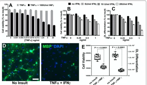
To determine the effects of inflammatory cytokines on actively differentiating OLs, we initially tested the effects of exposure to IFNγ alone on the viability of T3-induced differentiating OLs, an insult previously shown to cause apoptosis of OLs [8]. Following 48 h of IFNγ exposure, the viability of cells was determined by alamarBlue® (AB—a fluorescent indicator of metabolic activity) fluo-rescence quantification. At least 200 Units/ml of IFNγ was required to generate a 20 % reduction in differenti-ating OL viability when added alone (Fig. 1B). We used this concentration to test whether we could identify small molecules that could rescue OLs from IFNγ tox-icity. We tested the confirmed hit compounds identified in an acute OL differentiation assay (see [15]). We added test compounds 24 h following OPC plating and 1 h prior to the addition of INFγ insult to give compounds the best chance for efficacy; cells were assayed 48 h later for viability with AB. Since AB fluorescence is produced by incubation with live cells and is non-toxic, we were able to wash, fix, and immunostain the same cultures for MBP expression (see “Methods” section) and quantify MBP expression. Figure 1A shows the decrease in MBP immunostaining intensity caused by 200 U/ml IFNγ. Additional file 1: Table S1 includes the EC50 values for
efficacious concentration in protecting OLs from IFNγ toxicity (Fig. 1B) as well as for preserving MBP expres-sion (Fig. 1C).
In an attempt to increase the cytokine protection win-dow, we tested TNFα alone, up to 20 ng/ml, which had no measurable impact on viability of OLs (Fig. 2A). Since it is known that IFNγ and TNFα can combine to have syn-ergistic effects and are both present in MS lesions [8, 16], we titrated each cytokine against the other to determine possible synergistic effects for use in the OL protection assay. When TNFα was titrated against a fixed concen-tration of 100 U/ml IFNγ, the combination of 100 U/ ml IFNγ + 1.25 ng/ml TNFα resulted in a drop in OL viability of 46 % vs. only 11 % for 100 Units/ml of IFNγ alone (Fig. 2A). To further optimize the synergistic com-bination of TNFα and IFNγ we narrowed the range of concentrations and measured both OL viability and dif-ferentiation. We were able to titrate IFNγ down to 10 U/ ml in combination with 1 ng/ml TNFα and still repro-ducibly obtain ~30 % reduction of OL viability (Fig. 2B, E). This cytokine combination also resulted in ~50 %
reduction in OL differentiation as determined by quan-tification of MBP expression (Fig. 2C–E). Concentrations of TNFα above 1 ng/ml resulted in toxicity that reduced MBP to undetectable levels.
We used this newly developed cytokine protection assay (TNFα + IFNγ) to evaluate the activity of the hit compounds identified in the acute OL differentiation screen (see [15]). We generated dose response curves and EC50 values for each compound for viability and OL
[image:3.595.57.541.86.370.2]Page 4 of 12 Rosler et al. BMC Res Notes (2016) 9:444
of cytokines (Additional file 2: Figure S1F1, S1G1, S1H1).
This indicates that the mechanism of cytokine protection can be separated from the promotion of differentiation in the presence of inflammatory cytokines.
We then assessed the ability of QTP to protect OLs from the combined 1 ng/ml TNFα and 10U/ml IFNγ toxicity. QTP preserved OL differentiation in the pres-ence of both 1 ng/ml TNFα and 10 U/ml IFNγ demon-strated by imaging of MBP immunostaining (Fig. 3A). Figure 3B, C shows the dose–response relationships for QTP in both parameters of the cytokine protection assay. The EC50 values were determined to be 157 nM
for viability protection and 125 nM for OL differentia-tion protecdifferentia-tion. From this data, we chose 1.1 µM as a positive control assay concentration to be used in subse-quent assay screens. 1.1 µM QTP reproducibly rescued differentiating OLs from cytokine-induced toxicity by an average of 90 % (Fig. 3D) and restored their ability to dif-ferentiate by 60 % (Fig. 3E).
NCC Library screening for compounds that protect differentiating OLs from combined INFγ and TNFα insult Prior to library screening, NCC library compounds were prescreened at three concentrations for OL toxicity (see [15]). This allowed for the reduction of toxic concentra-tions and to potentially detect active compounds active at lower doses.
[image:4.595.58.542.87.366.2]OL toxicity assay) followed 1 h later by addition of 1 ng/ ml TNFα + 10 U/ml IFNγ. After 48-h incubation in the presence of cytokines, AB was added to cells and the fluorescence quantified to determine cell viability. Each compound concentration was screened in triplicate with data scaled relative to 0.1 % DMSO as the negative con-trol and 1.1 µM QTP as the positive concon-trol. A no insult
control was also included to assess the consistency of cytokine toxicity.
The criterion for a hit compound was protection of dif-ferentiating OLs from cytokine toxicity that was at least 50 % of the positive control, 1.1 µM QTP level or above. Similar to the OL differentiation assay, we included in our analysis the visualization of three SDs above the mean
Table 1 Confirmed cytokine protection hits from the NCC library screen
LO lipooxygenase
a Hit in acute OL differentiation assay (as determined in [15]) b Inconsistent, weak OL differentiation activity
c Not in NCC library
Drug class Compound AB screening
(% quetiapine) AB EC50 (μM) MBP EC50 (μM) Hit in OL diff a
SERM Raloxifene 92 0.03 0.05 •
Toremifene 73 0.02 0.01 •
Tamoxifen 117 0.02 0.01 •
Mestranol 93 0.92 0.79
Clomiphene 52 0.01 0.01
Tricyclic anti‑depressants Perphenazine 96 0.01 0.03 •
Fluphenazine 91 0.02 0.02 •
Prochlorperazine 110 0.04 0.04 •
Trifluoperazine 155 0.02 0.03 •
Quetiapine 130 0.15 0.13 •
Non‑tricyclic anti‑depressants Perospirone 85 0.22 0.15 •
Bupropion 71 3.89 3.86 •
Bifemelane 78 0.20 0.26
Cinanserin 64 0.97 1.59
Trazadone 55 7.18 5.42
Muscarinic Clemastinec 0.14 0.14 •
Benztropine 123 0.17 0.14 •
Donepezil 95 0.46 0.74 •
Vesamicol 151 1.83 2.26 •
Oxybutynin 92 0.67 0.54 •
Propantheline 91 2.95 1.25
Adrenergic Salmeterol 55 0.22 0.20 •
Betaxolol 52 5.56 6.21 •
Ion channel Ifenprodil 162 0.36 0.52 •
Benproperine 99 0.43 0.12 •
Proxymetacaine 98 1.23 0.78 •
DMPP 54 2.18 2.84 •
Anti‑fungal Bifonazole 105 0.44 b •
Clotrimazole 171 0.77 b
Econazole 90 0.34 b
Antidiabetic Pioglitazone 56 7.34 b
LO inhibitor MK886 51 0.31 0.19
Histamine H1 Antagonists Hydroxyzine Pamoate 106 0.15 0.11
Meclizine 97 0.12 0.10
Retinoids All‑trans‑retinoic acid 128 0.88 0.44
[image:5.595.56.540.102.570.2]Page 6 of 12 Rosler et al. BMC Res Notes (2016) 9:444
(Fig. 5A). Alongside this, we show the 50 % QTP thresh-old line (Fig. 5A). Control QTP versus DMSO values from the entire screen for OL differentiation were highly statistically significant per assay plate (Fig. 5B, C) as well as when averaged over entire screen (Fig. 5C, inset) indi-cating an acceptable screen window and reproducibility. In a screen of 727 FDA-approved drugs, we identified 44 actives (6 % primary hit rate) able to protect differentiat-ing OLs from cytokine toxicity at one or both concentra-tions tested (Fig. 5D). Additional file 3: Table S2 includes the data for all compounds tested in the cytokine protec-tion screen.
Independent confirmation and validation of identified hits In order to confirm the activity of the primary hits iden-tified in the cytokine protection assay, we purchased 40 fresh compounds from independent vendors and per-formed a dose response analyses (4 compounds were excluded from follow up). We confirmed 36 active com-pounds (90 % hit confirmation rate) and determined the EC50 values for both viability (AB fluorescence
quantifi-cation) and OL differentiation (quantification of MBP
immunostaining) protection in the presence of inflam-matory cytokines (Table 1).
Discussion
[image:6.595.60.540.88.344.2]conditions, such as those that would prevail in an MS lesion.
There are a number of advantages of adapting this assay for the purpose of screening compound libraries for com-pounds that protect OLs from toxic insult. The robust-ness, scale and reproducibility are amenable to HTS drug discovery. The assay uses minimally-passaged primary rat cells, obviating the need to produce large numbers of OPCs either through multiple passages or by deriva-tion from large-scale stem cell cultures. We designed the assay to preserve primary cell, in vivo-like characteristics in culture by limiting subculture to a single expansion. Thus, we avoided undesirable changes in cellular pheno-type (e.g. gene expression and/or unstable karyopheno-types) which multiple cell passages may introduce [3, 17–19]. We have also demonstrated that additional endpoints combined within the same assay can introduce interest-ing predictive cellular phenotypes. Although the OL pro-tection assay could be scored using OL differentiation as an image-based endpoint for identification of drugs that support OL differentiation in the presence of inflamma-tory mediators, we have demonstrated that OL viability is an endpoint that is highly predictive of differentiation (33 of 36 hit compounds). Viability can be rapidly and inexpensively assessed using alamarBlue®, permitting much higher throughput than the image-based OL differ-entiation assays. Moreover, alamarBlue® reagent is easily
removed, permitting subsequent staining and imaging for OL differentiation if desired.
It is important to note that we have only evaluated OL differentiation in the presence of just two cytokines, but this delineates just one of many relevant, potentially detrimental, cytokine combinations. This OL protection assay is highly flexible and should be useful for evaluat-ing a wide variety of other factors, such as examinevaluat-ing the effects of other cytokines known to be present in MS lesions (alone and in combination) and other inflamma-tory mediators such as myelin debris/breakdown prod-ucts that may have detrimental effects on OLs.
Confirmed hits group into multiple classes
[image:7.595.61.541.88.286.2]Page 8 of 12 Rosler et al. BMC Res Notes (2016) 9:444
differentiation assay and (2) antidiabetic, anti-retroviral, lipoxygenase inhibitor, histamine H1 antagonists and the retinoids which were only identified in the OL protec-tion screen (Table 1). Within these classes emerged hit compounds that have relevance to MS and may be repur-posed for therapeutic use in the treatment of MS.
Relevance to multiple sclerosis
We chose to screen the NCC library because it is a small, focused FDA-approved set of compounds with great clinical potential. Additionally we chose to screen at mul-tiple concentrations in hopes of identifying compounds that are more efficacious at lower concentrations. While this added time to the process, we felt that this increase in screening time was more than offset by rapid progress to clinical trials for any such identified drugs that are FDA-approved.
There are many examples where repurposed drugs are making an impact in the field of MS. A few of note are BG-12 (Tecfidera®), a modification of a psoriasis drug;
fingolimod (Gilenya®), originally developed for trans-plantation and alemtuzamab (Lemtrada™), originally an oncology drug. We found that a fair number of our hits aligned with current MS repositioning efforts in terms of patents, preclinical work, and clinical trials and are briefly cited here.
QTP, our positive control for OL protection, has been reported in the literature to provide protection from the development of EAE symptoms and cuprizone insult, both rodent models of demyelinating disease, as well as preventing myelin loss and promoting OL differentiation in vivo [20–24].
The SERMs represent a promising new class of com-pounds that protect OLs from cytokine toxicity and promote OL differentiation. Modulation of the estrogen receptor has been demonstrated to be effective in pre-venting demyelination in vivo and to stimulate differen-tiation and remyelination [25–28]. Specifically, raloxifene (Evista®) was shown to be effective in suppressing EAE [29]. Additionally, the pregnancy hormone estriol Fig. 5 Analysis of the cytokine OL protection assay of the NCC compound library. A Dot plot representation of the entire high‑throughput screen‑ ing data set used to identify promoters OL survival. The mean response is indicated by the blue solid line. The black dotted line delineates the value of three standard deviations above the mean. The red dotted line in delineates the ≥50 % positive control selection criteria. B Scatter plot representa‑ tion of the high‑throughput screening assay window. Using the QTP/DMSO ratio as the window of OL survival, the ratio of the positive (QTP) to negative (DMSO) controls is depicted in the scatterplot. The red line delineates the mean average ratio value for the entire NCC library screen = 1.3. The numbers above the scatter indicate the coefficient of variation (CV) for each plate. CV values <20 ± 5 were considered in the acceptable range.
Each point is an average value from each screening plate (n = 4 wells, mean ± SEM). Inset depicts consistency of cytokine insult for the entire library with a mean average value of 1.4. C An overlapping representation of raw AB values of DMSO and QTP for each library screening plate. The inset
[image:8.595.58.542.89.368.2](Trimesta™), has shown efficacy in a clinical trial of MS reducing the lesion numbers and volumes [30]. This indi-cates that modulation of the estrogen receptor pathway may be a viable target for OL differentiation and protec-tion. Interestingly, sexual dimorphism has been noted within OL preparations with OLs derived from males and females separately responding differently to sex hor-mones [31]. Our OL preparations were derived from ran-domly selected P7 rat pups and therefore the response to some sex hormone signaling pathways may show differ-ences from OLs isolated from one sex or the other.
A number of hits that we identified are covered in pat-ents for oligodendrocyte directed differentiation. Notably trifluoperazine (Stelatzine®), benztropine (Cogentin®), ipatropium (Atrovent®), and clemastine (Tavist®), (US 20140038949 A1) are patented for the use of neurotrans-mitter receptor modulating agents for inducing OL dif-ferentiation, as well as methods for treating subjects with such agents in a demyelinating disease.
Other hits have been evaluated or will soon be tested in human MS trials. QTP (Seroquel XR®), an antipsychotic approved for schizophrenia is currently in clinical trials to determine tolerability to people with RRMS and pro-gressive MS (Clinicaltrials.gov identifier: NCT02087631). Selective serotonin re-uptake inhibitors (SSRIs) such as nitalapram (Celexa®) and escitalopram (Lexapro®) have also been in human MS trials. Nitalapram is one of three other SSRIs tested as an add-on with fingolimod, a cur-rent immunomodulatory treatment for MS (REGAIN Trial NCT01436643) in RRMS patients with depression. Escitalopram, has been in a study to test improvement of depression in patients of MS or ALS (NCT00965497). Donepezil (Aricept®), used as a treatment for demen-tia, has been used in a study to improve cognitive dys-function in MS patients (NCT00315367) [32–34]. Oxybutynin (Ditropan XL®) is used currently in symp-tom management for MS to treat overactive bladder in MS (SONIC trial NCT00629642). Clemastine fumarate (Tavist®) originally approved as a first-generation antihis-tamine, is active in a current study to assess the drug as a remyelinating agent is RRMS patients (ReBUILD trial NCT02040298).
In preclinical studies, some of the compounds we iden-tified as having protective properties show great promise in MS. The peroxisome proliferator-activated receptor gamma agonist, pioglitazone (Actos®—a drug used for diabetes), shows protective effects in the brain, delays onset and severity in rodent EAE models [35, 36] and is used as an add-on therapy for RRMS for treatment of nerve pain [37]. MK886, a lipoxygenase inhibitor, may be a useful therapeutic disease modifying treatment in MS, and it has been demonstrated to attenuate neuron inflammation, motor dysfunction and axonal damage in
the mouse cuprizone-induced demyelination model [38]. Retinoic acid (Vesanoid®), an anti-cancer chemotherapy drug, has been cited to have a protective role in vitro against neuro-inflammatory insults in vitro [39].
The antihistamines, hydroxyzine (Atarax®) and
meclizine (Antivert®), identified from the OL protection screen are currently used for symptom management in MS. Hydroxyzine is used for alleviation of sensory symp-toms in MS and cited in a published pilot clinical trial in MS patients [40]. Meclizine, used to treat vertigo, motion sickness and nausea, also treats those symptoms MS, as well as vomiting and dizziness symptoms.
To our knowledge, the remainder of our confirmed hits has not been tested in any demyelinating diseases. Future studies should focus on testing these compounds both in vitro with human cells as well as in vivo models of MS.
Conclusion
Here, we have described a large-scale primary cell-based drug discovery OL protection assay, with paradigms relevant to the MS environment and performed with industry rigor. We hope that other groups will find this assay helpful for comprehensively examining the effects of these factors on oligodendrocyte differentiation and health, and for developing drug screens that more closely mimic the pathology of the MS lesion.
Methods
Page 10 of 12 Rosler et al. BMC Res Notes (2016) 9:444
AbD Serotech (Killington, UK). Falcon TC 96-well plates were purchase from Corning (Corning, NY). Additional file 4: Table S3 lists the primary antibodies and their dilu-tions used in this study.
Compounds
All compounds in the NIH Clinical Collection (NCC) library were supplied in DMSO at 10 mM in 96-well plates. Hit compounds were purchased as powders and stock solutions were dissolved in DMSO to 10 mM (see Additional file 3: Table S2 for complete listing of compounds).
Isolation and expansion of neonatal rat OPCs
All animal work was carried out in strict accordance with the recommendations in the Guide for the Care and Use of Laboratory Animals of the National Institutes of Health. The protocol was approved by the Institutional Use and Care and Use Committee at the Molecular Med-icine Research Institute (Sunnyvale, CA). Animals used for OPC isolation in these studies were euthanized by decapitation.
OPCs from brains of P6–P7 Sprague–Dawley rat pups (Charles River, Wilmington, MA, USA) were enriched by immunopanning as previously described [41]. Briefly, sin-gle cell suspensions were obtained from papain-digested neonatal brains and depleted of mature glial cells by suc-cessive panning with RAN-2- and GalC-coated plates, followed by enrichment for OPCs using O4-coated plates.
In vitro expanded OPCs were used in toxicity and cytokine protection assays. Acutely enriched OPCs were seeded into PDL-coated tissue culture flasks at 1000– 2000 cells/cm2 in differentiation media (DMEM, 100 U/
ml Penicillin/100 µg/ml Streptomycin, 2 mM l-Glu-tamine, 1 mM Na Pyruvate, 5 µg/ml Insulin, 5 µg/ ml N-acetyl-l-cysteine, 1× Trace Elements B, 10 ng/ ml d-Biotin, 100 µg/ml BSA, 100 µg/ml apo-Transfer-rin, 16 µg/ml Putrescine, 60 ng/ml Progesterone, 40 ng/ ml Sodium Selenite 5 ng/ml Forskolin, 10 ng/ml CNTF, 1 ng/ml NT-3) supplemented with 10 ng/ml PDGF-AA. Proliferating OPCs were fed every 2–3 days and supple-mented with 2–3× concentration of PDGF as needed to prevent OPC differentiation. After 6–7 days of prolif-eration, OPCs were harvested by trypsinization, plated into PDL-coated 96-well Falcon plates and centrifuged at 200×g to facilitate cell attachment, survival, and even distribution of cells.
Oligodendrocyte protection assay
Expanded OPCs were plated at 10,000 cells/well in dif-ferentiation media supplemented with 40 ng/ml T3 and incubated for 24 h at 37 °C, 10 % CO2 prior to addition of
test compounds. Adjusted concentrations of NCC library compounds were added to triplicate wells to account for toxicity. One hour after addition of test compounds, 10 Units/ml IFNγ + 1 ng/ml TNFα were added and cul-tures were incubated for 48 h. Controls were added in six replicate wells, negative control = 0.1 % DMSO; positive controls = no insults (no IFNγ and no TNFα) or 1.1 μM QTP. A 1:10 dilution of AB was added to each well, incu-bated 4 h at 37 °C, 10 % CO2 and fluorescence measured
as above. In some experiments, following AB quantifi-cation, cells were fixed with 4 % paraformaldehyde and immunostained for MBP expression as described below.
Immunofluorescence staining and imaging
Following compound treatment, media was removed leaving 50 μl/well using an ELx405 microplate washer (BioTek, Winooski, VT, USA), the automated plate washer was also used for all subsequent washing steps during the staining. Cells were fixed for 14 min with 4 % paraformaldehyde at room temperature (RT). Follow-ing fixation, plates were washed with 1 ml PBS leavFollow-ing 50 μl/well behind. Cells were then incubated in block-ing buffer (10 % normal goat serum (NGS) diluted in antibody buffer: 150 mM NaCl, 50 mM Tris Base, 1 % BSA, 100 mM l-lysine, 0.04 % sodium azide, pH 7.4), with 0.4 % Triton X-100 for 1 h at RT, then stained over-night at 4 °C with rat anti-MBP antibodies (Additional file 4: Table S3) diluted in blocking buffer with 0.08 % Triton X-100. The cells were washed and incubated with secondary antibodies and 0.3 μM DAPI for 1 h at RT. After a final wash, 100 μl of PBS was added to each well and plates imaged. Secondary antibody alone controls showed little to no background staining. Images were captured with a Nikon Eclipse TE-2000-U microscope, Zyla cMOS megapixel camera (ANDOR Technology, Belfast, UK), fitted with an automated stage controlled by NIS Elements AR software 4.0 (Melville, NY, USA). An air 10× lens was used to capture 2 images per well with 16 bit resolution, 2560 × 2160 pixels. Images for each assay run were captured using identical camera set-tings. Images were exported as TIFF files for analysis and quantification.
Image quantification
same field. The calculated extent of MBP expression (OL differentiation) was then scaled to internal plate nega-tive (0.1 % DMSO, set to 0) and posinega-tive controls (40 ng/ ml T3 set to 100 %) to generate percent OL differentia-tion scores relative to the efficacy of positive control. For the cytokine protection assay, the positive controls were either no insult or 1.1 μM QTP. This analysis consistently generated quantitatively similar results when scaled to internal plate negative and positive control wells.
Relative EC50 analysis
EC50 values were obtained by fitting the data to a
sigmoi-dal dose–response curve-fitting function (Prism, Graph-Pad, San Diego, CA, USA). Serial dilutions of eight to ten different concentrations with three or four data points per concentration were used for curve fitting. Experi-ments were repeated at least two times.
Statistical methods
For all experiments, assuming normal distribution, two-tailed t tests were used to evaluate comparisons between two groups and ANOVA was used when more than two groups were compared. For the quantitative analysis of in vitro differentiation, ANOVA with Bonferroni or Dun-nett correction was used.
Abbreviations
CNS: central nervous system; MS: multiple sclerosis; OL: oligodendrocytes; OPCs: oligodendrocyte precursor cells; DIV: days in vitro; MBP: myelin basic protein; DMSO: dimethylsulfoxide; HTS: high throughput screening; NCC: NIH clinical collection.
Authors’ contributions
ER conceived, designed and performed the experiments; acquired, inter‑ preted, and provided intellectual content to the study; KLW and DL provided statistical analysis of the data, and drafted the manuscript; DL, KLW, ER JT, JD, and TC participated in the design of the study, provided intellectual content to the study, helped revise the manuscript critically and revised its final version. All authors read and approved the final manuscript.
Author details
1 Translational Medicine Center, Myelin Repair Foundation, Sunnyvale, CA 94085, USA. 2 Teva Pharmaceuticals, Biologics and CNS Discovery, Redwood City, CA 94063, USA. 3 Alios BioPharma, South San Francisco, CA 94080, USA. 4 Rigel Pharmaceuticals, South San Francisco, CA 94080, USA. 5 NGM Biophar‑ maceuticals, Inc., South San Francisco, CA 94080, USA.
Additional files
Additional file 1: Table S1. Acute OL differentiation hit compounds tested in the IFNγ protection assay.
Additional file 2: Figure S1. Viability and OL differentiation EC50 curves for acute OL differentiation hits in cytokine protection assay.
Additional file 3: Table S2. NCC library cytokine protection screen data set.
Additional file 4: Table S3. Antibodies.
Acknowledgements
We are grateful to Nicole Edwards for excellent administrative and data analysis support. We thank Russel Rydel for helpful discussions and comments on the manuscript. We gratefully thank Ben Barres for his continual support, guidance and generosity for providing antibodies. We are profoundly grateful to all the individual donors who have donated their time and funds to the Myelin Repair Foundation and the Translational Medicine Center.
Competing interests
The authors declare that they have no competing interests.
Availability of supporting data
Detailed protocols are available on request from Leonoudakis and Lariosa— Willingham at [email protected] and karen.Lariosa‑Willing‑ [email protected].
Ethics approval and consent to participate
This study did not use any human subjects, human material or human data. All animal work was carried out in strict accordance with the recommenda‑ tions in the Guide for the Care and Use of Laboratory Animals of the National Institutes of Health. The protocol was approved by the Institutional Use and Care and Use Committee at the Molecular Medicine Research Institute (Sun‑ nyvale, CA).
Received: 5 May 2016 Accepted: 15 August 2016
References
1. Franklin RJ, ffrench‑Constant C, Edgar JM, Smith KJ. Neuroprotection and repair in multiple sclerosis. Nat Rev Neurol. 2012;8(11):624–34.
2. Ransohoff RM, Hafler DA, Lucchinetti CF. Multiple sclerosis—a quiet revolution. Nat Rev Neurol. 2015;11(3):134–42.
3. Deshmukh VA, Tardif V, Lyssiotis CA, Green CC, Kerman B, Kim HJ, Padmanabhan K, Swoboda JG, Ahmad I, Kondo T, et al. A regen‑ erative approach to the treatment of multiple sclerosis. Nature. 2013;502(7471):327–32.
4. Jepson S, Vought B, Gross CH, Gan L, Austen D, Frantz JD, Zwahlen J, Lowe D, Markland W, Krauss R. LINGO‑1, a transmembrane signaling protein, inhibits oligodendrocyte differentiation and myelination through intercellular self‑interactions. J Biol Chem. 2012;287(26):22184–95. 5. Lee S, Leach MK, Redmond SA, Chong SY, Mellon SH, Tuck SJ, Feng ZQ,
Corey JM, Chan JR. A culture system to study oligodendrocyte myelination processes using engineered nanofibers. Nat Methods. 2012;9(9):917–22. 6. Najm FJ, Madhavan M, Zaremba A, Shick E, Karl RT, Factor DC, Miller TE, Nevin ZS, Kantor C, Sargent A, et al. Drug‑based modulation of endog‑ enous stem cells promotes functional remyelination in vivo. Nature. 2015;522:216–20.
7. Navikas V, Link H. Review: cytokines and the pathogenesis of multiple sclerosis. J Neurosci Res. 1996;45(4):322–33.
8. Lin W, Harding HP, Ron D, Popko B. Endoplasmic reticulum stress modulates the response of myelinating oligodendrocytes to the immune cytokine interferon‑gamma. J Cell Biol. 2005;169(4):603–12.
9. Mann SA, Versmold B, Marx R, Stahlhofen S, Dietzel ID, Heumann R, Berger R. Corticosteroids reverse cytokine‑induced block of survival and dif‑ ferentiation of oligodendrocyte progenitor cells from rats. J Neuroinflam‑ mation. 2008;5:39.
10. Vartanian T, Li Y, Zhao M, Stefansson K. Interferon‑gamma‑induced oligodendrocyte cell death: implications for the pathogenesis of multiple sclerosis. Mol Med. 1995;1(7):732–43.
11. Vela JM, Molina‑Holgado E, Arevalo‑Martin A, Almazan G, Guaza C. Inter‑ leukin‑1 regulates proliferation and differentiation of oligodendrocyte progenitor cells. Mol Cell Neurosci. 2002;20(3):489–502.
12. Cannella B, Raine CS. The adhesion molecule and cytokine profile of multiple sclerosis lesions. Ann Neurol. 1995;37(4):424–35.
Page 12 of 12 Rosler et al. BMC Res Notes (2016) 9:444
• We accept pre-submission inquiries
• Our selector tool helps you to find the most relevant journal
• We provide round the clock customer support
• Convenient online submission
• Thorough peer review
• Inclusion in PubMed and all major indexing services
• Maximum visibility for your research
Submit your manuscript at www.biomedcentral.com/submit
Submit your next manuscript to BioMed Central
and we will help you at every step:
14. Selmaj K, Raine CS, Cannella B, Brosnan CF. Identification of lymphotoxin and tumor necrosis factor in multiple sclerosis lesions. J Clin Invest. 1991;87(3):949–54.
15. Lariosa‑Willingham KD, Rosler ES, Tung JS, Dugas JC, Collins TL, Leonoudakis D. A high throughput drug screening assay to identify compounds that promote oligodendrocyte differentiation using acutely dissociated and purified oligodendrocyte precursor cells. BMC Res Notes. 2016;9:419. doi:10.1186/s13104‑016‑2220‑2.
16. Ofengeim D, Ito Y, Najafov A, Zhang Y, Shan B, DeWitt JP, Ye J, Zhang X, Chang A, Vakifahmetoglu‑Norberg H, et al. Activation of necroptosis in multiple sclerosis. Cell Rep. 2015;10:1836–49.
17. Cole R, de Vellis J. Preparation of astrocyte, oligodendrocyte, and micro‑ glia cultures from primary rat cerebral cultures. 3rd ed. Totowa: Humana Press; 2001.
18. Najm FJ, Zaremba A, Caprariello AV, Nayak S, Freundt EC, Scacheri PC, Miller RH, Tesar PJ. Rapid and robust generation of functional oligo‑ dendrocyte progenitor cells from epiblast stem cells. Nat Methods. 2011;8(11):957–62.
19. Passaquin AC, Schreier WA, de Vellis J. Gene expression in astrocytes is affected by subculture. Int J Dev Neuroslci. 1994;12(4):363–72. 20. Bi X, Zhang Y, Yan B, Fang S, He J, Zhang D, Zhang Z, Kong J, Tan Q, Li XM.
Quetiapine prevents oligodendrocyte and myelin loss and promotes maturation of oligodendrocyte progenitors in the hippocampus of global cerebral ischemia mice. J Neurochem. 2012;123(1):14–20. 21. Chandran P, Upadhyay J, Markosyan S, Lisowski A, Buck W, Chin CL, Fox
G, Luo F, Day M. Magnetic resonance imaging and histological evidence for the blockade of cuprizone‑induced demyelination in C57BL/6 mice. Neuroscience. 2012;202:446–53.
22. Mei F, Guo S, He Y, Wang L, Wang H, Niu J, Kong J, Li X, Wu Y, Xiao L. Quetiapine, an atypical antipsychotic, is protective against autoimmune‑ mediated demyelination by inhibiting effector T cell proliferation. PLoS ONE. 2012;7(8):e42746.
23. Xiao L, Xu H, Zhang Y, Wei Z, He J, Jiang W, Li X, Dyck LE, Devon RM, Deng Y, et al. Quetiapine facilitates oligodendrocyte development and prevents mice from myelin breakdown and behavioral changes. Mol Psychiatry. 2008;13(7):697–708.
24. Xu H, Yang HJ, McConomy B, Browning R, Li XM. Behavioral and neuro‑ biological changes in C57BL/6 mouse exposed to cuprizone: effects of antipsychotics. Front Behav Neurosci. 2010;4:8.
25. Bebo BF Jr, Dehghani B, Foster S, Kurniawan A, Lopez FJ, Sherman LS. Treatment with selective estrogen receptor modulators regulates myelin specific T‑cells and suppresses experimental autoimmune encephalomy‑ elitis. Glia. 2009;57(7):777–90.
26. Elloso MM, Phiel K, Henderson RA, Harris HA, Adelman SJ. Suppression of experimental autoimmune encephalomyelitis using estrogen receptor‑ selective ligands. J Endocrinol. 2005;185(2):243–52.
27. Jung‑Testas I, Renoir M, Bugnard H, Greene GL, Baulieu EE. Demonstration of steroid hormone receptors and steroid action in primary cultures of rat glial cells. J Steroid Biochem Mol Biol. 1992;41(3–8):621–31.
28. Morales LB, Loo KK, Liu HB, Peterson C, Tiwari‑Woodruff S, Voskuhl RR. Treatment with an estrogen receptor alpha ligand is neuroprotec‑ tive in experimental autoimmune encephalomyelitis. J Neurosci. 2006;26(25):6823–33.
29. Li R, Xu W, Chen Y, Qiu W, Shu Y, Wu A, Dai Y, Bao J, Lu Z, Hu X. Raloxifene suppresses experimental autoimmune encephalomyelitis and NF‑ kappaB‑dependent CCL20 expression in reactive astrocytes. PLoS ONE. 2014;9(4):e94320.
30. Sicotte NL, Liva SM, Klutch R, Pfeiffer P, Bouvier S, Odesa S, Wu TC, Voskuhl RR. Treatment of multiple sclerosis with the pregnancy hormone estriol. Ann Neurol. 2002;52(4):421–8.
31. Swamydas M, Bessert D, Skoff R. Sexual dimorphism of oligodendrocytes is mediated by differential regulation of signaling pathways. J Neurosci Res. 2009;87(15):3306–19.
32. Amato MP. Donepezil for memory impairment in multiple sclerosis. Lancet Neurol. 2005;4(2):72–3.
33. Amato MP, Portaccio E. Is there a future for donepezil therapy in the treatment of multiple sclerosis‑related cognitive impairment? Expert Rev Neurother. 2011;11(9):1243–6.
34. O’Carroll CB, Woodruff BK, Locke DE, Hoffman‑Snyder CR, Wellik KE, Thaera GM, Demaerschalk BM, Wingerchuk DM. Is donepezil effective for multiple sclerosis‑related cognitive dysfunction? A critically appraised topic. Neurologist. 2012;18(1):51–4.
35. Feinstein DL, Galea E, Gavrilyuk V, Brosnan CF, Whitacre CC, Dumitrescu‑ Ozimek L, Landreth GE, Pershadsingh HA, Weinberg G, Heneka MT. Peroxi‑ some proliferator‑activated receptor‑gamma agonists prevent experi‑ mental autoimmune encephalomyelitis. Ann Neurol. 2002;51(6):694–702. 36. Pershadsingh HA, Heneka MT, Saini R, Amin NM, Broeske DJ, Feinstein
DL. Effect of pioglitazone treatment in a patient with secondary multiple sclerosis. J Neuroinflammation. 2004;1(1):3.
37. Kaiser CC, Shukla DK, Stebbins GT, Skias DD, Jeffery DR, Stefoski D, Katsamakis G, Feinstein DL. A pilot test of pioglitazone as an add‑on in patients with relapsing remitting multiple sclerosis. J Neuroimmunol. 2009;211(1–2):124–30.
38. Yoshikawa K, Palumbo S, Toscano CD, Bosetti F. Inhibition of 5‑lipoxyge‑ nase activity in mice during cuprizone‑induced demyelination attenuates neuroinflammation, motor dysfunction and axonal damage. Prostaglan‑ dins Leukot Essent Fatty Acids. 2011;85(1):43–52.
39. Mizee MR, Nijland PG, van der Pol SM, Drexhage JA, van Het Hof B, Mebius R, van der Valk P, van Horssen J, Reijerkerk A, de Vries HE. Astrocyte‑derived retinoic acid: a novel regulator of blood‑brain barrier function in multiple sclerosis. Acta Neuropathol. 2014;128(5):691–703. 40. Logothetis L, Mylonas IA, Baloyannis S, Pashalidou M, Orologas A,
Zafeiropoulos A, Kosta V, Theoharides TC. A pilot, open label, clinical trial using hydroxyzine in multiple sclerosis. Int J Immunopathol Pharmacol. 2005;18(4):771–8.