“ANALYSIS OF VIDEO HEAD IMPULSE TESTING IN
PATIENTS WITH VERTIGO”
Submitted in partial fulfillment of the requirements for the award of degree of
M.S. DEGREE BRANCH-IV OTORHINOLARYNGOLOGY
THE TAMILNADU DR. M.G.R. MEDICAL UNIVERSITY
UPGRADED INTITUTUE OF OTORHINOLARYNGOLOGY MADRAS MEDICAL COLLEGE
BONAFIDE CERTIFICATE
This is to certify that this dissertation entailed “ANALYSIS OF VIDEO HEAD
IMPULSE TESTING IN PATIENTS WITH VERTIGO” submitted by Dr. SINDHU
KALYANARAMAN, appearing for M.S. ENT., Branch IV Degree examination in May 2018 is a bonafide record of work done by her under my direct guidance and supervision in partial fulfillment of regulations of the Tamil Nadu Dr. M.G.R. Medical University, Chennai, Tamil Nadu, India.
GUIDE:
Prof. Dr. R. Muthukumar M.S., DLO, DNB Director and Professor of ENT
Upgraded Institute of Otorhinolaryngology Madras Medical College
Rajiv Gandhi Govt. General Hospital
DIRECTOR AND PROFESSOR OF ENT THE DEAN
Prof. Dr. R. Muthukumar M.S. DLO, DNB Prof. Dr. Narayana Babu MD, Dch, Upgraded Institute of Otorhinolaryngology Madras Medical College
Madras Medical College Rajiv Gandhi Govt. General Hospital Rajiv Gandhi Govt. General Hospital Chennai- 600003
DECLARATION
I solemnly declare that the dissertation “ANALYSIS OF VIDEO HEAD
IMPULSE TESTING IN PATIENTS WITH VERTIGO” is done by me at the
Madras Medical College and Government General Hospital, Chennai during 2015- 2017 under the guidance and supervision of Prof. Dr. R. MUTHUKUMAR M.S., D.L.O., DNB.
This dissertation is submitted to The Tamilnadu Dr. M.G.R. Medical University, towards partial fulfillment of regulation for the award of M.S. DEGREE IN OTORHINOLARYNGOLOGY (BRANCH-IV)
Dr. SINDHU KALYANARAMAN
M.S. E.N.T. postgraduate,
Upgraded Institute of Otorhinolaryngology, Madras Medical College
ACKNOWLEDGEMENT
I express my gratitude to Prof.Dr.NARAYANA BABU M.D. Dch, THE DEAN, Madras Medical College, for having permitted me to use the hospital material in this study.
I am immensely thankful to Prof.Dr.R.MUTHUKUMAR M.S. D.L.O. D.N.B., The Director and Professor, Upgraded Institute of Otorhinolaryngology, for his invaluable support and encouragement. His constant guidance and inquisitive analysis enabled me to conduct this study meticulously.
I am grateful to Prof.Dr.G.SANKARANARAYANAN MS.DLO.DNB.MNAMS., Professor, Upgraded Institute of Otorhinolaryngology for his valuable support and guidance during the study.
I am grateful to Prof.Dr.N. SURESHKUMAR MS., DLO., Professor, Upgraded Institute of Otorhinolaryngology for his valuable support and guidance during the study.
I would like to thank Assistant Professor Dr. C J Vikram for his
guidance in conducting the study.
I would like to thank Assistant Professor, Dr. K. Semmanaselvan,
Dr. Venugopal, and Dr. Shenbagavalli, Dr.Vivek for their support in
conducting the study.
I express my sincere thanks to all the Assistant Professor s, for their
thoughtful guidance.
I express my gratitude to my colleagues Dr. Maheshkumari and Dr.
Kalaimani for their guidance and constructive criticism throughout the
study.
I would like to extend a special thanks to Dr. K. Kumaresan, who
analyzed the statistical data of this study
I take immense pleasure to thank my parents for their unconditional
love, care and support that has led to the fulfillment of this study.
I am greatly indebted to my husband, Mr. Prashanna Sivaraman, for
helping me formulate the framework of this dissertation and for his constant
I thank my in-laws for their enormous support and encouragement throughout this study.
I am extremely grateful to my daughter for her sustained patience and support.
I express my appreciation for the generosity shown by all the patients who participated in the study.
TABLE OF CONTENTS
S.NO CONTENT PAGE NO
1. INTRODUCTION 1
2. AIMS OF THE STUDY 3
3. REVIEW OF LITERATURE 4
4. ANATOMY OF INNER EAR 11
5. ULTRASTRUCTRE OF THE VESTIBULAR ORGAN 17
6. PHYSIOLOGY OF INNER EAR 19
7. EVALUATION OF BALANCE DISORDERS 23
8. VESTIBULAR TEST BATTERY- ENG 31
9. VEMP 34
10. vHIT 35
11. INDIVIDUAL CONDITIONS 42
12. MATERIALS AND METHODS 56
13. RESULTS 59
14. ANALYSIS AND DISCUSSION 76
15. CONCLUSION 87
16.
ANNEXURE Abbreviation
Ethical committee Clearance Certificate Anti Plagiarism Screen Shot
Patient Proforma
1
INTRODUCTION
The vestibular system coordinates information about the position and motion of the
head. Along with visual and proprioceptive signals, the central nervous system
uses this data to synchronize the eyes, head, and body during movement and to
give a conscious perception of space orientation. Disturbances in this integrative
process, especially in the vestibular pathways, can lead to vertigo.
Vertigo is defined as an illusion of rotation of oneself or surrounding. It can be due
to peripheral vestibular or central causes. It is among the commonest symptoms
presented to physicians, with a lifetime occurrence of around 20% to 30% (1).
A distinct clear binocular vision is achieved by the occulomotor system in
conjugation with six other systems of supranuclear control, namely, the smooth
pursuit system, the saccadic system, the vestibular system, the fixation system, the
optokinetic system, and the vergence system. “The smooth pursuit system comes
into play while tracking an object of interest that is in movement. This system
cannot follow objects moving faster than 30–40°/s. Thus, faster moving objects
elicit saccades. Saccades are rapid, brief conjugate eye movements that are
2
optokinetic and other systems are used for gross movements to prevent retinal
slippage.
The Vestibulo- ocular reflex is one of the fastest reflexes in the body responsible
for foveating objects on the retina during movements of the head (150- 3000/s). Abnormality of VOR results in vertigo and nystagmus during head movements.
The bedside test for assessing the VOR is the Halmagyi- Curthoys head impulse
test. It consists of monitoring eye movements as the patient fixates on a still target
while the head is moved briskly in the lateral plane. Normal individuals can
maintain a steady gaze but patients with deficient VOR cannot keep up with
high-velocity head turns and generate „catch-up‟ saccades after head impulses toward
the damaged side.
Video head impulse test (vHIT) is a computerized technology that uses a goggle
with camera attached to capture the „catch-up‟ saccades with precision. It records
both overt and covert saccades and measures VOR gains individually in each of the
six semicircular canals. It can differentiate between peripheral and central lesions.
3
AIMS AND OBJECTIVES
Using Video Head Impulse Testing in patients with vertigo:
1. To differentiate between peripheral vestibular lesions and central lesions
2. To detect exactly which semicircular canal is affected in the particular ear.
3. To record saccades identifying both overt and covert saccades
4. To evaluate VOR gain and assess severity of the vestibular lesion
4
REVIEW OF THE LITERATURE
From time immemorial, for the diagnosis of vestibular disorders, laboratory tests
like ENG (caloric testing), rotational chair tests, scleral search coil and
posturography are being used.
In 1988, Halmagyi and Curthoys (2, 3) described a bedside test for testing the VOR, which consists of observing eye movements as the patient focuses on an earth
bound target while the head is moved quickly to either side stimulating the LSCC.
This test enabled better and quick diagnosis for patients with vestibular disorders
and also recorded the overt „catch-up‟ saccades.
Between 2009 and 2013, a video assisted (vHIT) testing of each of the six canals
individually was developed by Halmagyi and Curthoys (4). It is a physiological test which had the main advantage of recording VOR gain in each of the SCC thereby
specifying which canal is affected. It can record both overt and covert saccades.
To ensure that the objectives of this research are consistent with the functionality
of vHIT and also to validate and compare our test results, we have reviewed and
5
H.G. MacDougall et all (4) in July 2009 studied “an easy-to-use video HIT system (vHIT) as a clinical tool for identifying peripheral vestibular deficits and to
validate the diagnostic accuracy of vHIT by simultaneous measures with video and
search coil recordings for 16 subjects. The sensitivity and specificity of both the
reference and index test were 1.0 (95% confidence interval 0.69–1.0). vHIT
measures detected both overt and covert saccades as accurately as coils and
concluded that vHIT is equivalent to search coils in identifying peripheral
vestibular deficits but easier to use in clinics, even in patients with acute vestibular
neuritis.”
Blodow et al(5) in November 2012 studied “117 patients and a control group of 20 healthy subjects. The group of patients included vestibular neuritis (n = 52),
vestibular schwannoma (n = 31), Meniere‟s disease (n = 22) and bilateral
vestibulopathy (n = 12) and found that normal hVOR gain was at 0.96 _ 0.08,
while abnormal hVOR gain was at 0.44 _ 0.20 (79.1% of all cases). An abnormal
vHIT was found in VN (94.2%), VS (61.3%), MD (54.5%) and BV (91.7%). Three
conditions of refixation saccades occurred frequently in cases with abnormal
hVOR: isolated covert saccades (13.7%), isolated overt saccades (34.3%) and the
combination of overt and covert saccades (52.0%). They concluded that vHIT
6
refixation saccades. Since isolated covert saccades in hVOR changes can only be
seen with vHIT, peripheral vestibular disorders are likely to be diagnosed more
accurately with the vHIT.”
MacDougall et all (6) in January 2013 reported that “vHIT detects peripheral deficits of both vertical and horizontal semicircular canal function and is a new
tool for measuring dysfunction of individual semicircular canals in vestibular
patients.”
Eza-Nuñez et all (7) in June 2014 studied “a prospective work in 123 patients with different types of vestibular disease seen because of dizziness in which both vHIT
and caloric tests were performed the same day. The caloric test and the vHIT
results were fully coincident in 60 patients (48.7%), and the results for both were
normal in 36 patients. The correlation coefficient for canal paresis and gain
asymmetry was 0.67. They concluded that the assessment of LSCC function with
the vHIT needs to take into consideration not only the gain in VOR but also the
existence of refixation saccades.”
Albernaz et all (8) in June 2014 studied “a sample of 200 patients with a clinical
7
semicircular canals and abnormal responses of the anterior and posterior canals
were found in several patients, either alone or combined with altered responses in
the lateral canals.” The study concluded that additional research is required, along
with a comparison with other tests, to validate the role of vHIT.
Holger A. Rambold (9) July 2014 did “a retrospective study with 1063 patients subjected to vHIT and bithermal caloric irrigation on the same day and analyzed
with respect to side differences. Of those patients 13.3 % had pathological vHIT
and a caloric irrigation test, 4.6 % a pathological vHIT only and 24.1 % a
pathologic caloric test only. He concluded that the caloric irrigation and vHIT are
complimentary and should be used in a defined sequence to optimize the available
time for diagnosis of vertigo and dizziness patients.”
S.L. Bell et all (10) in November 2014 studied the “Sensitivity and specificity of the vHIT test relative to calorics was studied for a clinical sample of 51 patients (20
male, 31 female) who attended a private clinic for balance disorders. They found
sensitivity of 29 % with a specificity of 94 % and concluded that the vHIT is likely
to be a useful addition to the vestibular testing battery as it gives complementary
8
Mantokoudis et all (11) in December 2014 studied “26 acute vestibular syndrome patients using a video HIT device to differentiate peripheral and central
pathologies. vHIT VOR gains differ between peripheral and central causes of
AVS. PICA strokes were readily separated from neuritis using gain measures, but
AICA strokes were at risk of being misclassified based on VOR gain alone and
should be correlated with clinical findings and gain asymmetry.”
McCaslin et all (12) in March 2015 reported “13 patients with Ménière's disease and described how the combination of audiometry, video head impulse testing, and
caloric results may prove helpful in the diagnosis of Ménière's disease. They
concluded that ipsilesional abnormal caloric testing in the presence of normal
video head impulse testing is a pattern of findings observed in a cohort of patients
who have “definite” Ménière's disease”.
Hamilton et all (13) in May 2015 studied “33 patients with age less than 20 years of age with vertigo and studied the efficacy of vHIT. They concluded vHIT is an
effective test for evaluating semicircular canal function in children and offers
9
B. F. Esch et all (14) in April 2016 did a “cross-sectional study between May 2012 and May 2013 and analyzed 324 patients with dizziness who had completed caloric
testing and the vHIT. For the left and right vestibular system the mean vHIT gain,
sensitivity, specificity, positive and negative predictive values with 95 %
confidence intervals was calculated. They analyzed that vHIT is a very specific
rather than sensitive test for detecting vestibular hypofunction. In case of a normal
vHIT, additional caloric testing remains indicated but in case of an abnormal vHIT,
additional caloric testing is not necessary. They concluded that the vHIT is
clinically useful as a first test in determining vestibular hypofunction in dizzy
patients.”
Bansal et all (15)in March 2016 studied “25 young healthy subjects to obtain VOR gain data, correlate all planes in both sides of head (right and left) and assess the
test–retest reliability of VOR gain measure using vHIT and concluded that it shows
a good reliability of VOR gain functions in normal individuals and any significant
change in gain parameters would suggest a vestibular pathology and not a simple
random variation.”
10
complementary test for other vestibular test, especially caloric test. As this test is
not associated with nausea or vomiting and well tolerated by the patients, it is
recommended to perform the vHIT as a first test in each semicircular canal.
I. S. Curthoys & L. Manzari (17) reviewed in July 2017 that “the origin and rationale for vHIT; how it is carried out; pitfalls in testing; the validation of the
test; the interpretation of overt and covert compensatory saccades in patients with
vestibular loss; how the area VOR gain is calculated. They concluded that it can be
given to patients even during acute vertigo attacks and for testing young children.
It tests all six semicircular canals individually.
Fallahnezhad et all (18) in September 2017 did a pilot study of “Twenty-nine unilateral PSCC-BPPV patients with normal oculographic and caloric results in
which vHIT was performed on six canals, and VOR gain, gain asymmetry and
saccades were measured. Sixteen (55.17%) patients had abnormal posterior canal
VOR gains in the ipsilesional ear. VOR gains in both horizontal canals were within
normal limits. Superior canal VOR gains were mostly lower than normal and were
not correlated to PSC abnormalities (P>0.05). No corrective saccades could be
observed. He concluded that VOR gain in the direction of the posterior
11
ANATOMY OF INNER EAR
The balance system is a complicated sensory organization which involves the
coordination between the peripheral vestibular system, the visual system, postural
muscles, the brainstem, cerebellum and the cortex. This array of information from
these sources is processed by vestibular centers in the central nervous system to
allow the body to maintain balance and proper spatial orientation during motion.
THE LABYRINTH
The peripheral vestibular apparatus is situated in the inner ear and consists of a
bony and a membranous labyrinth. It is located in the otic capsule in the petrous
portion of the temporal bone. The bony labyrinth consists of:
Vestibule
Cochlea
The bony semicircular canals- Anterior, lateral and posterior canals
BONY LABYRINTH
The vestibule is the part of the bony labyrinth that lies between the cochlea and
bony semicircular canal. It is separated from the middle ear by the oval window.
The utricle and saccule, which are part of the membranous labyrinth, are located
12
The saccule lies in the spherical recess on the anteromedial wall, which is
perforated by inferior vestibular nerve in the macula cribrosa media. The elliptical
recess which is posteromedial lodges the utricle and perforated by macula cribrosa
superior (Mike‟s dot) for the superior vestibular nerve (19). The two recesses are separated by the crista vestibule (pyramid) which lodges the fossa cochlearis,
perforated by macula cribrosa inferior, supplying the vestibular end of the ductus
cochlearis. The medial wall also has the orifice of the vestibular aqueduct which
contains the endolymphatic duct.
The three semicircular canals lie perpendicular to each other. Each canal has an
ampullated end opening separately into the vestibule and a non ampullated end.
The non ampullated ends of the anterior and posterior canal form the crus
commune. Hence the three canals open by five openings into the utricle.
The canals are organized into functional pairs, such that both members of the pair
lie in the same plane. Any rotation in that plane is excitatory to one member of the
pair and inhibitory to the other (20). The pairs are:
right and left lateral- 300 to horizontal
right anterior and left posterior- 450 to vertical
left anterior and right posterior-450 to vertical
13
The bony cochlea is a spiral structure around the modiolus and contains three
compartments, namely, scala vestibule, scala tympani and scala media. The scala
tympani is connected with the subarachnoid space via the cochlear aqueduct.
The bony labyrinth is filled with a fluid called perilymph that is continuous with
and alike in composition to CSF- rich in sodium ions (FIG 1). The fluid is a filtrate
from the capillaries of the spiral ligament and is drained by the cochlear aqueduct
into the adjacent subarachnoid space.
14
MEMBRANOUS LABYRINTH
The membranous labyrinth consists of the utricle, saccule, the 3 semicircular ducts,
the cochlear duct (scala media) and the endolymphatic sac and duct. The
membranous labyrinth is bathed in endolymph which is similar in composition to intracellular fluid thus rich in potassium ions. It is produced by the stria vascularis
and gets absorbed through the endolymphatic sac in the dural space. (21)
UTRICLE AND SACCULE
The sensory epithelium is the macula (consisting of hair cells and few supporting
cells) for both utricle and saccule (FIG 2). The utricle senses motion in the
horizontal plane (forward-backward movement and left-right movement). The
saccule senses motions in the vertical plane (up-down movement).
The macula of the saccule is oriented vertically and the utricular macula
horizontally. The orientation of the stereocilia (hair cells) within the macula is
determined by the striola, which a curvilinear ridge that crosses the middle of
the macula. Thus, in the utricle, the kinocilia are sloping toward the striola, and in
the saccule they lie away from it.
Above the maccula is a gelatinous layer, on which is embedded the otolithic
membrane, with crystals of calcium carbonate- otoconia. The otoconia make the
otolithic membrane significantly heavier than the fluids surrounding it. Thus,
15
relative to the sensory epithelium. This results in a shearing motion between the
otolithic membrane and the macula which displaces the hair bundles generating a
[image:22.612.156.484.175.415.2]receptor potential.
FIG 2: Macula of utricle and saccule
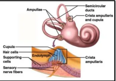
SEMICIRCULAR CANALS
The sensory epithelium or the crista of the semicircular canals is housed in
the ampulla, which is a globular expansion at the base. A gelatinous cupula is
found over the crista forming a barrier through which endolymph cannot pass (FIG
3). During angular rotation, the inertia of the endolymph creates a force which
displaces the cupula away from the direction of head movement. A receptor
16
macula, the hair cells in crista are arranged with their kinocilia pointing in the same
direction (22). In the lateral canals, the kinocilium of each hair cell faces the utricle, whereas it is reversed in the anterior and posterior canals. Thus, when the cupula
moves in a particular direction, the entire hair cell population is depolarized, from
its resting membrane potential (–50 and –70 mV). Hyperpolarization occurs in the
[image:23.612.113.496.275.544.2]opposite direction.
17 COCHLEAR DUCTS
Scala media has three boundaries, namely, basilar membrane with organ corti,
Reissners membrane, and the stria vascularis. It is connected to the saccule by the
ductus reunions.
ENDOLYMPHATIC DUCT
The saccule and utricle joins to form the utriculosaccular duct, which passes
through the vestibular aqueduct as the endolymphatic duct. This duct opens into
the sac lying between two layers of dura on the posterior cranial fossa.
ULTRASTRUCTRE OF THE VESTIBLAR ORGAN
VESTIBULAR SENSORY CELLS, TYPE I AND TYPE II
The macula and crista contain two types of sensory cells described ultra
structurally as type I and type II sensory cells. The type I cells are flask-shaped and
a nerve chalice formed by the diatal end of the afferent fibre of the vestibular nerve
surrounds it. These calyces have extensions that end on type II hair cells.
Type II cells, phylogenetically older cells, are cylindrical in shape and have similar
arrangement of stereocilium and kinocilium as type I cells. The upper end of each
18
the longest stereocilia is situated next to the kinocilium. The stereocilia contain
actin filaments for deflection.
THE VESTIBULAR GANGLION AND NERVE
The vestibular ganglion (Scarpa‟s ganglion) is made of bipolar neurons located in
the lateral part of the IAC. It consists of a superior and an inferior group of cells
related to the SVN and IVN.
SVN: Cristae of the anterior and lateral canals, the macula of the utricle and
the part of the macula of the saccule.
IVN: Crista of the posterior canal and the main portion of the macula of the
saccule.
The large nerve fibers are chiefly involved in the innervation of type I hair cells
located around the striola (23). The small fibers are more plentiful at the slope and
the marginal region of the maculae. In the IAM, the vestibular and cochlear nerves
unite. A tiny arterial branch from the AICA courses between the facial and
vestibular nerve on the brainstem. The human vestibular nerve contains efferent
19
PHYSIOLOGY OF BALANCE
Every motion in space can be divided into three rotational degrees of freedom
(yaw, pitch and roll) and three translational degrees of freedom (left–right, up–
down, for–aft). The vestibular system is intricately involved in the physiology of
balance and involves the following components:
The vestibuloocular reflex (VOR) for gaze stabilization
VSR/ VCR for head and body position in space
The vestibular cortex is involved in navigation
Autonomic function with respect to balance
VOR
The vestibuloocular reflex (24) is one of the fastest reflexes in our body used to
foveate images on the retina during head movements. This is made by producing
eye movements in the direction equal and opposite to head movement, thus
preserving the image on the visual field. As minor head movement is present all
the time, the VOR is mandatory for vision and balance. In patients whose VOR is
20
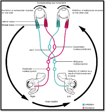
The basic principle of VOR generation for the LSCC is as follows (FIG 4):
During head rest, a resting discharge rate of 90 spikes/ sec is present.
Head rotates to the left.
Endolymph lags behind and moves to the right in the SCC due to inertia.
[image:27.612.126.471.306.671.2] The cupula turns to the right in each canal.
21
In the left LSCC, the stereocilia bends toward the kinocilium-
ampullopetal(towards the vestibule)
In the right LSCC, the stereocilia bends away from the kinocilium-
ampullofugal (away from the vestibule)
The discharge rate rises in the left ear to around 300 spikes/sec.
The discharge rate decreases in the right ear to 10-20 spikes/ sec.
The difference is positive to left as is interpreted by the vestibular nuclei
The left MLF is simulated and the right is inhibited.
This in turn simulates the ipsilateral oculomotor and contralateral abducens
nuclei. This moves the left medial rectus and right lateral rectus so that the
eyes move in an equal and opposite direction to preserve gaze stabilization.
EWALDS LAW (25)
1. “A stimulation of the semicircular canal causes a movement of the eyes in
the plane of the stimulated canal”
2. “In the horizontal semicircular canals, an ampullopetal endolymph
movement cases a greater stimulation than an ampullofugal one.”
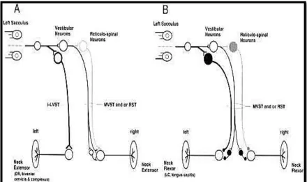
22 VSR/ VCR
The function of the VSR/ VCR is to stabilize the neck and body with respect to the
head in space. During head tilt to one side, both the SCC and otoliths are
stimulated, thus depolarizing the vestibular nerve and nucleus of that side. Signals
are transmitted through the medial and lateral vestibulospinal tracts towards the
spinal cord (FIG 5). Extensor movement is on the side to which the head is leaning,
and flexor on the opposite side. When the position of the body or neck changes,
[image:29.612.93.533.399.661.2]VSR/VCR tends to normalize the posture with respect to gravity.
23
EVALUATION OF BALANCE DISORDERS
Disorders of balance affects almost one fourth of the population and in 80% of
cases it is severe enough to see a physician. Dizziness is a non specific term and
can be divided into four general categories:
Vertigo- Peripheral or central- is defined as an illusion of rotation of either
oneself or environment
Disequilibrium- usually cerebellar cause
Light headedness- pre-syncope
Anxiety/ psychogenic
Good history taking is the most important factor in diagnosing vertiginous cases
(26)
. The symptoms that patients experience include vertigo, giddiness, nausea,
motion intolerance, imbalance and oscillopsia(blurring of vision during head
movement).
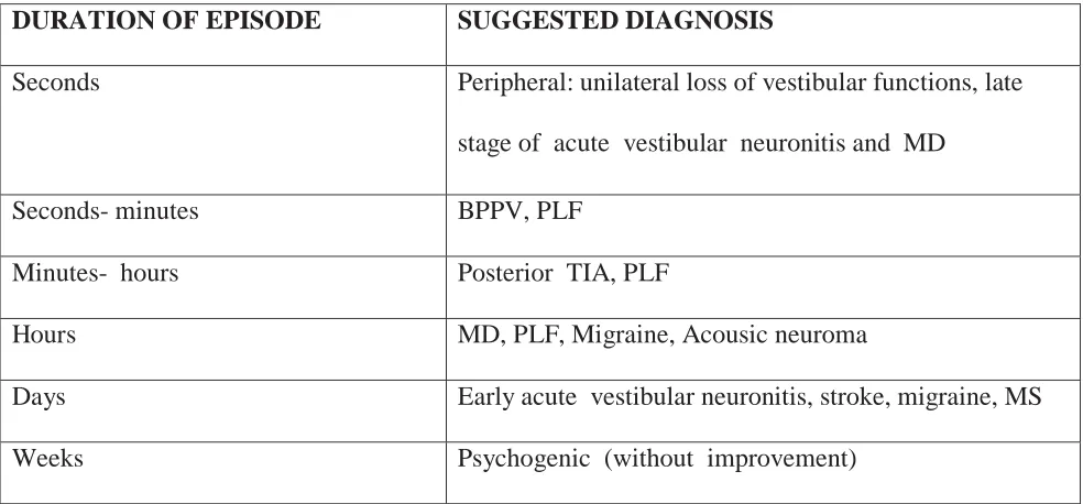
The duration of each episode is as important as the period of the overall disease
(TABLE 1). The trigger factors such as acoustic triggers (Tullio phenomenon),
coughing and straining should also be assessed carefully (27). Associated factors
24
involvement, and diplopia, facial numbness and dysarthria indicates central causes(
[image:31.612.72.564.167.396.2]TABLE 2, 3).
TABLE 1: Association with duration
DURATION OF EPISODE SUGGESTED DIAGNOSIS
Seconds Peripheral: unilateral loss of vestibular functions, late
stage of acute vestibular neuronitis and MD
Seconds- minutes BPPV, PLF
Minutes- hours Posterior TIA, PLF
Hours MD, PLF, Migraine, Acousic neuroma
Days Early acute vestibular neuronitis, stroke, migraine, MS
Weeks Psychogenic (without improvement)
TABLE 2: Trigger factors
TRIGGER FACTOR DIAGNOSIS
Turning the head to the right or left BPPV, Vestibular paroxysm
Coughing, pressing PLF
Tullio phenomenon PLF, Meniere‟s disease
When patient walks B/l vestiblopahthy
Food or sleep deprivation or certain foods (chocolate, cheese, red wine)
25
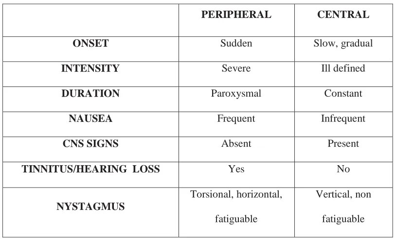
TABLE 3: Difference between Central and Peripheral vertigo
Clinical testing includes examination of ears, eye movement, positional maneuvers
and posture and gait. Otoscopic examination and fistula testing should be done.
Eye examination is done to assess the VOR by observing for spontaneous
nystagmus and positive head thrust. Also, other eye movements such as smooth
pursuit, saccades, optokinetic tests indicative of central lesions is done.
Spontaneous nystagmus is assessed when the eyes are in the central position. The
waveform (Jerk, see-saw, pendular nystagmus), the direction of nystagmus,
amplitude of beat, gaze evoked or induced should be evaluated. A nystagmus of
PERIPHERAL CENTRAL
ONSET Sudden Slow, gradual
INTENSITY Severe Ill defined
DURATION Paroxysmal Constant
NAUSEA Frequent Infrequent
CNS SIGNS Absent Present
TINNITUS/HEARING LOSS Yes No
NYSTAGMUS
Torsional, horizontal,
fatiguable
Vertical, non
26
peripheral vestibular origin is essentially horizontal, with a minor torsional
component. For central lesions, the nysagmus may be vertical or direction
changing.
Gaze-evoked nystagmus occurs after the acute stage of a peripheral lesion, wherein
the nystagmus is not observable in primary gaze but only on deviation of gaze to
the opposite side of the lesion, i.e. in the direction of the fast phase. This is called
Alexander’s law (28)
1st degree - nystagmus only in the direction of the fast phase
2nd degree- nystagmus in primary gaze
3rd degree- nystagmus in the direction of the slow, primary and fast
component
Gaze paretic nystagmus, which implies that the patient has difficulty in holding
gaze in an eccentric position in the orbit, indicates a central lesion. In Brun‟s
nystagmus occurring in vestibular schwanomma, there is gaze evoked and gaze
paretic nystagmus in the same person.
Smooth pursuit movement of the eyes occurs when a target is moving at a speed of
10- 150/ second. The patient should be assessed for a cogwheel or jerky appearance
27
vestibular disorder and contrarily, a broken pursuit is invariably central in origin.
Likewise, saccadic abnormalities of velocity, amplitude and conjugacy specify a
central lesion.
VOR reflexes are measured by head impulse test. Halmagyi and Curthoys
introduced the bedside physiologic head impulse test in which sudden, brisk,
discrete and unpredictable head thrusts are made, so that the VOR deficit is more
evident. The patient is instructed to fixate a target and the head is moved 150 to
right or left in velocities of 150- 3000 /second. A patient with a defective VOR will
produce „catch-up‟ or refixation saccades. Thus, a quick right head thrust will
make a patient with right-sided vestibulopathy initiate catch-up saccades towards
the left. The test is extremely useful for identifying acute unilateral peripheral
vestibular deficits, like vestibular neuritis.
Positional maneuvers are a crucial component of the clinical examination and must
be carried out in all patients with episodic vertigo, especially if provoked by
head-neck movements. The Dix Hallpike maneuver is the most commonly done
28
FIG 6: Dix hallpike maneuver
The following steps carried out for 3 minutes each and include:
1. The patient sits in the table
2. The patient head is held and turned 450 to right.
3. The patient is placed in supine position with head hanging at 300 below
horizontal
29
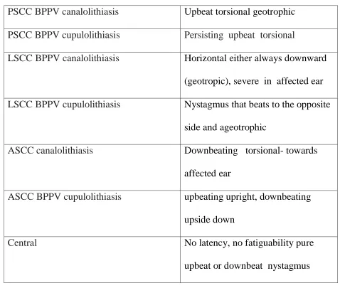
[image:36.612.71.549.161.566.2]Interpretations include:
TABLE 4: Dix Hallpike Interpretations
PSCC BPPV canalolithiasis Upbeat torsional geotrophic
PSCC BPPV cupulolithiasis Persisting upbeat torsional
LSCC BPPV canalolithiasis Horizontal either always downward
(geotropic), severe in affected ear
LSCC BPPV cupulolithiasis Nystagmus that beats to the opposite
side and ageotrophic
ASCC canalolithiasis Downbeating torsional- towards
affected ear
ASCC BPPV cupulolithiasis upbeating upright, downbeating
upside down
Central No latency, no fatiguability pure
upbeat or downbeat nystagmus
Clinical examination of balance, posture and gait should be done as part of balance
30
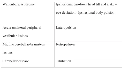
[image:37.612.72.544.145.408.2]Posture examination
TABLE 5: Posture
The Romberg test is a test of proprioception. The subject is asked to stand with
his/her feet together, eyes open and hands crossed. Then he/she closes the eyes and
observed for a full minute. A positive Romberg means that the patient shows a
tendency to actually fall and most patients with balance disorders show a small to
moderate increase in imbalance on eye closure. In acute phase of a peripheral
vestibular disorder the Romberg test will be positive. Patients with anterior lobe
cerebellar degeneration may show the characteristic trunk oscillation only on eye
closure.
Wallenburg syndrome Ipsilesional ear-down head tilt and a skew
eye deviation. Ipsilesional body pulsion.
Acute unilateral peripheral
vestibular lesions
Lateropulsion
Midline cerebellar-brainstem
lesions
Retropulsion
31
Tandem walking with eyes closed show a degree of unsteadiness in patients with
B/L loss of vestibular function. In ataxia, patients are unable to do this test. In the
Untenberger test, the patient is made to walk on the spot with the eyes closed. In
unilateral vestibular lesions the patient turns towards the affected side.
Complete neurological examination with motor, sensory, cranial nerve function
should be done. Cardiology and skeletal system examination is done when
appropriate.
VESTIBULAR TEST BATTERY
ENG
A pair of surface electrodes around the orbit is used to record the difference in
potential between the cornea and the retina. Given that the corneal region is
positive and the retina negative, the eye becomes a dipole. Two electrodes placed
at the external canthus detect movement of this dipole.
The following are recorded using ENG (31):
Spontaneous nystagmus- It is done when eyes are closed (when it is not
suppressed by optic fixity). If the intensity of nystagmus increases on
closing the eyes, it is suggestive of peripheral pathology. The direction and
32
Gaze nystagmus- the patient looks 300 away from midline. A gaze
nystagmus usually indicates a central pathology except in cases of acute
unilateral vestibulopathy.
Recording of saccades, smooth pursuit, optokinetic tests.
Positional test- Major disadvantage is that it cannot record torsional eye
movements.
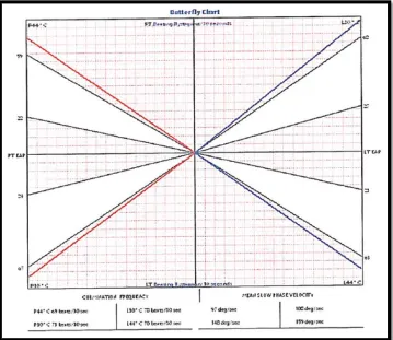
Bithermal caloric testing- The principle of the caloric test is that changes in
temperature in the EAC influence the level of activity of the LSCC. The
subject lies down with the head raised 300 above horizontal. The recording of caloric test is useful in that the canal paresis and direction preponderance
can be calculated with precision. It can also differentiate central from
vestibular pathology. Water irrigation of EAC is done at 300 and 440C in the order of left cold, Right cold, Left Warm, and Right Warm with each
syringing lasting 40 seconds. Cold irrigation induces horizontal nystagmus
beating in the opposite direction of irrigation, and ipsilaterally during warm
irrigation. Bilateral absence of caloric nystagmus in B/L vestibulpathy and a
reduced or absent response in one ear, as in vestibular neuritis, is few
33
FIG 7: Claussen butterfly chart
Scleral search coil and rotational chair testing are other tests for used to assess
VOR.
POSTUROGRAPHY
It is used to record the propioception of the patient.
Static- If the patient just stands quietly on the platform, with eyes open or
closed.
34
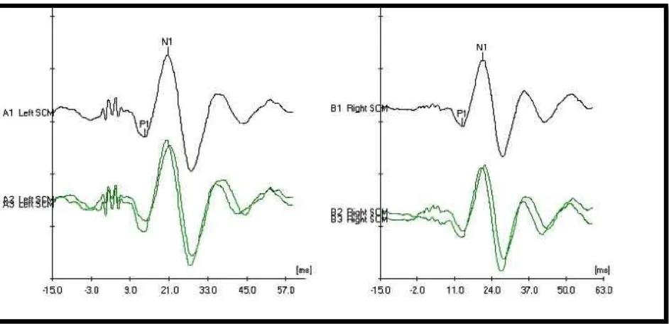
VEMP
Click sounds activate the vestibular labyrinth and an electromyographic (EMG)
potential can be identified from neck muscles due to vestibulocolic reflex (32, 33).
Each ear tested separately. The main response is an ipsilateral positive deflection at
13 ms (P13) and a negative deflection at approximately 23 ms (N23). The main
use is for the diagnosis of the SSCD and U/L vestibulopathy like vestibular
neuritis.
cVEMP- cervical VEMP- to detect saccule function
[image:41.612.75.546.391.619.2]oVEMP- ocular VEMP- to detect utricle function
FIG 8: VEMP GRAPH
Audiological tests like PTA, OAE, BERA, SISI, Tone decay tests and recruitment
35 vHIT
The video head impulse test is a documentation of the Halmaygi clinical head
impulse test. The test ascertains and very accurately documents the functional
status of each of the three individual semicircular canals of both sides. It can
evaluate the VOR of each of these semicircular canals at different high frequency
of stimulation which closely matches the range at which normal head movement
takes place. The vHIT evaluates the vestibular labyrinth at frequencies slightly
below 1Hz to about 7 Hz. It records both overt and covert saccades. It can record
the VOR gain of each semicircular canal.
To achieve perfect gaze stabilization, the speed at which the eyes move should
be exactly equal at which he head moves (34).
VOR GAIN= Speed of Eye movement
Speed of Head Movement
This is calculated by the computerized software using area under the graph. In
cases of defective VOR (gain <100%), the eyes fall short of the target. So to focus
and fix the gaze on the target, the eyes will jump to „catch- up‟ and refixate. Hence
the smoothness of eye movements is lost and the jerky movement is called
36 Covert saccades- During the head movements;
[image:43.612.121.430.152.526.2]Overt saccades- After the head movements.
FIG 9: Saccades
The vHIT overcomes the limitation of the clinical test by detecting covert saccades
accurately (FIG 9).
Covert saccades
37
Procedure
The test is performed in a bright lit room to maintain miotic pupil. The patient is
made to sit on an adjustable chair approximately one meter from the target. We
have used the ICS Impulse® using the Monocular Video Frenzel goggles. The
goggles have an inbuilt high speed USB camera and a nine axis motion sensor
which detects the right eye movement (FIG 10).
FIG 10: vHIT goggles
The goggles must be secured snugly and connected to the computer.
Next calibration is done to assess saccadic movement of the patient.
The computer data acquisition program tracks the centre of the pupil. The
patient is reminded to keep their eyes wide open and not to blink during the
head thrust.
The head thrust is given with high velocities – at least 1500/s upto 3000/s and
38
The operator places his hands on the top of the head of the patient and turns
the patient‟s head in an sudden, unpredictable, lateral head turn and stops
unexpectedly about 10- 150 from midline.
[image:45.612.122.470.205.442.2]FIG 11: Performing vHIT
39
For testing of the vertical canals- LARP and RALP, the patient‟s gaze must
be parallel with the plane of the canal being tested. The head impulse is
given in the vertical axis (FIG 11, 12).
During the impulse, the patient is asked to keep looking at the target on the
wall in front of them and if they lose the target, to return their eyes to the
target as quickly as possible. A standard 20 impulses on each side is given.
The vHIT result is evaluated on three parameters:
VOR gain- for lateral canal 80- 100% is normal while for vertical canal
70-100% is normal.
The presence or absence of saccades- consistency, latency, amplitude and
direction.
40
Illustrations of normal vHIT results in a HEX PLOT:
` FIG 13: Normal vHIT graph
The normal vHIT shows VOR gains in green in the hex plot, that is above
80%. The inner hex plot is the 50% VOR gain value. The graph surrounding it
[image:47.612.102.527.158.471.2]41 ABNORMAL vHIT RESULTS- an example
FIG 14: Abnormal vHIT graph
This shows an abnormal vHIT graph whereby the right posterior canal is affected
with VOR gain of 69%. The asymmetry for the posterior canal is 17%. All other
42 INDIVIDUAL CONDITIONS
MENIERE’S DISEASE
Meniere‟s disease is a tetrad of symptoms, namely, unilateral low tone
fluctuating-progressive sensorineural hearing loss, episodic vertigo (20 minutes to 24 hours),
tinnitus, and aural fullness. Meniere‟s syndrome is secondary to trauma, infection,
inflammation or neoplasm.
The American Academy of Otolaryngology–Head and Neck Surgery (AAOHNS)
Committee on Hearing and Equilibrium has classified it as (35):
Certain Meniere’s disease- With histopathologic confirmation of endolymphatic
hydrops.
Definite Meniere’s disease- two or more definitive episodes of vertigo with,
tinnitus, hearing loss or aural fullness.
Probable Meniere’s disease- one definitive episode of vertigo along with hearing
loss, tinnitus, or aural fullness.
Possible Meniere’s disease- there is vertigo but it is not associated with hearing
loss.
Meniere‟s disease is theorized to be caused by increased endolymphatic pressure.
Schunket proposed distention of the endolymphatic space with rupture of
Reissner‟s membrane. There is cross contamination between the two spaces which
43
the vestibule, which causes mechanical interference by inhibition of signal
generation.
The endolymphatic sac secretes glycoproteins and saccin which stimulates
production of endolymph from dark cells that distend the sac. The ensuing rapid
flow of endolymph from the sac results in an acute vertigo episode. Autoimmune
mechanism has also been suggested.
In acute attack of meniere‟s disease the patient undergoes irritative, paretic and
recovery phase. In early stages the patient is well between attacks. In late stages
the patient may develop persistent hearing loss, tinnitus, and postural imbalance.
Drop attacks or tumarkin's otolihtic crisis is due to acute otolithic dysfunction and
consists of a sensation of being pushed away with no vertigo or loss of
consciousness.
For the diagnosis of Menieres disease, a detailed history and thorough audiometric
evaluation is necessary. Pure tone audiometry indicates side and severity of
sensorineural hearing loss. Recruitment and SISI is positive and BERA is used to
rule out retrocochlear pathologies. Electronystagmography establishes vestibular
dysfunction, but may be normal in early Meniere‟s disease. Serology is used to
detect autoimmune etiology. Electrocochleography, cochlear microphonics, and
video head impulse testing and vestibular evoked myogenic potentials supplements
44
In ECochG, the summating potential and action potential ratio is < 0.4 in normal
individual whereas it is >0.45 in Meniere‟s. Oral glycerol dehydration test shows
considerable hearing improvement by audiogram and/or speech audiometry.
During acute phase, vestibular sedatives like phenothiazines, antihistamines are
used. Salt restricted diet and avoidance of smoking is advised. Diuretics and
vasodilators can be used as a prophylactic measure. Steroids are used in
autoimmune etiology. For intractable vertigo, various hearing preserving and
hearing sacrificing surgeries are done. These include intratympanic steroid/
gentamycin therapy, endolymphatic sac decompression, shunting, vestibular nerve
section and labyrinthectomy. Menniet device uses positive pressure to alter
endolymphatic pressure, thus bringing relief to the patient.
VESTIBULAR NEURITIS
Vestibular neuritis is a peripheral disorder (U/Lvestibulopathy) wherein there is
sudden, spontaneous, reduced or absent afferent input from one labyrinth, which is
deduced to result from a viral infection of the vestibular nerve. Studies have shown
latent infection of the superior and inferior vestibular ganglia by herpes simplex
virus type 1.The SVN is more commonly affected as it is longer and narrower than
45
Symptoms: Acute spontaneous vertigo, associated nausea and vomiting, postural
imbalance. It is aggravated by head movement. Patient unable to walk and swerve
towards affected side.
Signs: Spontaneous horizontal torsional unidirectional (towards unaffected side)
nystagmus. The head impulse test shows „catch-up‟ saccades with head thrusts on
the affected side (FIG 15). The compensatory saccades are in the same direction as
the fast phases of the nystagmus. Untenberger and Fukuda test is positive.
ENG creates a permanent record of the nystagmus. MRI of the brain is generally
not required. vHIT shows the side and nerve which is affected with great
precision. It also records the improvement over time, hence used for prognosis. As
cerebellar infarction is the main differential diagnosis of vestibular neuritis, vHIT
differentiates both conditions with the former having normal vHIT.
As vestibular neuritis is a result of viral infection of the vestibular nerve,
corticosteroid and antiviral treatments have therapeutic gain. Early mobilization
and vestibular rehabilitation therapy has been shown to facilitate compensation and
improve both postural and visual stability. Peripheral vestibular function recovers
in less than 50 percent of patients with vestibular neuritis though the acute
46
FIG 15: vHIT in left superior vestibular neuritis- Anterior and lateral canal
alone are affected.
BENIGN PAROXYSMAL POSITIONING VERTIGO
“BPPV is a disorder characterized by brief attacks of vertigo, with associated
nystagmus, precipitated by certain changes in head position with respect to
gravity” It is the most common peripheral cause of vertigo. Patients complain of
transient vertigo spells (5- 20 sec), associated with nausea, that is provoked by
characteristic head movements, such as turning to the affected side in bed, looking
47
BPPV is commonly an idiopathic disorder but may occur as a complication of head
trauma or vestibular neuritis (38).
It is a mechanical disorder wherein some of the statoconia embedded in the
utricular macula become dislodged and migrate into semicircular canals, most
commonly into the PSCC. The dislodged particles may either be freely floating in
the endolymph in the canal (canalolithiasis) or get deposited in the cupula
(cupuloloithiasis), which in turn stimulate the semicircular canal inappropriately, in
response to head movements.
In cases of PSCC or ASCC, the nystagmus will be vertical-torsional. In case of
LSCC, the nystagmus will be horizontal. It is not associated with hearing loss or
tinnitus.
The classical Dix- hallpike maneuver for diagnosing BPPV has the patient lying
back from a sitting to a head-hanging position with the head rotated 450 toward the side tested. The test turns the undermost posterior semicircular canal (SCC) in the
earth‟s gravity. This rotation produces the characteristic oculomotor response,
primarily a downbeating torsional geotrophic nystagmus with a latency period.
In a patient with LSCC BPPV canalolithiasis, nystagmus is horizontal and
geotropic towards the lowermost ear. In cupulolithiasis, the nystagmus is
48
BPPV may be efficiently treated by repositioning the otoconia from the SCC duct
into the vestibule using the Epley manoevre, especially for the posterior canal. The
Epley manoeuvre positively brings on a remission from symptoms in nearly all
patients. Other maneuvers like Semonts maneuver, Yakuvino maneuver etc are
done for specific cases. In the patient with severe, intractable symptoms, surgical
intervention by singular neurectomy or occlusion of the PSCC is done.
BILATERAL VESTIBULOPATHY
This is a chronic disorder where both the labyrinth and vestibular nerves are
affected. Symptoms include blurred vision during motion (oscillopsia) and postural
unsteadiness. The instability is worse in the dark. Rotatory vertigo is unusual.
Bilateral vestibulopathy may be due to exposure to ototoxic drug, e.g. gentamycin.
It can also be due to infection like meningitis, Meniere's disease, sarcoidosis or b/l
aural surgeries. Advanced age is another risk factor. “In about 1/3rd
of the cases, it
is idiopathic.”
B/L vestibulopathy is diagnosed by thorough history elicitation and clinical
examination such as Romberg testing and tandem walking. Laboratory tests like
49
vHIT diagnoses B/L vestibulopathy with precision and calculates how much each
canal is affected. In addition, it assesses recovery and compensation.
FIG 16: B/L vestibulopathy- All 6 canals are affected and within the
[image:56.612.118.492.275.590.2]50
Treatment involves identifying the cause and treating it, when probable.
Unfortunately, it is uncommon that the cause is recognized. The patient is asked to
avoid vestibular suppressants and ototoxins. Vestibular rehabilitation is vital for
speed recovery and compensation. A recent advance is the vestibular implant
which is implanted in a hole drilled above the tympanic portion of the fallopian
canal.
VESTIBULAR SCHWANNOMA
Vestibular schwannoma is a benign tumour which arises from schwann cells, the
support cell which envelope the lateral portion of the vestibular nerve in the IAM.
The etiology of this condition is not known although a defect of chromosome 22q
has been studied.
Histopathological examination of vestibular schwanomma shows two kinds of
tissue patterns. The Antoni A pattern in which there are tightly packed cells with
spindle-shaped and dense staining nuclei. A Verocay body is a twirled appearance
of Antoni type A cell. In Antoni B pattern there are a loose cellular collection of
vacuolated pleomorphic cells. Since the tumour develops from the nerve sheath, it
compresses but not invades the nerve on which it arose from. From the IAM it
51
The IAM is widened with the seventh and eight nerve stretched out. In the CPA, it
compresses the cerebellum and trigeminal nerve. When the fourth ventricle is
involved, it causes hydrocephalus.
Hence, the tumor can cause unilateral progressive hearing deficit, tinnitus, and
vertigo. Speech discrimination is poor. Further increase in tumor size causes
midfacial and corneal hypoesthesia, headache, ataxia and lower cranial nerve
symptoms
Gold standard investigation is MRI with gadolinium scan (39). Surgical intervention
is needed to remove the tumor by various routes like translabyrinthine,
retrosigmoid and middle cranial fossa approach.
CENTRAL CAUSES
CEREBELLAR STROKE
AICA region infarct (lateral portion of caudal pons, middle cerebellar
peduncle, anteroinferior aspect of cerebellar hemisphere)
I/L loss of sensation over the face
C/L loss of pain and temperature over the trunk and limbs
I/L facial weakness and limb ataxia,
I/L deafness and tinnitus,
52 I/L Horner‟s syndrome,
gaze-evoked nystagmus, fractured smooth pursuit
PICA region infarct- Wallenburg syndrome (40) (dorsolateral portion of
rostral pons, superior aspect of cerebellar hemisphere)
Diplopia
Dysarthria
C/L loss of pain and temperature sensation
I/L limb ataxia and tremor of the ipsilateral upper extremity
I/L horner‟s syndrome
Brainstem and cerebellar hemorrhage
Sudden onset of severe headache
Vertigo, nausea and vomiting.
Inability to stand or walk.
Any other related symptoms or signs depending on the site of the
hemorrhage.
There may be signs of increased intracranial tension. Deterioration may be due to
direct brainstem compression or obstructive hydrocephalus. Urgent neurosurgical
53
In these central conditions, vHIT is essentially normal with direction changing
nystagmus and skew deviation positive (FIG 17).
HINTS- Head Impulse negative, Nystagmus direction changing and Test of Skew
positive (41). HINTS appear to be more sensitive for stroke than early MRI in AVS.
FIG 17: Skew Deviation
VERTEBROBASILAR INSUFFICIENCY (VBI)
Transient VBI commonly results in vertigo. The vertigo is sudden in onset and has
associated brainstem or cerebellar symptoms like diplopia, dysarthria, and ataxia.
Sudden temporary B/L hearing loss strongly hints of VBI.
For diagnosis, radiological imaging should be obtained. CT is used as the initial
investigation, especially in an acute haemorrhage. MRI is helpful in an acute
infarct or small posterior fossa infarct. DWI is also useful. CT and MRI may also
54
generally normal but when U/L loss of vestibular or auditory function is noticed,
AICA infarct should be considered.
Neurological intervention is needed for stroke. In case of infarct, thrombolysis with
intravenous recombinant tissue-plasminogen activator or aspirin therapy is given.
Angioplasty or stenting may be necessary if a vertebral or basilar artery lesion is
identified. In case of hemorrhage, the blood pressure should be controlled.
Haemostatic agents may be used. Surgery is indicated in large hemorrhage more
than 3cm.
MULTIPLE SCLEROSIS
Multiple sclerosis is “an autoimmune disease of the central nervous system,
described by plaques of demyelination and neurodegeneration”. It causes both
spontaneous and provoked vertigo and postural imbalance. Multiple sclerosis has a
relapsing-remitting phase, in which there are distinct events of neurological
dysfunction.
An acute plaque of demyelination involving the vestibular nuclei causes
spontaneous vertigo. Provoked vertigo is due to plaques around the fourth
ventricle. As the disease progresses, many will have postural imbalance, optic
neuritis or central ocular motor abnormalities, such as nystagmus or a subtle
55
“There will be „catch-up‟ saccades following head impulses in the vertical
semicircular canal planes” and a reduction in visual acuity during vertical
head-shaking test.
MRI may be used for diagnosis. Plaques classically appear hyperintense on
T2-weighted and FLAIR MRI sequences. IV methylprednisolone is the basis of
treatment for acute relapses of MS, including those causing vertigo. Interferon
beta-1a, beta-1b or glatiramer acetate can be used in chronic cases. Vestibular
rehabilitation can be done in cases of postural imbalance.
VESTIBULAR MIGRAINE
“Migrainous vertigo is spontaneous or positional vertigo that is causally related to
migraine”. The condition is thought to result from multipart connections between
the vestibular, trigeminal and thalamocortical system.
Symptoms include periodic attacks of positional or spontaneous vertigo. Aura
includes photophobia or phonophobia in association with their vertigo attacks.
Migrainous vertigo is diagnosed by thorough history taking. Ergotamine and
sumatriptan are used to treat this condition. Some respond to prophylactic remedy
56
MATERIALS AND METHODS
STUDY DESIGN:
o Prospective study
STUDY SETTING:
o The study was conducted at Upgraded Institute of
Otorhinolaryngology, Rajiv Gandhi Government General Hospital,
Chennai – 600003.
STUDY PERIOD:
o The study was conducted from Oct 2015 to Sept 2017
STUDY SUBJECTS
o Patients with vertigo coming to Upgraded Institute of
Otorhinolaryngology, Rajiv Gandhi Government General Hospital, who
satisfy the inclusion criteria.
INCLUSION CRITERIA
o Age between 10 and 80 years
o Both sex ( male and female )
57
EXCLUSION CRITERIA
o Age below 10 and above 80 years
o Patients with cervical pathologies like cervical spondylosis or neck
trauma
o Patients with cardiac problems like carotid artery atherosclerosis
o Right blind eye
SAMPING AND SAMPING SIZE
o Non- probability (Convenient) sampling was used for the study.
Patient with vertigo presenting to the institute was selected
consequently, who satisfy the inclusion criteria.
o A total of 100 patients were included in the study.
DATA COLLECTION
o The data was collected from the patients using structured
questionnaire/ Case proforma.
o Thorough clinical examination was done before the test
o Audiogram and other relevant investigations were done before the
test.
o vHIT was done. The canal affected with individual mean VOR gain of
each canal with presence or absence of saccades was recorded for all
58
DATA ANALYSIS
o The data collected in the case Proforma was entered into Microsoft
excel sheet and data analysis was done using SPSS statistical
software
o Quantitative and Qualitative data was expressed in means and
proportions.
o Comparison was done using appropriate statistical methods and
appropriate tests of significance were used.
ETHICAL CONSIDERATIONS
o Before starting the study, the study protocol was approved by the
Institutional Ethical Committee
o Informed written consent was obtained from all patients of the
study participants before the study
o The information collected was only used for the study purpose and
59
RESULTS
Statistical analysis is done through Statistical Program for Social Sciences (SPSS
20) software. Descriptive statistical analysis was done to summarize the baseline
characteristic results. Mean, median and standard deviation has been calculated in
descriptive statistics. Pearson chi- square was used to assess statistically significant
parameters.
[image:66.612.64.522.379.592.2]AGE GROUP WISE DISTRIBUTION
Table-6: Percentage distribution of study participants by age group
Among the study patients, majority (50%) were in 41 – 60 years age group,
followed by 21 – 40 years age group (31%) and 61 – 80 years (16%). The mean
age of the patients was 45.38 years with median age 45 years.
AGE GROUP NUMBER/ PERCENT
OF PATIENTS PERCENT
<20 3 3
21-30 12 12
31-40 19 19
41-50 37 37
51-60 13 13
>60 16 16
60
FIG 18: Bar diagram showing age distribution of patients
SEX DISTRIBUTION
FIG 19: Bar diagram showing age distribution
0 5 10 15 20 25 30 35 40
<20 21-30 31-40 41-50 51-60 >60
Age_Group
Number of Patients
0%
50%
100% Male
Female
[image:67.612.179.445.467.689.2]61
Regarding sex distribution of participants, the males were 51% and females were
49%.
Table-7: Percentage distribution of study participants by sex
Sex Number Percentage
Male 51 51
Female 49 49
Total 100 100
CENTRAL/ PERIPHERAL PATHOLOGY
Table-8: Percentage distribution of study participants by Central/ Peripheral
disorders
Classification N %
Central 22 22
Peripheral 72 72
Central/Peripheral 6 6
Total 100 100
62
FIG 20: Pie chart showing % distribution of central/ peripheral pathology
Peripheral pathology was found to be 72%, central to be 22% and 6% was a
combination of both.
Table-9: Percentage distribution of study participants by Peripheral
disorders
Peripheral N %
BPPV 24 33.33
Meniere's disease 9 12.50
b/l vestibulopathy 10 13.89
Ototoxicity 2 2.78
Vestibular neuritis 13 18.06
Vestibular Schwanomma 7 9.72
other u/l vestibulopathies 7 9.72
Total 72 100
22%
72% 6%
63
[image:70.612.51.568.115.419.2]Fig 21: Pie chart showing % of individual disease in the study population
Table- 10: Percentage distribution of study participants by central disorders
Central N %
Vertebrobasilar insufficiency 6 27.27
Multiple sclerosis 5 22.73
Vestibular migraine 5 22.73
Cerebellar infarct 2 9.09
Cerebellar SOL 2 9.09
Internuclear opthalmoplegia 2 9.09
Total 22 100
vertebrobasilar insufficiency
6%
Multiple sclerosis 5%
INO 2%
Cerebellar infarct 2%
Cerebellar SOL 2%
Vestibular migraine 5% Peripheral BPPV 26% Peripheral Meniere's disease 10% Peripheral b/l vestibulopathy 11% Peripheral ototoxicity
2% Peripheral Vestibular
neuritis 14% Peripheral Vestibular
Schwanomma 8%
Peripheral other u/l vestibulopathy
7%