methods to control pros
The Tamil Nadu Dr. M.G.R Medical University, Chennai. In partial fulfilment
Dr. M. Ramanathan, M. Pharm, Ph.D.,
Department
PSG COLLEGE OF PHARMACY
methods to control prostate cancer cell proliferation
Dissertation Submitted to
adu Dr. M.G.R Medical University, Chennai. fulfilment for the requirement of the Degree of
MASTER OF PHARMACY (Pharmacology)
Submitted By A. Balasachidanandam
(Reg. No. 261525902)
Under the Guidance of
M. Ramanathan, M. Pharm, Ph.D.,
Department of Pharmacology
PSG COLLEGE OF PHARMACY
PEELAMEDU, COIMBATORE-641 004
MAY-2017
ate cancer cell proliferation
PSG College of Pharmacy, Peelamedu, Coimbatore - 641 004.
CERTIFICATE
This is to certify that the dissertation work entitled “Development of cucurbitacin derivative through in-silico and in-vitro methods to control prostate cancer cell proliferation” submitted by University Reg. No. 261525902 is a bonafide work carried out by the candidate under the guidance of Dr. M. Ramanathan, M. Pharm., Ph.D., and submitted to the Tamil Nadu Dr. M.G.R. Medical University, Chennai, in partial fulfillment for the Degree of Master of Pharmacy in Pharmacology at the Department of Pharmacology, PSG College of Pharmacy, Coimbatore, during the academic year 2016-2017.
DECLARATION
I do hereby declare that the dissertation work entitled “Development of cucurbitacin derivative through in-silico and in-vitro methods to control prostate cancer cell proliferation” submitted to the Tamil Nadu Dr. M.G.R. Medical University, Chennai, in partial fulfillment for the Degree of Master of Pharmacy in Pharmacology, was done by me under the guidance of Dr. M. Ramanathan, M. Pharm., Ph.D., at the Department of Pharmacology, PSG College of Pharmacy, Coimbatore, during the academic year 2016-2017.
This is to certify that the dissertation work entitled “Development of cucurbitacin
derivative through in-silico and in-vitro methods to control prostate cancer cell proliferation” submitted by University Reg. No 261525902 to the Tamil Nadu Dr. M.G.R. Medical University, Chennai in partial fulfillment for the Degree of Master of Pharmacy in Pharmacology is a bonafide work carried out by the candidate at the Department of Pharmacology, PSG College of Pharmacy, Coimbatore and was evaluated by us during the May 2017.
Examination Center: PSG College of Pharmacy, Coimbatore. Date:
Dedicated
Dedicated
Dedicated
Dedicated
To
To
To
To
Beloved Parents
Beloved Parents
Beloved Parents
Beloved Parents,,,,
Respectful Guide,
Respectful Guide,
Respectful Guide,
Respectful Guide,
&
&
&
&
The
The
The
The Almighty
Almighty
Almighty
Almighty
The joys, satisfaction and euphoria that comes along with successful completion
of my work would be incomplete unless I mention the names of those people who made
it possible.
I take this opportunity to render my profound sense of gratitude to my
indebtedness and respectful regards to my guide and beloved principal
Dr. M. Ramanathan, M.Pharm., Ph.D., for his support and encouragement during this work. I’m grateful for his support, valuable advice and unwavering guidance from the
beginning of the dissertation work.
I owe my sincere thanks to my PARENTS for their endless support, love and care, without their moral support I’m nothing and I dedicate all my achievements at
their feet.
I thank Mr. S. Divakar, M.Pharm., Dr. R. Ranjith Kumar, M.Pharm., Ph.D., Research Scholars, Dept. of Pharmacology, for greeting me with some of their
chemicals and helping me in my work.
It’s my pleasure to thank all other Staff members, Lab Technicians, Library Persons and Lab Attenders for their help and support during my project work.
I submit my sincere thanks to PSG Sons and Charities for all the facilities that were provided to me at the institute, enabling me to do the work of this magnitude.
I thank Tamilnadu pharmaceutical science welfare trust for awarding “G.Rangachari memorial award” scholarship to my project work.
I acknowledge with gratitude, the memorable company and co-operation
TABLE OF CONTENTS
Chapter No Content Page no.
1 Introduction 1
2 Literature review 8
3 Plan of study 30
4 Materials and methods 32
5 Results 43
6 Discussion 61
7 Summary and conclusion 65
LIST OF TABLES
Table No Title of tables Page no
1 Drugs used in prostate cancer treatment 3
2 Drugs available for prostate cancer 12
3 Drugs in clinical trial for prostate cancer 13
4 Cucurbitacins and their activity 26
5 Chemicals used in this study 32
6 Instruments used in this study 33
7 Primers for gene expression 40
8 Docking of cucurbitacin derivatives with prostate cancer targets 47
9 Interaction of cucurbitacin derivatives with AR 48
10 ADME property prediction 49
11 Lipinski Rule of five 50
12 % Inhibition of Cucurbitacin I in LNCaP and PC3 cells 51
13 % inhibition of Bicalutamide in LNCaP and PC3 cells 52
14 IC50 and selectivity oaf test and standard 52
15 % Apoptosis in cucurbitacin I treated LNCaP cells 53
16 Caspase activity of Cucurbitacin I in LNCaP cells 53
[image:8.612.82.530.109.628.2]LIST OF FIGURES
Figure
No.
Title of figures Page
No
1 Crystal structure of wild AR showing α-helices and β-strand
folding
11
2 Essential Hydrogen bond interactions of DHT 11
3 Caspase pathway 17
4 Interaction of bicalutamide with AR 55
5 Interaction of cucurbitacin I with AR 55
6 % Inhibition of cucurbitacin I 56
7 % Inhibition of bicalutamide 56
8a LNCaP cells treated with solvent DMSO 57
8b LNCaP cells treated with cucurbitacin I 57
9 Gene expressions 58
10 PSA gene expression 59
11 Bax gene expression 59
DHT : Dihydrotestosterone
AR : Androgen receptor
ADT : Androgen deprivation therapy
CAB : Combined androgen blockade therapy
GnRH : Gonadotropin releasing hormone
LHRH : Lutenizing hormone releasing hormone
PSA : Prostate specific antigen
LNCaP : Lymph node carcinoma of the prostate
PI3K : Phosphoinositide 3-kinase
Bcl2 : B-cell lymphoma 2
Bax : Bcl2 associated X protein
JAK/STAT : Janus kinase/signal transducer and activators of transcription
LBD : Ligand binding domain
TAU : Transactivation units
AF : Activation function
LBP : Ligand binding pocket
HSP : Heat shock proteins
PDB : Protein Data Bank
ER β : Estrogen receptor β TNF : Tumour necrosis factor
TST : Testosterone
APAF : Apoptotic protease activating factor
RPMI : Roswell park memorial institute
Asn : Asparagine
Gln : Glutamine
Arg : Arginine
Leu : Leucine
Phe : Phenylalanine
Met : Methionine
Trp : Typtophan
Val : Valine
Department of Pharmacology, PSGCOP Page 1
1. INTRODUCTION 1.1. Prostate cancer
Prostate cancer is the second most common malignancy among men in the world. Prostate cancer occurs in prostate gland, which is a small walnut-shaped gland that produces the seminal fluid that nourishes and transports sperm. Prostate cancer usually grows slowly and initially remains confined to the prostate gland, where it may not cause serious harm. While some types of prostate cancer grow slowly and may need minimal or no treatment, other types are aggressive and can spread quickly. Prostate cancer begins when cells in the prostate gland start to grow uncontrollably.
The prostate is below the bladder and in front of the rectum. The size of the prostate changes with age. In younger men, it is about the size of a walnut, but it can be much larger in older men. Just behind the prostate are glands called seminal vesicles that make most of the fluid for semen. The urethra, which is the tube that carries urine and semen out of the body through the penis, goes through the center of the prostate. Prostate cancer occurrence is high in developed countries and recorded the lowest in central Asian countries. (Ahmedin et al., 2011)
Causative factors for prostate cancer are multiple which include increasing age, race, ethnicity, family history, environmental pollution, diet and obesity, smoking, frequent sex and sexually transmitted diseases. (Osamu et al., 2002)
1.2. Types and treatment of prostate cancer
• Localized prostate cancer • Metastatic prostate cancer
Department of Pharmacology, PSGCOP Page 2 localized prostate cancer. For metastatic prostate cancer the therapy is aimed at reducing the circulating prostate specific antigen and the androgen level. This can be established by androgen deprivation therapy (ADT) and combined androgen blockade therapy (CAB).
ADT includes:
• Surgical castration - bilateral orchiectomy (removal of both the testes) and the standard castrate level of testosterone (≤ 20ng/dl) is achieved within 12hr.
• Pharmacological castration using Gonadotropin releasing hormone (GnRH) receptor agonist or GnRH receptor antagonist. There will be an initial rise in the testosterone concentration by using GnRH receptor agonist but after 2 to 4 weeks castration level of testosterone is maintained.
CAB includes:
• Pharmacological or surgical Castration.
• Androgen receptor (AR) antagonists are used to prevent AR activation by dihydrotestosterone. There are two types of AR antagonist, steroidal and non-steroidal. They competitively inhibit the binding of androgens to AR ligand binding pocket.
Department of Pharmacology, PSGCOP Page 3
Table 1: Drugs used in prostate cancer treatment Hormonal therapy
GnRH agonist Leuprolide, Goserilin, Triprorelin, Histrelin, Busrelin
GnRH antagonist Abarelix, Degarelix, Cetrorelix, Ganirelix Non-steroidal anti-androgens Flutamide, Bicalutamide, Nilutamide
Steroidal anti-androgens Cyproterone acetate, Megesterol, Medroxyprogesterone
DHT synthesis inhibitors Ketoconazole, Abiraterone acetate
Non hormonal therapy
Cytotoxic agents Docetaxel
1.3. Prostate cancer targets
1.3.1. AR mediated pathway
The normal development and maintenance of the prostate is dependent on androgen acting through the AR. AR remains important in the development and progression of prostate cancer. AR expression is maintained throughout prostate cancer progression, and the majority of androgen-independent or hormone refractory prostate cancers express mutated AR. Mutation of AR, especially mutations that result in a relaxation of AR ligand specificity, may contribute to the progression of prostate cancer and the failure of endocrine therapy. Similarly, alterations in the relative expression of AR co-regulators have been found to occur with prostate cancer progression and may contribute to differences in AR ligand specificity or transcriptional activity. Prostate cancer progression is also associated with increased growth factor production and an altered response to growth factors of prostate cancer cells. (Heinlein et al., 2004).
Department of Pharmacology, PSGCOP Page 4 5- α reductase to the most active metabolite 5-dihydrotestosterone (DHT). Testosterone (TST) and DHT exert their biological effects through binding to AR and inducing AR transcriptional activity. The androgen-induced transcriptional activation of AR is modulated by the interaction of AR with co-regulators and by phosphorylation of AR and AR co-regulators in response to growth factors. (Heinlein et al., 2002) (Buchanan et al., 2001). Approximately 80 -90% of prostate cancers are dependent on androgen at
initial diagnosis, and endocrine therapy of prostate cancer is directed toward the reduction of serum androgens and inhibition of AR. (Denis et al., 2000).
The prostate specific antigen is the biological marker for androgen mediated prostate cancer. The prostate specific antigen (PSA) test clearly provides the opportunity for clinically relevant prostate cancer to be detected. However, in some patients the PSA test may lead to investigations which can identify clinically insignificant cancers, which would not have become evident in a man's lifetime. In addition, a raised PSA may often indicate benign prostatic enlargement, and this may provide an opportunity for treatment of this condition before complications develop. (Kirby et al., 2016).
1.3.2. Estrogen receptor mediated pathway
Although AR signaling is the main molecular tool regulating growth and function of the prostate gland, estrogen receptor β (ER-β) is involved in the differentiation of prostatic epithelial cells and numerous anti-proliferative actions on prostate cancer cells. ER-β agonist is promising as an anticancer therapy and in the prevention of prostate cancer. (Paraskevi et al., 2014)
1.3.3. Phosphoinositide 3-kinase (PI3K) pathway
Department of Pharmacology, PSGCOP Page 5 functions, is perhaps the most commonly activated signaling pathway in human cancer. Emerging evidence demonstrates a key role for the PI3K-AKT-mTOR signaling axis in the development and maintenance of Castration resistant prostate cancer. This pathway, which is deregulated in the majority of advanced Prostate cancer, serves as a critical nexus for the integration of growth signals to downstream cellular processes such as protein synthesis, proliferation, survival, metabolism and differentiation, thus providing mechanisms for cancer cells to overcome the stress associated with androgen deprivation.(Pixu et al., 2009)(Edlind et al.,2014). Studies suggest that PI3K/Akt/mTOR signaling is upregulated in 30-50% of prostate cancers, often through loss of PTEN. Molecular changes in the PI3K/Akt/mTOR signaling pathway have been demonstrated to differentiate benign from malignant prostatic epithelium and are associated with increasing tumor stage, grade, and risk of biochemical recurrence. (Todd et al., 2016).
1.3.4. Bcl-2 over expression
Department of Pharmacology, PSGCOP Page 6 in prostate carcinoma cell lines and in a castrated-male rat model further established a connection between BCL-2 expression and prostate cancer progression. (Catz et al., 2003)
1.4. Cucurbitacin
1.4.1. Phytochemistry
Cucurbitacins which are structurally diverse triterpenes with cucurbitane skeleton, found in the members of Cucurbitaceae and several other plant families possess immense pharmacological potential. They differ from most other tetracyclic triterpenes by being highly unsaturated and containing numerous keto-, hydroxyl- and acetoxy groups. Chemically, cucurbitacins are ranked according to the presence of various functional groups on rings A and C, diversity in the side chain and stereo-chemical considerations. They are widely distributed in the plant kingdom, where they act as heterologous chemical pheromones that protect plants from external biological insults. Chemically, Cucurbitacins are ranked according to the presence of various functional groups on rings A and C, diversity in the side chain and stereochemical considerations. (Zheng et al., 2007)
1.4.2. Mechanism of anti-cancer activity
80-Department of Pharmacology, PSGCOP Page 7 90% of prostate cancers are dependent on androgen at initial diagnosis, and endocrine therapy of prostate cancer is directed toward the reduction of serum androgens and inhibition of AR (Heinlein et al., 2004).
Department of Pharmacology, PSGCOP Page 8 2. LITERATURE REVIEW
2.1. Role of AR in prostate cancer
The normal development and maintenance of the prostate is dependent on
androgen acting through the AR. AR remains important in the development and
progression of prostate cancer. AR expression is maintained throughout prostate cancer
progression, and the majority of androgen-independent or hormone refractory prostate
cancers express AR. Mutation of AR, especially mutations that result in a relaxation of
AR ligand specificity, may contribute to the progression of prostate cancer and the
failure of endocrine therapy by allowing AR transcriptional activation in response to
anti-androgens or other endogenous hormones. Similarly, alterations in the relative
expression of AR co-regulators have been found to occur with prostate cancer
progression and may contribute to differences in AR ligand specificity or
transcriptional activity. Prostate cancer progression is also associated with increased
growth factor production and an altered response to growth factors by prostate cancer
cells. The kinase signal transduction cascades initiated by mitogenic growth factors
modulate the transcriptional activity of AR and the interaction between AR and AR
co-activators. The inhibition of AR activity through mechanisms in addition to androgen
ablation, such as modulation of signal transduction pathways, may delay prostate
cancer progression. (Heinlein et al., 2004)
2.2. AR structure and functions
AR is a nuclear receptor encoded by a single gene located on human
X-chromosome at Xq11-12 region that spans more than 90kb and contains 8 exons.
Human AR is a 900-920 amino acid containing protein and the variations are due to the
polymorphism in the length of polyglutamine (CAG repeats) and polyglycine (GGN
Department of Pharmacology, PSGCOP Page 9 Like other nuclear receptors AR is also divided into 4 regions
• Variable amino terminal domain or Activation function 1 (AF1), • DNA binding domain,
• Hinge Region,
• Ligand binding domain (LBD) / Activation function-2 (AF-2).
Two isoforms of AR (A&B) are expressed in humans. AR-B isoform is predominantly
expressed over AR-A isoform and the ratio of AR-B to AR-A isoform is 10:1 (Nigel et
al., 2010).
2.2.1. Structure and function of AR amino terminal domain
The amino terminal domain approximately contains half the molecule of AR
and is required for full transcriptional activity. The activity of AR depends on various
transactivation units (TAU) within the amino terminal domain. The amino terminal
domain of AR contains two transactivation units namely TAU-1 and TAU-5. TAU-1
appears to be important in ligand dependant activation and TAU-5 is responsible for
constitutive activation of AR. The 23FQNLF27 motif is involved in the ligand dependant
intramolecular amino/carboxyl terminal interaction which is essential for full activation
of AR because this interaction is essential for receptor stabilization, prevents ligand
dissociation from AR, increases the DNA binding affinity, creates surface area for
recruitment of co-activators etc. This ligand dependant amino/carboxyl terminal
interaction occurs between the first motif of amino terminal domain with helices 3, 4
and 12 of AF-2 domain overlapping the binding site of p160/SRC family of
co-activators (Scott et al., 2007). The second motif WXXLF is responsible for constitutive
activation of AR in the absence of ligand or amino terminal domain. The 433WXXLF437
Department of Pharmacology, PSGCOP Page 10 2.2.2. Structure and Function of AR DNA binding domain
DNA binding domain of AR is highly conserved and plays a major role in AR
nuclear localization, receptor dimerization, specific and non-specific DNA binding.
DNA binding domain is made of 3 α-helices having 2 zinc finger motifs and a short C-
terminal extension. Each zinc finger consists of 4 cysteine residues and a Zn-1 ion. The
first zinc finger (P-box) is involved in the sequence recognition and directly binds to
major groove of hormone response element (HRE) in the DNA. The second zinc finger
(D-box) is involved in the stabilization of DNA bound AR interactions and mediate
DNA dependant intermolecular protein-protein interactions (homo dimerization) (Scott
et al., 2007).
In AR the carboxy terminal extension amino acids provide an additional dimer
interface along with second zinc finger that is required for selective and high affinity
binding of AR to specific androgen response elements (Edward et al., 2002).
2.2.3. Structure and functions of hinge region
Hinge region is flexible and separates the DNA binding domain from Ligand
binding domain. The hinge region of AR has a transcriptional inhibiting motif made of
highly basic amino acids and has an attenuating effect on AF-2 by interfering with the
recruitment of co-activators. Deletion of the inhibiting motif enhances the
intramolecular N/C terminal interaction which is necessary for AR transcriptional
function as discussed in Variable amino terminal domain but impairs the hormone
dependant nuclear translocation because of the deletion of nuclear localization signal
Department of Pharmacology, PSGCOP Page 11 2.2.4. Structure and function of LBD
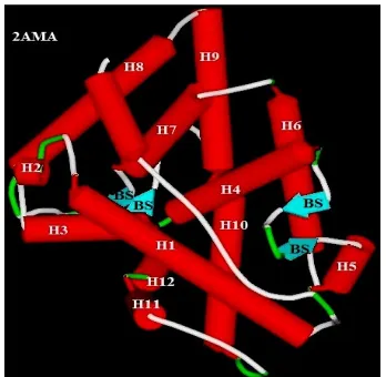
The LBD of AR consists of 12 α-helices and 1 β-sheet that fold to form triple
layered α-helical sandwich which creates a hydrophobic ligand binding pocket in which
ligand binds (Fig.1).
Agonist binding to LBD causes helix-12 to lay over the ligand binding pocket
and exposes the groove necessary for intramolecular N/C interaction. Antagonist
binding displaces the helix-12 away from ligand binding pocket and unveils the binding
surface for co-repressor NcoR/SMRT interaction and inhibition of AR transcriptional
[image:22.612.233.407.325.495.2]activity. (Osguthorpe et al., 2011).
Fig. 1: Crystal structure of wild AR showing α-helices and β-strand folding
[image:22.612.206.437.535.689.2]Department of Pharmacology, PSGCOP Page 12 2.2.5. Ligand dependant AR activation
The ligand dependant activation is required for maximum activation of full
length AR. Upon androgen binding to AR LBP, AR dissociates from heat shock
proteins (HSP), undergoes ligand dependant intramolecular amino-carboxy terminal
interaction, phosphorylation, nuclear translocation, DNA dependant homodimerization,
co-activator recruitment, binding to specific androgen response elemnts and initiation
of transcription. AR resides predominantly in cytoplasm is associated with HSP and
chaperons. HSP also have active role in transcriptional activation of the receptor
because HSP 90 binds to AR LBD and keeps it in an active conformation so that AR is
able to bind androgens. Androgen binding to LBP results in the dissociation of HSP
from AR, helix 12 to fold over the LBP to enclose the ligand unmasking the necessary
grooves to form intramolecular amino-carboxy terminal interaction, dimerization and
exposes nuclear localization signal. Phosphorylation of AR also plays an important role
in transcriptional activity of AR. The newly translated AR is phosphorylated at serine
residues 506, 641, 653 as a post-translational modification which increase the ligand
acquiring properties. Antagonist binding does not enhance receptor phosphorylation.
After translocation into nucleus AR undergoes DNA dependant intermolecular
[image:23.612.114.518.560.726.2]homodimerization (Jin et al., 2009).
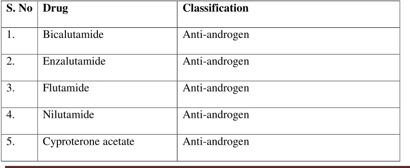
Table 2: Drugs available for prostate cancer
S. No Drug Classification
1. Bicalutamide Anti-androgen
2. Enzalutamide Anti-androgen
3. Flutamide Anti-androgen
4. Nilutamide Anti-androgen
Department of Pharmacology, PSGCOP Page 13
6. Abiraterone acetate Anti-androgen and CYP17A1 inhibitor
7. Cabazitaxel Taxane derivative and anti-microtubular agent
8. Docetaxel Taxane derivative and anti-microtubular agent
9. Degarelix Testosterone inhibitor
10. Goserelin acetate LHRH agonist
11. Leuprolide acetate LHRH agonist
12. Triprorelin acetate LHRH agonist
13. Mitoxantrone hydrochloride Anti-tumor antibiotic
14. Sipulencel – T Cellular immunological agent
15. Histrelin GnRH inhibitor
16. Buserelin acetate GnRH inhibitor
17. Ketoconozole Inhibitor of CYP17 and TST synthesis
18. Radium 223 dichloride Radiopharmaceutical
19. Prednisone Corticosteroid
20. Hydrocortisone Corticosteroid
21. Dexamethasone Corticosteroid
22. Denosumab Monoclonal antibody
23. Zoledonic acid Bisphosphonate
[image:24.612.114.512.61.546.2]Ref: (National cancer institute) (Gerald et al., 2017)
Table 3: Drugs in clinical trial for prostate cancer
S. No Drug Phase
1. Vaccine-Based Immunotherapy Regimen (VBIR) I
2. AZD8186 I
Department of Pharmacology, PSGCOP Page 14
4. Abiraterone Acetate With or Without Cabazitaxel II
5. ARN-509+Abiraterone acetate+Leuprolide with Stereotactic,
Ultra-Hypofractionated Radiation
II
6. JNJ-56021927 in Combination with Abiraterone Acetate and
Prednisone Versus Abiraterone Acetate and Prednisone in Subjects
with Chemotherapy-naive Metastatic Castration-resistant Prostate
Cancer
III
Ref: (Memorial Sloan Kettering cancer centre, 2017)
2.3. The role of ER-β in prostate cancer
ER-β is encoded by chromosome locus 14q22–24 and it is expressed in both
stromal and luminal epithelial cells of the human prostate. ERα is expressed mainly in
prostate stroma. (Fixemer et al., 2003). As a member of the nuclear receptor family,
ERβ acts individually, forming homodimers (ERβ/β) or heterodimers (ERβ/α).
Ligand-induced dimerization leads to translocation of dimer to the nucleus, binding with
co-regulatory proteins and interaction with responsive elements (binding sites) in the
promoter regions including nuclear factor-κB (NF-κB) and activator protein 1 (AP-1).
ERβ binds indirectly to these alternative binding sites through the recruitment of
cofactors to the receptor. (Heldring et al., 2007). However, less is known about the
interactions between ERβ and transcriptional cofactors. ERβ/β and ERα/β homo- and
hetero dimers, respectively, exhibit anti-proliferative effects as they activate different
target genes. Interestingly, ERα/β heterodimer is more stable than the ERβ/β
homodimer. Overall, ER dimerization is a crucial step in defining ER signaling.
Interestingly, prostate morphogenesis occurs under the control of androgens and
is modulated by estrogens (Marker et al., 2003). However, ERβ is not required in early
Department of Pharmacology, PSGCOP Page 15 in the life of newborn mice following ERα expression (Omoto et al., 2005). Moreover,
in the developing rodent prostate gland, ERα-induced excessive estrogenic exposure
leads to permanent alternation of the gland including squamous metaplasia,
inflammation and epithelial dysplasia as reported by an in utero study (Prins et al.,
2006). Notably, the developmental pattern for ERβ in the human prostate is different
from the rodent. Apparently, ERβ is the only detectable ER in the developing human
fetal prostate. However, by year 11 post-natally, expression of ERβ is restricted to the
basal epithelial cells and prostate stromal compartments, similar to adult human
prostate. (Shapiro et al.,2005). Thus, in the developing human prostate, ERβ is the
predominant ER in both stromal and epithelial cells (Adams et al., 2002).
In the adult human prostate, ERβ is characterized as an important mediator of
epithelial differentiation (Imamov et al., 2004). The mechanisms through which ERβ
maintain differentiation involve the degradation of hypoxia-inducible factor 1α
(HIF-1α) (Mak et al., 2010). ERβ enhances transcription of prolyl hydroxylase
domain-containing protein 2 (PHD2) that hydroxylates HIF-1α and marks HIF for destruction
by the von Hippel-Lindau tumor suppressor (VHL) (Mak et al., 2013). Additionally,
ERβ appears to have antiproliferative actions which are independent from the
alternations of systemic androgen concentration and the activation of ERα, as
documented in aromatase-knockout mice treated with ERβ-specific agonists
(McPherson et al., 2007). There, ERβ seemed to have a suppressive role in the
proliferation process, stimulating the differentiation of adult prostate epithelial cells.
2.4. Role of Bcl-2 protein family in regulation of apoptosis in prostate cancer
In-vitro studies have highlighted the role of Bcl-2 proteins as important
Department of Pharmacology, PSGCOP Page 16 family, was discovered by studies of t(14:18) chromosomal translocations, which are
frequent in non-Hodgkin lymphomas and follicular lymphomas. The name Bcl-2 (B
cell lymphoma/leukemia gene2) signifies the close association of this gene to these
malignancies in which enhanced expression was initially believed to arise solely as a
result of these translocations, resulting in the juxta-position of the Bcl-2 gene to a
potent enhancer element sequence of the Ig H gene. Several genes have been identified
and designated as the Bcl-2 family based on their sequence homology to Bcl-2. These
genes include both positive and negative regulator of apoptosis.(Chaudhary et al.,1999)
2.5. Role of PI3K/Akt/mTOR pathway in prostate cancer
One pathway with a prominent role in prostate cancer is the PI3K/Akt/mTOR
pathway. Current estimates suggest that PI3K/Akt/mTOR signaling is upregulated in
30-50% of prostate cancers, often through loss of PTEN. Molecular changes in the
PI3K/Akt/mTOR signaling pathway have been demonstrated to differentiate benign
from malignant prostatic epithelium and are associated with increasing tumor stage,
grade, and risk of biochemical recurrence. Multiple inhibitors of this pathway have
been developed and are being assessed in the laboratory and in clinical trials, with
much attention focusing on mTOR inhibition. Current clinical trials in prostate cancer
are assessing efficacy of mTOR inhibitors in combination with multiple targeted or
traditional chemotherapies, including bevacizumab, gefitinib, and docetaxel. (Todd et
al., 2009)
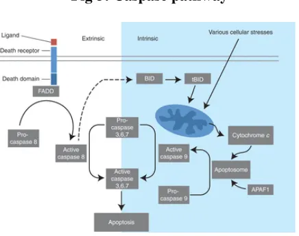
2.6. Caspase pathway
Caspases involved in apoptosis have been subclassified by their mechanism of
action and are either initiator caspases (caspase-8 and -9) or executioner caspases
Department of Pharmacology, PSGCOP Page 17 caspases that subsequently coordinate their activities to demolish key structural proteins
and activate other enzymes.
The extrinsic apoptosis pathway is activated through the binding of a ligand to a
death receptor, which in turn leads, with the help of the adapter proteins
(FADD/TRADD), to recruitment, dimerization, and activation of caspase-8. Active
caspase-8 then either initiates apoptosis directly by cleaving and thereby activating
executioner caspase (-3, -6, -7), or activates the intrinsic apoptotic pathway through
cleavage of BID to induce efficient cell death. The intrinsic or mitochondrial apoptosis
pathway can be activated through various cellular stresses that lead to cytochrome c
release from the mitochondria and the formation of the apoptosome, comprised of
APAF1, cytochrome c, ATP, and caspase-9, resulting in the activation of caspase-9.
Active caspase-9 then initiates apoptosis by cleaving and thereby activating executioner
[image:28.612.210.428.415.587.2]caspases (Fig 3).
Fig 3: Caspase pathway
2.7. Cucurbitacin
Cucurbitacins are found in many of the cucurbitaceous plants (Kaushik et al.,
2015). This diverse group of compounds may prove to be important lead molecules for
Department of Pharmacology, PSGCOP Page 18 can prove to be of immense significance in generating scientifically validated data with
regard to their efficacy and possible role in various diseases. Medicinal and toxic
properties of these compounds have stimulated a continuing interest in them. (Kupchan
et al., 1978). Many genus of Cucurbits viz. Trichosanthes, Cucurbita, Cucumis and
Citrullus are affluent in cucurbitacins. These compounds have also been discovered in
other plant families like Scrophulariaceae, Cruciferae, Datiscaceae, Primulaceae,
Rubiaceae etc. The diversity of cucurbitacins lies in variety of its side chain derivatives
that contribute to their disparate pharmacological actions. (Stuppner et al., 1993). The
bitter taste of plant species like cucumber has been attributed to the presence of
cucurbitacins.
The first cucurbitacin was isolated as a crystalline substance in 1831 and was
named α-elaterin. Certain plant species rich in cucurbitacins like Momordica hold
coveted position in different system of traditional medicines for curative effects in
metabolic disease like diabetes. All cucurbitacins contain a basic 19-(10→9β
)-abeo--10α-lanost-5--ene ring skeleton. A common feature among all compounds in the
category of Cucurbitacins is the presence of 5, (6) --double bond. The difference of
Cucurbitacins from steroidal nucleus lies in the fact that in basic structure of
Cucurbitacins a methyl group is located at C-9 rather than C-10. (Dinan, et al., 2001)
Most of the Cucurbitacins are tetracycline, but some representatives have an extra ring
due to formal cyclization between C--16 and C--24 as in cucurbitacins S and T.
(Gamlath et al., 1998) The Cucurbitacins differ from most of the other tetracyclic
triterpenes by being highly unsaturated and contains numerous keto--, hydroxyl--, and
acetoxy--groups. (Jorn et al., 1998) Certain Cucurbitacins have been discovered in the
Department of Pharmacology, PSGCOP Page 19 1991). Cucurbitacin B inhibits ATP citrate lyase which is a key enzyme that plays
crucial role in cancer cell metabolism in prostate cancer (Gao et al., 2014).
The structural composition of following Cucurbitacins are known and have been
designated by the letters: A, B, C, D, E, F, G, H, I, J, K, L, O, P, Q, R and S.
Cucurbitacin I caused reduction of growth in breast and prostate carcinoma cell lines
(MDA-MB-231, MDA-MB-468, Panc-1), in vitro, as well as in nude mice xenograft
models (Sun et al., 2008). Cucurbitacin Q induces apoptosis more potently in human
and murine tumors. Furthermore, in HeLa cells, cucurbitacins inhibited DNA, RNA,
and protein synthesis (Witkowski et al., 1984). The 23, 24-dihydrocucurbitacin B and
cucurbitacin R, inhibit proliferation and/or induce apoptosis in colon cancer cell lines.
Cucurbitacin E inhibits the proliferation of prostate cancer cells and caused disruption
of the cytoskeleton structure of actin and vimentin (Duncan et al., 1996). Cucurbitacin
A and I act by inhibition of only JAK2 and STAT3 respectively. It has been reported
that cucurbitacin E inhibited tumor angiogenesis by inhibiting JAK-STAT3 and
mitogen activated protein kinases (MAPK) signaling pathways. It has been reported
that cucurbitacin B exerts an anticancer effect by inhibiting telomerase via
down-regulating both the human telomerase reverse transcriptase and c-Myc expression in
breast cancer cells (Kaushik et al., 2015).
2.7.1. Cucurbitacin and Anti-inflammatory activity
Cucurcitacin analogues viz. Cucurbitacin R and DHCB have been reported to
possess anti-inflammatory potential and their action is reported to be mediated by
inhibition of tumor necrosis factors (TNF)-α and other mediators of inflammation such
as nitric-oxide synthase-2 and cyclo-oxygenase-2 (Escandell et al.,2008). Cucurbitacins
B, D, E and I have been reported to inhibit cyclo-oxygenase -2 enzymes with no effect
Department of Pharmacology, PSGCOP Page 20 24-dihydrocucurbitacin D (DHCD) have been hypothesized to get mediated through
blocking of NF-κ B activation thereby obstructing the release of nitrous oxide. DHCD
can be taken up as probable lead and appraised for providing a promising
anti-inflammatory agent. (Park et al., 2004).
2.7.2. Cucurbitacin and Antitumor activity
Very less information is available on the role of Cucurbitacins at molecular
level which has lead to slow advancement in the development of Cucurbitacins as
anti-cancer agents. (Kee et al., 2008). In relation to cancer, targets of Cucurbitacin actions
involve growth inhibition, arrest of cell cycle at G2/M phase and induction of apoptosis
in cancer cell. (Liu et al., 2000). The mechanisms underlying anti-tumorigenic
potentials of Cucurbitacins involve inhibition of Janus kinase/Signal Transducer
Activator of Transcription 3 (JAK/STAT3) signaling pathway whose activation is
required for the proliferation and sustainment of cells. (Bowman et al., 2000). The role
of Cucurbitacin I in suppressing phosphotyrosine STAT3 in cancer cell lines and
cancerous lung cells of humans has been reported. (Blaskovich et al., 2003). Although
Cucurbitacin B, E, and I act by inhibiting the activation of both JAK2 and STAT3,
Cucurbitacin A and I acts by inhibition of only JAK2 and STAT3 respectively. (Sun et
al., 2005). It has been reported that Cucurbitacin E inhibited tumor angiogenesis by
inhibiting JAK-STAT3 and mitogen activated protein kinases (MAPK)- signaling
pathways (Dong et al., 2010). The role of interference with actin cytoskeleton has been
attributed to anti-proliferative effects of Cucurbitacin B and E. The anti-proliferative
activities have been correlated directly with the disruption of the F-actin cytoskeleton.
(Duncan et al., 1996). It has been proposed that the combination of Cucurbitacin B with
docetaxel may augment the chemotherapeutic effects by suppression STAT3 in patients
anti-Department of Pharmacology, PSGCOP Page 21 tumor effects since they have been reported to contain Cucurbitacin C. (Higashio et al.,
2002) It has been reported that cucurbitacin B exerts an anticancer effect by inhibiting
telomerase via down-regulating both the human telomerase re verse transcriptase and
c-Myc expression in breast cancer cells. (Duangmano et al., 2010)
2.7.3. Cucurbitacin and Anti-artherosclerotic activity
There have been reports on Cucurbitacin B and E in glycosidic form to exhibit
inhibitory effect on lipid oxidation products like- malonaldehyde (MDA) and
4-hydroxynonenal (4-HNE). (Tannin-Spitz et al.,2007). These reports bolster the
therapeutic role of Cucurbitacins in artherosclerosis, which involves modification of
lipoproteins by involvement of- MDA and 4-HNE. (Saba et al., 2010)
2.7.4. Cucurbitacin and Anti-diabetic activity
There have been a plethora of reports on the role of Cucurbitacins for their
cytotoxic, hepatoprotective, cardiovascular, and antidiabetic effects. (Park et al., 2004).
Cucurbitane triterpenoids present in momordica fruits are noted for antidiabetic and
anticancer activities, this may provide leads as a class of therapeutics for diabetes and
obesity. (Tan et al., 2008). The 5’-adenosine monophosphate-activated protein kinase
(AMPK) pathway is suggested as a probable mechanism for the stimulation of GLUT4
translocation by triterpenoids from M. charantia. It is particularly interesting in relation
to diabetes and obesity because activation of AMPK increases fatty acid oxidation,
inhibits lipid synthesis, and can improve insulin action. An analogue of
23,24-dihydrocucurbitacin F from Hintonia latiflora has been reported to possess significant
hypoglycemic and antihyperglycemic effects. The probable mechanism underlying--
antihyperglycemic effect could be stimulation of insulin release and regulation of
Department of Pharmacology, PSGCOP Page 22 2.7.5. Miscellaneous activity of cucurbitacin
It has been reported that the concentration of Cucurbitacin C in the leaves is an
important parameter in spider mite resistance in Cucumis sativus, perhaps by acting as
an antagonist of a spider mite ecdysteroid receptor. (Balkema et al., 2003). The steroid
like resemblance of Cucurbitacin D may possess therapeutic effects via inhibition of
Na+/K+-ATPase. (Chen et al.,2010). The role of Cucurbitacins as preventive and radical
scavenging antioxidant has also been reported. Cucurbitacins have also been reported to
possess adaptogenic activity. Cucurbitacins have been reported to increase the rat
capillary permeability and to demonstrate antifertility effects in female mice. (Shohat et
al.,1972). Cucurbitacin D has been reported to inhibit ovulation in mice. There has been
protective role of Cucurbitacins acting as allomones in many plant species. Role of
Cucurbitacins as anti-feedants for few insects, birds and as kairomones (Cucurbitacin
B, E, D, I and L) for diabroticite beetles have been reported. (Subbiah. 2011). It is
reported that Cucurbitacins act via Cuc receptors located on the maxillary palpi. They
arrest the searching behavior of diabroticite beetles and produce a compulsive feeding
behavior. Role of Cucurbitacin B and D in controlling diabrotic beetles can be an
interesting approach. (Escandell et al.,2007).
2.7.6. Cucurbitacin I (JSI-124) induces apoptosis via p53 pathway in HepG2 cells
JSI-124 inhibited the proliferation and induced Hoechst 33258-stained
chromatin condensation in HepG2 cells in a concentration- and time-dependent manner.
Flow cytometry revealed that 1.00 µmol/L JSI-124 treatment increased the apoptotic
rate significantly in HepG2 cells compared with the control cells. Furthermore, JSI-124
significantly enhanced the mRNA expressions of p53 and its downstream apoptotic
factors, including Bax and Fas, but did not change the gene expression of the p53 tumor
Department of Pharmacology, PSGCOP Page 23 increased the levels of p53 and cleaved caspase-3 proteins. Conclusion JSI-124 induces
the apoptosis of HepG2 cells through the activation of p53 and its downstream
pro-apoptotic factors. (Wu et al., 2017)
Cucurbitacin I dose- and time-dependently inhibited the proliferation of five OS
cell lines. Following cucurbitacin I treatment, STAT3 was inactivated and analysis of
Mcl-1, cleaved PARP and caspase-3 indicated apoptosis induction. Expression of cell
cycle regulator proteins, such as phospho-cyclin D1, c-Myc and survivin, were
suppressed. Finally, cucurbitacin I potently inhibited the tumor growth of human OS
143B cells in nude mice. In-vitro and in-vivo results suggest that STAT3 inhibition by
cucurbitacin I will be an effective and new approach for the treatment of Osteosarcoma.
(Oi et al., 2016 )
2.7.7. Cucurbitacin I and cardio-toxicity
The mechanisms of cucurbitacin-I-induced cardiotoxicity are examined by
investigating the role of MAPK-autophagy-dependent pathways. After being treated
with 0.1-0.3µM cucurbitacin-I for 48h, H9c2 cells showed a gradual decrease in the cell
viabilities, a gradual increase in cell size, and mRNA expression of ANP and BNP
(cardiac hypertrophic markers). Cucurbitacin-I concentration-dependent apoptosis of
H9c2 cells was also observed. The increased apoptosis of H9c2 cells was paralleling
with the gradually strong autophagy levels. Furthermore, an autophagy inhibitor,
3-MA, was used to block the cucurbitacin-I-stirred autophagy, and then the hypertrophy
and apoptosis induced by 0.3µM cucurbitacin-I were significantly attenuated. In
addition, cucurbitacin-I exposure also activated the MAPK signaling pathways,
including ERK1/2, JNK, and p38 kinases. Interestingly, only the ERK inhibitor U0126,
but not the JNK inhibitor SP600125 and p38 MAPK inhibitor SB203580, weakened the
Department of Pharmacology, PSGCOP Page 24 findings suggest that cucurbitacin-I can increase the autophagy levels of H9c2 cells,
most likely, through the activation of an ERK-autophagy dependent pathway, which
results in the hypertrophy and apoptosis of cardiomyocytes. (Wu et al., 2016)
2.7.8. Mechanism of cucurbitacin I in gastric cancer cells
Deng et al., for the first time systematically studied the underlying molecular
mechanisms of Cu-I-induced gastric cancer cell death both in vitro and in vivo. In this
study, they show that Cu-I markedly inhibits gastric cancer cell growth by inducing
G2/M phase cell cycle arrest and apoptosis at low nanomolar concentrations via a
STAT3-independent mechanism. Notably, Cu-I significantly decreases intracellular
GSH/GSSG ratio by inhibiting NRF2 pathway to break cellular redox homeostasis, and
subsequently induces the expression of GADD45α in a p53-independent manner, and
activates JNK/p38 MAPK signaling. Interestingly, Cu-I-induced GADD45α and
JNK/p38 MAPK signaling form a positive feedback loop and can be reciprocally
regulated by each other. Therefore, the present study provides new insights into the
mechanisms of antitumor effects of Cu-I, supporting Cu-I as an attractive therapeutic
drug in gastric cancer by modulating the redox balance. (Deng et al., 2016)
2.7.9. Cucurbitacin I and Breast cancer
Qi et al used JSI-124 (Cucurbitacin I), a selective JAK/STAT3 signaling
pathway inhibitor, to investigate the role of STAT3 in tumor angiogenesis of a human
BC cell line in vitro. JSI-124 inhibited cell viability, proliferation, adhesion, migration
and tube formation of a human BC cell line MDA-MB-468. After transfection with
pMXs-Stat3C, a dominant active mutant, the inhibitory effects of JSI-124 on
MDA-MB-468 were abolished. Furthermore, JSI-124 reduced the phosphorylation of STAT3.
Department of Pharmacology, PSGCOP Page 25 line in vitro through the reduction of STAT3 phosphorylation. In addition, JSI-124
could reduce VEGF transcription and secretion, suggesting that JSI-124 is also
involved in the inhibition of the VEGF autocrine loop in the tumor microenvironment.
(Qi et al., 2015)
The small GTPase Rac1 has been widely implicated in mammary tumorigenesis
and metastasis. Previous studies established that stimulation of ErbB receptors in breast
cancer cells activates Rac1 and enhances motility via the Rac-guanine nucleotide
exchange factor P-Rex1. As the Janus tyrosine kinase 2 (Jak2)/signal transducer and
activator of transcription 3 (Stat3) pathway has been shown to be functionally
associated with ErbB receptors, Lopez et al asked if this pathway could mediate
P-Rex1/Rac1 activation in response to ErbB ligands. They found that the anticancer agent
cucurbitacin I, a Jak2 inhibitor, reduced the activation of Rac1 and motility in response
to the ErbB3 ligand heregulin in breast cancer cells. However, Rac1 activation was not
affected by Jak2 or Stat3 RNA interference, suggesting that the effect of cucurbitacin I
occurs through a Jak2-independent mechanism. Cucurbitacin I also failed to affect the
activation of P-Rex1 by heregulin. Subsequent analysis revealed that cucurbitacin I
strongly activates RhoA and the Rho effector Rho kinase (ROCK) in breast cancer cells
and induces the formation of stress fibers. Interestingly, disruption of the RhoA-ROCK
pathway prevented the inhibitory effect of cucurbitacin I on Rac1 activation by
heregulin. Lastly, they found that RhoA activation by cucurbitacin I is mediated by
reactive oxygen species (ROS). The ROS scavenger N-acetyl L-cysteine and the
mitochondrial antioxidant Mito-TEMPO rescued the inhibitory effect of cucurbitacin I
on Rac1 activation. In conclusion, these results indicate that ErbB-driven Rac1
Department of Pharmacology, PSGCOP Page 26 they established that the inhibitory effect of cucurbitacin I on Rac1 activity involves the
alteration of the balance between Rho and Rac. (Lopezet al., 2013)
2.7.10. Cucurbitacin I and colon cancer
Song et al., examined the chemopreventive potential of cucurbitacin I, a natural
component extracted from plants of the Cucurbitaceae family, in the colon cancer cell
line COLO205. They hypothesized that cucurbitacin I would prevent colon cancer cell
migration and invasion, and sensitize colon cancer cells to chemotherapy. The data
demonstrated that exposure of the COLO205 cells to cucurbitacin I significantly
decreased cell viability. Furthermore the data demonstrated for the first time that in the
COLO205 cells, cucurbitacin I could suppress the cell migration and invasion, and
harbor chemosensitization activity against colon cancer. The anticancer activity of
cucurbitacin I was accomplished by downregulating p-STAT3 and MMP-9 expression.
Collectively, the results suggest that cucurbitacin I may be a potent adjuvant
chemotherapeutic agent for colon cancer with anti-migration, anti-invasion and
chemosensitizing activities. (Song et al., 2015). Cucurbitacin-I reduced colon cancer
cell proliferation by enhancing apoptosis and causing cell cycle arrest at the G2/M
[image:37.612.116.527.560.679.2]phase. (Kim et al., 2014).
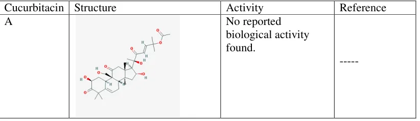
Table 4: Cucurbitacins and their activity
Cucurbitacin Structure Activity Reference
A No reported
biological activity found.
Department of Pharmacology, PSGCOP Page 27
B Anti-proliferative
activity in prostate and breast cancer.
Gao et al.,2014. Duangmano et al.,2010.
C No reported
biological activity found.
---
D Inhibits proliferation
and induce apoptosis of T-cell leukemia cells correlating
NF-κB inhibition and down-regulation of the expression of antiapoptotic proteins Bcl-xL and Bcl-2
Ding et al.,2011
E Cucurbitacin
E-induced disruption of the actin and vimentin cytoskeleton in prostate carcinoma cells and inhibits tumor angiogenesis through VEGFR2 mediated JAK2/ STAT3 signaling pathway.
Duncan et al., 1996.
Dong et al., 2010.
F Cucurbitacin F
induces cell cycle G2/M arrest and apoptosis in human soft tissue sarcoma cells.
Department of Pharmacology, PSGCOP G H I J K L
Department of Pharmacology, PSGCOP
No reported biological activity found. ---No reported biological activity found. ---Anti-tumor activity against colon cancer, gastric cancer, osteosarcoma breast cancer. Deng al., Qi Ren al., et al., Kim 2014. al., No reported biological activity found. ---No reported biological activity found. ---No reported biological activity found. ---Page 28 --- ---
Deng et al.,2016, Qi et al.,2015, Ren et
al.,2014, Song et al.,2015. Kim et al 2014. Oi et al.,2016.
---
---
Department of Pharmacology, PSGCOP Page 29
O No reported
biological activity found.
---
P No reported
biological activity found.
---
Q Selective STAT3
activation inhibitor with potent antitumor activity
Sun et al., 2008.
R Reduces the
inflammation and bone damage associated with adjuvant arthritis in Lewis rats by
suppression of tumor necrosis factor-alpha in T lymphocytes and macrophages.
Escandell et al., 2007.
S No reported
biological activity found.
---
U No reported
biological activity found.
Department of Pharmacology, PSGCOP Page 30 3. OBJECTIVES AND PLAN OF STUDY
3.1 Objectives
To identify Cucurbitacin derivative, which is having high affinity towards
prostate cancer targets by in-silico method.
To evaluate the cytotoxic activity of the cucurbitacin derivative in prostate
cancer cell lines by MTT assay.
To elucidate the apoptotic mechanism of cucurbitacin in prostate cancer cell line
by caspase assay, acridine orange\ethidium bromide staining methods and gene
expression studies.
3.2 PLAN OF STUDY
3.2.1 Phase І
3.2.1.1 In-Silico study
In-silico study comprises of molecular docking of cucurbitacin derivatives (A –
U) with prostate cancer target proteins like AR, ER-β, PI3K-α, Bcl2 and AKT.
Cucurbitacin which is having Glide score more than the standard in concerned
protein will be studied further.
Selected cucurbitacins will be examined in QikProp for its ADME properties
and toxicity.
One lead molecule will be taken for in-vitro studies in cancer cell lines.
3.2.2 Phase ІІ
3.2.2.1 In-Vitro study
Culturing LNCaP and PC3 cell lines using RPMI + 10% FBS and DMEM +
Department of Pharmacology, PSGCOP Page 31
MTT assay of standard bicalutamide and cucurbitacin will be done in LNCaP
and PC3 cell lines to evaluate the IC50 value.
Determination of apoptosis mechanism by ethidium bromide/acridine orange
staining method and caspase 3, 8, 9 assay.
PCR studies – to detect the expression of genes such as Bax, Bcl2 and PSA
Department of Pharmacology, PSGCOP Page 32
4. MATERIALS AND METHODS
4.1. Materials
[image:43.612.153.484.162.682.2]4.1.1. Chemicals used in this study
Table 5: Chemicals used for this study
S. No Chemical Manufacturer
1. Cucurbitacin I Phytolab
2. DMSO Thermo fisher scientific
3. DMEM Gibco
4. RPMI Gibco
5. Trypsin Sigma
6. Phosphate buffer saline Gibco
7. FBS Sigma
8. MTT Himedia
9. Dihydrotestosterone Himedia
10. Bicalutamide Santa cruz
11. Acridine orange Himedia
12. Ethidium bromide Himedia
13. Caspase 3 assay kit Biovision
14. Caspase 8 assay kit Biovision
15. Caspase 9 assay kit Biovision
16. TRI reagent Sigma
17. Chloroform Nice chemicals
Department of Pharmacology, PSGCOP Page 33
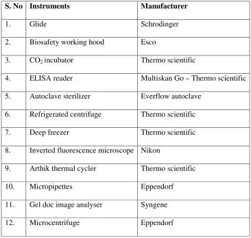
Table 6: Instruments used in this study
S. No Instruments Manufacturer
1. Glide Schrodinger
2. Biosafety working hood Esco
3. CO2 incubator Thermo scientific
4. ELISA reader Multiskan Go – Thermo scientific
5. Autoclave sterilizer Everflow autoclave
6. Refrigerated centrifuge Thermo scientific
7. Deep freezer Thermo scientific
8. Inverted fluorescence microscope Nikon
9. Arthik thermal cycler Thermo scientific
10. Micropipettes Eppendorf
11. Gel doc image analyser Syngene
12. Microcentrifuge Eppendorf
4.2. Methods
4.2.1. In-silico screening
4.2.1.1. Molecular Modelling
The cucurbitacin derivatives are downloaded from the PubChem compound
database and are prepared for docking using LigPrep (Ligprep, Schrödinger). LigPrep
helps to convert 2D structure to 3D representation. LigPrep can also produce a number
of structures from each input structure with various ionization states, tautomers,
stereochemistries, and ring conformations, and eliminate molecules using various
Department of Pharmacology, PSGCOP Page 34
present. Subsequently the structures were optimized by means of OPLS-2005 using a
default setting in LigPrep.
4.2.1.2. Protein preparation
Proteins used in the study are downloaded from the RCSB protein data bank
(PDB). Protein structures are prepared by using Maestro software (Maestro,
Schrödinger) and aligned using the protein structure alignment module in Prime (Prime,
Schrödinger). Bond order and formal charges were added for heterogroups and
hydrogens were added to all atoms in the system. Protein was inspected visually for
accuracy in the χ2 dihedral angle of Asn and His residues and the χ3 angle of Gln, and rotated by 180˚ when needed to maximize hydrogen bonding. The proper His tautomer
was also manually selected to maximize hydrogen bonding. All Asp, Gln, Arg and Lys
residue were left in their charged state. Water molecules for crystallization were
removed from the complex except in the active site. A brief relaxation was performed
on structure using the protein preparation module in Maestro with the “Refinement
only” option. This is a two – part procedure that consists of optimizing hydroxyl and
thiol torsion in the first stage followed by an all-atom constrained minimization carried
out with the impact refinement module (Impref) using the OPLS-2005 force field to
alleviate steric clashes that may exist in the original PDB structures. The minimization
was terminated when the Root Mean Square Deviation (RMSD) reached a maximum
cutoff of 0.30Å.
4.2.1.3. Grid generation and ligand docking
Grids were defined by centering them on the ligand in the crystal structure using
the default box size setting in Glide. Scaling of van der Waals radii of protein atom's
partial atomic charge of less than 0.25 in 1.0. Hydrogen bond constraints were not
Department of Pharmacology, PSGCOP Page 35
calculations were performed using the “Extra precision” (XP) mode of Glide program.
Glide uses a hierarchical series of filter to search for possible locations of the ligand in
the active site region of the receptor. The initial filters test the spatial fit of the ligand to
the defined active site and examined the ligand-receptor interactions using a grid-based
method. Poses that pass these initial screens enter the final stage of the algorithm,
which involves evaluation and minimization of a grid approximation to the OPLS-AA
non-bonded ligand-receptor interaction energies. Final scoring is then carried out on the
energy-minimized poses. The minimized poses are restored using Schrödinger’s
proprietary Glide score (G score) scoring function. G score is a modified version of
ChemScore, but includes a steric-clash term and adds buried polar terms devised by
Schrödinger to penalize electrostatic mismatches.
G Score = (a x vdW) + (b x Coul) + Lipo + Hbond + Metal + BuryP + RotB + Site
Where, vdW – van der Waal energy, Coul – Coulomb energy, Lipo – Lipophilic contact
term, Hbond – Hydrogen bond, Metal – Metal binding term, BuryP – penalty for buried
polar groups, RotB – penalty for freezing rotatable bonds, Site – polar interaction of the
active site.
4.2.1.4. ADMET property Prediction
Many drugs often fail to enter the market as a result of poor pharmacokinetic
profiles. Thus, it has become imperative nowadays to design lead compounds which
can be easily orally absorbed, easily transported to their desired site of action, not easily
metabolized into toxic metabolic products before reaching the targeted site of action
and easily eliminated from the body before accumulating in sufficient amounts that
may produce adverse side effects. The sum of the above mentioned properties is often
referred to as ADME (absorption, distribution, metabolism and elimination) properties,
Department of Pharmacology, PSGCOP Page 36
toxicity issues). The inclusion of pharmacokinetic considerations at earlier stages of
drug discovery programs using computer-based methods is becoming increasingly
popular. The rationale behind in silico approaches are the relatively lower cost and the
time factor involved, when compared to standard experimental approaches for ADMET
profiling. As an example, it only takes a minute in an in silico model to screen 20,000
molecules, but takes 20 weeks in the “wet” laboratory to do the same exercise. A set
of ADMET-related properties (a total of 46 molecular descriptors) were calculated by
using the QikProp program (Schrödinger) running in normal mode. QikProp generates
physically relevant descriptors, and uses them to perform ADMET predictions. An
overall ADME-compliance score – drug-likeness parameter (indicated by #stars), was
used to assess the pharmacokinetic profiles of the test compounds. The #stars parameter
indicates the number of property descriptors computed by QikProp that fall outside the
optimum range of values for 95% of known drugs.(QikProp, Schrodinger). The
methods implemented were developed by Jorgensen and Duffy and among the
calculated descriptors are: the logarithm of predicted binding constant to human serum
albumin, log KHSA (range for 95% of drugs: -1.5 to 1.2) the logarithm of predicted
blood/brain barrier partition coefficient, log B/B (range for 95% of drugs: -3.0 to 1.0)
the predicted apparent Caco-2 cell membrane permeability (BIPcaco − 2) in Boehringer– Ingelheim scale, in nm s-1 (range for 95% of drugs: < 5 low, > 100 high) the predicted apparent Madin-Darby canine kidney (MDCK) cell permeability in nm s-1 (< 25 poor, >
500 great) calculated from the number of hydrogen bond acceptors (HBA), donors
(HBD) and the predicted IC50 value for blockage of HERG K+ channels, log HERG
(concern < −5) the predicted skin permeability, log Kp (−8.0 to −1.0 for 95% of
drugs) and the number of likely metabolic reactions, #metab (range for 95% of drugs:
Department of Pharmacology, PSGCOP Page 37
4.2.2 In-vitro screening
4.2.2.1. Mammalian cell culture
The LNCaP and PC3 cell lines are obtained from NCCS, Pune. LNCaP cells are
maintained in Roswell Park Memorial Institute (RPMI) medium supplemented with
10% FBS and penicillin–streptomycin, whereas PC-3 cells were maintained in the
Dulbecco’s Modified Eagle Medium (DMEM) containing 10% FBS, penicillin–
streptomycin, L-glutamine, pyruvate sodium. Cells were grown at 37°C with 5% CO2.
PC3 cells are grown in culture flasks with polystyrene surface. The LNCaP cells are
grown in culture flasks with corning Cell BIND surface. 0.25 % trypsin is used for
detachment of the cells. (Claudia et al., 2007).
4.2.2.2. Maintaining and storage of cell lines
LNCaP cells show a steady growth rate with a doubling time of 60 hrs whereas
PC3’s doubling time is 48 hrs. The cells reached confluence in 6 to 7 days and were
passed to get cells for experiment and also to store liquid nitrogen. Passaging was done
as follow as.
Culture medium was removed from T25 flask by decanting into a clean
container inside the laminar airflow hood, cells were rinsed with Phosphate Buffer
Solution (PBS) to remove the traces of serum. 2ml of trypsin was added to the flask
containing cells and incubate at 37˚C for 2 to 2.5 minutes. As soon as, cells started
dislodging from the surface, the flask was rinsed with culture medium to arrest
trypsination. The suspension of cells was collected in a sterile 15ml centrifuge tube
using serological pipette and the cells were pelleted at 1500rpm for 5 mins. The cell
pellet was resuspended in PBS and again centrifuged. The resulted pellet is resuspended
in Culture medium and a part of it was seeded back into a sterile flask. The remaining
Department of Pharmacology, PSGCOP Page 38
in a cryovial and frozen at -80˚C for short term storage and at liquid nitrogen for long
time storage.
4.2.2.3. Drug Preparation
Cucurbitacin I was subjected to solubility test with different organic solvents
and found to be dissolved in DMSO, 50mM concentration was prepared. The desired
doses such as 0.1, 0.5, 0.10 and 50 mm were prepared from the stock using DMSO.
4.2.2.4. Anti-proliferative assay
Approximately 1000 to 3000 cells/well were added in 96 well plate from well
grown culture, the viability is tested using tryptan blue dye with the help of
haemocytometer, 95% viability should be confirmed. The anti-proliferative activity is
measured by adding standard and test compounds (0.1 nM to 500 nM). The
proliferative activity of the standard and test compound was evaluated without 1nM
DHT. After 24 hours, fresh medium containing the drug replaces the old medium and
incubated for another 24 hour. At the end of 48th hr 10µl of 3-[4, 5-dimethyl
thiazol-2-yl] 2, 5- diphenyl tetra-zolium bromide (MTT) is added and the plates were incubated
for an additional 4 hour. The formazon crystals were dissolved in 100µl of DMSO/well.
The optical density was measured at 570nm. (Sanchez et al., 2006). By plotting dose
response curve the IC50 value will be calculated. The cell viability can be calculated
using the formula,
Cell viability = (O.D of test cells/O.D of control) Χ 100
4.2.2.5. Dose finalization
Based on the IC50 value of test compound of different doses in LNCaP and