A PROSPECTIVE ASSESSMENT STUDY OF
THYROID DYSFUNCTION IN MODERATE
TO SEVERE COPD
DISSERTATION SUBMITTED FOR
M.D GENERAL MEDICINE BRANCH – I
APRIL
2018
THE TAMILNADU
CERTIFICATE FROM THE DEAN
This is to certify that this dissertation entitled “A Prospective
Assessment Study of Thyroid Dysfunction in Moderate to Severe COPD” is the bonafide work of Dr VIJAYARAGAVAN. R in partial fulfillment of the university regulations of The Tamil Nadu Dr. M.G.R.
Medical University, Chennai, for M.D General Medicine Branch
I examination to be held in April 2018.
DR. D. MARUTHUPANDIYAN, M.S,
THE DEAN,
Madurai Medical College,
Government Rajaji Hospital,
CERTIFICATE FROM THE HOD
This is to certify that the dissertation entitled “A Prospective
Assessment Study Of Thyroid Dysfunction In Moderate To Severe COPD ” submitted by Dr.VIJAYARAGAVAN.R , to the Tamil Nadu Dr. M.G.R. Medical University, Chennai, in partial fulfillment of the
requirement for the award of degree of Doctor Of Medicine (M.D)
Branch- I General Medicine, is a bonafide research work carried out
by him under my direct supervision & guidance.
Dr.V.T.PREMKUMAR M.D
Professor and HOD,
Department of General Medicine,
Government Rajaji Hospital,
Madurai Medical College,
CERTIFICATE FROM THE GUIDE
This is to certify that the dissertation entitled “A Prospective
Assessment Study Of Thyroid Dysfunction In Moderate To Severe COPD ” submitted by Dr.VIJAYARAGAVAN.R , to the Tamil Nadu Dr. M.G.R. Medical University, Chennai, in partial fulfillment of the
requirement for the award of degree of Doctor Of Medicine (M.D)
Branch- I General Medicine, is a bonafide research work carried out
by him under my direct supervision & guidance.
DR C.DHARMARAJ M.D,D.C.H
Professor Of Medicine,
Department Of General Medicine,
Government Rajaji Hospital,
Madurai Medical College,
DECLARATION
I, Dr VIJAYARAGAVAN R solemnly declare that, this dissertation entitled“A Prospective Assessment Study Of Thyroid
Dysfunction In Moderate To Severe COPD” is a bonafide record of
work done by me at the Department of General Medicine, Government
Rajaji Hospital, Madurai under the guidance of Professor
DR C.DHARMARAJ M.D,D.C.H in Department of General Medicine, Madurai Medical college, Madurai from September 2016 to November 2016. I also declare that this bonafide work or a part of this work was not submitted by me or any others for any award, degree, diploma to any
other University Board either in India or in abroad
This dissertation is submitted to The Tamil Nadu Dr. M.G.R.
Medical University, Chennai in partial fulfillment of the rules and
regulations for the award of Degree of Doctor of Medicine (M.D.) General Medicine Branch-I examination to be held in April 2018.
Place: Madurai DR VIJAYARAGAVAN R
ACKNOWLEDGEMENT
I would like to thank the Dean DR D. MARUTHU PANDIYAN( M.S ) , Madurai Medical college for permitting me to utilise the hospital facilities for the dissertation. I also extend my sincere thanks to
Dr.V.T.PREMKUMAR M.D., Head of the department and Professor of Medicine for his constant support during the study. I would like to
express my deep sense of gratitude and thanks to my unit Chief
Dr.C.DHARMARAJ M.D, D.C.H., my guide and Professor of Medicine, for his valuable suggestions and excellent guidance during the
study.
I thank the Assistant Professors of my Unit Dr. M. RAJKUMAR M.D.,DR SENTHUR RAJA PANDIAN M.D,D.M., for their help and constructive criticisms.
I offer my special thanks to Dr. J. SANGUMANI M.D, D.DIAB ,
Head of the department of Endocrinology for his kind co-operation and
valuable guidance.
I offer my special thanks to Dr. PRABAKARAN M.D., Head of the department of Thoracic medicine for his valuable guidance and
support.
I am greatly indebted to my beloved Professors,
Dr M.NATARAJAN M.D., Dr G. BHAGYALAKSHMI M.D, Dr J.SANGUMANI, M.D., Dr C.DHARMARAJ, M.D. D.C,H., and Dr. R.PRABHAKARAN, M.D., for their valuable suggestions throughout the course of study.
I offer my special thanks to Dr.SRIDHAR M.D, D.M., Assistant Professor of the department of Endocrinology for his kind co-operation
and valuable guidance.
I thank all the patients who participated in this study for their
extreme patience and kind co-operation.
I wish to acknowledge all those, including my Post graduate
colleagues, my parents who have directly or indirectly helped me to
complete this work with great success.
Above all I thank the Lord Almighty for his kindness and
CONTENTS
S.NO CONTENTS PAGE NO
1 INTRODUCTION 1
2 AIMS & OBJECTIVES 3
3 REVIEW OF LITERATURE 4
4 MATERIALS & METHODOLOGY 59
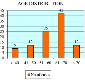
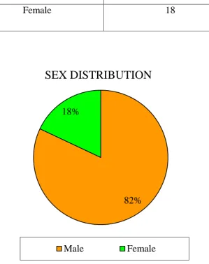
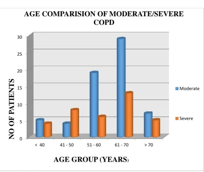
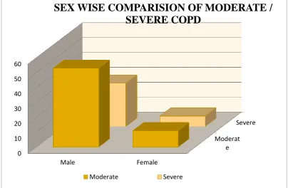
5 RESULTS & INTERPRETATION 65
6 DISCUSSION 82
7 SUMMARY 85
8 CONCLUSION 88
9 ANNEXURES
BIBILIOGRAPHY PROFORMA
1
INTRODUCTION
Chronic Obstructive Pulmonary Airway Disease is a Preventable &
Treatable Disease which is characterised by irreversible airflow
limitation of progressive in nature occurring due to chronic
inflammatory response affecting the lung parenchyma & airways to
noxious stimulants& various toxic pollutants in the atmosphere. The
Global Burden Of Disease Society States that COPD will become the
“THIRD” leading cause of death by 2020. COPD is a chronic systemic
disease affecting the vital organ systems in the body, since it is not
confined to affect only the respiratory system. COPD often leads to
Anaemia, Osteoporosis, Ischemic Heart Disease, Muscle Wasting ,
Depression & various other systemic ailments.
Extrapulmonary effects of COPD often culminates in significant
morbidity & mortality which affects the quality of life in the COPD
individuals. Endocrinological disorders occurs in COPD as a result of
Hypoxia, Hypercapnia, Iatrogenic usage of Glucocorticoids, Systemic
inflammatory mediators such as IL 6, INF gamma,Tumour necrosis factor
alpha , IL 1. Among the Endocrinological disorders , Thyroid disease is
2
Thyroid hormone has vital role in metabolism of carbohydrates ,
proteins, lipids, membrane bound enzymes & regulation of
thermogenesis. As the severity of COPD increases, impaired Thyroid
function in the form of Sub Clinical Hypothyroidism, Overt
Hypothyroidism, Nonthyroid Illness Syndrome can manifest.
Hypothyroidism adversely affect the quality of life in COPD individuals.
Hypothyroid in COPD leads to decreased respiratory drive, Acute
on chronic alveolar hypoventilation, Decreased lung volumes, Depression
of respiratory centres to its stimulants, Upper airway obstruction,
Respiratory Failure & frequent exaceberations. Hypothyroidism causes
inspiratory & expiratory muscle weakness due to impaired expression of
myosin heavy chains IIb & decreased neuromuscular transmission.
Diaphragmatic dysfunction & myopathy can occur. Severity of
hypothyroidism linearly correlates with muscle weakness & myopathy.
All these factors results in frequent exaceberations of COPD which has
significant role in affecting the quality of life in COPD individuals.
Hence, determining the functional state of Thyroid gland is of remarkable
3
AIMS & OBJECTIVES
To assess the prevalence of Thyroid dysfunction in moderate to
severe COPD patients.
To measure the relationship between COPD severity with the
4
REVIEW OF LITERATURE
Chronic Obstructive Pulmonary Disease Is characterised by
progressive development of chronic air flow limitation which is not fully
reversible as defined by GOLD (Global Initiative Of Obstructive Lung
Disease). COPD is a preventable & treatable disease as well . COPD
includes two broad categories which are Chronic Bronchitis and
Emphysema. By definition, Chronic Bronchitis is characterized by
chronic cough with expectoration for more than three months in two
consecutive years . Emphysema is described as abnormal persistent
distension of the air spaces distal to the terminal bronchioles associated
with destruction of their walls without any obvious fibrosis.
Epidemiology
In India, COPD being the second most common lung disorder
successive to the Pulmonary Tuberculosis. Its incidence is encountered
mainly in the fourth decade. It has an equal incidence among people
living in rural and urban areas. COPD manifestation is usually rare
5 Risk factors
1) Tobacco smoking
2) Air pollution – Indoor & Outdoor Pollutants
3) Familial and Genetic factors
4) Recurrent Respiratory Tract Infections in childhood
5) Occupational hazards due to inhalation of noxious particlulates -
organic or Inorganic dust fumes.
Tobacco smoking
“It is the most predominant risk factor associated with a
progression of COPD”. It mainly contains vapourised chemicals, serious
noxious particulates, carcinogens which are suspended in the gaseous
medium. Tobacco smoked is directly proportional to the detrimental
effects observed in COPD patients. Pack years, smoking index are the
most vital parameters which are used for categorizing the smoking
exposure and possibility of the disease outcome.
Sustained and chronic cigarette smokers are more susceptible to
impaired ciliary motility, goblet cell hyperplasia with its tenacious
secretions due to hypertrophy of the respiratory secretory epithelium.
Reid index is often used as a pathological index used to assess the
severity of the Chronic Bronchitis. It is the ratio between the thickness of
6
cartilage and the epithelium. (Normal = 0.44). Passive smoking and
inhalation of environmental noxious particulates resulted in the
deterioration of pulmonary function. But their role in the manifestation
of COPD still being uncertain.
Pack years:
Number of packets of x Numbers of years of
Cigarettes smoked per day Smoking
Smoking index:
Number of cigarettes x Number of years of
Smoked per day Smoking
Air pollution
Due to urbanisation, industrialized urban areas are loaded with
COPD patients. The heavy air pollutants includes mainly sulphur dioxide
and wooden logs used for cooking, indoor pollution made by burning of
cow dung cakes all contributing significantly to atmospheric pollution .
Occupational hazards
COPD is more prevalent among people who were working in
places where organic or inorganic or noxious pollutants are enriched in
7
Recurrent respiratory infection in childhood
Precipitating infections are the main culprit factors for acute
exacerbation in chronic background of obstructive pulmonary airway
disease. It is the factor which resulted in a significant increase in the
morbidity and mortality. Release of proteolytic enzymes such as
Metalloproteinases, Serinase, Lysosomal enzymes from the first line
defence mechanism cells - Neutrophils contributes to the extensive lung
pathology. Recurrent Viral respiratory tract infections during the infancy
period is pivotal in causing airway obstruction in the later life
Genetic mechanism
Randomised control Studies have suggested monozygotic twins are
prone to develop emphysema & chronic bronchitis due to genetic
predisposition.
Alpha 1 Antitrypsin is a serine protease inhibitor & an acute phase
reactant. Alpha 1 Antitrypsin deficiency in COPD patients is one to two
percent . The deficiency level rises to 50 percent in severe monozygotic
diseased people which usually affects younger age group. Alpha 1
antitrypsin inhibits elastase, collagenase and other proteolytic enzymes
& offers protection to lung in clearing the secretions. Alpha 1 antitrypsin
deficiency is the strongest genetic factor in development of chronic
8
predominantly involve lower lobes , which is usually pan lobular or pan
acinar but in smokers it affects the upper lobe and it is of centri lobular or
centri acinar type. Protease inhibitors variants that encode alpha 1
antitrypsin have been recognized. Two alleles such as S & Z alleles are
associated with reduced and markedly reduced alpha 1 antitrypsin levels
respectively.
Treatment with Alpha 1 Antitrypsin enzyme augmentation therapy
is available in recent times with linkage analysis of earlier onset disease
among family members have evidenced various spirometric variations
linked to appropriate regions of the chromosomes. Determinants of
specific genetic coding regions yet to be identified
Acute exacerbation precipitating factors
Infections - Streptococcus pneumonia, Haemophilus influenza,
Moraxella catarrahalis , Chlamydia pneumoniae
Exposure to noxious stimulants
Pneumothorax
Pulmonary thromboembolism, Myocardial infarction
Pathology of COPD
In the entity of Chronic Bronchitis, there is hypertrophy of goblet
cells which secrete mucus in the respiratory epithelium. In COPD
9
mucosal and the mucosal regions of the epithelium. Fibrosis involving
the peribronchus & accumulation of mucus plug in the intralumen of the
bronchus also contributes to the COPD pathology .
Emphysema is categeorized according to the involvement of
airway distal to the terminal bronchiole. Panacinar signifies the
involvement of whole acinus involving central and peripheral portions. In
centriacinar involves the respiratory bronchioles without involving the
periphery. An entity known as paraseptal involving the airspaces at the
lobule periphery nearer to the pleura. Irregular pattern almost associated
with scarring occurs in post pulmonary tuberculosis.
Pathophysiology of COPD
Two determinants of COPD as Chronic Bronchitis and
Emphysema often frequently coexist each other & there may be
domination of one entity over the other. Narrowing of the airway is a
common pathology in both categories. Besides basic pathophysiology of
the airways, there is loss of elastic recoiling capacity of lung
parenchyma in Emphysema leads to poor radial support to airways.
Increased work of breathing experienced by COPD patients is due to
alteration in the pressure and airflow pattern. Spirometry helps to
determine the major pulmonary functional parameters such as FVC and
10
in the FEV1 contributes towards the major morbidity. Responsiveness to
the inhaled bronchodilators is maximum in COPD as compared to
Bronchial asthma. Imbalance between the elastic recoil of the alveolar sac
and resistance offered by the airway towards the airflow determines the
reduction in FEV1 and FEV1/FVC. Residual volume and increase in the
ratio of residual volume to total lung capacity, is mainly due to trapping
of air. Total lung capacity is increased resulting in hyperinflation of lung
which is one of the late manifestations of COPD. Hyperinflation
preserves airflow during peak expiration. It is due to increase in the lung
volume which increases the elastic recoiling pressures & leading to
enlargement of airway thereby decreasing the resistance of the airways.
Hyperinflation is the compensatory mechanism to relieve the airway
obstruction. However flattened position of the diaphragm resulted in the
various adverse effects. The opposition zone between the abdominal wall
and diaphragm is decreased, effective abdominal pressure during
inspiration to the chest wall is not applied. Abnormal movement due to
hindering rib cage impairs the inspiration.Muscle fibers of the flattened
diaphragm are abnormally shorter than the normal diaphragm, resulting in
the less capability of inspiratory pressures.
According to Laplace‟s law ,
11
P = Trans pulmonary pressure producing the tidal breathing.
T = Tension gradient of the flattened diaphragm
R = Radius of the curvature
The flattened diaphragm has to generate tension to overcome the
trans pulmonary pressure to generate normal tidal volumes. In COPD,
partial pressure of O2 is not altered until FEV1 falls to 50%. Cardiac
complications like Pulmonary hypertension, Right heart failure,
CorPulmonale occurs when FEV1 is less than 25% of predicted value
with reduction in the partial pressure of oxygen to less than 55 mm of hg.
Mismatch between perfusion-ventilation & disproportionate ventilation
are the penultimate features of COPD. It reflects the diverse pathological
abnormality within the parenchyma & the smaller bronchi.
Pathogenesis
Airflow limitation is due to obstruction in the smaller airways and
Emphysema is the major pathological sequeale in COPD. Smaller
airways surrounded by fibrosis contributes significantly to morbidity and
mortality. Collagenase activity increases during the pathogenesis in
COPD. It is the major culprit resulting in accumulation of collagen
surrounding the airway. Potential mechanism through which fibrosis is
induced by activation of fibrogenic cytokines by growth factors such as
12
inflammatory cells in the distal air spaces due to chronic exposure to
cigarette smoke. These cells damage the matrix of the lung parenchyma
by releasing more potent proteinases like Elastases. The interaction
between cell and the matrix is lost, resulting in the apoptosis of the
structural cells in the lung. Extracellular matrix forms a integrity between
smaller airways & lung parenchyma. It is offered mainly by elastin a
predominant component of elastic fibers. The imbalance between
degrading enzymes and inhibitors involved in Elastin biology determines
the abnormal permanent distension of air spaces. Alpha 1 antitrypsin
deficiency patients are more prone for Emphysema due to lack of
neutrophil elastase inhibitor . Inactivation of histone deacetylase2
resulted in the acetylation and more heterochromatin which exposes the
transcription sites involving many pro inflammatory cytokines resulting
in recruitment of Neutrophils. Cigarette smoke recruits the suppressor
Tcells leading to production of macrophage elastase. Cleavage
component of elastin acts as a signaling chemokine which traverse the
destructive hypothesis. Ciliary dysfunction caused by cigarette smoke
traverse the fertile background for bacterial infection along with increase
in Neutrophill count. In final stage, there is an copious inflammatory
response suggesting that mechanisms of smoking induced disease differs
from inflammation resulting after cessation of smoking.. Finally the
13
degrading the matrix of lung parenchyma with defect in the cell
anchoring leads to apoptosis. Reparative capacity of the damaged alveoli
remains questionable.
Potential stimulus for constriction of pulmonary vasculature is
hypoxia. Cross sectional area of pulmonary vasculature is reduced in
COPD patients due to alteration in the vascular smooth musculature of
artery & arteriole of pulmonary vessels. Acidosis and the polycythaemia
due to chronic hypoxia culminates in right heart failure.
Clinical features Chronic bronchitis
Cough along with sputum production forms the mainstay of
Chronic bronchitis. History of chronic smoking is always present.
Predominant cough during winter months is the earlier manifestation.
Increase in the frequency of bronchial infection favours chronic
bronchitis. Insufficient respiration occurs during acute exacerbation.
Prominent bronchovascular markings is of pathognomic feature in
chronic bronchitis variant of COPD.
Over weight and cyanosis picture favours the terminology „blue
bloaters‟. Crepitation and polyphonic wheeze with resonant lung is
14
resulting in polycythaemia is common. Pulmonary vascular pathology
and right sided heart failure are more prevalent in chronic bronchitis.
Emphysema
History of dyspnoea with minimal cough and scanty production of
sputum is the main clinical feature of Emphysema. The Asthenic body
build with evidence of loss of weight noted in the physical examination.
The predominant usage of accessory muscles involved in respiration
resulted in sternal lift in anterosuperior direction in each phase of
inspiration. Tachypnea with prolonged expiratory phase through pursed
lip is characteristic of emphysema. Leaning forward with extension of his
arms to brace himself in sitting posture is known as tripod position. The
intercostals spaces retract each other during each inspiration in the lower
region of hemithorax which can be felt by palpation is known as
Hoover‟s sign. Apical impulse is usually visualized in the region between
xiphoid and subxiphoid areas. Hyperresonant percussion note with absent
or reduced cardiac dullness is a classical feature seen in COPD. The
upper margin of liver is usually shifted to a lower level than the normal.
Added sounds and abnormal air entry makes the clue to differentiate from
normal variant – compensatory emphysema. Since partial pressure of
oxygen is maintained in emphysema, they are called as pink puffers.
15
the inspiratory phase of the respiration. Since inflammatory cytokines
such as tumor necrosis factor alpha is elevated resulting in malnutrition
which is manifested as muscle wasting and it is an independent poor
prognostic variable in COPD. Incidence of right sided heart failure and
pulmonary hypertension is rare among emphysematous patients due to
maintenance in the partial pressure of oxygen in normal range. Diffusing
capacity with carbon monoxide used to differentiate chronic bronchitis
and emphysema decreased in emphysema and normal or slight variation
in chronic bronchitis. Advanced disease is manifested in form of
universal wasting more predominantly in bitemporal areas. Digital
clubbing is usually not a significant finding in COPD. Its presence should
16 COPD assessment
It requires a confined stratification taking into account of all the
factors. Four groups have been stratified by GOLD guidelines
Group A - low risk, less symptoms
Group B - low risk, more symptoms
Group C - high risk, less symptoms
Group D - high risk, more symptoms
Stage 3 or 4 with airflow limitation assessed by Spirometry is
given a label of high risk. Acute exacerbations more than 2 in no in the
previous year are hospitalisation requiring exacerbations are also taken
into account.
Symptoms are mainly assessed by
COPD Assessment test
COPD Control questionare
Chronic Respiratory Questionare.
St George Respiratory Questionare.
Parameters which predicts the prognosis involves BODE Index
B Body Mass Index
O Obstruction( Assessed by Spirometry)
17
E Exercise tolerance
GOLD CLASSIFICATION OF COPD BY SPIROMETRY
STAGES CHARCTERISTICS
(POST BRONCHODILATOR
RESPONSE)
Mild FEV1 / FVC ≤ 70%, FEV1 > 80%
Moderate FEV1 / FVC ≤ 70%, FEV1 50-80%
Severe FEV1 / FVC ≤ 70%, FEV1 30-50%
Very Severe FEV1 < 30% PREDICTED (OR)
FEV1 <50% PREDICTED WITH
RESPIRATORY FAILURE
Spirometry
Staging of COPD have been classified in similar manner from mild
to very severe COPD. FEV1/FVC ratio of < 0.7 is also applicable. FEV1
is the main predictor in COPD staging.
Comorbidities
Cardio vascular disorders
Skeletal muscle dysfunction
Nutritional deficiencies
18
Depression
Metabolic Syndrome
Osteoporosis
Cachexia
Lung malignancies
Hypercarbia related complications
Investigations used in diagnosing COPD Spirometry – Pulmonary Function Test
Spirometry is the most commonly used lung function screening
study. It generally should be the clinician's first option, with other studies
being reserved for specific indications. Most patients can easily perform
spirometry when coached by an appropriately trained technician or other
health care provider. The test can be administered in the ambulatory
setting, physician's office, emergency department or inpatient setting.
“The indications for spirometry are diverse” .Spirometry requires a
voluntary manoeuvre in which a seated patient inhales maximally from
tidal respiration to total lung capacity and then rapidly exhales to the
fullest extent until no further volume is exhaled at residual volume. “The
manoeuvre may be performed in a forceful manner to generate a forced
vital capacity (FVC) or in a more relaxed manner to generate a slow vital
19
expiratory SVC and expiratory FVC are essentially equal. However, in
patients with obstructive small airways disease, the expiratory SVC is
generally higher than the FVC.
This difference might, however, be due partly to the difficulty in
maintaining a maximum expiratory effort for an extended time period
20
The Spirometer can measure only the lung volumes that the subject
can exchange with it. As is the case with many pulmonary function tests,
the subject must be conscious and cooperative and understand the
instructions for performing the test. The VT, IRV, ERV, IC, and VC can
all be measured with a Spirometer (as can the Forced Expiratory Volume
in 1 second [FEV1], Forced Vital Capacity [FVC] and Forced Expiratory
Flow [FEF25–75%]). The RV, FRC, and the TLC cannot be determined
with a spirometer because the subject cannot exhale the lungs completely.
The gas in a spirometer is at ambient temperature, pressure and water
vapour saturation and the volumes of gas collected in a spirometer must
be converted to equivalent volumes in the body. Other kinds of
spirometers include rolling seal and bellows spirometers. These
spirometers are not water-filled and are more portable.
“A spirogram is a graphic representation of bulk air movement
depicted as a volume-time tracing or as a flow-volume tracing. Values
generated from a simple spirogram provide important graphic and
numeric data regarding the mechanical properties of the lungs including
airflow (Forced Expiratory Volume in 1 second [FEV1] along with other
timed volumes) and exhaled lung volume (FVC or SVC). The
measurement is typically expressed in liters for volumes or in liters per
21
gas that is saturated with water vapour. Data from a spirogram provide
important clues to help distinguish obstructive pulmonary disorders that
typically reduce airflow such as asthma and emphysema, from restrictive
disorders that typically reduce total lung volumes including pulmonary
fibrosis and neuromuscular disease”
Forced Expiratory Volume in 1 Second
“The FEV1 is the most widely used parameter to measure the
mechanicalproperties of the lungs. In normal persons, the FEV1 accounts
for the greatestpart of the exhaled volume from a spirometric maneuver
and reflects mechanical properties of the large and the medium-sized
22
85% of the FVC. This parameter isreduced in obstructive and restrictive
disorders. In obstructive diseases, FEV1 is reduced disproportionately to
the FVC, reducing the FEV1/FVC ratio below the lower limit of normal
and indicates airflow limitation. In restrictive disorders, the FEV1, FVC,
and total lung capacity are all reduced, and the FEV1/FVC ratio is normal
or even elevated.”
Forced Vital Capacity
FVC is a measure of lung volume and is usually reduced in
diseases that cause the lungs to be smaller. Such processes are generally
termed restrictive and can include disorders of the lung parenchyma such
as pulmonary fibrosis or of the bellows, including kyphoscoliosis,
neuromuscular disease, and pleural effusion. However, a reduction in
FVC is not always due to reduced total volumes and can occur in the
setting of large lungs hyperinflated due to severe airflow obstruction and
air trapping, as in emphysema. In this setting, the FVC is decreased due
to reduced airflow, air trapping, and increased residual volume, a
phenomenon referred to as pseudo restriction. Reduced FVC can occur
despite a normal or increased total lung volume. Therefore, FVC is not a
reliable indicator of total lung capacity or restriction, especially in the
setting of airflow obstruction. The overall accuracy of the FVC for
23
FEV1 Forced Expiratory Volume in 1 second
LLN Lower limit of normal
TLC Total lung capacity
VC Vital Capacity
Adapted from American Thoracic Society: Lung function testing:
24
Measurement of Lung Volumes not measurable with Spirometry.
The Residual Volume (RV), Total Lung Capacity (TLC) and related
volumes cannot be measured directly so special techniques are required to
record these volumes. There are several accepted methods for
determining these volumes, which are frequently referred to as 'static lung
volumes'. These methods include helium dilution, nitrogen washout and
body plethysmography. The FRC is usually determined and RV (which
is equal to FRC minus ERV) and the TLC (which is equal to VC plus
RV) are then calculated from volumes obtained by spirometry.
Nitrogen-Washout Technique
In the nitrogen-washout technique, the person breathes 100%
oxygen through a one-way valve so that all the expired gas is collected.
The concentration of nitrogen in the expired air is monitored with a
nitrogen analyzer until it reaches zero. At this point all the nitrogen is
washed out of the person's lungs. The total volume of all the gas the
person expired is determined, and this amount is multiplied by the
percentage of nitrogen in the mixed expired air, which can be determined
with the nitrogen analyzer. The total volume of nitrogen in the person's
lungs at the beginning of the test can thus be determined. Nitrogen
constitutes about 80% of the person's initial lung volume, and so
25
lung volume. If the test is begun at the” end of a normal expiration, “the
volume determined is the FRC.
Total Volume expired X %N2 = original volume of N2 in the lungs
original volume of N2 in the lungs X 1.25 = original Lung Volume
2) Imaging
Xray
Bronchial wall thickening manifested by tramline Shadows
with dominant
broncho vascular markings suggest chronic bronchitis.
Hyperlucent lung fields with no peripheral vascular
markings, emphysematous bullae, low level diaphragm,
tubular heart suggest Emphysema.
HRCT - Dilated main pulmonary artery and its branches is more
prominent when COPD progresses towards cor pulmonale
DLCO - diffusion lung capcity carbon monoxide
12 leads electrocardiogram
2D Echocardiography
Sputum examination
26 Complications of COPD
Pneumothorax
Pulmonary artery hypertension
Polycythemia
Cor Pulmonale
Acute and chronic on chronic respiratory failure
Right sided heart failure
Recurrent episodes of acute exacerbations
Potential markers used in prognosis of COPD
Alpha 1 antitrypsin deficiency
CFTR gene mutation
MBI2 genes
Fibrinogen, C reactive protein and other acute phase reactants
during exacerbation.
Potential measures to reduce the mortality in COPD can be reduced
by following measures by
Cessation of smoking
27 Smoking cessation
Pharmacological measures
Nicotine Replacement Therapy - Transdermal patch, chewing
gums, lozenges, inhalers, nasal spray.
Non Nicotine pharmacotherapy:
Bupropion 150 mg per day * 3 days followed by bd * 7 – 12
weeks.
Drug Should be started 1 week before quit date. Adverse effects
are dizziness, headache, insomnia ,nausea, xerostomia, hypertension,
seizures.
Avoid monoamine oxidase inhibitors to prevent serotonin
syndrome.
Varenicline 0.5 mg per day * 3 days followed by BD * 4 days ,then
1 mg BD * 12-24 weeks .start 1 week before quit date.
Domiciliary oxygen therapy
There are three forms of domiciliary supplementary oxygen
therapy,
Long term control oxygen therapy for atleast 15 hours daily in
28
In exercise related hypoxemia portable oxygen therapy is
supplemented.
Short term and short burst oxygen therapy as a palliative treatment
for temporary relief .
Criteria for long term oxygen therapy
“Absolute indications – COPD , Hypoxemia , Edema , FEV1 <
1.5 l , FVC <2l , Pao2 < 55 mm hg , PaCo2 > 45 mm hg , P pulmonale >
3 mm in Lead II, III, avf, Pulmonary Hypertension , Cor Pulmonale ,
Right ventricular hypertrophy, Polycythemia with Erythrocytosis with
hematocrit > 56% , desaturation < 96% on exercise , refractory dyspnea
associated with cardiac failure. Relative indications – as mentioned above
but without Edema or Paco2 >45mm hg”.
Palliative
FEV1 is the strongest predictor of survival in long term oxygen
therapy. It has been shown to affect the polycythemia which occurs
during chronic hypoxemia. It reduces both hematocrit and red cell mass.
However with persistent smoking exposure which results in chronic
elevation of carboxy hemoglobin decreases the effectiveness of long term
oxygen therapy in correcting polycythemia. It showed a marked decrease
in pulmonary artery Pressure with breathing controlled oxygen therapy. It
29
change in mood or quality of life. It showed a sustained improvement in
exercise endurance in patients with COPD breathing supplemental
oxygen. It is also associated with improvement in the sub maximal work
rate, with improvement in walking distance and ability to perform daily
activities .with 6 months of long term oxygen therapy, there is a
remarkable reduction in the mortality associated with COPD.
Long term management in COPD
Bronchodilator therapy is the treatment to reduce the symptoms
and improves the exercise capacity in COPD. The principle symptomatic
bronchodilators can be divided into three groups based on their
pharmacological properties.
1.Inhaled beta 2 agonists are preferred over oral preparations. It showed significant improvement in bronchodilation.in chronic bronchitis & the
decline in FEV1 was more reduced in those patients who used
continuous beta 2 agonists. However bronchodilator benefits are less
compared to asthma due to structural damages in airways.
2.Anticholinergics have time to peak effect of 30 to 60 minutes in most COPD patients , which is slower than beta 2 agonists but have a
somewhat longer time of effectiveness of 6 to 10 hours compared with
beta 2 agonists. There is a conflicting evidence regarding the effects of
30
increase in maximum exercise, ventilation and reduction in oxygen
consumption at any given workload.
3. Theophyllines
The bronchodilator property of theophyllines is relatively limited
in patients with COPD. Non bronchodilator effects of theophyllines such
as improving right ventricular performance and their anti inflammatory
actions are of questionable clinical significance. It has a narrow
therapeutic index with experienced side effects.theophylline metabolism
is increased by cigarette smoking , anti convulsant drugs , rifampicin.
Decreased by congestive cardiac failure , respiratory acidosis , liver
cirrhosis , viral infection , old age, arterial hypoxemia , on drugs like
erythromycin , ciprofloxacin , Cimetidine.
4. Glucocorticoids
Chronic inflammation in large and small airways is a characteristic
feature of COPD. The use of corticosteroids in COPD remains
contentious particularly the prediction of which patients will respond to
this treatment.
5. Pulmonary Rehabilitation
The restoration of the individual to the medical , emotional , social
and vocational potential of which he/she is capable. The main aim of
31
exercise and immobility due to dyspnea and allow the patient to cope
with the disease. Exercise training programmes have taken two
approaches the first is to attempt to improve cardiorespiratory fitness by
aerobic exercises of 20 to 30 minutes duration atleast three times per
week. It has been suggested that due to training effect ,it is usually
restricted to those patients with mild to moderate exercise limitation.
Second approach is to improve their Anaerobic fitness. In patients with
very severe COPD, there are no established guidelines for pulmonary
rehabilitation programmes, but carefully supervised exercise condition in
the hospital setting , with oxygen supplementation should be considered
in those who develop hypoxemia during exercise. Respiratory muscle
training and ventilatory assist devices have been used to reduce the
ventilator limitation during exercise. The presence of resting hypercarbia
is not a contraindication to pulmonary rehabilitation. Education of
patients in understanding the various components of the disease is
intuitively valid. Mood disturbance particularly depression are very
common in patients with advanced disease.
Exercise training
Expiratory flow rates during tidal breathing in patients with severe
COPD are close to the maximum expiratory flow volume relationship. An
32
COPD through dynamic hyper inflation.at the expense of increase in
respiratory work , since tidal volume operates in a less compliant range of
the pressure volume relationship and hence initiation of inspiration
requires additional inspiratory pressures to overcome elastic recoiling of
the diseased airways Continuous positive airway pressure overcomes the
increased recoil Pressure at the end of expiration , thus reducing breathing
workload.
Controlled breathing techniques
It attempts to diminish the breathlessness by training the patients
to breath efficiently. It mainly aims for
Restoration of diaphragm to a more normal position and function.
Decrease the respiratory rate by using a breathing pattern that
diminishes air trapping, Improving the respiratory duty cycle.
To diminish breathing workload.
To reduce dyspnea and alleviate patient anxiety.
The effects of different postures on respiratory muscle function
have also been assessed. Diaphragmatic breathing exercises have been
used to improve diaphragm function and are thought to be most helpful in
33 Nutrition
Weight loss ( Cachexia) is common in COPD patients particularly
those with severe airway obstruction. Those patients with less than 90%
of their ideal body weight are generally considered to be malnourished.
Weight loss has been associated with a higher mortality in these patients,
it would therefore seem logical to give nutritional support to patients with
COPD. The weight gain is lost soon after cessation of nutritional support
and any improvement in Peripheral muscle performance and exercise
capacity are also small and of short duration. However if sustained weight
gain can be achieved this may improve survival.& the theoretical
complication of carbohydrate based diet increasing carbondioxide
production and hence hypercapnia in patients with COPD does not
appear to be a problem. Obesity should be discouraged in patients with
COPD in order to avoid additional strain on the cardiorespiratory system
& appropriate dietary advice should be given.
Vaccination
Influenza & Pneumococcal vaccination are recommended for
patients with COPD, although the specific evidence for this in COPD
34 Lung transplantation
Indications
Age<50 years for heart-lung transplantation or double lung
transplantation
Age<60 years for single lung transplantation
Patients with an estimated life expectancy of less than 18 months
Contraindications
Malnutrition is a relative contraindication
Ideally the recipients should be within 15 kg of their ideal body
weight
Recurrent or persistent pulmonary infections - contraindication to
Single lung transplantation.
Other considerations :
Previous thoracic surgery increases the risk of haemorrhage
Cor pulmonale is not a contraindication to single lung
transplantation
Psychological stability is necessary
35 Lung volume reduction surgery
The rationale for this technique is to reduce the volume of over
inflated emphysematous lung by 20 to 30% with the aim of improving
the elastic recoiling of the lungs, diaphragm dynamic ability , chest wall
mechanics and gas exchangeThe technique is usually performed via a
median sternotomy, without the need for cardiopulmonary bypass.
Careful selection is necessary on the basis of a distended thorax,
predominantly upper lobe disease and severe functional disability despite
a programme of pulmonary rehabilitation. The improvements that have
occuered upto 6 months after surgery are better than conventional
medical treatment with bronchodilators and corticosteroids.
Thoracoscopic laser pneumoplasty has been developed as an
alternative to the more conventional excisional surgery. The Nd: Yag
laser appears to be a safer technique than Co2 laser. It relies on the fact
that at operation the lung that remains represents the most affected areas
and would absorb most energy; thus scarring and contraction would be
concentrated at these sites. COPD & its complication are related to the
chronic history. Management of exacerbations requires ventilator support
with controlled oxygen therapy with or without assisted ventilation. Non
36
THYROID DISORDER
History
“Hypothyroidism was described for the first time in London
(1870)”. It was named Myxedema. In 1888. It was found out that
cretinism, Myxedema & Post Thyroidectomy changes were a result of
loss of function of thyroid gland. Kendall isolated Thyroxin hormone in
1914. Harrington synthesized “Thyroxine” in 1926. However; synthesis
of Thyroxine was done in large scale in 1949. Later it became a
universally accepted therapy for hypothyroidism.
THYROID GLAND
Anatomy of Thyroid Gland
Thyroid gland has a midline isthmus lying horizontally just below
the Cricoid Cartilages Right & left lateral lobes that extend superiorly
together, in front of neck giving the appearance of butterfly. The gland is
enclosed by pre tracheal fascia under the strap neck muscles which
makes the gland move up with the deglutition.
Histology of Thyroid
Thyroid gland is divided by thin fibrous septa into Pseudolobules.
These pseudolobules are composed of follicles or acini which are
37
cuboidal epithelium. Protenaceous colloidal material filled within the
lumen of follicles called as Thyroglobulin. The peptide sequences of T4
and T3 are stored and synthesized as component of Thyroglobulin.
Embryology of Thyroid Gland
Develops from the ectoderm of the pharyngeal floor with some
contribution from the lateal pharyngeal pouches. Thyroglossal duct,
which extends from the foramen caecum near the base of the tongue to
the isthmus of the thyroid arise as descent of the midline thyroid anlagen.
The posterior aspect of the thyroid gland becomes associated with the
parathyroid gland & the para follicular C cells during the development,
which are derived from ultimo-bronchial body, which become
incorporated into its substance · While they undergo malignant
transformation, the C cells are the source of the calcitonin & leads to
medullary thyroid carcinoma. At about 10-12 weeks of gestation, the
foetal thyroid begins to concentrate & organify Iodine. Maternal TSH and
T4 do not cross the placenta, but the maternal TRH crosses the placenta.
The major source of thyroid hormone in the foetal life is T4 from the
foetal thyroid itself. · The functional unit is foetal pituitary – thyroid axis
38 Physiology of Thyroid Gland
“Thyroid secretes hormones – Thyroxin (T4), Triiodothyronine (T3) &
Calcitionin”. Thyroid follicles secretes only the first two hormones &
termed as “Thyroid Hormones”. Calcitonin is chemically & biologically
different, secreted from parafollicular C cells. It regulates calcium
metabolism along with Parathorrmone (PTH). Iodine enters the thyroid in
the form of inorganic or organic iodide which is oxidized by peroxidise
enzyme. Subsequent reactions results in formation of thyroxin. The only
source of T4 is thyroid gland. Thyroid secretes 20% of T3; extra
glandular tissues produce the remaining amount by peripheral conversion
of T4 into T3.
CHEMISTRY AND SYNTHESIS OF THYROID HORMONE
Both T4 and T3 are product of 2 molecules of tyrosine with iodine
containing derivatives of Thyronine. Thyroxine (T4) - 3, 5, 3‟, 5‟ –
Tetraiodothyronine T3 - 3, 5, 3‟ – Triiodothyronine. Thyroid hormones
are synthesized & stored in thyroid follicules as part of Thyroglobulin
molecules, a glycoprotein synthesized in thyroid cells. There are 5 steps
in synthesis of thyroid hormones.
1. IODIDE UPTAKE / IODIDE TRAPPING: Iodine from peripheral circulation is taken into follicles by active transport Na + I – symporter.
39
activates & large storage inhibits this trap. This process is mediated by
TSH. “Percholarate, Thiocyanates, Nitrates inhibits this trapping”.
2. OXIDATION AND IODINATION : Iodide trapped by follicular cells is transported by active transporter across the apical membrane by
“Pendrin” & oxidized by thyroid peroxidise enzyme in the follicular
membranes & forms Iodinium ions (I+) or hypoiodous acid (HOI) or
enzyme linked hypoiodate (E-OI) with the help of H2O2. These various
forms of iodine bind with Thyroglobulin and forms Monoiodothyrosine
(MIT) and Diiodotyrosine (DIT).
3. COUPLING : Pairs of iodinated tyrosine residues forms T3 and T4 by coupling. Coupling is a oxidative reaction which is catalysed by the
same thyroid peroxidise. TSH regulates this coupling process as well.
4. STORAGE AND RELEASE : Tyrosine residues are stored as Colloids. These are taken back into follicular cells by Endocytosis which
undergo lysosomal proteolysis then released as T4 and T3. At rest,
follicles filled with colloids are flat / cuboidal cells where TSH stimulated
follicles are filled with columnar cells with lack of colloids.
5. PERIPHERAL COVERSION OF T4 TO T3 : Conversion occurs in kidney & liver. One third of T4 undergoes conversion & most of T3 in
40
functions except brain & pituitary which take up as T4 and converts in to
T3 by their own cellular mechanisms.
Relation between T3 and T4
Physiologically Thyroid secretes higher amont of T4 comperd to
T3. Normally T4 is the major circulating form as it bound with plasma
proteins 15 times more. T3 is five times more potent than T4. “T3 acts
very faster than T4”. ·Peak effect of T3 comes earlier (1-2 days) whereas
peak effect of T4 comes later (6-8 days).· T3 is more tightly bound to the
nuclear receptors than T4. About one third of T4 is converted to T3 in
peripheral tissues, liver and kidney, by D1 type of 5‟ Deiodinase (D1
type 5‟ DI) and released in to circulation. T3 is also generated within the
cells like skeletal muscles, brain, pituitary and heart, by another enzyme
type called type 2 deiodinase (D2 type 5‟ DI). T4 is converted to
metabolically active T3 or inactive reverse T3 (r T3). T4 & T3 are
metabolized in liver by conjugation with Glucuronate and Sulfate.
Enzyme inducers such as Phenobarbitone, Carbamazepine & phenytoin
increase the metabolic clearance of the hormones without decreasing the
proportion of free hormones in the circulation. Finally, T3 is an active
form. T4 is a transport form ie .precursor of T3. Normal daily secretion of
T3 – 10 - 30 mcgm. T4- 60-90 mcgm. T3 and T4 bound with 3 plasma
41
albumin (Transthyretin) & Albumin. Plasma t ½ of T3 is 1-2 days; of T4
is 6-7 days. The half life is increased in hypothyroidism and shortened in
hyperthyroidism due to enhanced and blunted metabolism respectively.
Only source of T4 is Thyroid gland
Clinical Scenarios with altered concentration of TBG
INCREASED TBG DECREASED TBG
1. New born 1. Phenytoin
2. OCP / Estrogens / Tamoxifen 2. Acromegaly
3. Biliary cirrhosis 3. Androgens
4. Chronic Active hepatitis 4. Nephrotic syndrome
5. Acute Intermittent Porphyria 5 Large doses of glucocorticoids
6. Pregnancy 6. Chronic liver disease
REGULATION OF THYROXIN SECRETION
Thyroid hormone secretion is regulated by Hypothalamo - Pituitary
– Thyroid axis. Thyrotropin releasing hormone (TRH) from
hypothalamus stimulates anterior pituitary to secrete TSH, this in turn
stimulates thyroid gland as a result thyroxin is released from thyroid
follicles. T3 & T4 are then released into circulation. T3 and T4 by the
negative feedback mechanism directly control both hypothalamus and
42 Thyrotropin Releasing Hormone
TRH is a major positive regulator for pituitary TSH synthesis and
release. TRH production starts in fetus as early as 30 days of the IUF .It
undergoes rapid degradation in the serum. It reaches pituitary by a
pathway consisting of TRH fibres that enter median eminence & release
TRH into portal system. TRH also reach pituitary by direct diffusion from
hypothalamus or through cerebrospinal fluid via sub arachnoid process.
The Anterior Pituitary
Anterior lobe contain active cells that produce TSH. TSH cells are
part of the lineage that is dependent on home box transcription factor pit
– 12. Fetal pituitary TSH synthesis can be detected by 13 weeks but
remain low till 18 weeks, then it increases dramatically in pituitary & in
serum. This is followed by increase in the serum total and free T4 levels.
TSH Action
TSH regulates thyroid gland function through TSH-R, a seven
transmembrane G protein – coupled receptor (GPCR). The TSH – R is
coupled to the sub unit of Stimulatory G protein (G), activates adenylyl
cyclise, leading to increased production of C - AMP. “TSH also
stimulates phosphatidylinositol turnover by activating phospholipase C.
The functional role of TSH – R is exemplified by consequences of
43
cause congenital hypothyroidism and thyroid hypoplasia”. Dominant gain
of function mutations cause sporadic or familial hyperthyroidism that is
characterized by hyperthyroidism that is characterized by thyroid cell
hyperplasia, goitre & autonomous function. This mimics the changes
induced by TSH covalent binding or the interactions with
thyroid-stimulating immounglobulin (TSI) in Grave‟s disease. Activating TSH-R
mutations occur as somatic events, leading to clonal selection and
expansion of the affected thyroid follicular cell and autonomously
functioning thyroid nodules. Although TSH is the dominant hormonal
regulator of thyroid gland growth and function, many growth factors,
secreted in the thyroid gland regulates the synthesis of thyroid hormone.
They are endothelia, transforming growth factor (TGF), epidermal growth
factor and insulinlike growth factor I (IGF-1). The quantitative roles of
these factors are not well understood, but they are important in selected
disease states. “In Acromegaly, increased levels of growth hormone and
IGF-1 are associated with goiter and predisposition to Multinodular
Goiter (MG)”. Certain interleukins (ILs) & cytokines produced in
association with autoimmune thyroid disease induce thyroid growth,
whereas others lead to apoptosis. Iodine deficiency upregulates the NIS.
It increases blood flow to thyroid and iodine uptake. Transient inhibition
of thyroid iodide organification, by excess iodide itself is called
44
resumes and the gland escapes from this inhibitory effect; the suppressive
action of high iodide may persist in patients with underlying autoimmune
thyroid disease.
CAUSES OF HYPOTHYROIDISM Primary
Subtotal or Total Thyroidectomy,
Iatrogenic - External beam radiotherapy for Hodgkin‟s lymphoma.
Congential Hypothyroidism: TSHR mutation, Dyshormonogenesis,
Aplasia or ectopic thyroid gland.
Infiltrative disorders like sarcoidosis, scleroderma, cystinosis,
amyloidosis, hemochromatosis & Reidel‟s thyroiditis.
Autoimmune hypothyroidism: atrophic thyroiditis, Hashimoto‟s
thyroiditis.
Drugs : Sunitinib, Iodine excess (including iodine – containing
contrast media & Amiodarone), Antithyroid drugs, Interferon,
Cytotoxics , Aminoglutethimide, Lithium and P-AminoSalicylic
acid, Deficiency of iodine.
Transient
Withdrawal of Thyroxine treatment
Post treatment or Subtotal Thyroidectomy for Graves‟ disease
Subacute Thyroiditis
45 Secondary
Hypothalamic disease : Infiltrative disorders, tumors, trauma,
idiopathic.
Hypopituitarism:- Tumors, pituitary surgery / irradiation /
infiltrative disorders. Isolated TSH deficiency, Genetic forms of
combined pituitary hormone deficiencies Sheehan‟s syndrome, trauma
Dexarotene treatment.
CLINICAL PRESENTATION OF HYPOTHYROID DISORDERS: Symptoms
Fatigue , lethargy, Dry skin , Tiredness, Weakness, Lastittude,
Hair loss& Constipation
Signs
Puffiness of face, hands & feet, Diffuse alopecia,
PseudoMyotonic reflexes, Weight gain with poor appetite, Dry coarse
skin; Cold extremities, Serous effusions, Difficulty in concentrating
and poor memory, Dyspnoea, Peripheral edema, Bradycardia,
Menorrhagia, Hoarseness, Carpal tunnel syndrome.
Clinical Examination
Examination is normal in most of the hypothyroids. Some patients
have clinical signs such as typical hypothyroid facies suggestive of overt
hypothyroidism. Skin may be cold, dry, rough & scaly. Peripheral
46
brittle and thickened. Some patients have Madarosis (loss of hair in the
lateral third of the eyebrows). Patients may have sinus bradycardia with
diastolic hypertension. Blood pressure may be normal (or) low in
subclinical hypothyroidism. The thyroid gland may be rubbery, enlarged
& firm. It is non-tender, commonly no bruit is heard. Thyroid may be
normal in size also. Patients can have memory loss & slow speech. A
polyneuropathy like carpel tunnel syndrome with involvement of some
peripheral nerves in form of parasthesia may be seen.
LABORATORY DIAGNOSTIC EVALUATION Measurement of Thyroid Hormones
“TSH levels changes dynamically in response to alterations of T4
and T3. First approach to thyroid testing is to first find out whether TSH
is normal, suppressed or elevated”. In rare exceptions, a normal TSH
level excludes a primary thyroid dysfunction. The enhanced sensitivity &
specificity of TSH assays have greatly improved laboratory assessment of
thyroid function. Immune chemi-luminometric assays - ICMAs for TSH
are sensitive enough to identify between the suppressed values that occur
with Thyrotoxicosis and with the lower limit of the reference ranges.
Extremely sensitive -fourth generation assays can detect low TSH levels
(0.004 mU/L). The TRH stimulation test is now obsolete because of the
47
TSH to rise after an intravenous bolus of 200-400g TRH. “The finding of
an abnormal TSH level should then be followed by circulating thyroid
hormone levels to correctly diagnose hypothyroidism (elevated TSH) or
hyperthyroidism (Suppressed TSH)”. Radio immunoassays are widely
available for serum totalT4 & totalT3. T4 and T3 are highly
protein-bound. Medications, illness, genetic factors Can influence protein
binding. So Free hormone levels which correspond to the biologically
activity should be measured next. This is because total thyroid hormone
level is not affected by changes in serum binding protein affinity. Serum
TSH level is the first line of investigation in the diagnosis of primary
hypothyroidism and hyperthyroidism. However the test is not diagnostic
in secondary thyroid disorders.
Thyroid hormones level in various clinical scenarios
Condition Free T3 Free T4 TSH
Subclinical
Hypothyroidism
Normal Normal Increased
Subclinical
Hyperthyroidism Normal Normal Low
Primary
Hyperthyroidism Increased Increased Undetectable
Primary(overt)
48
Condition Free T3 Free T4 TSH
Secondary
Hyperthyroidism Increased Increased
Normal /
Increased
Secondary
Hypothyroidism Low or Normal Low
Low or
Normal
T3 Toxicosis Well Increased Normal Undetectable
Drugs influencing metabolism and Thyroid hormone functions Metabolic Process Increased Decreased
Binding Proteins Heroin, Estrogen,
Clofibrate
Androgens,
Glucocorticoids,
Phenytoin
Carbamazepine
T4 synthesis / release Iodine Lithium, Iodide
T4 Metabolism Rifampicin
TSH Secretion Amiodarone Phenytoin,
Glucocorticoids,
Dopamine agonist
THYROID HORMONE RESISTANCE
This syndrome is characterized by elevated FT3, FT4 but with
49
have Goitre, Short stature, mental retardation & ADHD in children . The
differential diagnosis is TSH secreting pituitary tumour. Treatment is by
suppressing TSH with Bromocriptine, DT4, Tri iodo-thyroacetic acid &
Octreotide. Thyroid ablation by either radioiodine or surgery is tried in
refractory cases.
SCREENING RECOMMENDATIONS FOR THYROID DISORDERS
Annual TFT for diabetic patients,
Type 1 DM women in first trimester of pregnancy & post delivery,
Patients with hyperlipidaemia,
Monthly assessment in patients on Amiodarone & lithium
History of post partum thyroiditis,
Atrial fibrillation patients,
Annual TFT in Turner‟ Syndrome, Down‟s syndrome, Addison‟s
disease, because of high prevalence of hypothyroidism.
ATYPICAL THYROID FUNCTION TESTS
TEST CAUSE
Detectable TSH, Elevated FT3,FT4
Heterophile Antibodies, TSH
secreting pituitary tumor, Thyroid
Hormone Resistence
50
TEST CAUSE
Normal FT4, Suppressed TSH T3 Toxicosis
Suppressed TSH, Normal FT3,FT4
Excess Thyroxine Replacement
Subclinical Thyrotoxicosis
Sick Euthyroid,
SICK EUTHYROID SYNDROME / NON THYROIDAL ILLNESS SYNDROME (NTIS)
Low T4 and T3 with normal (or) low TSH . Low concentration of
thyroid hormones in all tissues. Found in starvation, severe systemic
illness, cardiac failure, liver failure, infections, malignancy, adrenal
hypofunction. Benefit of thyroxine replacement is controversial .
Treatment – manage the underlying illness.
ATYPICAL CLINICAL SITUATION Struma Ovarii
Ovarian Teratoma with hyperfunctioning Thyroid tissue. There is no
thyroid enlargement. Diagnosed by Radioiodine Scan.
Thyrotoxicosis Factitia
Without thyroid enlargement, increased FT4, low TSH, decreased
uptake in scintigraphy. Differentiated from thyroditis, by TBG -
51 Choriocarcinoma of Testes
Associated with Thyrotoxicosis & Gynaecomastia – Elevated
HCG.
Transient Hyperthyroidism of Hyperemesis Gravidarum
Increased beta HCG level is the most accepted mechanism. LH,
FSH, TSH and beta HCH are glycoprotein hormones. It contains a
specific beta subnit with a common alpha subunit. TSH level is decreased
& serum free T4 raised. TFT returns to normal after recovery from
hyperemesis gravidarum. Anti thyroid drugs are not needed.
Trophoblastic Tumours
Theses tumours secrete HCG. HCG is structurally similar to TSH
and eventually stimulates thyroid. So there may be mild Thyrotoxicosis.
AMIODARONE AND THYROID FUNCTION
Amiodarone - Benzofuronic derivative has structural similarity to
thyroxin. High iodine content is present in Amiodarone (39% by weight).
Daily optimal intake of iodine is around 150 – 200 µgm/ day. But in
Amiodarone dose 200 – 700 mg per day, 7-21 mg of iodine enters the
body. T 1/2 of Amiodarone is 52.6 + 23.7 days. Thyroid function
abnormality occurs in 50% of patients of chronic Amiodarone intake. In
52
manifest. Amiodarone Induced Thyrotoxicosis (AIT) occurs in areas with
low iodine intake. AIT develops even after several months of
discontinuation of Amiodarone owing to long half life. Hypothyroidism is
common in females & in patients with positive thyroid auto antibodies.
TFT should be done every 6 months in patients on Amiodarone therapy.
Thyrotoxicosis due to iodine excess is AIT type I. Thyrotoxicosis due to
toxic effect of Amiodarone is AIT type II.
Feature AIT Type I AIT Type II
Etiology Iodine excess Thyroiditis
Vascularity Normal / increased Decreased
Goitre Frequent Infrequent
Thyroid antibodies Positive Negative
Late Hypothyroidism No Possible
Thyroid clinical signs Present Absent
IL 6 Normal Highly elevated
Thyroglobulin Normal / Mild
elevation Highly Elevated
Radioiodine Uptake Normal Decreased
SUBCLINICAL HYPOTHYROIDISM
TSH level is raised but free thyroid hormones are normal