R E S E A R C H
Open Access
Genomic characterization of explant tumorgraft
models derived from fresh patient tumor tissue
David J Monsma
1, Noel R Monks
1, David M Cherba
1, Dawna Dylewski
1, Emily Eugster
1, Hailey Jahn
1,
Sujata Srikanth
1, Stephanie B Scott
1, Patrick J Richardson
1, Robin E Everts
2, Aleksandr Ishkin
3, Yuri Nikolsky
3,
James H Resau
4, Robert Sigler
5, Brian J Nickoloff
6and Craig P Webb
1*Abstract
Background:There is resurgence within drug and biomarker development communities for the use of primary tumorgraft models as improved predictors of patient tumor response to novel therapeutic strategies. Despite perceived advantages over cell line derived xenograft models, there is limited data comparing the genotype and phenotype of tumorgrafts to the donor patient tumor, limiting the determination of molecular relevance of the tumorgraft model. This report directly compares the genomic characteristics of patient tumors and the derived tumorgraft models, including gene expression, and oncogenic mutation status.
Methods:Fresh tumor tissues from 182 cancer patients were implanted subcutaneously into immune-compromised mice for the development of primary patient tumorgraft models. Histological assessment was performed on both patient tumors and the resulting tumorgraft models. Somatic mutations in key oncogenes and gene expression levels of resulting tumorgrafts were compared to the matched patient tumors using the
OncoCarta (Sequenom, San Diego, CA) and human gene microarray (Affymetrix, Santa Clara, CA) platforms
respectively. The genomic stability of the established tumorgrafts was assessed across serialin vivogenerations in a representative subset of models. The genomes of patient tumors that formed tumorgrafts were compared to those that did not to identify the possible molecular basis to successful engraftment or rejection.
Results:Fresh tumor tissues from 182 cancer patients were implanted into immune-compromised mice with forty-nine tumorgraft models that have been successfully established, exhibiting strong histological and genomic fidelity to the originating patient tumors. Comparison of the transcriptomes and oncogenic mutations between the tumorgrafts and the matched patient tumors were found to be stable across four tumorgraft generations. Not only did the various tumors retain the differentiation pattern, but supporting stromal elements were preserved. Those genes down-regulated specifically in tumorgrafts were enriched in biological pathways involved in host immune response, consistent with the immune deficiency status of the host. Patient tumors that successfully formed tumorgrafts were enriched for cell signaling, cell cycle, and cytoskeleton pathways and exhibited evidence of reduced immunogenicity.
Conclusions:The preservation of the patient’s tumor genomic profile and tumor microenvironment supports the view that primary patient tumorgrafts provide a relevant model to support the translation of new therapeutic strategies and personalized medicine approaches in oncology.
Keywords:Tumorgrafts, Genomics, Translational models
* Correspondence:[email protected]
1
Laboratory for Translational Medicine, Van Andel Research Institute, Grand Rapids, MI, USA
Full list of author information is available at the end of the article
Background
Although direct transfer xenografts of fresh human tumors, or primary “tumorgrafts”, have been reported as early as the 1970s for testing of new pharmaceut-ical agents [1-4], only recently has there been a resur-gence of interest in this alternative to the more traditional cell line xenograft models. This has been due, in part, to the growing realization that drugs which work in the traditional cell line xenograft mod-els rarely exhibit comparable efficacy in patients with the anatomically/pathologically equivalent tumor [5-7]. Indeed, it has been highlighted that the failure of drugs during clinical trials is linked to a lack of test-ing in clinically relevant preclinical models [8]. The advent of “precision medicine” (PMed), utilizing novel molecular techniques to design patient tumor-specific therapeutic regimens, requires preclinical cancer mod-els that closely reflect the originating human disease to allow evaluation and rapid translation of these mo-lecular based approaches to therapy selection in the clinic [9].
While there have been a growing number of studies reporting comparative chemotherapeutic responses be-tween tumorgraft models and patients [10-13], there is limited data addressing genomic similarities between tumorgraft models and the originating patient tumors [14-16]. Previous studies have reported excellent con-cordance in expression profiles between patient tumors and tumorgraft models of similar histotypes [17,18]; however, the comparisons were not made in a pairwise fashion between each tumorgraft and the originating tumor from the donor patient.
Given the added cost and time required to develop primary patient tumorgrafts relative to the more clas-sical cell line xenografts, a detailed cellular and mo-lecular characterization of the tumorgraft models showing a high degree of equivalence to the originat-ing patient’s tumor in the human host could provide the justification for the routine use of this model, even at the ectopic subcutaneous site, in translational studies and personalized medicine applications. Our goal was to compare the genomic profiles of a panel of diverse tumorgraft models to the patient tumor tis-sue from which the models were derived. To address inter-disease variability, the panel of tumorgraft mod-els selected for this study contains a spectrum of neo-plastic diseases, rather than a limited set of disease types [14-16,19]. Our long term objective is to de-velop this panel of mixed-type tumorgraft models to evaluate new treatment strategies based on the mo-lecular characterization of an individual disease and the subsequent treatment with targeted therapy,
inde-pendent of the tumor histology and anatomical
location.
Methods Human subjects
Ethical approval for this study was granted by the Insti-tutional Review Boards at both the Van Andel Research Institute (VARI) and the collaborating clinical institu-tions: Spectrum Health Hospitals, Grand Rapids, MI; Saint Mary's Health Care, Grand Rapids, MI; Oncology Care Associates, St. Joseph, MI; Mary Crowley Cancer Research Centers, Dallas, TX. Written, informed consent was obtained from each patient or guardian prior to study enrollment. Eligibility for tumorgraft development was not limited by cancer type, cancer staging, or ana-tomical location. The tumor tissue used for tumorgraft development was deemed excess to that required for the patient’s diagnosis and standard of care and treatment.
Tumor processing
The tumor tissue was subdivided into a portion for gen-omic analysis and a portion for implantation into immune-compromised mice. Tissue for genomic analysis was snap frozen (30–60 s) in chilled isopentane (dry ice) and stored at−80° C. Tumor tissue for implantation into mice was maintained in 6 ml transport media, [RPMI 1640 media (Invitrogen, Carlsbad, CA), 1% penicillin/ streptomycin (Invitrogen), and 50 units of heparin (Sigma, St. Louis, MO)/ml media] at 4 °C until implant-ation. Samples from distant sites were shipped on wet ice and were implanted within 24 hours of surgical resection.
Tumorgraft development
All animal studies were approved by the VARI Institu-tional Animal Care and Use Committee (IACUC) and used 6–8 week old athymic nu/nu mice from the VARI breeding colony. Food and water was available ad libi-tum for the duration of the studies. Mice for each tumorgraft model were gender matched to the donor pa-tient. Body weights of the mice were recorded weekly during tumorgraft development. Tumorgraft volumes (½ x length x depth x height) were measured 1x/week when volumes ≤50 mm3 and 3x/week at tumor vol-ume >50 mm3. Mice were euthanized and subcutane-ous tumorgrafts harvested following IACUC guidelines.
inserted into the pocket and the incision closed using a surgical staple. Immediately following surgery, the mouse received a single dose of the analgesic Ketoprofen (5 mg/kg body weight). Mice were monitored for health and tumor growth for the duration of the study. A tumorgraft model that failed to develop within 6 months in the 1st generation mice was discontinued and the mice euthanized.
When a 1st generation tumorgraft reached a volume
of ≥1500 mm3 the mouse was euthanized and the
tumorgraft was aseptically harvested. The tumorgraft was subsequently divided into pieces for four applica-tions: 1) 5 pieces (≤3 millimeters (longest axis) were directly transplanted into 2nd generation mice; 2) 18 pieces (≤3 millimeters (longest axis) were cryopre-served (RPMI 1640 media (Invitrogen), 10% fetal
bo-vine serum (Invitrogen), 10% dimethyl sulfoxide
(Sigma, St. Louis, MO), and 50 units heparin (Sigma)/ml RPMI 1640 media), using a “Mr. Frosty” Freezing Con-tainer (Thermo Scientific, Waltham, MA) at −1°/min cooling rate, in a−80 °C freezer, to allow for subsequent reestablishment of the tumorgraft model; 3) a single piece (3 mm x 5 mm x 5 mm) was snap frozen for future genomic/proteomic analysis; and 4) remaining tissue was formalin fixed.
The 2nd generation mice were monitored for health and tumorgraft growth characteristics as with the 1st generation mice. Once a total of ~50 tumorgraft frag-ments, from either 1stor 2ndgeneration mice, had been cryopreserved, all remaining mice were euthanized and any tumorgrafts harvested (snap frozen and formalin fixed) and stored for subsequent analyses. Two osteosar-coma models were grown out to a 4th generation and used to examine longitudinal changes such as genetic drift and mutation status.
For a tumorgraft model to be considered fully estab-lished the following three criteria needed to be met: 1) 1st generation growth, 2) 2nd generation tumor growth following direct (fresh) transplant of a tumor fragment from a 1st generation mouse, and 3) re-growth of the cryopreserved tumor fragment. To test cryo-viability, three 1st generation tumor fragments, frozen in liquid nitrogen for a minimum of four weeks, were thawed rapidly, rinsed in sterile phosphate buffered saline (Invitrogen) containing 1% penicillin/streptomycin (Invitrogen), and implanted into immune-compromised mice, as described above. In the event that the cryo-preserved tissue failed to develop into a tumorgraft within 6 months, the model was deemed unsuccessful and the mice euthanized. While the preservation of the histopathological features of the primary patient tumor in the resulting tumorgraft models was assessed, it was not a requirement for a successful model development.
Data storage and management
For this study, all clinical, preclinical, histological, and genomic data was stored and analyzed using the XenoBase-BioIntegration Suite (XB-BIS) (http:// xbtransmed.com/). Data analysis is accomplished by in-tegrating molecular, preclinical and clinical data, facili-tating integrated analysis within this single platform as described in the pertinent sections below.
Histological analysis of tumorgrafts and patient tumors
Histopathological evaluation was performed on five mi-cron hematoxylin and eosin (H&E) stained sections of formalin-fixed paraffin-embedded (FFPE) pieces of pa-tient tumors and of subsequent 1st and 2nd generation tumorgrafts. Tissue sections were reviewed by two inde-pendent pathologists (BJN & RS) to compare the patho-logical features of the tumorgrafts and compared directly with those of the corresponding patient tumor.
Mutational analysis using the sequenom OncoCarta assay
For mutation detection, the OncoCarta mutation panel was used according to manufacturer’s protocol (Seque-nom, San Diego, CA). The OncoCarta™ Panel v1.0 (Sequenom, San Diego, CA) has the capacity to detect 238 mutations in 19 oncogenes.
Ten representative pairs of 1st generation tumorgrafts and originating patient tumors and 1st - 4th generation series of an osteosarcoma tumorgraft model were screened using the MassARRAY system and OncoCarta Panel v1.0 (Sequenom, San Diego, CA) [20,21]. Tumor DNA was isolated from either snap frozen of formalin-fixed paraffin embedded tissue using an initial Proteinase K digestion followed by salt ethanol precipitation. The DNA was further purified using the QIAamp DNA micro Kit (Qiagen, Valencia, CA). Only samples in which the H&E sections, from above and below the processed tissue, contained ≥50% of viable malignant cell content (assessed by nuclei vs. surface area) were submitted for analysis of oncogenic mutations.
The spectra from the MassARRAY Compact Analyzer were loaded into MassARRAY Typer Analyzer software 4.0.4.20 (Sequenom). The analysis software automates the identification of mutants by comparing ratios of the wild type peak to that of all suspected mutants and adjusting these peaks for adducts it detects in the spectrum [20,21]. All mutations detected by this auto-mated process were visually reviewed to identify "real" mutant peaks and to remove all artifact peaks due to salt peaks or other background peaks.
Gene expression analysis
head and neck, and genitourinary cancers, and genomes of two musculoskeletal models across four tumorgraft generations was performed using the Human U133 Plus 2.0 GeneChipW (Affymetrix, Inc, Santa Clara, CA). The resulting gene expression data were stored and analyzed in XB-BIS. RNA was isolated from snap-frozen patient tumors and tumorgraft tissue that was assessed for vi-able malignant cell content, as described above.
RNA was isolated by chloroform extraction, precipita-tion in isopropyl alcohol, a 70% ethanol wash and sus-pension in nuclease free water. A final RNA purification was performed using RNeasy Mini-/Micro-KitTM (Qia-gen) with an on-column DNase 1 digestion. RNA quan-tity and purity was determined using a NanoDrop Spectrophotometer (NanoDrop, Wilmington, DE); RNA quality was determined with an Agilent 2100 Bioanalyzer (Agilent, Foster City, CA).
Labeled one round cDNA was generated either by the Affymetrix method: Gene-Chip One-Cycle Target Label-ing and Control Reagent (Affymetrix) or by the NuGEN method: WT-Ovation Pico RNA Amplification system and FL-Ovation cDNA Biotin Module (NuGEN Tech-nologies, Inc., San Carlos, CA). The resulting amplifica-tion products were quantified and hybridized to
GeneChipW Human Genome U133 Plus 2.0 Arrays
(Affymetrix, Inc). Hybridization, washing, staining and scanning of the arrays were performed according to manufacturer’s instructions (Affymetrix, Inc.). The resulting .cel files were loaded into the Affymetrix Ex-pression Console Version 1.1, batch processed using the Affymetrix MAS5 algorithm with a target value of 500. The resulting pivot table of normalized expression inten-sities was then exported into XB-BIS for subsequent analysis.
Comparative analysis of genomes of tumorgrafts and matched patient tumors
Genomic similarity between the tumorgraft and its matched patient tumor were assessed by determination of Pearson correlation coefficients and by unsupervised cluster analysis. For determination of Pearson correl-ation coefficients, gene expression data was filtered to remove those Affymetrix probes deemed absent and/or with normalized intensities less than 100 in≥50% of the sample set of 24 matched tumor/tumorgraft pairs (n = 25,806 probe sets); the gene expression data was not censored for Affymetrix x_at probes.
For unsupervised cluster analysis, the selection of the significant variance probes was made to emphasize the differences in the matched tumor pairs, which is dimin-ished by large numbers of low intensity, non-significant probes that are similar both between pairs and across the tumor panel. The probe sets were filtered to include only probe sets with an intensity≥150, called present in
≥20% samples, and exhibited significant variance (coeffi-cient of variance >1). Affymetrix x_at probes were excluded from the final data used for the cluster analysis.
Initial unsupervised clustering of the 24 tumorgraft/ patient tumors, identified that the choice of RNA amplifi-cation methods, Affymetrix and NuGEN, was a significant confounding variable (data not shown). Subsequently, the 48 data sets were clustered independently, based on method of RNA amplification (Affymetrix, 28 genomes; NuGEN, 20 genomes). The resulting Affymetrix amplifi-cation probe set (n = 6,845) and NuGEN amplifiamplifi-cation probe set (n = 6,265) were loaded into the programming language R version 2.11.1. The distance matrix was cre-ated using Euclidean distance default parameters and complete linkage with the default parameters were uti-lized for the hclust function. The unrooted dendrogram was constructed using the phylo object and plotted with default parameters.
To determine whether the differential expression in the genomes between the tumorgrafts and the originat-ing patient tumors was random or represent a gain or loss of specific pathways, a paired t-test (p<0.05) was employed on the set of 25,806 probes, generated above, to identify those genes that were differentially expressed, 2-fold or greater, between tumorgrafts and their matched tumors. No multiple testing correction was ap-plied because the sequential pathway enrichment ana-lysis is expected to remove or diminish the effects of random significant gene selection. The resulting genes up-regulated (n = 17) or down-regulated (n = 395) in tumorgrafts, as compared to the originating patient tumors (Additional file 1: Table S1), were analyzed for canonical pathway enrichment and ontology enrichment using the MetaCore™ pathway analysis suite (GeneGo-Thomson Reuters, St. Joseph, MI) using p-values of hypergeometric distribution as enrichment and ranking method [22]. GeneGo’s data import methods automatic-ally resolve duplicate gene mapping and unmapped Affy-metrix probes. The attrition in the mapping of the Affymetrix probes in Additional file 1: Table S1 ranged from 30% to 35%. In addition, the gene lists were ana-lyzed for enrichment in broadly defined protein func-tions, i.e. transcription factors, secreted proteins, kinases, receptors, proteases, etc. using MetaCore™’s Interactome workflow.
Analysis of genomes of tumors that successfully formed tumorgrafts compared to tumors that failed to
form tumorgrafts
that did not form tumorgrafts. These 32 genomes were selected since they were all amplified by the Affymetrix method, thus removing the confounding effect of the RNA amplification method.
For the analysis of the tumorgraft-forming tumor omes against the non-tumorgraft forming tumor gen-omes, a student’s two-tailed t-test (p<0.05) was employed to identify genes that were differentially expressed, 2-fold or greater, between the two classes. No multiple testing correction was applied because the se-quential pathway enrichment analysis is expected to re-move or diminish the effects of random significant gene selection. The resulting list of up-regulated (n = 491) and down-regulated (n = 691) genes in the tumorgraft-forming tumors, compared to the non-tumorgraft form-ing tumors (Additional file 2: Table S2), were submitted to MetaCore™ for enrichment analysis and interaction analysis, as described above. The attrition in the map-ping of the Affymetrix probes in Additional file 2: Table S2 ranged from 28% to 36%.
Results
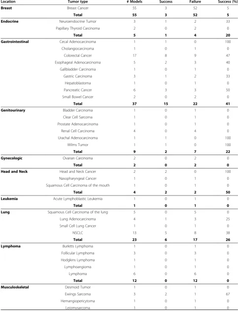
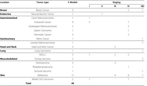
Forty-nine tumorgraft models, representing 18 distinct cancer pathologies have been developed by direct im-plantation of patient tumor tissue into immune-compromised nude mice giving the overall take rate of 27% (Table 1). These 49 models were distributed across eight different cancer location categories, based on the NCI Cancer by Body Location/System (Table 2). Tumor-graft models of the gastrointestinal tract and skin ac-count for ~50% of successfully established tumorgraft models. There were an additional three models that formed 1st generation tumorgrafts but that failed to re-establish tumorgrafts following cryopreservation of 1st generation tumorgraft fragments, giving a 94% success rate in tumorgraft model re-establishment following cryopreservation. No association was identified between tumorgraft development and the time between surgical resection and implantation (p = 0.86 by simple t-test on representative sample set, data not shown).
The take rate (the ability of donor tumor to success-fully be propagated in mice) was closely related to tumor stage at time of resection. Forty-three percent (35/82) of tumors with a pathological staging of Stage III and Stage IV tumors were successfully propagated in mice, accounting for 73% of fully developed models (Table 2). Only 10% (6/60) of the donor tumors from Stage I or II cancers successfully propagated formed tumorgrafts in mice, accounting for only 12% of the fully developed models. Pathological staging information for the remaining 22% of the tumor tissues implanted into mice (40/182) which formed tumorgrafts was not obtained. A contingency table analysis with stage III and IV as one condition and stage I and II the second condition
supported the hypothesis with a p-value = 1.2 E-5 and an odds ratio of 6.7 for this data set (data not shown).
Tumorgraft development was also found to be related to cancer type. Of the human tumor samples used to de-rive the panel of 49 tumorgraft models, a success rate of
>40% was observed for skin (65%), musculoskeletal (54%), head and neck cancers (50%), and gastrointestinal cancers (41%) (Table 1). Lung (26%), genitourinary (22%), and endocrine (20%) cancers exhibited lower take rates for tumorgraft formation. Cancers of the breast (5%), predominantly early stage (data not shown), and the lymphatic system (0%) proved the most challenging to form tumorgrafts using the athymic nude mouse strain and methodologies adopted here. No tumorgrafts were established from tumors of gynecologic, leukemic, or neurologic origins; however, these finding are not ne-cessarily representative of the true take rate for these tumor types as ≤2 attempts for each of these cancer types were attempted.
Hematoxylin and eosin stained sections from matching patient tumors and 1st and 2nd generation tumorgrafts from pancreatic cancer, breast cancer, lung cancer and cervical cancer models were examined for histological similarities. In all 4 different tumorgraft models, the ini-tial patient tumors were characterized by histological features consistent with the anatomical site of origin (Figure 1 A-D). The 1st generation tumor grafts resembled the original patient tumor, including in most instances retention of peritumoral stroma which became progressively replaced in the 2ndgeneration tumorgrafts by malignant cell overgrowth. In addition, the relative state of differentiation of the respective cancer type was retained following engraftment, including production of mucin and retention of atypical glandular structures for the adenocarcinomas, even after 302 and 177 days for the pancreatic and breast cancers, respectively. In the lung cancer and cervical cancer models, the 1stand 2nd generation tumors revealed viable malignant appearing cells. The 2nd generation tumorgrafts retained essential diagnostic features at 286 and 345 days, respectively, for the lung and cervical cancer models.
Table 1 Summary of Tumorgraft Development
Location Tumor type # Models Success Failure Success (%)
Breast Breast Cancer 55 3 52 5
Total 55 3 52 5
Endocrine Neuroendocrine Tumor 3 1 2 33
Papillary Thyroid Carcinoma 2 0 2 0
Total 5 1 4 20
Gastrointestinal Cecal Adenocarcinoma 1 1 0 100
Cholangiocarcinoma 1 0 1 0
Colorectal Cancer 17 8 9 47
Esophageal Adenocarcinoma 5 2 3 40
Gallbladder Carcinoma 1 0 1 0
Gastric Carcinoma 3 1 2 33
Hepatoblastoma 1 0 1 0
Pancreatic Cancer 6 3 3 50
Small Bowel Cancer 2 0 2 0
Total 37 15 22 41
Genitourinary Bladder Carcinoma 1 0 1 0
Clear Cell Sarcoma 1 0 1 0
Prostate Adenocarcinoma 1 0 1 0
Renal Cell Carcinoma 4 0 4 0
Urachal Adenocarcinoma 1 1 0 100
Wilms Tumor 1 1 0 100
Total 9 2 7 22
Gynecologic Ovarian Carcinoma 2 0 2 0
Total 2 0 2 0
Head and Neck Head and Neck Cancer 2 2 0 100
Nasopharyngeal Cancer 1 0 1 0
Squamous Cell Carcinoma of the mouth 1 0 1 0
Total 4 2 2 50
Leukemia Acute Lymphoblastic Leukemia 1 0 1 0
Total 1 0 1 0
Lung Squamous Cell Carcinoma of the lung 5 0 5 0
Lung Adenocarcinoma 4 1 3 25
Small Cell Lung Cancer 1 0 1 0
NSCLC 13 5 8 38
Total 23 6 17 26
Lymphoma Burkitts Lymphoma 1 0 1 0
Follicular Lymphoma 3 0 3 0
Hodgkins Lymphoma 1 0 1 0
Lymphoangioma 1 0 1 0
Lymphoma 6 0 6 0
Total 12 0 12 0
Musculoskeletal Desmoid Tumor 1 0 1 0
Ewings Sarcoma 3 2 1 67
Hemangiopericytoma 1 0 1 0
the patient tumor, but showed a single KRAS mutation in the tumorgraft. In the osteosarcoma model VARI-LTM-021, no mutations were identified in the patient tumor with any genetic drift identified in the 1st - 4th generation tumorgrafts (data not shown).
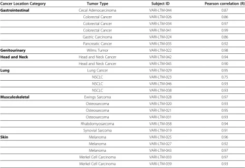
Paired analysis of the 24 tumorgraft/tumor pairs exhibited a high degree of similarity, with Pearson cor-relation coefficients ranging from r = 0.75 to 0.99 with a median coefficient of 0.93 (Table 4). There were only four pairs, three gastrointestinal and one lung, that had Pearson values r<0.90; the NSCLC model VARI-LTM-023 had the Pearson correlation coefficient of r = 0.75.
For this model, the originating patient tumor RNA had a RIN (RNA Integrity Number) of 4.9 implying degraded RNA, which was likely to be the primary factor driving the low correlation and would typically be excluded from the molecular analysis.
[image:7.595.57.546.101.243.2]Of the 28 genomes amplified by the Affymetrix method, 12 of the 14 tumorgraft/donor tumor pairs co-clustered in an unsupervised analysis (Figure 2A). Of the 20 genomes amplified by the NuGEN method, 7 of 10 tumorgraft/ tumor pairs coclustered (Figure 2B). 4 of the 5 tumorgraft/tumor pairs that did not co-cluster in an unsupervised fashion exhibited Pearson correlation
Table 1 Summary of Tumorgraft Development(Continued)
Osteosarcoma 3 3 0 100
Rhabdomyosarcoma 3 1 2 33
Synovial Sarcoma 1 1 0 100
Total 13 7 6 54
Neurologic Neuroblastoma 1 0 1 0
Total 1 0 1 0
Skin Melanoma 18 11 7 61
Merkel Cell Carcinoma 2 2 0 100
Total 20 13 7 65
Final Total 182 49 133 27
Table 2 Distribution of Tumorgraft Models across Cancer Location Categories and Staging
Location Tumor type # Models Staging
I II III IV ND
Breast Breast Cancer 3 2 1
Endocrine Neuroendocrine Tumor 1 1
Gastrointestinal Cecal Adenocarcinoma 1 1
Colorectal Cancer 9 2 7
Esophageal Adenocarcinoma 2 2
Gastric Carcinoma 1 1
Pancreatic Cancer 3 3
Genitourinary Wilms Tumor 1 1
Urachal Adenocarcinoma 1 1
Head and Neck Head and Neck Cancer 2 2
Lung Lung Carcinoma 1 1
NSCLC 5 1 2 2
Musculoskeletal Ewings Sarcoma 2 2
Osteosarcoma 3 3
Rhabdomyosarcoma 1 1
Synovial Sarcoma 1 1
Skin Melanoma 11 4 6 1
Merkel Cell Carcinoma 2 1 1
[image:7.595.56.548.445.734.2]coefficients r≤0.90 (Table 4). The tumorgraft and origin-ating patient tumor from the NSCLC model VARI-LTM-023 co-clustered despite a low Pearson correlation coefficient of r = 0.75, suggesting significant variance in respect to other tumors in the cohort.
[image:8.595.59.539.90.382.2]The genomic profiles of two representative osteosar-coma models VARI-LTM-020 and VARI-LTM-021 were observed over 4 generations to assess the potential for genomic drift over time in vivo. The Pearson correlation coefficients were very high, ranging from r = 0.98-0.99 Figure 1Histological resemblance between patient tumor and resulting tumorgraft following sequential grafting into
immunocompromised mice.Representative sections stained with hematoxylin and eosin of indicated tumor samples (Scale bars indicate 100μm).A) Adenocarcinoma of pancreas with desmoplastic stromal reaction in original patient tumor. 1stgeneration tumorgraft (total time
in vivo, 217 days) with preservation of atypical glandular structures with inspissated mucinous material surrounded by fibrous stroma. 2nd
generation tumorgraft (303 days) with similar histology.B) Adenocarcinoma of colon with numerous atypical glands containing mucinous material and necrotic debris with surrounding fibrovascular connective tissue. 1stgeneration tumorgraft after 101 days reveals dilated atypical
ducts with mucin and necrotic debris in lumens comprised of multiple columnar layers of atypical epithelium surrounded by fibrovascular stroma. 2ndgeneration graft after 177 days with similar histology.C) Lung carcinoma with prominent fibrovascular connective tissue in original patient
neoplasm that becomes progressively overgrown by malignant cells in 1st(216 days) and 2nd(286 days) generation tumor grafts.D) Cervical
carcinoma with fibromucinous stroma in original patient neoplasm, with expansion of large islands of malignant cells surrounded by fibrovascular stroma in 1st(282 days) and 2nd(345 days) generation tumorgrafts.
Table 3 Oncogene mutations detected in tumorgrafts and originating patient tumors
Model Tumor Type Gene Mutation Primary Tumor Mutation Status Tumorgraft Mutation Status
VARI-LTM-041 Colorectal Cancer KRAS G12V TRUE TRUE
VARI-LTM-041 Colorectal Cancer PIK3CA R88Q TRUE TRUE
VARI-LTM-041 Colorectal Cancer PIK3CA E545K TRUE TRUE
VARI-LTM-044 Cecal Adenocarcinoma KRAS G13D TRUE TRUE
VARI-LTM-026 Colorectal Cancer KRAS G12D FALSE TRUE
VARI-LTM-086 Melanoma BRAF V600E TRUE TRUE
VARI-LTM-027 Melanoma NRAS Q61R TRUE TRUE
[image:8.595.57.538.606.732.2]between all 4 generations in both models (data not shown). The largest decrease in Pearson correlation co-efficient occurred between the human tumor to 1st gen-eration mouse tumorgraft (VARI-LTM-020, r = 0.91; VARI-LTM-021, r = 0.94). Genomic fidelity was then maintained in subsequent mouse to mouse generation comparisons. This stability in the genomes across mouse tumorgraft generations, along with the absence of add-itional somatic oncogene mutations suggests the main-tenance of genomic stability during serial in vivo
passages and time in vivo for at least up to 186 days (VARI-LTM-021).
Paired analysis of all 24 tumorgrafts to their matching tumors identified 25,806 gene probes that were present in at least 50% of the 48 samples. 395 genes were decreased and 17 genes increased two-fold or greater in the tumorgrafts (Additional file 1: Table S1) compared to the patient tumors (Figure 3A, green and red circles respectively).
Of the 395 genes down-regulated in the murine tumorgrafts, there was a significant enrichment in pathways related to immune response (Figure 3B). En-richment scores were extremely high, with the
top-scored classical complement pathway showing a –log p-value of 25. Of the 17 genes up-regulated in the tumorgrafts, there were only 1–2 genes per map, sug-gesting no enrichment for any canonical pathway (data not shown). An independent GO biological pathway enrichment analysis was conducted using the down regulated probe list and 14 of the most signifi-cantly enriched top 100 pathways p<4.0E-9 were associated with immune response [23]. Classification of the down-regulated genes with regards to enrich-ment in protein function, identified a borderline sig-nificance (p = 0.05) in receptors and ligands in the tumorgrafts compared to the patient tumors. For the up-regulated genes, the only significant enrichment in protein function was for proteases, accounting for only two of the 17 genes.
[image:9.595.56.539.111.442.2]Comparative analysis of the patient tumors that formed tumorgrafts to patient tumors that did not form tumorgrafts identified 491 genes that were up-regulated and 691 genes down-regulated 2-fold or greater in those tumors that formed tumorgrafts compared to non-tumorgraft forming tumors (Additional file 2: Table S2). The up-regulated genes in the tumors that formed
Table 4 Pearson correlation coefficient of each individual tumorgraft and originating patient tumor based on Affymetrix gene expression data
Cancer Location Category Tumor Type Subject ID Pearson correlation (R)
Gastrointestinal Cecal Adenocarcinoma VARI-LTM-044 0.87
Colorectal Cancer VARI-LTM-026 0.86
Colorectal Cancer VARI-LTM-034 0.97
Colorectal Cancer VARI-LTM-041 0.99
Gastric Carcinoma VARI-LTM-024 0.86
Pancreatic Cancer VARI-LTM-035 0.92
Genitourinary Wilms Tumor VARI-LTM-022 0.98
Head and Neck Head and Neck Cancer VARI-LTM-042 0.94
Head and Neck Cancer VARI-LTM-045 0.90
Lung Lung Cancer VARI-LTM-029 0.95
NSCLC VARI-LTM-023 0.75
NSCLC VARI-LTM-046 0.93
NSCLC VARI-LTM-038 0.93
Musculoskeletal Ewings Sarcoma VARI-LTM-028 0.97
Osteosarcoma VARI-LTM-020 0.93
Osteosarcoma VARI-LTM-021 0.95
Osteosarcoma VARI-LTM-031 0.93
Rhabdomyosarcoma VARI-LTM-058 0.94
Synovial Sarcoma VARI-LTM-019 0.91
Skin Melanoma VARI-LTM-025 0.96
Melanoma VARI-LTM-027 0.92
Melanoma VARI-LTM-043 0.97
Merkel Cell Carcinoma VARI-LTM-033 0.97
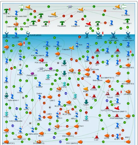
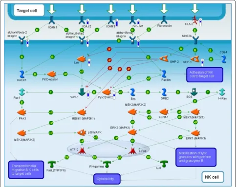
tumorgrafts were enriched for cell cycle, cell signaling pathways and cytoskeleton remodeling (Figure 4A). One key canonical pathway map, enriched with up-regulated genes that were elevated in those tumors that formed tumorgrafts, is for WNT signaling (Figure 5); genes up-regulated in this pathway map included VEGF-A, Frizzled, β-Catenin, c-Myc, and FAK-1. The down-regulated genes in the tumorgraft-forming tumors were enriched for immune response pathways (8/10 pathways, Figure 4B). One key pathway map enriched with
down-regulated genes is that of the role of integrins in NK cell cytotoxicity (Figure 6); note that in the context of this study, the upper Target Cell in this pathway corresponds to the relevant tumorgraft-forming cells.
Discussion
[image:10.595.57.542.89.544.2]The goal of this study was to create a panel of tumor-graft models, developed directly from patient tumor tis-sue from a wide range of heterogeneous tumor types of various stages. Prior investigations have focused on Figure 2Unsupervised cluster analysis of genomes of tumorgrafts and originating patient tumors.Unsupervised clustering was
specific cancers, e.g. Secondary liver cancer [15] lung [24], pancreatic cancer [16], pediatric osteosarcomas [14], or pediatric rhabdomyosarcoma [25], rather than a spectrum of malignancies. To examine the suitability of the panel of tumorgraft models generated in our lab as molecularly-relevant preclinical models, we directly compared a number of phenotypic and genotypic fea-tures of the tumorgrafts to the originating patient tumors from which they were derived.
[image:11.595.59.539.87.551.2]We have fully developed 49 models, spanning 18 dif-ferent cancer types. A critical component of our devel-opment process is the ability of the model to re-establish following cryopreservation, alleviating the need for con-tinual propagation, reported by some investigators [16]. Cryopreservation significantly reduces the cost and ne-cessary resources, extending the life of the tumor model, and minimizing genetic drift that could occur following long-term continuousin vivopropagation.
Our overall tumor take rate is similar to previous reports [3,5]. A number of strategies have been tested to improve tumorgraft take rates, including the severe combined immunodeficient mouse strains CB17SC-M-F
scid−/− [9,25,26] and CB17/Icr scid [12], suppression of the immune system of recipient CBA/CaJ mice by thym-ectomy, whole body irradiation, administration of 1-β -D-arabinofuranosylcytosine [14,25], and use of orthotopic
models [27]. However, these strategies could increase the cost of model development with minimal improvement in tumorgraft development over the use of subcutaneous implantation into naive athymic nu/nu mice reported in this study and by other investigators [2,16].
[image:12.595.59.538.90.570.2]It should be noted use of the athymic nu/nu mouse for use in the development of such tumorgraft does have limitations. The use of more immunodeficient mouse Figure 4Enrichment of canonical pathway maps in genes up and downregulated in tumorgraft forming tumors compared to
strains, e.g. NOD/scid or NOD/scid gamma (NSG), are necessary for successful engraftment of certain tumor types e.g. leukemia [28,29], may better resemble the human microenvironment, and could provide better consistency in growth rates following implantation of human tumor tissue into mice. However, in the context of developing a panel of tumorgraft models across a
wide range of tumor types, the athymic nu/nu mouse has proven a robust model.
[image:13.595.56.541.86.596.2]with Stage III or IV cancer at time of tissue acquisition which was successfully propagated in the murine host. Overall, ~75% of the 49 tumors that formed tumorgrafts were from patients that were diagnosed either Stage III or IV.
In this study, breast cancers had a take rate of only 6% following subcutaneous implantation into mice. Dobro-lecki et al [27] reported success in developing breast cancer models by orthotopically implanting the breast cancer tissue into the mammary fat pads of mice. While more labor intensive than the subcutaneous model, an orthotopic approach should be considered if there is need for a tumorgraft model of a specific cancer that cannot be established using the subcutaneous model.
An important conclusion from this study is that these subcutaneous (predominantly ectopic with the exception of melanomas) tumorgraft models recapitulate the
expression profiles of the original patient’s tumor within its natural site of site of metastatic spread. This provides some evidence that refutes the lack of relevance of the subcutaneous model for drug discovery and develop-ment, which may rather reflect the use of genetically drifted in vitro cell lines in the classical xenograft con-text. The primary value for any tumor model is to recap-itulate the in-vivo malignant tissue state, and accurately predict the efficacy of therapeutic agents in a timely and cost-effective manner. In contrast with commonly used animal models in which established human cancer cell lines are injected subcutaneously and fail to accurately predict human clinical trial results [8], this human tumorgraft model system is more likely to be a success-ful surrogate preclinical model for several reasons.
[image:14.595.56.542.90.474.2]First, tumor bearing tissue fragments are transplanted containing not only malignant cells, but also supporting Figure 6Immune response pathway map enriched with genes down-regulated in the tumorgraft forming patient tumors.The
stromal tissue in an anatomically correct hierarchy. Second, the transferred tumors include cells post en-graftment retaining their essential cell to cell interac-tions and relative state of differentiation as assessed by their histological appearance. Third, because of the high success rate in establishing not only 1st, but also subsequent generation tumorgrafts, there is expansion of the number of tumorgrafts available for testing and the ability to verify retention of histological hallmarks in a serial fashion. Fourth, assessment of tumor growth is relatively straightforward given the subcutaneous location of the xenografts. Finally, since portions of tumors can be cryopreserved and successfully engrafted post thawing, testing of therapeutic agents, or combination therapies not originally considered, can be studied with this experimental and scalable in vivo
model system.
In the era of molecularly targeted oncology agents, the effective translation of results in the mouse model to the clinic requires the genome of the tumorgraft model to be high comparable to the original tumor [30]. Signifi-cant changes in DNA fidelity or RNA expression levels significantly diminish the translational value of a model. Entire genome characterization of RNA expression ra-ther, than a focused RT-PCR analysis of a select cohort of genes [16], allows for the assessment of transcrip-tome wide alterations and associated biological systems that occur during the growth of the transplanted human tumors.
The high Pearson correlations seen between the gen-omes of the 24 pairs of tumorgraft and originating pa-tient tumors implies that the donor tumor genome is largely maintained in the tumorgraft model. Similarly, genotypic fidelity has also recently been reported in a panel of 25 human breast cancer tumorgrafts [27] and on a smaller scale in secondary liver cancers using quantitative PCR analysis of 21 genes related to onco-genesis and cell cycle [15]. The lower Pearson correla-tions observed in the gastrointestinal cancers could be an artifact of the tissue harvest and the high levels of digestive enzymes present in the patient tumors within the gastrointestinal tract. High levels of RNases present in pancreatic cancer are reported to complicate the ex-traction of high quality RNA [31]. This finding high-lights the importance of an optimized, rapid tissue collection protocol, especially for genomic profiling. It should be noted that poor RNA quality, for genomic profiling does not necessarily impact the ability of the tissue to form a viable tumorgraft. The high degree of clustering by matched tumor/tumorgraft pairs (79%) supports the high degree of similarity of the gen-omes of the patient tumor and resulting mouse tumorgraft even when the opportunity for RNA deg-radation may occur.
Genomic stability is an important characteristic to consider prior to the long term use of a tumorgraft model. The high correlation in gene expression across four tumorgraft generations observed in this study and similar findings of others [3,16,27,30] and absence of drift in somatic mutations in known oncogenes, suggest that the genomes of tumorgraft models are stable. Histo-logical integrity of patient-derived tumorgrafts has also been demonstrated for up to ten generations in immune-compromised mice [4] and up to 30 passages in mice without significant changes in growth and mor-phological characteristics [15]. One study reported tumorgraft models cultured in vivo for 10–12 genera-tions before regenerating the models from earlier cryo-preserved generations [3].
The role of somatic oncogene mutations in tumorigen-esis, pathogenesis and disease progression can have a profound influence on therapy [32]; mutations in onco-genes RAS [33] and B-RAF [34] are commonly found in a variety of cancers. Therefore it is imperative that such mutations are maintained in the tumorgraft models. Al-though only a few oncogenic mutations were observed, those identified in the patient tumors were conserved in the matching tumorgraft, consistent with other reports [16] and consistent with known mutations in specific tumor types [20,21].
There are a number of possible explanations for the enrichment of canonical immune pathways within those genes down-regulated in the tumorgrafts compared to patient tumors. First, a loss of tumor cell immune path-ways and proteins not necessary for growth and develop-ment when transplanted from the immunecompetent patient into the immunocompromised host. Second, there is an intentional tumor tissue response to trans-plantation to down regulate those genes responsible for its immunogenic signature presented to the vestigial im-mune defenses of the immunocompromised mouse. A third explanation, and most likely, is that the gene ex-pression changes are due to a loss of circulating human immune cells which infiltrate and support tumor de-velopment, when the tumor was transplanted from the human into the mouse as earlier reported by Neale et al [17].
endometrium transplanted into nude mice was corre-lated with a high VEGF content. Transgenic expression of VEGF into human islet cells followed by transplant into the livers increased the rate of tissue revasculariza-tion, survival and function [39]. A recent study demon-strated the pre-treatment of human ovarian tissue with VEGF and vitamin E prior to implantation into the back muscle of immunodeficient mice demonstrated enhanced growth, compared no pre-treatment of the ovarian tissue [40].
The downregulation of immunological pathways in the tumors that formed tumors would suggest that loss of immune pathways is a mark of late stage tumors and provides a selective advantage supporting the establish-ment of the tumors in a foreign host or location. This downregulation of immunological pathways could be a reflection of the loss of expression signature of patient immune infiltrating cells [17]. Of particular note is the downregulation of natural killer (NK) cell ligands, as identified as a pathway enriched for genes down-regulated in tumors that formed tumorgrafts compared to those tumors that failed to form tumorgrafts (Fig-ure 4B). Although athymic nu/numice cannot generate mature T lymphocytes, they maintain the ability to mount a response to T-independent antigens [41] and do produce NK cells as a component of their vestigial immune system. The downregulation of the NK ligands may therefore promote human tumor development in immune compromised mice. While there is little evi-dence demonstrating the interaction of human integrins and murine NK receptors, Kanwar et al reports interspe-cies cross-talk via the demonstration that the ectopic ex-pression of human intercellular cell adhesion molecule-1 (ICAM-1) in mouse EL-4 tumors inhibitedin vivotumor growth [42]. The mechanisms of NK cell recognition of self and immunosurveillance in both mouse and humans are complex and involve numerous receptors that recognize many cell surface ligands [43]. Experimental evidence clearly supports the view that murine NK cells can inhibit human tumor cell development and metasta-sis bothin vitroandin vivo[44-50].
This study reports the development of a series of tumorgraft models, representing a broad spectrum of cancer types, derived from molecularly characterized fresh patient tumor tissue. The genomes of these tumorgraft models exhibit a high degree of correlation to their originating patient tumors which is maintained over multiple generations of in vivo passages. The loss of immune function pathways and gain of cell prolif-eration pathways suggests a logical selective pressure for certain tumors to form tumorgrafts, which could be prescreened in future studies to identify those most likely to develop into useful preclinical models for translational studies.
Conclusions
The maintenance of the genome in the tumorgraft, from the human to murine host, supports the concept that tumorgraft models can provide an invaluable and robust tool in the evaluation of precision medicine methodolo-gies and novel treatment stratemethodolo-gies prior to use in clin-ical trials. An important point is that a subcutaneous primary patient tumorgraft models can recapitulate the expression profiles of the original“orthotopic”disease in the human and allow for reasonably high-throughput preclinical screens for drug efficacy. These models may therefore be useful in reducing the rate of attrition in drug development that has been in part been attributed to the use of the classical cell line xenografts.
Additional files
Additional file 1:Table S1. The spreadsheet contains the results of the differential gene expression analysis comparing each patient’s tumor with the paired tumorgraft.This file contains two spreadsheet tabs. The first tab displays the 17 gene probesets up-regulated in tumorgrafts (Group 2, column C) relative to the originating patient tumors (Group 1, column B), and the second tab displays the 395 gene probes down-regulated. The official gene name, mean normalized intensity in Groups 1 and 2, the associated fold-change in gene expression (Group 2/Group 1), the calculated pairwise t-test statistic, the corresponding p-value, the False Discovery Rate, and the Affymetrix probeset identifiers are shown.
Additional file 2:Table S2. The spreadsheet contains the results of the differential gene expression analysis comparing the patient tumors that successfully developed tumorgrafts with those that did not.This file contains two spreadsheet tabs. The first sheet displays the 491 gene probesets up regulated in the tumors that successfully formed tumorgrafts (Group 2, column C) relative to those that did not (Group 1, column B) and the second sheet displays the 691 gene probes down regulated. The official gene name, mean normalized intensity in Groups 1 and 2, the associated fold-change in gene expression (Group 2/Group 1), the calculated student’s t-test statistic, the corresponding p-value, the False Discovery Rate, and the Affymetrix probeset identifiers are shown.
Competing interests
CPW has disclosed a potential conflict of interest in relation to his invention of the XB-BIS informatics system used to analyze some of the data presented in this manuscript. He is also a consultant for the company TransMed Systems who acquired licensing rights to XB-BIS. Some third parties have acquired some of the described tumorgraft models for commercial projects, but these remain available for academic (noncommercial) purposes.
Authors’contributions
DJM supervised the animal work, analyzed the data, interpreted the results, and wrote the manuscript. NRM interpreted the results and revised the manuscript. DMC, PJR, AI, and YN analyzed the data. DD and SBS carried out the animal work. EE, HF, SS, and REE carried out the laboratory experiments. JR, RS, and BN analyzed the histopathological images. CPW defined the research project, interpreted the results, and revised the manuscript. All authors read and approved the final manuscript.
Acknowledgements
Wallraven (Gradalis, Inc, Carrollton, TX) for their clinical contributions to this study.
Author details
1Laboratory for Translational Medicine, Van Andel Research Institute, Grand Rapids, MI, USA.2Sequenom Inc, San Diego, CA, USA.3GeneGo-Thomson Reuters, St. Joseph, MI, USA.4Program in Biospecimen Science, Van Andel Research Institute, Grand Rapids, MI, USA.5Research Essential Services, LLC, Plymouth, MI, USA.6Laboratory of Cutaneous Oncology, Van Andel Research Institute, Grand Rapids, MI, USA.
Received: 14 March 2012 Accepted: 18 June 2012 Published: 18 June 2012
References
1. Cobb LM:The behaviour of carcinoma of the large bowel in man following transplantation into immune-deprived mice.Br J Cancer1973,
28:400–411.
2. Fiebig HH, Schuchhardt C, Henss H, Fiedler L, Lohr GW:Comparison of tumor response in nude mice and in the patients.Behring Inst Mitt1984,
74:343–352.
3. Garber K:From human to mouse and back: 'tumorgraft' models surge in popularity.J Natl Cancer Inst2009,101:6–8.
4. Houghton JA, Taylor DM:Maintenance of biological and biochemical characteristics of human colorectal tumours during serial passage in immune-deprived mice.Br J Cancer1978,37:199–212.
5. Fiebig HH, Widmer K-H, Fiedler L, WIttekind C, Lohr GW:Development and Characterization of 51 Human Tumor Models for Large Bowel, Stomach and Esophageal Cancers.Dig Surg1984,1:225–235.
6. Johnson JI, Decker S, Zaharevitz D, Rubinstein LV, Venditti JM, Schepartz S, Kalyandrug S, Christian M, Arbuck S, Hollingshead M, Sausville EA:
Relationships between drug activity in NCI preclinical in vitro and in vivo models and early clinical trials.Br J Cancer2001,84:1424–1431.
7. Voskoglou-Nomikos T, Pater JL, Seymour L:Clinical predictive value of the in vitro cell line, human xenograft, and mouse allograft preclinical cancer models.Clin Cancer Res2003,9:4227–4239.
8. Ellis LM, Fidler IJ:Finding the tumor copycat. Therapy fails, patients don't. Nat Med2010,16:974–975.
9. Morton CL, Houghton PJ:Establishment of human tumor xenografts in immunodeficient mice.Nat Protoc2007,2:247–250.
10. Boven E, Winograd B, Berger DP, Dumont MP, Braakhuis BJ, Fodstad O, Langdon S, Fiebig HH:Phase II preclinical drug screening in human tumor xenografts: a first European multicenter collaborative study. Cancer Res1992,52:5940–5947.
11. Hidalgo M, Bruckheimer E, Rajeshkumar NV, Garrido-Laguna I, De Oliveira E, Rubio-Viqueira B, Strawn S, Wick MJ, Martell J, Sidransky D:A Pilot Clinical Study of Treatment Guided by Personalized Tumorgrafts in Patients with Advanced Cancer.Mol Cancer Ther2011,8:1311–1316.
12. Peterson JK, Tucker C, Favours E, Cheshire PJ, Creech J, Billups CA, Smykla R, Lee FY, Houghton PJ:In vivo evaluation of ixabepilone (BMS247550), a novel epothilone B derivative, against pediatric cancer models.Clin Cancer Res2005,11:6950–6958.
13. Houghton PJ, Morton CL, Tucker C, Payne D, Favours E, Cole C, Gorlick R, Kolb EA, Zhang W, Lock R,et al:The pediatric preclinical testing program: description of models and early testing results.Pediatr Blood Cancer2007,
49:928–940.
14. Meyer WH, Houghton JA, Houghton PJ, Webber BL, Douglass EC, Look AT:
Development and characterization of pediatric osteosarcoma xenografts. Cancer Res1990,50:2781–2785.
15. Mischek D, Steinborn R, Petznek H, Bichler C, Zatloukal K, Sturzl M, Gunzburg WH, Hohenadl C:Molecularly characterised xenograft tumour mouse models: valuable tools for evaluation of new therapeutic strategies for secondary liver cancers.J Biomed Biotechnol2009,
2009:437284.
16. Rubio-Viqueira B, Jimeno A, Cusatis G, Zhang X, Iacobuzio-Donahue C, Karikari C, Shi C, Danenberg K, Danenberg PV, Kuramochi H,et al:An in vivo platform for translational drug development in pancreatic cancer. Clin Cancer Res2006,12:4652–4661.
17. Neale G, Su X, Morton CL, Phelps D, Gorlick R, Lock RB, Reynolds CP, Maris JM, Friedman HS, Dome J,et al:Molecular characterization of the pediatric preclinical testing panel.Clin Cancer Res2008,14:4572–4583.
18. Whiteford CC, Bilke S, Greer BT, Chen Q, Braunschweig TA, Cenacchi N, Wei JS, Smith MA, Houghton P, Morton C,et al:Credentialing preclinical pediatric xenograft models using gene expression and tissue microarray analysis.Cancer Res2007,67:32–40.
19. Jimeno A, Feldmann G, Suarez-Gauthier A, Rasheed Z, Solomon A, Zou GM, Rubio-Viqueira B, Garcia-Garcia E, Lopez-Rios F, Matsui W,et al:A direct pancreatic cancer xenograft model as a platform for cancer stem cell therapeutic development.Mol Cancer Ther2009,8:310–314.
20. Pearce M, Culinan A, Hogg G, Hosseni D, Ehrich M:Mutation profiling in tumor samples using the Sequenom OncoCarta™Panel.Nature Methods
2009,6:vii–viii.
21. Thomas RK, Baker AC, Debiasi RM, Winckler W, Laframboise T, Lin WM, Wang M, Feng W, Zander T, MacConaill L,et al:High-throughput oncogene mutation profiling in human cancer.Nat Genet2007,39:347–351. 22. Nikolsky Y, Kirillov E, Zuev R, Rakhmatulin E, Nikolskaya T:Functional
analysis of OMICs data and small molecule compounds in an integrated "knowledge-based" platform.Methods Mol Biol2009,563:177–196. 23. Zheng Q, Wang XJ:GOEAST: a web-based software toolkit for Gene
Ontology enrichment analysis.Nucleic Acids Res2008,36:W358–W363. 24. Fiebig HH, Neumann HA, Henss H, Koch H, Kaiser D, Arnold H:
Development of three human small cell lung cancer models in nude mice.Recent Results Cancer Res1985,97:77–86.
25. Houghton JA, Williams LG, Torrance PM, Houghton PJ:Determinants of intrinsic sensitivity to Vinca alkaloids in xenografts of pediatric rhabdomyosarcomas.Cancer Res1984,44:582–590.
26. Thompson J, George EO, Poquette CA, Cheshire PJ, Richmond LB, de Graaf SS, Ma M, Stewart CF, Houghton PJ:Synergy of topotecan in combination with vincristine for treatment of pediatric solid tumor xenografts.Clin Cancer Res1999,5:3617–3631.
27. Dobrolecki LE, Landis MD, Zhang X, Huang J, Lai Q, Wong H, Contreras A, Vahdat LD, Lewis MT CCJ:Preclinical utility of xenografted human breast cancer tumor models. InKeystone Symposia-Stem Cells, Cancer and Metastasis; March 6–11, 2011. Silverthorne, CO: Keystone, CO; 2011. 28. Zorzoli A, Di Carlo E, Cocco C, Ognio E, Ribatti D, Ferretti E, Dufour C,
Locatelli F, Montagna D, Airoldi I:Interleukin-27 inhibits the growth of pediatric acute myeloid leukemia in NOD/SCID/Il2rg−/−mice.Clin Cancer Res2012,18:1630–1640.
29. Malaise M, Neumeier M, Botteron C, Dohner K, Reinhardt D, Schlegelberger B, Gohring G, Gruhn B, Debatin KM, Corbacioglu S:Stable and reproducible engraftment of primary adult and pediatric acute myeloid leukemia in NSG mice.Leukemia2011,25:1635–1639.
30. Garber K:Personal mouse colonies give hope for pancreatic cancer patients.J Natl Cancer Inst2007,99:105–107.
31. Vilardell F, Iacobuzio-Donahue CA:Cancer gene profiling in pancreatic cancer.Methods Mol Biol2010,576:279–292.
32. Harris TJ, McCormick F:The molecular pathology of cancer.Nat Rev Clin Oncol2010,7:251–265.
33. Irahara N, Baba Y, Nosho K, Shima K, Yan L, Dias-Santagata D, Iafrate AJ, Fuchs CS, Haigis KM, Ogino S:NRAS mutations are rare in colorectal cancer.Diagn Mol Pathol2010,19:157–163.
34. Shepherd C, Puzanov I, Sosman JA:B-RAF inhibitors: an evolving role in the therapy of malignant melanoma.Curr Oncol Rep2010,12:146–152. 35. Hu T, Li C:Convergence between Wnt-beta-catenin and EGFR signaling
in cancer.Mol Cancer2010,9:236.
36. Ott PA, Adams S:Small-molecule protein kinase inhibitors and their effects on the immune system: implications for cancer treatment. Immunotherapy2011,3:213–227.
37. Singh RR, Cho-Vega JH, Davuluri Y, Ma S, Kasbidi F, Milito C, Lennon PA, Drakos E, Medeiros LJ, Luthra R, Vega F:Sonic hedgehog signaling pathway is activated in ALK-positive anaplastic large cell lymphoma. Cancer Res2009,69:2550–2558.
38. Nisolle M, Casanas-Roux F, Marbaix E, Jadoul P, Donnez J:Transplantation of cultured explants of human endometrium into nude mice.Hum Reprod2000,15:572–577.
39. Shimoda M, Chen S, Noguchi H, Matsumoto S, Grayburn PA:In vivo non-viral gene delivery of human vascular endothelial growth factor improves revascularisation and restoration of euglycaemia after human islet transplantation into mouse liver.Diabetologia2010,
53:1669–1679.
41. Pelleitier M, Montplaisir S:The nude mouse: a model of deficient T-cell function.Methods Achiev Exp Pathol1975,7:149–166.
42. Kanwar JR, Berg RW, Yang Y, Kanwar RK, Ching LM, Sun X, Krissansen GW:
Requirements for ICAM-1 immunogene therapy of lymphoma.Cancer Gene Ther2003,10:468–476.
43. Stewart CA, Vivier E:Strategies of Natural Killer (NK) Cell Recognition and Their Roles in Tumor Immunosurveillance. InHow the Immune System Recognizes Self and Nonself Volume. Edited by Kitamura D. New York: Springer; 2008:37–81.
44. Ohyama C, Kanto S, Kato K, Nakano O, Arai Y, Kato T, Chen S, Fukuda MN, Fukuda M:Natural killer cells attack tumor cells expressing high levels of sialyl Lewis x oligosaccharides.Proc Natl Acad Sci U S A2002,
99:13789–13794.
45. Ma D, Luyten GP, Luider TM, Niederkorn JY:Relationship between natural killer cell susceptibility and metastasis of human uveal melanoma cells in a murine model.Invest Ophthalmol Vis Sci1995,36:435–441.
46. Hansson M, Bakacs KK, Kiessling R, Klein G:Intra- and interspecies reactivity of human and mouse natural killer (NK) cells.J Immunol1978,121:6–12. 47. Nieswandt B, Hafner M, Echtenacher B, Mannel DN:Lysis of tumor cells by
natural killer cells in mice is impeded by platelets.Cancer Res1999,
59:1295–1300.
48. Welsh RM Jr:Mouse natural killer cells: induction specificity, and function.J Immunol1978,121:1631–1635.
49. Shouval D, Rager-Zisman B, Quan P, Shafritz DA, Bloom BR, Reid LM:Role in nude mice of interferon and natural killer cells in inhibiting the tumorigenicity of human hepatocellular carcinoma cells infected with hepatitis B virus.J Clin Invest1983,72:707–717.
50. Carroll JL, Nielsen LL, Pruett SB, Mathis JM:The role of natural killer cells in adenovirus-mediated p53 gene therapy.Mol Cancer Ther2001,1:49–60.
doi:10.1186/1479-5876-10-125
Cite this article as:Monsmaet al.:Genomic characterization of explant tumorgraft models derived from fresh patient tumor tissue.Journal of Translational Medicine201210:125.
Submit your next manuscript to BioMed Central and take full advantage of:
• Convenient online submission
• Thorough peer review
• No space constraints or color figure charges
• Immediate publication on acceptance
• Inclusion in PubMed, CAS, Scopus and Google Scholar
• Research which is freely available for redistribution