R E V I E W
Open Access
When mites attack: domestic mites are not just
allergens
Yubao Cui
Abstract
Domestic mite species found in indoor environments and in warm or tropical regions are well known for causing allergic disorders. However, little is known about human acariasis, in which mites invade and parasitize the human body in various tissues from the gastrointestinal tract to the lung. Here, we summarize the reported cases of human acariasis of pulmonary, intestinal, oral (anaphylaxis), urinary, otic, and vaginal systems. Because the clinical symptoms of acariasis often overlap with other disease symptoms leading to frequent misdiagnosis, we highlight the need for more attention on these infections.
Keywords:Human acariasis, Pulmonary acariasis, Intestinal acariasis, Urinary acariasis, Oral mite anaphylaxis (Pancake Syndrome), Otoacariasis, Vaginal acariasis
Background
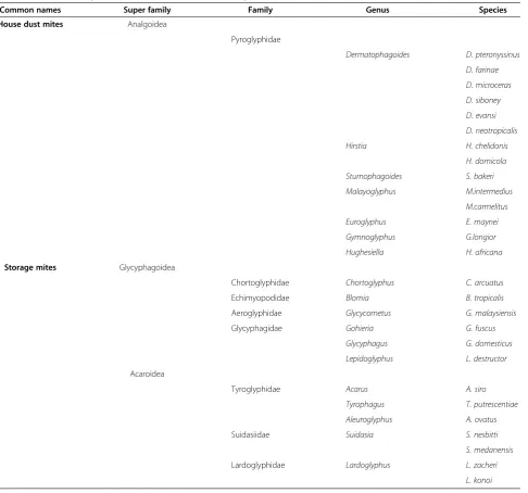
“Domestic mites” refers to a variety of small (0.5 mm) arthropods found living in close proximity to humans (Table 1). They include house dust mites and storage mites. Typically belonging to the family Pyroglyphidae (Analgoidea, Astigmata, Acariformes), the house dust mites are commonly found in human homes. Some other mites can be present in house dust, especially several species from the families Glycyphagidae, Aeroglyphidae Chortoglyphi-dae, EchimyopodiChortoglyphi-dae, TyroglyphiChortoglyphi-dae, LardoglyphiChortoglyphi-dae, and Suidasiidae, as well as species of the families Tarsonemidae and Cheyletidae. The latter are typically regarded as storage mites because they occur widely in-and contribute to the deterioration in quality of—stored products like grains, foodstuffs, and Chinese medicinal herbs [1,2]. They are also found in mattresses, pillows, and upholstery. Both the pyroglyphid mites and other mite species that are found in house dust can provoke an IgE antibody response in people world-wide [3-16]. These mites have been world-widely studied because their feces, eggs, and extracts from their bod-ies are potent allergens. Indeed, approximately 10% of the total population and 90% of asthmatic patients are allergic to domestic mites [17].
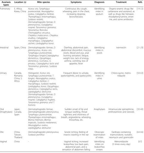
Although domestic mites are well known allergens, they are also responsible for other, non-allergic, symp-toms in humans, called acariasis. Little is known about acariasis, in which mites invade and parasitize the hu-man body in various tissues from the gastrointestinal tract to the lung. Mites, throughout their lives or during individual life cycle stages, commonly parasitize animals, and some free-living mites can invade a host only occa-sionally. When domestic mites invade a human body, it is believed to be a non-specific invasion differing from parasitization of an animal, because the non-specific in-vasion occurs only occasionally and lacks specific symp-toms and are not caused by specific mite species; in contrast, specific invasions are caused by specific para-site species occurring in specific hosts and producing specific symptoms [18]. To date, few reports of human acariasis have been published in English medical journals. However, acariasis has been described for pulmonary, intestinal, oral (anaphylaxis), urinary, otic, and vaginal systems. Importantly, the clinical symptoms of acaria-sis are non-specific and often overlap with other dis-ease symptoms, leading to frequent misdiagnosis and, likely, many missed cases. In this review, we present the types of acariasis that have been described, and summarize the current knowledge about these little known and little-understood infestations (Table 2).
Correspondence:[email protected]
Department of Laboratory Medicine, Yancheng Health Vocational & Technical College, Jiefangnan Road 263, Yancheng, Jiangsu Province 224006, P.R. China
Review
Pulmonary acariasis
Pulmonary acariasis is a non-specific infestation of hu-man lungs by free-living mites. In the 1930s, mites were observed in human sputum [4]. Subsequent experiments demonstrated that free-living mites can invade animal lungs and live in the respiratory tract. Indeed, Carter et al.[19] detected mites in sputum from 60.71% (17/28) of asthmatic patients. Interestingly, the authors thought the mites were derived from contamination of the test vessel because the detected mite species were present in dust samples from the same hospital and wards. After ruling out possible contamination, the authors repeat-edly tested the sputum samples and obtained the same
results. Among those 17 patients, one had severe asthma and had mites in his lungs for more than 7 months. Blood examination showed increased eosinophil num-bers in all 17 patients. After treating patients with ar-senic, the numbers of mites in their sputum samples increased, which showed that mites in the lungs were driven out, before the numbers of mites decreased and patients’symptoms resolved [19].
[image:2.595.59.546.98.552.2]Since that initial report, a number of other cases of pulmonary acariasis have been documented. In 1947, mites were detected in sputum of 3 out of 28 patients with tropical eosinophilia in East Africa [20]. In China, Gao et al.[21] reported for the first time that Tyrophagusand Tarsonemus were found in sputum of a bronchiectasis
Table 1 The taxonomy of domestic mites and common species
Common names Super family Family Genus Species House dust mites Analgoidea
Pyroglyphidae
Dermatophagoides D. pteronyssinus
D. farinae
D. microceras
D. siboney
D. evansi
D. neotropicalis
Hirstia H. chelidonis
H. domicola
Sturnophagoides S. bakeri
Malayoglyphus M.intermedius
M.carmelitus
Euroglyphus E. maynei
Gymnoglyphus G.longior
Hughesiella H. africana
Storage mites Glycyphagoidea
Chortoglyphidae Chortoglyphus C. arcuatus
Echimyopodidae Blomia B. tropicalis
Aeroglyphidae Glycycometus G. malaysiensis
Glycyphagidae Gohieria G. fuscus
Glycyphagus G. domesticus
Lepidoglyphus L. destructor
Acaroidea
Tyroglyphidae Acarus A. siro
Tyrophagus T. putrescentiae
Aleuroglyphus A. ovatus
Suidasiidae Suidasia S. nesbitti
S. medanensis
Lardoglyphidae Lardoglyphus L. zacheri
patient [21]. Ryu et al. [22] reported that a 23-year-old medical student showed a positive reaction on a skin test forParagonimus westermani, and twoTarsonemus florico-lusmites were subsequently found by sputum examination and identified morphologically. This was the first human infection with Tarsonemus reported in Korea. Further, in 2007 a new process for identifying mites in sputum was de-veloped. Martínez-Girón R et al. [23] demonstrated that dust mites artificially introduced into sputa could be
identified after sputa were liquified with bleach and the li-quid sample was observed under the microscope. Their ap-proach offers a time- and cost-saving tool for identifying dust mites in sputum, but the test is not commonly applied in the clinic because acariasis is not well recognized.
[image:3.595.57.541.100.616.2]Van Woerden [24] proposed that asthma in house dust mite-sensitive patients may be caused by recurrent in-halation of live dust mites that are able to live for some time in the bronchioles of the lung. The mites may
Table 2 A simple summary of reported cases of human acariasis
Acariasis types
Location (s) Mite species Symptoms Diagnosis Treatment Refs
Pulmonary E. Africa, Korea, China
Acarus siro, Tyrophagus putrescentiae, Aeuroglyphus ovatus, Caloglyphus berlesei, Thyreophagus entomophagus, Suidasia nesbitti,
Dermatophagoides farinae, D. pteronyssnius, Euroglyphus maynei, Tarsonemus granarius, Tarsonemus floricolus Chortoglyphus arcuatus, Cheyletus malaccensis. Cheyletus eruditus, Caloglyphus
mycophagus, Largoglyphus zacheri, Lardoglyphus konoi.
Continuous dry cough, wheezing, pain in the chest,
increasing dyspnea, bronchiectasis
Identifying mites in sputum
Organo-arsenic drugs like carbarsone and acetarsol, as
well as drugs like hetrazan, thiodiphenyl-amine, emet-ine, and some antibiotics
[19-22]
Intestinal Spain, China Dermatophagoides farinae,D. pteronyssinus,Acarus siro, Tyrophagus putrescentiae, Tyrophagus longior,Carpoglyphus domesticus,Glycyphagus domesticus, G.ornatus, G. privatus,Carpoglyphus lactisand Tarsonemus granarius, Suidasia mites
Diarrhea, abdominal pain, abdominal discomfort, mucous
stools, blood and pus, anal burning sensation, fatigue, weight loss, lack of energy, asthma, vomiting, loss of
appetite, fever
Identifying mites in
stools
ivermectin [21-29]
Urinary Canada,
Romania China, S. Africa
Histiogaster, Acarus siro, Tyrophagus putrescentiae, T. longior, Aleuroglyphus ovatus, Caloglyphus berlesei, C. mycophagus, Suidasia nesbitti, Lardoglyphus konoi, Glycyphagus domesticus, Carpoglyphus lactis, Lepidoglyphus destructor, Dermatophagoides farinae, D. pteronyssinus, Euroglyphus maynei, Caloglyphus hughesi, Tarsonemus granarius and T. hominis
Frequent desire to urinate, pyelonephritis, and pyelocystitis
Identifying mites in
urine
Chloro-quine, metro-nidazole
[58-63]
Oral (Anaphylaxis)
Japan, Canada, Spain
Dermatophagoides farinae, Dermatophagoides pteronyssinus, Tyrophagus putrescentiae, Thyreophagus entomophagus, Blomia freemani, Blomia tropicalis,Suidasia medanensis, Aleuroglyphus ovatus, Lepidoglyphus destructor
Sudden onset of lip and tongue swelling, throat tightness and shortness of breath, angioedema, wheezing,
rhinorrhea, etc.
Anaphylaxis Intramuscular epinephrine, antihistamines and steroids
[30-40]
Otic China,
Taiwan, Thailand
Dermatophagoides pteronyssinus, Suidasia pontifica
Severe itching, feeling of insects crawling in the ear
Otoscopic examination
of mites
Eardrops containing triamcinolone, nystatin, neomycin and gramicidin
[64-66]
Vaginal China - Vaginal itching, increased
leukorrhea, low back pain, abdominal pain, and a sensation of abdomen falling
Identifying mites on leukorrhea
smears
Metronidazole (3 times every day)
provide their own food source by excreting proteolytic enzymes—including the protein Der p 1, a major aller-gen—that free cells from the basement membrane to in-crease epithelial shedding. Shed respiratory epithelial cells can then be consumed by mites. However, this loss of respiratory epithelium can provoke sensitization to the dust mite proteins and other allergens, which later results in asthma symptoms. Indeed, the association be-tween pulmonary acariasis and asthma has been demon-strated across studies.
Several papers published in Chinese reported on the etiology, pathology, diagnosis, and treatment of pulmon-ary acariasis. Their work suggested that the occurrence of pulmonary acariasis is related to occupation, with in-dividuals who work in production, processing, and stor-age of food and herbs having higher risks of infection [25-27]. A few reports from China indicated that the in-fection rate and prevalence were highest in people aged 36-45 years, the second-highest rates were in 26-35 and 16-25 year-old people [28,29]. Further, males were more commonly infected. However, these patterns may be in-fluenced by occupational characteristics.
There are no specific clinical manifestations that point to pulmonary acariasis, beyond detection of mites in sputum. Patients with mild cases exhibit cold-like or bronchitis-like symptoms. Patients with severe cases often appear to have tuberculosis, pleurisy, or asthma, exhibiting symptoms such as cough, increased sputum volume, chest pain, shortness of breath, fatigue, fever, ir-ritability, blood in sputum, and hemoptysis. A few pa-tients have a severe cough in the morning and evening, accompanied by back pain, headache, dizziness, abdom-inal pain, and diarrhea. Except for increased eosinophil counts, no abnormalities are detected for red blood cells, hemoglobin, platelets, or liver function. A chest x-ray may show enhanced shadow in the hilar region and in-creased marking in the depth. Thus, pulmonary acariasis is often misdiagnosed as bronchitis, hilar lymphadenop-athy, lung fluke disease, tuberculosis, or pleurisy [30-34]. Clinicians therefore proposed that, along with chest x-ray and blood counts, occupational history can be used for differential diagnosis. Treatment for pulmonary acariasis in-cludes organoarsenic drugs like carbarsone and acetarsol, as well as drugs like hetrazan, thiodiphenylamine, emetine, and some antibiotics [30-34].
Chenet al. [35] reported typical nodular foci devel-oped in the lungs of guinea pigs six days after five mite species, Aeuroglyphus ovatus, Suidasia nesbitti, Dermatophagoides farinae, Tyrophagus putrescntae, and Acarus siro, were injected into the tracheas. The foci occurred in all parts of the lobes, and were found to be yellow in fresh tissues and ranged from one to five millimeters in diameter. The pathological changes were as-sociated mainly with bronchial and peribronchial lesions.
Eosinophil infiltration was not observed in the lungs 20 days later. Multiple multinucleate giant cells grew and there was a striking formation of foreign body granuloma with the in-volvement of blood vessels. The parasitic mites were usually found in association with some arterioles. It was therefore demonstrated that guinea pigs offer an appropriate animal model for the study of pulmonary acariasis and that the five mites-species possess the same pathogenesis [35].
Intestinal acariasis
Intestinal acariasis is caused by ingestion of mite-contaminated foods. This invasion of the human gastro-intestinal tract can cause symptoms including diarrhea, abdominal pain, and burning sensation around the anus. The first case of intestinal acariasis was reported by Hinman & Kammeier [36], who detected Tyrophagus longior(Tyroglyphidae) in human intestine [36]. Three cases of intestinal acariasis were described in Spain; one of these wasSuidasia mites [37]. Several cases of diarrhea were reportedly caused by Carpoglyphus lactis, presumed to have been transmitted through contaminated imported sugar [18,38]. Other studies have identified cases of abdominal pain, diarrhea, fatigue, and pyohemofecia attributable toDermatophagoides farinae,D. pteronyssinus, Acarus siro,Tyrophagus putrescentiae,Carpoglyphus domes-ticus,Glycyphagus domesticus, G. privatus, andTarsonemus granarius[39-41]. Interestingly, an allergic intestinal acaria-sis syndrome has also been described [42]. Occasionally mite eggs, rather than (or in addition to) mites, have been found in stools [43,44]. Indeed, Werneck et al. indicated that stool samples containing mite eggs, which were some-times accompanied by adult mites, may often be misidenti-fied as helminth ova, leading to erroneous treatment with far-reaching consequences [44].
Both direct fecal smear and a saturated salt solution floating method can be used for detection of mites in larva, adult, live, dead, or egg stage. Zhanget al. developed an avidin-biotin system enzyme-linked immunosorbent assay (ABC-ELISA) to aid diagnosis of intestinal acariasis [45]. Further, Li [46] suggested the broad-spectrum antipar-asitic drug ivermectin as the first choice for treatment of human intestinal acariasis [46].
Oral mite anaphylaxis (Pancake Syndrome)
et al.[55] summarized 36 cases with oral mite anaphyl-axis in Japan. Of those, 34 had ingested okonomiyaki or takoyaki, Japanese pancakes prepared at home using mixes that were previously opened and stored for months at ambient temperature. Microscopic examination of those mixes revealed contamination with mites such as Dermatophagoides farinae, Tyrophagus putrescentiae, and Dermatophagoides pteronyssinus[55].
Matsumoto et al.indicated that two cases who devel-oped systemic anaphylaxis shortly after eating food con-taminated by a storage mite, Tyrophagus putrescentiae, were sensitive to storage mites but not to food allergens [49]. Similarly, Blanco et al. [56] investigated sixteen patients with respiratory allergies to dust mites and re-ported three of six food challenges with contaminated flours resulted in systemic reactions. Microscopic examin-ation of four flours implicated in allergic reactions revealed a high degree of mite contamination: Dermatophagoides farinae in one case and Thyreophagus entomophagus in three cases [56]. Other reports identifiedBlomia freemani and Thyreophagus entomophagus in wheat flour as the source of anaphylaxis [51,57]. Thus, ingestion of foods con-taminated with mites may induce systemic anaphylactic re-actions in patients with respiratory allergy to mites [56]. Systemic anaphylaxis can occur after the ingestion of heated or unheated mite-contaminated foods, and the most common symptoms are breathlessness, angio-edema, wheezing, and rhinorrhea, beginning between 10 and 240 minutes after eating [50].
Urinary acariasis
Urinary acariasis results from the presence of mites in the human urinary system. The first report of mites detected in human urine was published in 1938. These mites were identified as Histiogaster of the Tyroglyphidae family of storage mites [58]. Since then, other cases of urinary acaria-sis have been reported [59,60]. One report described 7 cases with primary infection, pyelonephritis, and pyelocystitis resulting from numerous mites in the urinary sediment and, in some cases, also their eggs—some of which were motile, others were encrusted with salts [61]. One report indicated that, in a case with a few mite eggs in the urine, a six-legged mite larva emerged after the eggs were squashed on the slide. The authors suggest that the possibility of gut or bladder mite infection should be entertained only after repeated identification of mites in urine or stool samples from a symptomatic patient with no other cause for the symptoms and where the possibilities of contamination and spurious infection have been excluded [62].
Interestingly, a study in China of a sampling of individ-uals across different occupations indicated that 3.46% (69/ 1994) of urine samples contained adults, larvae, or eggs of mites [63]. Mites can damage urethral epithelia because they are good at digging. Furthermore, they can also invade
loose connective tissue and small blood vessels in the urin-ary tract and cause localized ulcers. Undoubtedly, mites de-tected in urine under a microscope would contribute to diagnosis of this disease. Both chloroquine and metronida-zole produce good responses for human urinary acariasis [46]. However, the pathogenesis of urinary acariasis remains uncertain.
Otoacariasis
Less commonly, otoacariasis—or mite infestation in the ear—has been described. Mites were observed on the crusts taken from the radical mastoidectomy cavity and on the earwax from the external auditory canal of a fe-male peasant in China; nearly all stages of the life cycle of the parasite were observed [64]. Similarly, a 70-year-old man in Taiwan presented with a 2-month history of pruritus and a sense of fullness in the right ear, and otoscopic examination revealed a number of mites and mite eggs in the right external auditory canal, which were identified as the house dust mite Dermatopha-goides pteronyssinus [65]. This patient was treated with eardrops containing triamcinolone, nystatin, neomycin and gramicidin. Finally, in Thailand the external auditory canal of a 57-year-old woman was infested with >20 mites [66].
Vaginal acariasis
In what is probably a similar mechanism to that for urinary acariasis, mites can parasitize the vagina. Changet al.[67] described two cases of vaginal acaria-sis whose main symptoms were vaginal itching, in-creased leukorrhea, low back pain, abdominal pain, and a sensation of abdomen falling. Microscopic exam-ination detected mites on leukorrhea smears [67]. Both patients were treated with metronidazole (3 times every day), which resulted in resolution of the infection.
Conclusions
to shed light on the occurrence of mite invasion and its associated symptoms.
Competing interests
The author declares that He has no competing interest.
Acknowledgments
This work was supported by National Sciences Foundation of China (NSFC 30060166, NSFC81001330, NSFC31272369).
Received: 16 May 2014 Accepted: 13 August 2014 Published: 29 August 2014
References
1. Fang W, Cui YB:A survey of stored product mites of traditional Chinese medicinal materials.Pan-Pac Entomol2009,85:174–181.
2. Cui YB, Li CP, Wang J, Yang QG, Tian Y:Acaroid mites (Acari: Astigmata) in Chinese medicinal herbs.Ann Trop Med Parasitol2003,97:865–873. 3. Wong SF, Chong AL, Mak JW, Tan J, Ling SJ, Ho TM:Molecular
identification of house dust mites and storage mites.Exp Appl Acarol
2011,55:123–133.
4. Sander I, Zahradnik E, Kraus G, Mayer S, Neumann HD, Fleischer C, Brüning T, Raulf-Heimsoth M:Domestic mite antigens in floor and airborne dust at workplaces in comparison to living areas: a new immunoassay to assess personal airborne allergen exposure.PLoS One2012,7:e52981. 5. Cui Y, Zhou P, Peng J, Peng M, Zhou Y, Lin Y, Liu L:Cloning, sequence
analysis, and expression of cDNA coding for the major house dust mite allergen, Der f 1, in Escherichia coli.Braz J Med Biol Res2008,41:380–388. 6. Cui Y, Zhou Y, Ma G, Yang L, Wang Y, Shi W:Cloning, bioinformatics
analysis, and expression of the dust mite allergen Der f 5 of Dermatophagoides farinae.Braz J Med Biol Res2012,45:746–752. 7. Cui Y, Zhou Y, Shi W, Ma G, Yang L, Li L:Molecular cloning, expression,
sequence analyses of dust mite allergen Der f 6 and its IgE-binding reactivity with mite allergic asthma patients in southeast China.Mol Biol Rep2012,39:961–968.
8. Cui YB, Zhou Y, Wang N, Teng FX, Yu LL, Bian YH, Song JX, Yang L, Zhang CB:Expression, cloning, and IgE-binding of the full-length dust mite allergen Der f 8.Immunol Res2014. Epub ahead of print PMID 24838264.
9. Cui Y, Zhou Y, Ma G, Shi W, Yang L, Wang Y:Establishment of two novel ELISA methods forDermatophagoides farinae-specific IgE detection with recombinant group 2 allergen.Ann Clin Lab Sci2012,42:392–396. 10. Cui YB, Cai HX, Zhou Y, Gao CX, Shi WH, Yu M, Li L:Cloning, expression,
and characterization of Der f 7, an allergen ofDermatophagoides farinae from China.J Med Entomol2010,47:868–876.
11. Cui YB, Cai HX, Li L, Zhou Y, Gao CX, Shi WH, Yu M:Cloning, sequence analysis and expression in E. coli of the group 3 allergen of Dermatophagoides farinae.Chin Med J (Engl)2009,122:2657–2661. 12. Cui Y, Peng J, Zhou P, Peng M, Qian S:Bioinformatics studies on the
group 2 allergens ofDermatophagoides farinaefrom China.Asian Pac J Allergy Immunol2007,25:199–206.
13. Cui Y:Immunoglobulin E-Binding Epitopes of Mite Allergens: From Characterization to Immunotherapy.Clin Rev Allergy Immunol2013. Epub ahead of print PMID 24218295.
14. Cui Y:Structural biology of mite allergens.Mol Biol Rep2013,40:681–686. 15. Cui YB, Zhou Y, Shi W, Ma G, Yang L, Yungang W:Cloning, expression, and
analysis of the group 2 allergen fromDermatophagoides farinaefrom China.Ann Braz Acad Sci2010,82:1–11.
16. Cui YB, Gao CX, Zhou Y, Peng M, Lin Y, Peng JL:Phylogenetic analysis of house dust mites.Central European J Med2010,5:69–74.
17. Derewenda U, Li J, Derewenda Z, Dauter Z, Mueller GA, Rule GS, Benjamin DC:The crystal structure of a major dust mite allergen Der p 2, and its biological implications.J Mol Biol2002,318:189–197.
18. Wen TH:Non-specific mite infestation.Zhongguo Ji Sheng Chong Xue Yu Ji Sheng Chong Bing Za Zhi2005,23:374–378.
19. Carter HF, Wedd G, D’Abrera VSE:The occurrence of mites (Acarina) in human sputum and their possible significance.Indian Med Gaz1944, 79:163–168.
20. Wilson HT:Tropical eosinophilia in East Africa.Br Med J1947,1:801–804. 21. Gao JM, Liu MH, Wei BX:A case of mites in patients with respiratory
disease and observation on its life cycle and resistance.China Med J
1956,30:1048–1052.
22. Ryu JS, Ree HI, Min DY, Ahn MH:A human case of house dust mite Tarsonemus floricolus collected from sputum.Korean J Parasitol2003, 41:171–173.
23. Martinez-Giron R, van Woerden HC, Ribas-Barcelo A:Experimental method for isolating and identifying dust mites from sputum in pulmonary acariasis.Exp Appl Acarol2007,42:55–59.
24. van Woerden H:Dust mites living in human lungs–the cause of asthma?
Med Hypotheses2004,63:193–197.
25. Li CP:An epidemiological survey of pulmonary acariasis in different occupations.Chin Occup Med2000,27:23–25.
26. Zhao YQ, Zhen TM, Cheng P, Deng XL, Sun CH, Wang HW, Wang JX: Investigation and control of pulmonary acariasis pathogeny and its epidemic status.Chin J Hygienic Insecticides Equipments2007,13:435–438. 27. Zhao YQ, Deng XL, Zhen TM, Cheng P, Sun CH, Gong MQ, Wang HW, Wen
GX, Qou JX, Wang HF, Liu LJ:Pathogeny and epidemic status of pulmonary acariasis of Shandong Province, China.J Pathogen Biol2009, 4:43–45.
28. Zhang XL, Liu JM, Zhang MS, Xia LZ, Xia LL:A survey on etiology and epidemiology of human pulmonary acariasis in Shenzheng City.Chin J Parasitic Dis Control1993,6:236–237.
29. Wang HH, Zhao FH, Wang DQ, Zhao GJ, Cheng WC:Epidemiological survey on pulmonary acariasis in different occupations.Chin J Parasit Dis Con2003,16:1.
30. Liu YC, Guo YH:Progress in pulmonary acariasis.Chin J Parasitic Dis Control
1997,10:307–308.
31. Li CP, Chen XB:Progress in human pulmonary acariasis.Acta Academiae Medicinae Bengbu1986,11:56–60.
32. Liu H, Ning YQ:18 cases of pulmonary acariasis.Anthol Med2001,2001:4. 33. Chen XB, Sun X, Hu S:Clinical features of human pulmonary acariasis and
its treatment.Clin Med Sci1991,11:101–102.
34. Li CP, Liang G:A summary of symptoms of human pulmonary acariasis and its treatment.J Qiqihar Med Coll1991,12:177–179.
35. Chen XB, Zhang SF, Sun X, Hu SF, Yao M, Guo CL:Pathological observation of experimental pulmonary acariasis.J Bengbu Med Coll1990,15:179–182. 36. Hinman E, Kampmeier RH:Intestinal acariasis due toTyroguphas longior
Gavais.Amer J Trop Med1934,1934:355.
37. Martinez Maranon R, Hoffmann A:3 cases of human intestinal mite infestation in the South of Veracruz.Rev Invest Salud Publica1976,36:187–201. 38. Zhou HF:Carpoglyphus lactisand intestinal acariasis.Jiangsu Medical
Journal1986,12:44.
39. Li CP, Wang J:Intestinal acariasis in Anhui Province.World J Gastroenterol
2000,6:597–600.
40. Zhao XQ:A case of intestinal acariasis.Jiangsu Med J1984,10:29. 41. Li CP, Wang KX, Xu GX, Li L, Wang J, Li ZD:An epidemiological survey of
intestinal acariasis.Chines J Parasitol Parasitic Dis1996,14:63–65. 42. Scala G:House-dust mite ingestion can induce allergic intestinal
syndrome.Allergy1995,50:517–519.
43. Uga S, Kimura D, Kimura K, Margono SS:Intestinal parasitic infections in Bekasi district, West Java, Indonesia and a comparison of the infection rates determined by different techniques for fecal examination.
Southeast Asian J Trop Med Public Health2002,33:462–467.
44. Werneck JS, Carniato T, Gabriel A Jr, Tufik S, Andrade SS:Mites in clinical stool specimens: potential misidentification as helminth eggs.Trans R Soc Trop Med Hyg2007,101:1154–1156.
45. Zhang RB, Huang Y, Li CP, Cui YB:Diagnosis of intestinal acariasis with avidin-biotin system enzyme-linked immunosorbent assay.World J Gastroenterol2004,10:1369–1371.
46. Li CP:Treatment of human intestinal acariasis.World Chinese J Digestol
2000,8:919–920.
47. Erben AM, Rodriguez JL, McCullough J, Ownby DR:Anaphylaxis after ingestion of beignets contaminated withDermatophagoides farinae.
J Allergy Clin Immunol1993,92:846–849.
48. Castillo S, Sanchez-Borges M, Capriles A, Suarez-Chacon R, Caballero F, Fernandez-Caldas E:Systemic anaphylaxis after ingestion of mite-contaminated flour.J Allergy Clin Immunol1995,95:304. 49. Matsumoto T, Hisano T, Hamaguchi M, Miike T:Systemic anaphylaxis after
eating storage-mite-contaminated food.Int Arch Allergy Immunol1996, 109:197–200.
51. Wen DC, Shyur SD, Ho CM, Chiang YC, Huang LH, Lin MT, Yang HC, Liang PH:Systemic anaphylaxis after the ingestion of pancake contaminated with the storage mite Blomia freemani.Ann Allergy Asthma Immunol2005, 95:612–614.
52. Hara A, Fukahori S, Nakata H, Fukushima C, Matsuse H, Kohno S:A case of anaphylaxis caused by mite-contaminated Okonomi-yaki.Arerugi2006, 55:574–577.
53. Tay SY, Tham E, Yeo CT, Yi FC, Chen JY, Cheong N, Chua KY, Lee BW: Anaphylaxis following the ingestion of flour contaminated by house dust mites–a report of two cases from Singapore.Asian Pac J Allergy Immunol2008,26:165–170.
54. Adachi YS, Itazawa T, Okabe Y, Higuchi O, Ito Y, Adachi Y:A case of mite-ingestion-associated exercise- induced anaphylaxis mimicking wheat-dependent exercise-induced anaphylaxis.Int Arch Allergy Immunol2013,162:181–183.
55. Takahashi K, Taniguchi M, Fukutomi Y, Sekiya K, Watai K, Mitsui C, Tanimoto H, Oshikata C, Tsuburai T, Tsurikisawa N, Minoguchi K, Nakajima H, Akiyama K:Oral mite anaphylaxis caused by mite-contaminated okonomiyaki/ pancake-mix in Japan: 8 case reports and a review of 28 reported cases.
Allergol Int2014,63:51–56.
56. Blanco C, Quiralte J, Castillo R, Delgado J, Arteaga C, Barber D, Carrillo T: Anaphylaxis after ingestion of wheat flour contaminated with mites.
J Allergy Clin Immunol1997,99:308–313.
57. Iglesias-Souto J, Sanchez-Machin I, Iraola V, Poza P, Gonzalez R, Matheu V: Oral mite anaphylaxis byThyreophagus entomophagusin a child: a case report.Clin Mol Allergy2009,7:10.
58. McCulloch AE:Acariasis of the Urinary Tract Caused by Histogaster.
Can Med Assoc J1938,38:351–352.
59. Blazquez A, Bellvert F:Vesical infestation by mites.Arch Esp Urol1962, 16:37–40.
60. Fossati C:on a Case of the Presence of Acarids in Urinary Sediment.
Acta Med Ital Med Trop Subtrop Gastroenterol1963,18:219–221. 61. Pitariu T, Popescu IG, Banescu O:Acarids of pathological significance in
urine.Rev Ig Bacteriol Virusol Parazitol Epidemiol Pneumoftiziol Bacteriol Virusol Parazitol Epidemiol1979,24:55–59.
62. Dini LA, Frean JA:Clinical significance of mites in urine.J Clin Microbiol
2005,43:6200–6201.
63. Li CP, Cui YB, Wang J, Yang QG, Tian Y:Acaroid mite, intestinal and urinary acariasis.World J Gastroenterol2003,9:874–877.
64. Liu AQ, Jing WD, Li F:Infestation and multiplication of Acarids in the radical mastoidectomy cavity and the external auditory canal (a case report).J Bethune Medical University1985,11:97–99.
65. Liao EC, Chang KC:Images in clinical medicine. Mites in the external auditory canal.N Engl J Med2012,367:e19.
66. Samung Y, Apiwathnasorn C, Wonglakorn S, Phayakkaphon A:The first reported case of mites,Suidasia pontifica, in the external ear canal of a Thai agricultural worker.J Trop Mol Parasitol2006,29:65–67.
67. Chang DP, Hu XY, Yu NC:Two cases of vaginal acariasis.People’s Military Surgeon1998,41:117.
doi:10.1186/1756-3305-7-411
Cite this article as:Cui:When mites attack: domestic mites are not just allergens.Parasites & Vectors20147:411.
Submit your next manuscript to BioMed Central and take full advantage of:
• Convenient online submission
• Thorough peer review
• No space constraints or color figure charges
• Immediate publication on acceptance
• Inclusion in PubMed, CAS, Scopus and Google Scholar
• Research which is freely available for redistribution