BMC Developmental Biology (2001) 1:9 http://www.biomedcentral.com/1471-213X/1/9
BMC Developmental Biology (2001) 1:9
Research article
Rhythmic expression of Nocturnin mRNA in multiple tissues of the
mouse
Yunxia Wang
1
, David L Osterbur
2
, Pamela L Megaw
3
, Gianluca Tosini
4
,
Chiaki Fukuhara
4
, Carla B Green
5
and Joseph C Besharse*
6
Address: 1PEL-FREEZ Clinical Systems, LLC, 9099 North Deerbrook Trail, Brown Deer, WI, 53223, USA, 2Biological Laboratories Library, Harvard University, 16 Divinity Ave, Cambridge, MA 02138, USA, 3School of Human and Biomedical Sciences, Division of Science and Design, University of Canberra, ACT 2601, Australia, 4Neuroscience Institute, Morehouse School of Medicine, Atlanta, GA, USA, 5Department of Biology and NSF Center for Biological Timing, University of Virginia, Charlottesville, USA and 6Department of Cell Biology, Neurobiology and Anatomy, Medical College of Wisconsin, Milwaukee, USA
E-mail: Yunxia Wang - [email protected]; David L Osterbur - [email protected];
Pamela L Megaw - [email protected]; Gianluca Tosini - [email protected]; Chiaki Fukuhara - [email protected]; Carla B Green - [email protected]; Joseph C Besharse* - [email protected]
*Corresponding author
Abstract
Background: Nocturnin was originally identified by differential display as a circadian clock regulated gene with high expression at night in photoreceptors of the African clawed frog, Xenopus laevis. Although encoding a novel protein, the nocturnin cDNA had strong sequence similarity with a C-terminal domain of the yeast transcription factor CCR4, and with mouse and human ESTs. Since its original identification others have cloned mouse and human homologues of nocturnin/CCR4, and we have cloned a full-length cDNA from mouse retina, along with partial cDNAs from human, cow and chicken. The goal of this study was to determine the temporal pattern of nocturnin mRNA expression in multiple tissues of the mouse.
Results: cDNA sequence analysis revealed a high degree of conservation among vertebrate
nocturnin/CCR4 homologues along with a possible homologue in Drosophila. Northern analysis of mRNA in C3H/He and C57/Bl6 mice revealed that the mNoc gene is expressed in a broad range of tissues, with greatest abundance in liver, kidney and testis. mNoc is also expressed in multiple brain regions including suprachiasmatic nucleus and pineal gland. Furthermore, mNoc exhibits circadian rhythmicity of mRNA abundance with peak levels at the time of light offset in the retina, spleen, heart, kidney and liver.
Conclusion: The widespread expression and rhythmicity of mNoc mRNA parallels the widespread expression of other circadian clock genes in mammalian tissues, and suggests that
nocturnin plays an important role in clock function or as a circadian clock effector.
Introduction
Circadian rhythms, synchronized to external environ-mental cycles such as day and night, occur in a broad range of physiological and behavioral processes.
Endog-enous oscillators or clocks capable of sustained oscilla-tion through multiple cycles control such rhythms in the absence of external cues [1,2]. Molecular-genetic
analy-sis of circadian rhythms in Drosophila, Neurospora and
Published: 25 May 2001
BMC Developmental Biology 2001, 1:9
This article is available from: http://www.biomedcentral.com/1471-213X/1/9 (c) 2001 Wang et al, licensee BioMed Central Ltd.
BMC Developmental Biology (2001) 1:9 http://www.biomedcentral.com/1471-213X/1/9
more recently in vertebrate systems has led to the con-clusion [3,4,5] that rhythms of gene expression are of central importance both in the sustained generation of rhythmicity (clock genes) and in the control of output pathways (clock controlled genes).
Recently, the search for components of the vertebrate circadian system has led to the identification of
homo-logues of period [6,7,8,9,10,11,12] and timeless
[13,14,15,16], originally characterized as central clock genes in Drosophila, as well as Clock and Bmal1 (Cycle), identified initially in the mouse [17,18,19,20,21]. In Dro-sophila CLOCK and BMAL1 regulate transcription of pe-riod (per) and timeless (tim) in a cycle in which the PER and TIM proteins dimerize, enter the nucleus, and nega-tively regulate their own transcription [22,23]. This pat-tern of rhythmic gene transcription appears to be of central importance to the clock mechanism. In addition, rhythmic regulation of the transcription of "clock con-trolled" genes such as tryptophan hydroxylase is impor-tant in the regulation of overt rhythms downstream of the clock [24,25].
Among vertebrates, circadian oscillators have been for-mally identified in the suprachiasmatic nucleus of the mammalian brain [26], the retina [27,28,29], and the pineal gland of non-mammalian vertebrates [30,31]. Each of these systems controls behavioral, physiological or neuroendocrine rhythms that are of physiological im-portance to the organism. However, it has recently be-come apparent that rhythmic gene expression occurs
more broadly. For example, in Drosophila the circadian
oscillator controlling behavioral rhythmicity can be lo-calized to a small set of lateral neurons in the brain [32]
while circadian transcription of the clock gene, period,
occurs in multiple tissues and organs even when isolated to an in vitro setting [33]. The recent identification of pe-riod gene homologues in mammals has led to a similar finding of rhythmic expression in multiple tissues [7,8,10,11,12]. In one case, sustained rhythmicity has been demonstrated in tissue culture [34].
The nocturnin gene was discovered in a differential dis-play screen for circadian gene expression using the retina
of the African clawed frog, Xenopus laevis [35,36]. The
gene encodes a protein with a leucine repeat domain and a domain homologous to the carbon catabolite repres-sion 4 protein (CCR4), a transcription co-activator in yeast [37]. Analysis of the EST database also revealed hu-man transcripts with extensive sequence similarity to the same domain in yeast CCR4 and NOCTURNIN [36]. CCR4 is thought to affect gene transcription through in-teractions with other proteins in the yeast
transcription-al apparatus [37]. In Xenopus retina, nocturnin was
found to exhibit high amplitude rhythmicity in which
most, if not all, of the nighttime increase in mRNA could be accounted for as increased gene transcription [36].
Although the nocturnin gene appears to encode a
poten-tially important component of the circadian regulatory
system in the Xenopus eye, its position within or
down-stream of the circadian clock mechanism has not been determined. In addition, its importance in mammalian circadian regulation and in systems outside of the eye has not been evaluated. Here we report that a mouse homologue of nocturnin is expressed in a circadian pat-tern in multiple tissues including retina, spleen, kidney, heart and liver. Widespread rhythmic expression of
mouse nocturnin (mNoc) parallels the pattern seen for
other clock-related genes in the mouse, indicating that nocturnin is broadly associated with other circadian reg-ulatory components.
Results
Homologues of Xenopus Nocturnin
Blast analysis of public databases reveals a large number
of coding sequences with significant similarity to
Xeno-pus nocturnin (xNoc). As we originally reported [36], XNOC protein is similar to a large, C-terminal domain of the transcriptional regulator, CCR4, but appears to be lacking in several regulatory domains critical to CCR4 function. Recently, however, mouse and human cDNAs encoding homologues (Fig. 1A) of the same domain of CCR4, but comparable in size to XNOC [38,39] have been reported (Accession number # AAD56547 and AAD56548). Furthermore, availability of the complete
genome ofDrosophila melanogaster [40] has revealed a
coding sequence (AAF54601.1) with significant similari-ty to XNOC. In addition to these, we have recently added a complete coding sequence of mouse nocturnin cDNA
from retina (mNoc) along with partial coding sequences
for human (hNoc), bovine (bNoc), rat (rNoc) and chick-en (cNoc).
Among the sequences illustrated in Figure 1, NOC-TURNIN shows a high level of conservation throughout its coding sequence. As aligned, xNoc is 66% and 65% identical to HNOC and MNOC respectively. The aa iden-tity drops to 36% when compared to DNOC. Among the vertebrate species this conservation is particularly strik-ing beginnstrik-ing at aa 67. This methionine (not aligned in the Drosophila sequence) corresponds to the fourth
co-don of Xenopus [36] and mouse exon II [39]; the ATG at
simi-BMC Developmental Biology (2001) 1:9 http://www.biomedcentral.com/1471-213X/1/9
larity, Xenopus [36] and mouse [39] genes have a simple
3 exon structure with very similar boundaries of the sec-ond and third exons.
Previously, an unusual leucine zipper-like domain was
identified in Xenopus nocturnin [36]. The third leucine
in the Xenopus sequence is either a tyrosine (mouse and
rat) or a phenylalanine (human, cow and chicken) in oth-er voth-ertebrates (see bar in Fig. 1). Furthoth-ermore, the alanine in position 4 of the second heptad is replaced by a proline in all five species. The latter proline is adjacent to a conserved proline identified earlier in XNOC (Fig. 1). These proline residues are not compatible with the coiled-coil structure charateristic of leucine zipper
mo-Figure 1
Comparison of the conceptual amino acid sequences of nocturnin homologues from chicken (CNOC), cow (BNOC), rat, (RNOC), Xenopus (XNOC), mouse (MNOC), human (hNoc) and Drosophila (DNOC). Sequences were ana-lyzed using the Clustal W Alignment procedure. Gaps indicated with dots are inserted to achieve optimum alignment. Dark gray and light gray highlights indicate amino acid identities and similarities respectively. The horizontal bar marks the position of the heptad leucine repeat in Xenopus and the asterisks indicate the position of the leucines. The positions of introns 1 and 2 based on the Xenopus gene [36] are indicated by arrows. Note that both mouse [39] and human (from data in public databases of the National Library of Medicine) appear to have a similar gene structure based on 3 exons. The XNOC sequence is from GenBank accession number U74761, HNOC is from NP036250.1 [39], and DNOC is from AAF54601.1 [40]. Our complete
[image:3.612.54.550.93.476.2]BMC Developmental Biology (2001) 1:9 http://www.biomedcentral.com/1471-213X/1/9
tifs [42]. However, conservation of this region of the pro-tein suggests that it is functionally important, perhaps serving as a protein interaction domain as is the case for a leucine rich-region in CCR4 [32,37,43]
mNoc is Expressed in Multiple Tissues of the Mouse
The principal goal of this study was to analyze mNoc
mRNA expression. We used C3H/He mice because they are useful for studying clock activity based on rhythmic release of melatonin; C3H/He is one of the few mouse strains that synthesizes melatonin rhythmically [29,44]. In Northern analysis using single stranded probes
gener-ated from the mNoc 3' UTR or from exon II, we found
that in contrast to our prior work in Xenopus, mNoc is
expressed as a single mRNA of about 3 kb (Fig. 2). The only variations from this pattern was diffuse hybridiza-tion of the probe above the 3 kb posihybridiza-tion when gels
con-tained higher levels of mNoc mRNA (Fig. 2, liver and
kidney; Fig. 3, 4 and 5 at ZT 12) and diffuse hybridization below the 3 kb band specifically in spleen (Figs. 2, 5C); the latter may reflect RNA degradation.
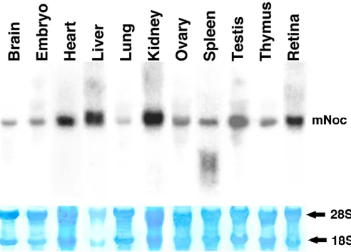
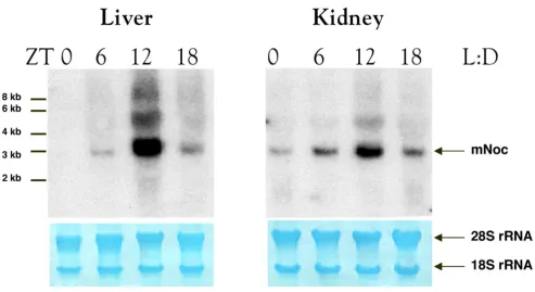
mNoc is expressed (Fig. 2) in retina, brain, heart, liver,
lung, kidney, ovary, skeletal muscle (data not shown), pineal gland (data not shown), testis and thymus. It ap-pears to be expressed at the highest levels in liver, then testis, kidney and retina. Lung has the lowest expression
level of those tested. In addition, mNoc mRNA is
ex-pressed at early embryonic stages (Fig. 2).
mNoc is Rhythmically Expressed in Multiple Tissues
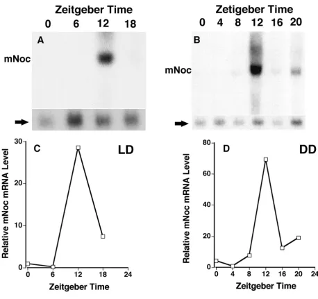
Northern analysis of retinal RNA shows that, as was the
case in Xenopus, mNoc exhibits a rhythm of mRNA
[image:4.612.58.551.86.439.2]abundance. Peak expression occurs at the time of light
Figure 2
BMC Developmental Biology (2001) 1:9 http://www.biomedcentral.com/1471-213X/1/9
offset (Fig. 3A). However, the amplitude of the rhythm is approximately 2 fold compared to the greater than 10
fold amplitude seen in Xenopus retina. Rhythmicity with
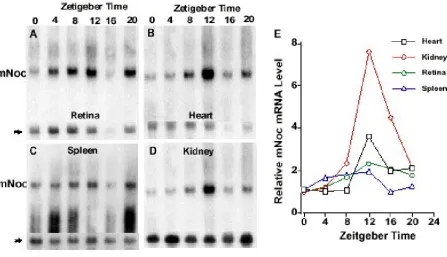
a similar peak at ZT12 is also seen in heart (Fig. 3B), spleen (Fig. 3C), kidney (Fig. 3D), and liver (Fig. 4). The amplitudes of the rhythms in heart, spleen and kidney, as determined by phosphor imaging, reflect 2 to 5 fold changes between minimum and maximum. In contrast, the magnitude of the day-night difference in liver repre-sents a nearly 30 fold change (see Fig. 4). Although the overall pattern of rhythmicity is similar in these tissues, baseline expression during the day is evident in retina, heart, and spleen and in part accounts for the lower am-plitude in these tissues.
Rhythms of mNoc mRNA Abundance are Circadian in Na-ture
In order to investigate the endogenous rhythmicity of
mNoc expression, C3H/He mice were maintained in
constant darkness for 36 hours before sampling for rhythmic changes in DD. Samples were then taken in darkness at 6 time points referenced to the normal LD
cycle in which they had been maintained (referred to as
Zeitgeber Time). mNoc from all five tissues shows
rhyth-mic changes in mRNA abundance (Figs. 4 and 5). Fur-thermore, liver tissue exhibits a high amplitude rhythm with virtually no mRNA detectable in the day-time as was the case in LD (Fig. 4B). The other four tissues all ex-hibit a higher level of daytime expression than in LD (Fig. 5). Unlike other tissues, spleen RNA exhibits a dif-fuse zone of hybridization centered at 1.0-1.2 kb, which may reflect RNA degradation.
mNoc mRNA appeared to reach higher levels at ZT12 in
DD than LD in all tissues except retina (compare Figs
3,4,5), suggesting that light may suppress mNoc mRNA
level. This was particularly striking in liver where the
ra-tio of mNoc/β-actin as determined by phosphorimaging
[image:5.612.51.551.84.353.2]was greater at ZT12 in DD than in LD (see Fig. 4D). This difference appears to be significant in that it was repro-ducible in an independent replication of the experiment in which LD and DD samples were analyzed on the same blot.
Figure 3
BMC Developmental Biology (2001) 1:9 http://www.biomedcentral.com/1471-213X/1/9
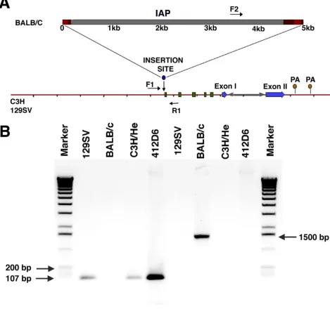
C3H/He and 129SV Mice Lack a Transposable IAP Element in the Nocturnin Gene
Laboratory strains of mice are heterogeneous in the pres-ence of a transposable intracisternal A-particle (IAP)
el-ement in the nocturnin/CCR4 gene. During the course of
our work on the mNoc cDNA it was reported [38,39] that a transposable IAP of viral origin, present in about 1000 copies throughout the mouse genome, is found in the
first intron of the mNoc/CCR4 gene. In DBA/2, BALB/c,
C57Bl/6 and C57Bl/10 mice, transcriptional read
through from the IAP insert to the mNoc/CCR4 open
[image:6.612.68.527.84.516.2]reading frame resulted in hybrid transcripts (3, 6 and 10 kb) whose abundance increased in aging mice. Appar-ently, insertion of the IAP element in the first intron oc-curred relatively recently because the insert was found to be lacking in some strains of mice [39]. We confirmed the lack of an IAP element in 129/SV and C3H/He (the strain used in the rhythmic analysis above) mice through
Figure 4
BMC Developmental Biology (2001) 1:9 http://www.biomedcentral.com/1471-213X/1/9
a combination of genomic PCR and partial sequencing of a genomic clone. As shown in Figure 6, genomic PCR us-ing primers from sites in the intron adjacent to the IAP position revealed only intronic sequence in C3H/He and 129/SV mice. This was further confirmed for 129/SV mice by the lack of an IAP element in genomic sequence from this region as well (data not shown). Although the IAP element is absent in these two strains of mice, we confirmed the presence of the IAP sequence by PCR in BALB/c (Fig. 6B) and C57/Bl6 (data not shown) mice as reported [39].
Rhythmic Expression of mNoc in BALB/c Mice
We tested the hypothesis that the IAP element in Intron I of BALB/c mice ([14,36] see Fig. 6) would disrupt
rhythmic expression of mNoc. Six week old BALB/c mice
were kept in our animal facilities for two weeks and liver and kidney tissues were obtained for Northern analysis at 6 hour intervals through a light-dark cycle. As shown in Figure 7, mNoc in BALB/c mouse liver and kidney clearly exhibits rhythmicity similar to that seen in C3H/
He mice with a prominent mRNA band at approximately 3 kb. Less abundant larger bands are also seen above 4 kb (liver and kidney) and 8 kb (liver only). Although larg-er mRNA bands have been reported to reflect hybrid transcripts including components of the IAP element in aging BALB/c mice, it is unclear whether this would ex-plain the larger bands in Figure 7. The bands in Figure 7 are smaller than the 6 and 10 kb bands reported in old mice [38,39] and follow a rhythmic pattern similar to that of the 3 kb band. The larger bands could reflect splicing intermediates that are seen only during the
peri-od of maximal transcription of mNoc. Although it is
pos-sible that altered transcription of mNoc as a consequence
[image:7.612.78.526.105.362.2]of an interaction between aging and the IAP insert may alter the rhythmic pattern of expression [39], our data clearly indicate that the mere presence of the IAP ele-ment in mice 8 weeks of age has little or no impact on rhythmic expression. Comparable results have been ob-tained using C57/Bl6 and CBA/J mice (data not shown).
Figure 5
BMC Developmental Biology (2001) 1:9 http://www.biomedcentral.com/1471-213X/1/9
Figure 6
C3H/He and 129/SV mice lack an intracisternal A-particle (IAP) insert in the first intron of the nocturnin/CCR4
BMC Developmental Biology (2001) 1:9 http://www.biomedcentral.com/1471-213X/1/9
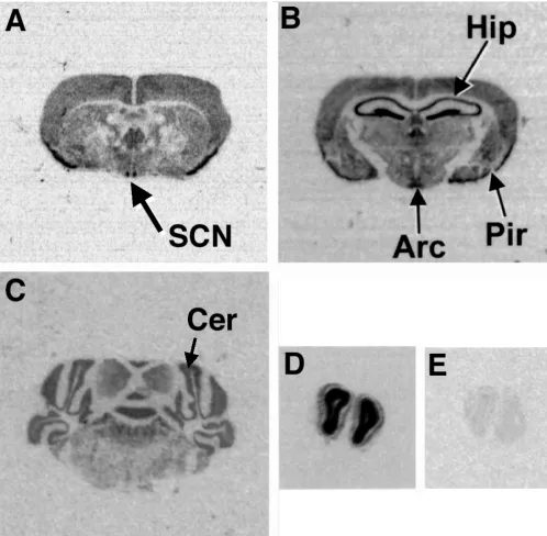
Expression of mNoc in the Brain
As shown in Figure 2, mNoc mRNA is expressed in tissue
from the mid-brain, which contains the hypothalamus including the suprachiasmatic nucleus. In several at-tempts at temporal Northern analysis in LD using sam-ples excised from the midbrain of C3H/He and C57/Bl6 mice, we saw hints of low amplitude rhythmicity, but the results were variable and could have resulted from sam-pling error (data not shown). We, therefore, examined
brain expression of mNoc mRNA further using in situ
hybridization of tissue from C57/Bl6 mice fixed at ZT 4,
ZT 12, and ZT 20. mNoc transcripts were detected (Fig.
8) in the suprachiasmatic nucleus (SCN), the ventral hy-pothalamic nucleus, arcuate nucleus (Arc), the piriform cortex (Pir), the hippocampus (Hip), the cerebellum, the subiculum, the internal granule layer of the olfactory bulbs and the pineal gland. Although we saw variability at different sample times (Table 1) in the intensity of the hybridization in several brain regions, the observations were of a qualitative nature and the magnitude of the changes was not great. Although the in situ hybridization data suggest low amplitude rhythmicity in these brain re-gions (including the SCN), we have not detected
[image:9.612.59.553.88.357.2]rhyth-mic expression in the brain comparable to the high amplitude rhythmicity detected in peripheral tissues.
Figure 7
mNoc mRNA is expressed rhythmically in 8 week old BALB/c mouse liver (A) and kidney (B) in a light dark cycle (LD). Tissues for RNA extraction were collected at Zeitgeber Times (ZT) 0 (24), 6, 12 and 18 with lights on at ZT 0 and off at ZT12. Images of the methylene blue stained 28S and 18S rRNA bands on the same blot are shown below as loading controls.
Table 1: Semi-quantitative analysis of mNocexpression in differ-ent brain regions at three times of day. (++ = Strong hybridiza-tion; + = Weak hybridizahybridiza-tion; - = no hybridization).
Brain Region ZT4 ZT12 ZT20
Olfactory Bulb
Internal Granule Layer ++ ++ ++
Lateral olfactory tract
---Piriform cortex ++ +
-Hippocampus ++ ++ +
Hypothalamus
Suprachiasmatic nucleus + +
-Arcuate nucleus + +
-Ventromedial hypothalam-ic
Nucleus + +
-Subiculum + + +
Cerebellum ++ ++ +
[image:9.612.314.552.504.694.2]BMC Developmental Biology (2001) 1:9 http://www.biomedcentral.com/1471-213X/1/9
Discussion
Our principal findings are that both the structure of the putative NOCTURNIN protein and circadian expression of its mRNA are conserved in the mouse. In addition, partial cDNA sequences and database analysis reveals
xNoc homologues in Drosophila, human, rat, cow, and
chicken. xNoc was originally identified as the product of
a differential display screen for circadian clock-regulated genes [35,36] in the retina of the African clawed frog, a
[image:10.612.57.556.86.575.2]system known to exhibit circadian clock activity in an in
Figure 8
BMC Developmental Biology (2001) 1:9 http://www.biomedcentral.com/1471-213X/1/9
vitro setting [27,45]. High amplitude circadian
regula-tion of the xNoc mRNA with peak abundance at night
was found to be a defining feature of the gene. Nuclear run-on assays showed that the high amplitude circadian
rhythm of xNoc was controlled at the level of gene
tran-scription. Furthermore, xNoc was found to be expressed
in photoreceptors, the site of a retinal circadian oscillator
[28]. To further our understanding of nocturnin, we
ini-tiated analysis of mammalian homologues of xNoc.
The main features of the putative NOCTURNIN protein in Xenopus were a leucine repeat domain and a CCR4 homology domain [36]; both regions are evident in the other sequences. However, the unusual leucine-repeat domain, originally identified in XNOC, is not well con-served and exhibits significant deviations from the clas-sic leucine zipper model [42]. The principal changes are the substitution of either tyrosine or phenylalanine for leucine at the beginning of the third heptad repeat and the addition of a second proline adjacent to the first in the second heptad repeat. Both changes, although con-served in five different species, are deviations from the classical leucine zipper model. Proline residues, as point-ed out previously [36] are expectpoint-ed to disrupt the coilpoint-ed- coiled-coil structure of the protein. The conservation of this do-main, including the prolines, suggests that it is a func-tionally important motif. Although its function is unknown, one possibility is that it serves as a protein in-teraction domain. For example, the leucine-rich domain in CCR4, mediates interaction with other proteins of the basal transcription apparatus [43].
The other conserved structural feature of nocturnin is a domain with homology to the C-terminus of yeast CCR4, a factor required for the transcription of genes including ADH2 (the glucose repressible alcohol dehydrogenase II; [43]). CCR4 is a multi-domain protein, substantially larger than nocturnin [37,43]; its estimated molecular weight is 94.5 kDa compared to 43.9 kDa for XNOC. CCR4 is thought to interact with other proteins via a leu-cine rich domain in the middle of the molecule. It is of some interest that while XNOC, MNOC and HNOC all align with the C-terminal domain, alignment of the leu-cine zipper-like domain of XNOC with the leuleu-cine rich region of CCR4 is relatively poor. Furthermore, regulato-ry domains, such as the glucose repressed activation do-main and glucose independent activation dodo-main, found in the amino-terminal half of CCR4 [37] are not present in NOCTURNIN. The fact that CCR4 has been character-ized as a transcriptional co-activator has led to the spec-ulation that nocturnin serves a similar function. However, the lack of key activation domains in nocturnin that are required for the function of CCR4 suggests that yeast CCR4 may not be the best model for delineating NOCTURNIN function. We believe that one of the keys
to understanding NOCTURNIN function is to identify its putative binding partners.
xNoc was identified on the basis of its high amplitude
cir-cadian expression in the Xenopus retina [36]. However,
in several additional Xenopus tissues we were unable to
detect xNoc mRNA by Northern analysis. A major
find-ing of this study is that mNoc mRNA is detected in most,
if not all, tissues of the adult mouse. Furthermore, iden-tification of ESTs derived from mouse and human em-bryonic cDNA libraries along with our Northern data on mouse embryo RNA indicate that mNoc is expressed ear-ly in development. Recentear-ly, earear-ly and ubiquitous
ex-pression of xNoc has also been detected during Xenopus
embryogenesis (Green, unpublished). Furthermore, rhythmic increases of mRNA abundance that persists in constant darkness, have been seen in mouse retina, liver, kidney, heart and spleen. This provides evidence that the
rhythmic changes in mNoc mRNA are controlled by one
or more circadian oscillators. Although this study has emphasized retinal and non-neural tissues, Northern
analysis has revealed mNoc mRNA in brain and pineal
tissue. In addition, we have found (see below) that noc-turnin is expressed in multiple brain regions including the suprachiasmatic nucleus (SCN), the site of the cen-tral circadian oscillator controlling behavioral rhythmic-ity [26].
The widespread circadian expression of mNoc mRNA in
multiple tissues of the mouse parallels that of the
Dro-sophila central "clock gene", period, which was recently
characterized in mammals. The period gene is
rhythmi-cally expressed in multiple tissues as well as in the cen-tral "clock" controlling behavior in both Drosophila [33] and mouse [7,8,10,11,12]. At present we do not know if nocturnin plays a role as a central component of the cir-cadian clock mechanism or as a "clock controlled" gene, perhaps coupling clock activity to an unidentified physi-ological rhythm. However, widespread expression and
rhythmic regulation of mNoc argues against a limited
role in rhythmic physiology of the retina or in the regula-tion of its melatonin output rhythm suggested by the
ear-lier work on the Xenopus eye. It seems more likely that
nocturnin is coupled to circadian function in a general way as either a central clock component or as a down-stream effector.
During the course of our work on mNoc, it was reported
[38,39] that a transposable intracisternal A-particle (IAP) element of viral origin is found in the first intron of
the mouse nocturnin/CCR4 gene. Furthermore, it was
reported that in DBA/2, BALB/c, C57Bl/6 and C57Bl/10 mice, transcriptional read through from the IAP
tran-scriptional start site to the nocturnin/CCR4 open
BMC Developmental Biology (2001) 1:9 http://www.biomedcentral.com/1471-213X/1/9
abundance increased in parallel in aging mice. This
re-port raised the immediate concern that disrupted mNoc
transcription might modify its rhythmic expression pat-tern and function. However, insertion of the
transposa-ble IAP in the nocturnin/CCR4 gene was apparently a
recent event, occurring after the origin of modern mouse strains because some mouse strains lack the insert [39]. Our genomic sequencing and PCR experiments confirm this finding by demonstrating that the IAP element is present in Balb/c and C57/Bl6 mice but lacking in 129/ SV and C3H/He mice. Thus, the rhythmic expression of
mNoc as a single mRNA species in C3H/He mice appears
to reflect the wildtype condition for this gene.
The IAP insert appears to strongly affect the expression
of mNoc mRNA in aging mice. The multiple hybrid forms
of mNoc mRNA [38] along with the recent report of an
absence of rhythmic expression [39] of mNoc mRNA in
mice containing the IAP element have raised the possi-bility that differences among mouse strains could
pro-vide a basis for understanding nocturnin function. It is
well known that strains of inbred mice have different rhythmic phenotypes. Perhaps the best understood is the lack of the ability to produce melatonin in some strains such as BALB/c and C57Bl/6 and the production of nor-mal rhythms of melatonin in others such as the C3H/He and CBA [44,46,47,48,49]. Recently, we initiated studies
directed at analysis of mNoc expression in BALB/c mice
with the goal of determining if the IAP insertion has a
di-rect consequence on rhythmic mNoc expression. Our
analysis shows that the presence of the IAP element has
no impact on mNoc expression or its rhythmicity in mice
up to 8 weeks of age. Although it is possible that altered
expression of the mNoc gene during the process of aging
may affect the rhythmic phenotype, our data indicate that the IAP insert itself cannot be regarded as a specific insertional mutation with direct consequences on rhyth-micity.
Although nocturnin was originally identified as a
rhyth-mic gene product in photoreceptors, the most striking rhythmicity identified in the mouse is in the peripheral tissues such as liver and kidney. In parallel with the
find-ings of this study we recently identified nocturnin as a
rhythmic gene product in rat liver and kidney based gene array analysis of over 9000 rat Unigenes (Kita, et al., un-published). Interestingly, in the latter study rNoc was identified among a group of clock-regulated genes that included Per1, Per2, Per3, Bmal1, and D-binding protein
(DBP). Independent clock driven pathways may be
criti-cal in the function of many tissues and organs as suggest-ed by the widespread expression of clock genes in peripheral tissues [7,8,10,11,12]. Support for this conclu-sion comes from the recent finding that circadian oscilla-tion of gene expression in the liver is entrained by food
intake independently of the central oscillator in the brain [50]. An understanding of the rhythmic function of noc-turnin may come from analysis of its role in rhythmic physiology of the liver and kidney.
Materials and Methods
Animals and tissue collection
C3H/He mice, wild type (+/+) at the rd locus, were orig-inally obtained from Dr. Michael Menaker at University of Virginia and then maintained as a breeding colony in ventilated environmental compartments within a
tem-perature-controlled animal facility (24 -25°C) on a 12
hour light:12 hour dark cycle (LD), except as noted. BALB/c, CBA/J and C57/Bl6 mice were purchased from Charles River Laboratories (Wilmington, MA) or Jack-son Laboratories and maintained under similar condi-tions. Experimental protocols were approved by the Institutional Animal Care and Use Committee and follow all federal guidelines. Mice were sacrificed by cervical dislocation following exposure to carbon dioxide or an overdose halothane anesthesia. Tissues for RNA extrac-tion in LD were collected at ZT0, ZT6, ZT12 and ZT18 in light (standard room fluorescent light) or dim red light (Kodak Wratten #2, filters). Those for constant dark (DD) experiments were collected in dim red light at ZT0, ZT4, ZT8, ZT12, ZT16 and ZT20 referenced to the LD cy-cle immediately before DD treatment. Bovine, chicken, rat (Sprague Dawley obtained from the ARC) and human retinal tissue (obtained from the Eye Research Institute at Medical College of Wisconsin) were immediately fro-zen on dry ice after dissection and stored at -80°C.
Total RNA and Genomic DNA isolation
Total RNA was extracted by the TRIZOL reagent proto-col (GIBCO/BRL, Rockville, MD), and then dissolved in
DEPC-treated water before storage at -80°C. QIAamp
Tissue Kits (Qiagen Inc., Santa Claita, CA,) were used for genomic DNA extraction from liver tissues according to the kit protocol.
cDNA Library Screening and DNA Sequencing
Mouse nocturnin cDNA (mNoc) clones were obtained by
screening a mouse BALB/c retinal cDNA library from ATCC (ATCC# 77448, Rockville, MD; reference [51]) us-ing the standard protocol published in Sambrook, et al, 1989 [52] with probes from a human EST (T87026) pur-chased from Research Genetics, Incorporated
(Hunts-ville, AL). mNoc clones were custom sequenced by
BMC Developmental Biology (2001) 1:9 http://www.biomedcentral.com/1471-213X/1/9
Biosystems, Foster City, CA). MacVector software was used for sequence analysis in this study.
Genomic Library Screening and Analysis
mNoc bacterial artifical chromosome (BAC) clones were
obtained from Research Genetics, Incorporated (Hunts-ville, AL) by custom screening of a 129/SV BAC library with the "whole cell PCR" protocol. BAC DNA was isolat-ed by a Qiagen Plasmid Maxi Kit and sequencisolat-ed using the ABI Prism 310. For the genomic PCR in Figure 6 the forward primer F1 (5'-AGTGACTGTCCTTCCTCTGT-3) is located upstream (5') and the reverse primer, R1 (5'-AACACAGTGAGACGCTGTCT-3') is located down-stream (3') of an intracisternal A-particle (IAP) element (reference [39]) identified in the nocturnin/CCR4 gene. The forward primer F2 (5'-TGATGTCCAGGGCGTCAA-TA-3') is located in the IAP element itself [39]. The se-quences for F1 and R1 are based on sese-quences from the BAC clone characterized in Figure 6A while the sequence of F2 is from the IAP element (reference [39]). These three primers were used for genomic PCR with Taq DNA polymerase (Promega). All of the resulting PCR products were cloned into pCRII-TOPO (Invitrogen, Carlsbad, CA) and sequenced as above.
Probe Preparation and Labeling
Single strand PCR probes for Northern hybridization
representing 553 bp of 3' UTR or Exon II of mNoc were
generated using a modification of the single-strand DNA
protocol of Bednarczuk, et al. [53], including 32P-dCTP
(40 mCi/ml; NEN Life Science Products, Boston, MA) in the reaction mixture. The primers for the 3'UTR probe were 5'-AACCATGCAGGTACAGTC-3' (bp 1557-1575 of
the mNoc cDNA, forward) and
5'-GTTTGGAAGAGGCT-TCAAC-3' (bp 2128-2147, reverse); for the Exon II probe they were 5'-ACCAGTCGACTCTACAGTGC-3' (bp 355-374, forward) and 5'-GGCTGGAAGGTGTCAAAG-3' (bp741-759, reverse). Random primed probes were pre-pared using the Random Primers DNA Labeling Kit (GIBCO/BRL, Rockville, MD). Radioactive probes were purified through NucTrap gel filtration columns (Strata-gene, La Jolla, CA).
Northern Blot Analysis
Ten µg (or less as specified) of each RNA sample was
sep-arated on 1.0% formaldehyde-agarose gels using stand-ard procedures [35]. Northern blot analysis was carried out according to QuikHyb hybridization solution proto-col (Stratagene, La Jolla, CA). Nylon membranes were stripped by washing twice for 10 min in boiling 0.01X SSPE (0.18 M NaCl/10 mM phosphate, pH 7.4/1 mM EDTA, 0.5% SDS) and rehybridized with probes made
from mouse β-actin cDNA [54]. Hybridization signals
were quantitated using a Storm PhosporImager and
Im-ageQuant software (Molecular Dynamics) using a previ-ously described method [36].
5' RACE and PCR reactions
Total RNA used as a template in 5'-RACE and RT-PCR was treated with RNase-free DNase I (Promega, Madi-son, WI) and subsequently phenol-chloroform extracted. RNasin Ribonuclease Inhibitor (Promega) was used in both 5'-RACE and RT-PCR reactions. 5'-RACE was per-formed according to kit protocol (GIBCO/BRL, Rock-ville, MD). The reverse Transcription System (Promega) coupled with Taq DNA Polymerase (Promega) was used for RT-PCR.
Degenerative PCR was carried out using Taq DNA Polymerase (Promega) with 5'-GATGGGAAAC(A/ G)GCACCAG(C/T)(A/C)GAC-3' and 5'-GC(G/C)AG(A/ G)ATGTTCCACTGCAT(G/C)AC-3' as forward and re-verse primers respectively. The resulting PCR products were cloned into pCRII-TOPO and sequenced with an ABI Prism 310 sequencer.
Brain In Situ Hybridization
C57/Bl6 mice were decapitated following an overdose of halothane anesthesia. The brain was removed, frozen on dry ice and stored at -80° until sectioning. In situ hybrid-ization followed the protocol of Fukuhara, et al. [55]. {α
-35S} UTP (1250 Ci/mmol; NEN Life Science Products,
Boston, MA) labeled probes were obtained by in vitro
transcription. A mouse nocturnin cDNA fragment (450
bp) cloned into the pBluescript KS (+) vector (Strata-gene) was linearized with XhoI or XbaI for antisense or sense probes, and radiolabeled using T7 or T3 RNA polymerase respectively. Serial coronal cryostat sections
(20 µm thick) were hybridized overnight at 55°C and
washed at 57°C. Slides were exposed to Kodak Biomax
film for 6 days at room temperature.
GenBank Accession Numbers
Sequences completed for this work have been placed in GenBank. Newly assigned GenBank Accession numbers
for these sequences are AF199491 (mNoc), AF199492
(hNoc, genomic fragment), AF199493 (hNoc, RT-PCR
product), AF199494 (hNoc, EST T87026), AF199495
(rNoc, 5' RACE product), AF199496 (rNoc, RT-PCR
product), AF199497 (bNoc) and AF199498 (cNoc).
Acknowledgements
The authors thank Brooke Steenhard and Sheila Baker for helpful discus-sions and comments on the manuscript. This work was supported by Na-tional Institutes of Health Research Grants EY02414 (JCB), Core Grant for Vision Research EY01931 (JCB), NINDS38483 (GT) and an award from the Alcon Research Institute, Ft. Worth, TX (JCB).
References
BMC Developmental Biology (2001) 1:9 http://www.biomedcentral.com/1471-213X/1/9
2. Pittendrigh CS: Circadian systems: Entrainment,In Handbook of Behavioral Neurobiology, Vol. 4. Biological Rhythms, Edited by J. Aschoff, New York, Plenum, 1981, :95-124
3. Aronson BD, Johnson KA, Loros JJ, Dunlap JC: Negative feedback defining a circadian clock: Autoregulation of the clock gene frequency.Science 1994, 263:1578-1584
4. Hall JC: Tripping along the trail to the molecular mechanism of biological clocks.Trends in Neurosci 1997, 18:230-240 5. Wilsbacher LD, Takahashi JS: Circadian rhythms: molecular
ba-sis of the clock.Curr.Opin.Genet.Dev. 1998, 8:595-602
6. Albrecht U, Sun ZS, Eichele G, Lee CC: A differential response of two putative mammalian circadian regulators, mper1 and mper2, to light.Cell 1997, 91:1055-1064
7. Sakamoto K, Nagase T, Fukui H, Hirikawa K, Okada T, Tabaka H, Sato K, Miyake Y, Ohara O, Kako K, et al: Miltitissue circadian expres-sion of rat period homolog (rPer2) mRNA is governed by the mammalian circadian clock, the suprachiasmatic nucleus in the brain.Journal of Biological Chemistry 1998, 273:27039-27042 8. Shearman LP, Zylka MJ, Weaver DR, Kolakowski LF Jr, Reppert SM:
Two period homologs: Circadian expression and photic reg-ulation in the suprachiasmatic nuclei.Neuron 1997, 19 :1261-1269
9. Shigeyoshi Y, Taguchi K, Yamamoto S, Takekida S, Yan L, Tei H, Mori-ya T, Shibata S, Loros JJ, Dunlap JC, et al: Light-induced resetting of a mammalian circadian clock is associated with rapid in-duction of the mPer1 transcript.Cell 1997, 91:1043-1053 10. Sun ZS, Albrecht U, Zhuchenko O, Bailey J, Eichele G, Lee CC:
RIGUI, a putative mammalian ortholog of the Drosophila period gene.Cell 1997, 90:1003-1011
11. Tei H, Okamura H, Shigeyoshi Y, Fukuhara C, Ozawa R, Hirose M, Sakaki Y: Circadian oscillation of a mammalian homologue of the Drosophila period gene.Nature 1997, 389:512-516 12. Zylka MJ, Shearman LP, Weaver DR, Reppert SM: Three period
ho-mologs in mammals: Differential light responses in the su-prachiasmatic circadian clock and oscillating transcripts outside the brain.Neuron 1998, 20:1103-1110
13. Koike N, Hida A, Numano R, Hirose M, Sakaki Y, Tei H: Identifica-tion of the mammalian homologues of the Drosophila time-less gene, Timetime-less1.Federation of European Biochemical Societies
1998, 441:427-431
14. Sangoram AM, Saez L, Antoch MP, Gekakis N, Staknis D, Whiteley A, Fruechte EM, Vitaterna MH, Shimomura K, King DP, et al: Mamma-lian circadian autoregulatory loop: A timeless ortholog and mPer1 interact and negatively regulate CLOCK-BMAL1-in-duced transcription.Neuron 1998, 21:1101-1113
15. Takumi T, Nagmine Y, Miyake S, Matsubara C, Taguchi K, Takekida S, Sakakida Y, Nishikawa K, Niwa S, Okumura H: A mammalian or-tholog of Drosophila timeless, highly expressed in SCN and retina, forms a complex with mPER1.Genes Cells 1999, 4:67-75 16. Zylka MJ, Shearman LP, Levine JD, Jin XW, Weaver DR, Reppert SM:
Molecular analysis of mammalian timeless. Neuron 1998,
21:1115-1122
17. Darlington TK, Wager- Smith K, Ceriani MF, Staknis D, Gekakis N, Steeves TDL, Weitz CJ, Takahashi JS, Kay SA: Closing the circadian loop: CLOCK-induced transcription of its own inhibitors per and tim.Science 1998, 280:1599-1603
18. Gekakis N, Staknis D, Nguyen HB, Davis FC, Wilsbacher LD, King DP, Takahashi JS, Weitz CJ: Role of the CLOCK protein in the mam-malian circadian mechanism.Science 1998, 280:1564-1569 19. Hogenesch JB, Gu YZ, Jain S, Bradfield CA: The
basic-helix-loop-helix-PAS orphan MOP3 forms transcriptionally active com-plexes with circadian and hypoxia factors.Proceedings of the Na-tional Academy of Sciences of the United States of America 1999, 95 :5474-5479
20. King DP, Zhao YL, Sangoram AM, Wilsbacher LD, Tanaka M, Antoch MP, Steeves TDL, Vitaterna MH, Kornhauser JM, Lowrey PL, et al: Po-sitional cloning of the mouse circadian Clock gene.Cell 1997,
89:641-653
21. Rutila JE, Suri V, Le M, So WV, Rosbash M, Hall JC: CYCLE is a sec-ond bHLH-PAS clock protein essential for circadian rhyth-micity and transcription of Drosophila period and timeless. Cell 1998, 93:805-814
22. Dunlap JC: Molecular basis for circadian clocks. Cell 1999,
96:271-290
23. Green CB: How cells tell time.Trends in Cell Biology 1998, 8 :224-230
24. Bernard M, Klein DC, Zatz M: Chick pineal clock regulates sero-tonin N -acetyltransferase mRNA rhythm in culture. Proceed-ings of the National Academy of Sciences of the United States of America
1997, 94:304-309
25. Green CB, Besharse JC: Tryptophan hydroxylase expression is regulated by a circadian clock in Xenopus laevis retina.Journal of Neurochemistry 1994, 62:2420-2428
26. Klein DC, Moore RY, Reppert SM: Suprachiasmatic Nucleus. The Mind's Clock.New York: Oxford University Press. 1991 27. Besharse JC, Iuvone PM: Circadian clock in Xenopus eye
con-trolling retinal serotonin N-acetyltransferase. Nature 1983,
305:133-135
28. Cahill GM, Besharse JC: Circadian clock functions localized in Xenopus retinal photoreceptors .Neuron 1993, 10:573-577 29. Tosini G, Menaker M: Circadian rhythms in cultured
mamma-lian retina.Science 1996, 272:419-421
30. Deguchi T: A circadian oscillator in cultured cells of chicken pineal gland .Nature 1979, 282:94-96
31. Takahashi JS, Murakami N, Nikaido SS, Pratt BL, Robertson LM: The avian pineal, a vertebrate model system of the circadian os-cillator: cellular regulation of circadian rhythms by light, sec-ond messengers, and macromolecular synthesis. Recent Progress in Hormone Research 1989, 45:279-352
32. Frisch B, Hardin PE, Hamblen-Coyle MJ, Rosbash M, Hall JC: A pro-moterless period gene mediates behavioral rhythmicity and cyclical per expression in a restricted subset of the Drosophi-la nervous system.Neuron 1994, 12:555-570
33. Plautz JD, Kaneko M, Hall JC, Kay SA: Independent photorecep-tive circadian clocks throughout Drosophila. Science 1997,
278:1632-1635
34. Balsalobre A, Damioloa F, Schibler U: A serum shock induces cir-cadian gene expression in mammalian tissue culture cells. Cell 1998, 93:929-937
35. Green CB, Besharse JC: Use of a high stringency differential dis-play screen for identification of retinal mRNAs that are reg-ulated by a circadian clock.Molecular Brain Research 1996, 37 :157-165
36. Green CB, Besharse JC: Identification of a novel vertebrate cir-cadian clock-regulated gene encoding the protein nocturnin. Proceedings of the National Academy of Sciences of the United States of America 1996, 93:14884-14888
37. Draper MP, Liu HY, Nelsbach AH, Mosley SP, Denis CL: CCR4 is a glucose-regulated transcription factor whose leucine-rich re-peat binds several proteins important for placing CCR4 in its proper promoter context.Molecular and Cellular Biology 1994,
14:4522-4531
38. Puech A, Dupressoir A, Loireau M-P, Mattei M-G, Heidmann T:
Characterization of two age-induced intracisternal A-parti-cle-related transcripts in the mouse liver, transcriptional read-through into an open reading frame with similarities to the yeast CCR4 transcription factor.Journal of Biological Chemis-try 1997, 272:5995-6003
39. Dupressoir A, Barbot W, Loireau M-P, Heidmann T: Characteriza-tion of a mammalian gene related to the yeast CCR4 general transcription factor and revealed by transposon insertion. Journal of Biological Chemistry 1999, 274:31068-31075
40. Adams MD, Celniker SE, Holt RA, Evans CA, Gocayne JD, Amanati-des PG, Scherer SE, Li PW, Hoskins RA, Galle RF, et al: The genome sequence of Drosophila melanogaster. Science 2000,
287(5461):2185-2195
41. Kozak M: Ana analysis of 5'-non-codingsequences from 699 vertebrate mRNAs.Nucleic Acid Res 1987, 15:8125-8148 42. Landschultz WH, Johnson PF, McKnight SL: The leucine zipper: a
hypothetical structure common to a new class of DNA bind-ing proteins.Science 1988, 240:1759-1764
43. Malvar T, Biron RW, Kaback DB, Denis CL: The CCR4 protein from Saccharomyces cerevisiae contains a leucine-rich re-peat region which is required for its control of ADH2 gene expression.Genetics 1992, 132:951-962
44. Goto M, Oshima I, Tomita T, Ebihara S: Melatonin content of the pineal gland in different mouse strains .Journal of Pineal Research
1989, 7:195-204
BMC Developmental Biology (2001) 1:9 http://www.biomedcentral.com/1471-213X/1/9
46. Ebihara S, Marks T, Hudson DJ, Menaker M: Genetic control of melatonin synthesis in the pineal gland of the mouse.Science
1986, 231:491-493
47. Goto M, Oshima I, Hasegawa M, Ebihara S: The locus controlling pineal serotonin N -acetyltransferase activity (Nat-2) is lo-cated on mouse chromosome 11.Molecular Brain Research 1994,
21:349-354
48. Roseboom PH, Namboodiri MAA, Zimonjic DB, Popescu NC, Rod-riguez IR, Gastel JA, Klein DC: Natural melatonin 'knockdown' in C57BL/6J mice: rare mechanism truncates serotonin N -acetyltransferase.Molecular Brain Research 1998, 63:189-197 49. Yoshimura T, Nagabukuro A, Matsuda Y, Suzuki T, Kuroiwa A, Iigo M,
Namikawa T, Ebihara S: Chromosomal mapping of the gene en-coding serotonin N -acetyltransferase to rat chromosome 10q32.3 and mouse Chromosome 11E2.Cytogenetics and Cell Ge-netics 1997, 79:172-175
50. Stokkan K-A, Yamazaki S, Tei H, Sakaki Y, Menaker M: Entrainment of the circadian clock in the liver by feeding. Science 2001,
291:490-493
51. Farjo Q, Jackson AU, Xu J, Gryzenia M, Skolnick C, Agarwal N, Swa-roop A: Molecular characterization of the murine neural ret-ina leucine zipper gene, Nrl.Genomics 1993, 18:216-222 52. Sambrook J, Fritsch EF, Maniatis T: Molecular Cloning: A
Labora-tory Manual.2 ed. Cold Spring Harbor, New York: Cold Spring Harbor Laboratory Press. 1989
53. Bednarczuk TA, Wiggins RC, Konat GW: Generation of high effi-ciency, single stranded DNA hybridization probes by PCR. Bi-otechniques 1991, 10:478
54. Alonso S, Minty A, Buckingham M: Comparison of three actin coding sequences in the mouse; evolutionary relationships between actin genes of warm blooded vertebrates.Journal of Molecular Evolution 1986, 23:11-22
55. Fukuhara C, Dirden JC, Tosini G: Circadian expression of Period 1, Period 2, and arylalkylamine N-acetyltransferase mRNA in the rat pineal gland under different light conditions. Neuro-science Letters 2000, 286:167-170
Publish with BioMedcentraland every scientist can read your work free of charge
"BioMedcentral will be the most significant development for disseminating the results of biomedical research in our lifetime."
Paul Nurse, Director-General, Imperial Cancer Research Fund
Publish with BMc and your research papers will be:
available free of charge to the entire biomedical community peer reviewed and published immediately upon acceptance cited in PubMed and archived on PubMed Central yours - you keep the copyright
[email protected] Submit your manuscript here:
http://www.biomedcentral.com/manuscript/
BioMedcentral.com