JOURNAL OFVIROLOGY, Sept. 2002, p. 9046–9059 Vol. 76, No. 18 0022-538X/02/$04.00⫹0 DOI: 10.1128/JVI.76.18.9046–9059.2002
Copyright © 2002, American Society for Microbiology. All Rights Reserved.
Ahi-1
, a Novel Gene Encoding a Modular Protein with WD40-Repeat
and SH3 Domains, Is Targeted by the
Ahi-1
and
Mis-2
Provirus Integrations
Xiaoyan Jiang,
1Zaher Hanna,
1,2Mohammadi Kaouass,
1Luc Girard,
1and Paul Jolicoeur
1,3,4*
Laboratory of Molecular Biology, Clinical Research Institute of Montreal, Montreal, H2W 1R7 Quebe´c,1De´partement de
Me´decine2and De´partement de Microbiologie et d’Immunologie,3Universite´ de Montre´al, Montre´al, H3C 3J7 Quebe´c,
and Department of Medicine, Division of Experimental Medicine, McGill University,
Montreal, H3G 1A4 Quebe´c,4Canada
Received 21 March 2002/Accepted 13 June 2002
TheAhi-1locus was initially identified as a common helper provirus integration site in Abelson pre-B-cell lymphomas and shown to be closely linked to the c-mybproto-oncogene. Since no significant alteration of c-myb
expression was found in Abelson murine leukemia virus-induced pre-B-lymphomas harboring a provirus inserted within theAhi-1locus, this suggested that it harbors another gene whose dysregulation is involved in tumor formation. Here we report the identification of a novel gene (Ahi-1) targeted by these provirus inser-tional mutations and the cloning of its cDNA. The Ahi-1 proviral insertions were found at the 3ⴕend of the gene, in an inverse transcriptional orientation, with most of them located around and downstream of the last exon, whereas another insertion was within intron 22. In addition, another previously identified provirus insertion site, Mis-2, was found to map within the 16th intron of theAhi-1gene. The Ahi-1cDNA encodes a 1,047-amino-acid protein. The predicted Ahi-1 protein is a modular protein that contains one SH3 motif and seven WD40 repeats. TheAhi-1gene is conserved in mammals and encodes two major RNA species of 5 and 4.2 kb and several other shorter splicing variants. TheAhi-1gene is expressed in mouse embryos and in several organs of the mouse and rat, notably at high levels in the brain and testes. In tumor cells harboring insertional mutations inAhi-1, truncatedAhi-1/viral fused transcripts were identified, including some splicing variants with deletion of the SH3 domain. Therefore,Ahi-1is a novel gene targeted by provirus insertion and encoding a protein that exhibits several features of a signaling molecule. Thus,Ahi-1may play an important role in signal transduction in normal cells and may be involved in tumor development, possibly in cooperation with other oncogenes (such as v-abland c-myc) or with a tumor suppressor gene (Nf1), since Ahi-1 insertion sites were identified in tumors harboring v-abldefective retroviruses or a c-myctransgene or in tumors exhibiting deletion ofNf1.
The Abelson murine leukemia virus (A-MuLV) is a defec-tive, replication-incompetent retrovirus. To replicate in vivo and in vitro, A-MuLV requires a nondefective helper MuLV (51). The Abelson virus complex is highly lymphomagenic and induces oligoclonal pre-B-cell lymphoma in susceptible mice (1, 3, 42, 45). However, the expression of the v-abloncogene in target cells does not appear to be sufficient for tumor forma-tion (17, 50) and addiforma-tional genetic events are thought to be required. We have reported that the long terminal repeat (LTR) of the helper Moloney MuLV is involved in the devel-opment of lymphoma induced by A-MuLV (50). In fact, the role of the Moloney provirus as an insertional mutagen was demonstrated by the identification of a common helper provi-rus integration site, designatedAhi-1(for Abelson helper in-tegration site 1), in 16% of A-MuLV-induced pre-B-cell lym-phomas (41). All Moloney proviruses for which the precise integration site within this region could be mapped were found to be in the same orientation, suggesting a specific molecular requirement for this genetic event to be involved in malignant
transformation in the presence of v-abl. Ahi-1was mapped to mouse chromosome 10 in close linkage to the c-myb proto-oncogene (41). By using long-range restriction mapping, we mapped the c-myb andAhi-1 regions within a 120-kbp DNA fragment, with theAhi-1locus being located ca. 35 kbp down-stream of c-myb (22). Interestingly, theMis-2 region, which represents another MuLV integration site, has also been mapped in the same region, ca. 160 kbp downstream of c-myb and ca. 120 kbp downstream ofAhi-1(62). We tested whether Moloney provirus integration in theAhi-1locus enhances the expression of c-myb by acis-acting mechanism, by examining c-mybgene expression in A-MuLV-induced pre-B-lymphomas (22). Our data did not reveal any clear evidence for such c-myb overexpression in the tumors which were tested.
In view of these findings, we have hypothesized that a novel gene within the Ahi-1 locus is targeted by provirus insertion and that its dysregulation contributes to tumor development. We report here the identification of that gene,Ahi-1, which is the target of these Ahi-1 and Mis-2 provirus insertions and which encodes a protein containing an SH3 motif and a WD40-repeat domain.
MATERIALS AND METHODS
Mice and viruses.MMTVD/myc transgenic (Tg) mice have been described
previously (36). They were bred on CD1 background. Newborn (⬍48 h) mice
* Corresponding author. Mailing address: Laboratory of Molecular Biology, Clinical Research Institute of Montreal, 110 Pine Ave., West, Montreal, H2W 1R7 Quebec, Canada. Phone: (514) 987-5569. Fax: (514) 987-5794. E-mail: [email protected].
9046
on November 8, 2019 by guest
http://jvi.asm.org/
were inoculated with Moloney MuLV (105PFU/ml) intraperitoneally, as
de-scribed previously (41).
DNA extraction and restriction endonuclease digestion.DNA extraction, di-gestion with restriction endonucleases, separation of DNA fragments by agarose
gel electrophoresis, and hybridization with32P-labeled probes by the method of
Southern were performed as described previously (41).
Molecular cloning of mouse genomic DNA.The molecular cloning of the
provirus with the adjacentAhi-1sequences has been described elsewhere (41).
To initiate chromosome walking, cosmid and phage libraries were constructed by
partial digestion of C57BL/6 mouse liver DNA withSau3A and ligation into the
BamHI site of the pWE15 and EMBL-3 vector, respectively, as described
else-where (41, 48, 62). Molecular clones (Cos1, Cos2, Cos3, and pSW4) were isolated
by screening these libraries with mouseAhi-1probes E1.9 and G (Fig. 1A). To
analyze these clones in more details and to derive additional probes necessary for
a finer mapping of this region, a restriction map of theAhi-1region was derived
and additional single-copy fragments were isolated and subcloned (Fig. 1A). This map was confirmed by digesting normal mouse cellular DNA with various re-striction endonucleases (P.L. Pharmacia, Montreal, Quebec, Canada) and hy-bridizing them with different probes derived from this region (data not shown), essentially as previously described (41, 48).
Exon amplification. Exon amplification to identify coding sequences in genomic DNA was performed as described by Buckler et al. (4). In brief, two
cosmid clones (Cos1 and Cos3), representing 70 kbp ofAhi-1region around the
integration sites, were digested withBamHI andBglII and subcloned as pools
into the vector pSPL1. In addition, another 14 individualBamHI and BglII
fragments derived from this region were cloned into pSPL1. Clones with inserts in sense and antisense orientations were selected and transfected into Cos-7 cells, and resulting RNAs were analyzed by reverse transcription-PCR
(RT-PCR) with specific primers derived from the vector as described previously (4). Finally, amplified exons were purified by electrophoresis on 1% agarose gels and
cloned into a dT-tailed (31)EcoRV-digested pBluescript II KS(⫹) plasmid
vector (Stratagene). Several criteria were used to distinguish real exons from false-positive clones. These criteria included nucleotide sequencing and compar-ison to DNA sequences in the databases (that is, the National Center for Bio-technology Information [NCBI] and European Bioinformatics Institutes [EBI] databases), detection of RNA transcripts by Northern blot analysis, and screen-ing of a library to identify positive cDNA clones.
Isolation of cDNA clones and sequencing.A rat braingt11 cDNA library was screened by using the RT-PCR fragment A (Fig. 2B). Two other mouse testis and
thymus cDNA (the Uni-Zap XR library [Stratagene] libraries and the 5⬘stretch
library [Clontech]) were also screened with cDNA probe E1.3 in order to get a
full-length cDNA. Furthermore, the missing 5⬘end of the cDNA was cloned by
5⬘RACE (rapid amplification of cDNA ends) as described previously (16) into
a dT-tailed vector. In addition, mouse brain RNA was used to amplify mouse cDNA sequences by RT-PCR with specific primers (that is, 276 and 296) derived from the rat cDNA sequences (Fig. 2A). Several clones were isolated: seven overlapping fragments from the rat brain cDNA library, eight overlapping clones, and five full-length clones from the mouse testis and thymic cDNA libraries, respectively. These clones were subcloned into pGEM-3, analyzed with restric-tion endonucleases, and sequenced by the dideoxynucleotide Sanger’s method (49) in both directions.
Probes.All probes containing cDNA or genomic fragments derived from the
[image:2.587.59.521.85.335.2]Ahi-1region were excised from the vector with appropriate restriction endo-nucleases, and the inserts were separated from the vector by agarose gel elec-trophoresis. The DNA fragments were recovered by electroelution and ethanol precipitation and used directly as hybridization probes. Probes were labeled by
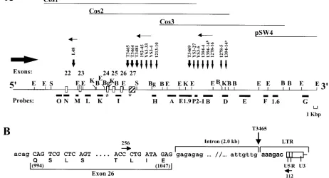
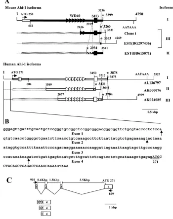
FIG. 1. Mapping of the MuLV proviruses within theAhi-1locus. (A) Structural organization of the proviruses integrated at the 3⬘end of the Ahi-1gene. The restriction endonuclease map of 80 kbp of theAhi-1region was determined with phage (pSW4) and cosmid (Cos1, Cos2, and Cos3) clones and confirmed by Southern blot analysis of mouse DNA. The localization of the proviruses relative to the exons is shown. The intron-exon boundaries of exons 25 and 26 have been confirmed by sequencing genomic fragments. The provirus integration within the ratAhi-1locus in tumor 1213-10 was detected with probe H but could not be mapped precisely because of the lack of restriction map in the rat. Symbols: open and strippled boxes, coding and noncoding exons, respectively; line, intronic sequences; closed horizontal boxes, single-copy fragments subcloned and used as probes; thin vertical arrows, sites of provirus integration in A-MuLV-induced pre-B-lymphomas; large vertical arrows, sites of provirus integration in T-cell thymomas of Mo-MuLV-infected MMTVD/myc Tg mice; horizontal thin arrows, orientation of provirus (5⬘to 3⬘); horizontal thick arrow,
orientation ofAhi-1transcription; asterisks, stop codons. Restriction sites: B,BamHI; Bg, BgII; E,EcoRI; K,KpnI; S,SmaI. Not all sites are indicated. (B) Site of the provirus integration within theAhi-1gene in tumor T3465. Sequences were obtained from a PCR product (⬃2.0 kbp) generated with primer 256 localized in exon 26 and primer 112 within the Moloney MuLV LTR. The sequences of the intron-exon boundaries are shown, respectively, in small and capital letters and the viral sequences in boldface. Encoded amino acids are shown in the lower line and the numbers refer to the amino acid position in the sequence shown in Fig. 3.
on November 8, 2019 by guest
http://jvi.asm.org/
incubating the denatured fragments in the presence of hexamers and the Klenow fragment of polymerase 1 (13).
RNA expression by Northern analysis.RNA was isolated from various tissues by the method of Chomczynski and Sacchi (6), separated on 1% formaldehyde-agarose gels, transferred onto nylon membranes, and hybridized as described
above. Washings were performed for 15 min at room temperature in 2⫻SSC
(1⫻SSC is 0.15 M NaCl plus 0.015 M sodium citrate), for 1 h at 65°C in 0.1⫻
SSC–0.5% sodium dodecyl sulfate, and in 0.1⫻SSC three times for 2 min each
time at room temperature. The oligonucleotide used to detect the 18S rRNA was
5⬘-ACGGTATCTGATCGTCTTCGAACC.
PCR and RT-PCR analysis.Total RNAs from the mouse tumor cells, from
Jurkat cells or from mouse thymus (5g) were transcribed with Moloney MuLV
reverse transcriptase as described previously (16). PCR was performed on the cDNAs obtained by using the following primers. For analysis of the different
splicing forms of the 5⬘-untranslated human Ahi-1 sequences, the primers used
were primer 910 (5⬘-GGGAGTTGATTTGCACTGCTC) and primer 899 (5⬘-A
TCTCCTTTGCCATTTCTTCAG), which are located on exons 1 and 4,
respec-tively (see Fig. 5C). For analysis of the novel sequences of exon 24 present at 3⬘
end of the AK000076 cDNA clone, we used primers 897 (5⬘-AGACCGACAG
TCACTTTGCTG; exon 23/24) and 898 (5⬘-TCCCAATATTTTATGAGTTTC
AAAG; exon 24). For analysis of the 3⬘end Ahi-1/viral fused transcripts, in Mis-2
and Ahi-1 (including L48) rearranged tumors, the primer 828 within the U5 LTR
(5⬘-AGTGATTGACTACCCGTCAGC), and a primer in exon 16 (primer 276,
5⬘-GAGAACAATGGTTGCGTTTTG), exon 17 (primer 277, 5⬘-CGAGTCTC
TGTTTCACAAGC), exon 19 (primer 264, 5⬘-CACCAGGCCCTGCAAAG
AAG), or exon 22 (primer 265, 5⬘-GTTAAAGGAGGGGACGCTC) were used.
The PCR products obtained were purified on agarose gels, cloned, and se-quenced. To confirm the presence of the novel sequences at the C terminus in the mouse EST clones BB615071 (exon 20) and BG29736 (exon 24), a primer in
either exon 20 (primer 1311, 5⬘-CTGTCACAAATTCTCTGGTCAGG) or exon
24 (primer 1312, 5⬘-CATTCCTTGGTCCAGCAGCAG) and the primer 264
(exon 19) were used.
PCR analysis was used to map the provirus integration sites precisely in T-cell
tumors arising in Moloney MuLV-infected MMTVD/myc Tg mice. The Moloney
MuLV R LTR primers 110 (5⬘-TAAGCTAGCTTGCCAAACCTACAGGTG)
and 112 (5⬘-CCCGAGCTCAATAAAAGAGCCCACAAC) and the Ahi-1
spe-cific primer 256 (5⬘-CCGCAAAGTCACCCTGATAGAG; exon 26) and primer
276 (exon 16) were used. Fragments of⬃2.0 and⬃2.2 kbp were amplified from
T3465 and 1213-19 cell DNA, respectively, purified on agarose gels, cloned, and sequenced.
Sequence analysis.Homology searches were carried out by computer-assisted nucleotide and protein blasts on the NCBI and EBI Web sites by using the human and mouse genome databases. Search for known domains were done by using the conserved domain database at NCBI with the latest versions of SMART v.3.3, Pfam v6.6, PSPOR II, and ScanProsite programs.
Nucleotide sequence accession numbers.The nucleotide sequence accession
number of the full-length mouseAhi-1cDNA (Fig. 3A) is AY133241; the
ac-cession number of clone I of mouseAhi-1cDNA (Fig. 5A) is AY133242; the
accession number of the untranslated 5⬘-end of humanAhi-1cDNA (Fig 5B) is
AY133243; and the accession numbers of fragments 1, 2, 4, and 5 of the ratAhi-1
genome (Fig. 6A) are AY133244, AY133245, AY133246, and AY133247, respec-tively.
RESULTS
Detection of proviruses integrated within theAhi-1region in additional types of murine tumors.TheAhi-1locus was pre-viously identified as a common provirus integration site in pre-B lymphoma induced by the Abelson virus complex (series YS3, 1278 and 1394, Fig. 1A) (41). To determine whether the
Ahi-1 locus was also targeted by provirus in MuLV-induced
leukemias of other hematopoietic cell lineages, we studied T-cell tumors arising in MMTVD/myc Tg mice infected with
[image:3.587.126.457.73.309.2]Moloney MuLV (16).EcoRV-digested tumor DNAs (n⫽28) were screened by the Southern technique withAhi-1probes D and I (Fig. 1A). Novel tumor-specific fragments, in addition to the normal germ line fragment (23 kbp), were detected in four tumors (T3464, T3465, T3481, and T3469). In addition, PCR
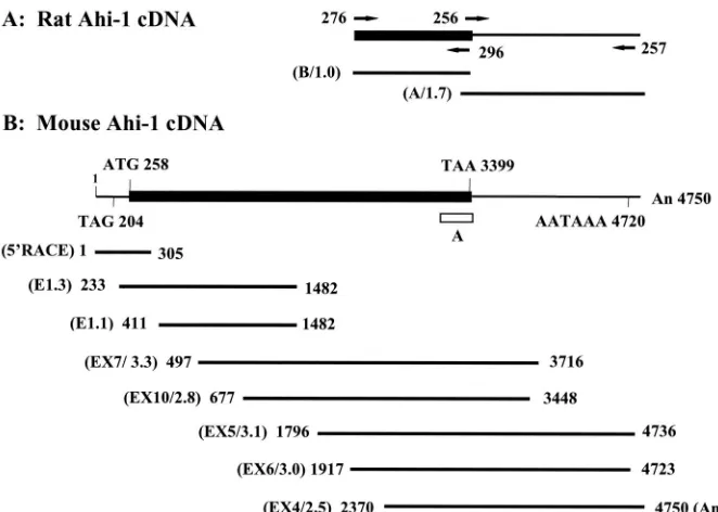
FIG. 2. Structure of theAhi-1cDNA clones. (A) Two overlapping rat cDNA clones, obtained from a brain cDNA library, are shown with their positions from 5⬘to 3⬘. Specific primers used for generating mouse cDNAs are indicated. (B) The completeAhi-1cDNA sequence was derived with eight overlapping mouse cDNA clones from two testis cDNA libraries (E and EX) and by 5⬘RACE. The methionine initiation codon, the termination codon, and the consensus polyadenylation signal are indicated. Symbols: thin line,Ahi-1untranslated region; closed box,Ahi-1coding sequences; horizontal lines,Ahi-1overlapping cDNA clones; open box, the location of the RT-PCR product-A. The numbers refer to the position of the nucleotides in Fig. 3. Numbers in parentheses represent the names of each clone (E or EX) with their respective lengths given in kilobase pairs.
9048 JIANG ET AL. J. VIROL.
on November 8, 2019 by guest
http://jvi.asm.org/
analysis confirmed and precisely mapped the integration in tumor T3465 within intron 26 (Fig. 1B), and RT-PCR analysis revealed the integration of a provirus between exons 22 and 23 in the L48 cell line (Fig. 1A) (see below, Fig. 7E), previously established from Moloney MuLV infected MMTVD/myc Tg
mice (16). The proviruses inserted at theAhi-1locus in T-cell leukemia were found at a frequency of⬃14% and to be inte-grated in the same orientation as those found in A-MuLV-induced lymphomas (Fig. 1A). Provirus integrations within Ahi-1 were also observed in 1 (i.e., 1213-10) of 20 distinct DNAs (5%) from Moloney MuLV-induced rat thymomas (se-ries 1213, Fig. 1A). These insertions were mapped in the same clusters of provirus integrations as those found in Abelson virus-induced tumors (Fig. 1A). Therefore, rearrangements of
theAhi-1 locus appear to be involved in the development of
lymphomas from different cell lineages.
Identification ofAhi-1coding sequences by exon amplifica-tion.To identify a putative gene located within theAhi-1locus, a bidirectional chromosome walking was initiated from the
Ahi-1 region first identified (41) by using cosmid and phage
libraries. Sequences spanning almost 80 kbp of theAhi-1 re-gion were cloned (Fig. 1A) and used to identify coding se-quences within this region by using an exon amplification method (4). A total of five putative exons ranging from 58 to 300 bp were recovered by this technique. One of the RT-PCR products fragment A (160 bp; Fig. 2B), generated from the 4-kbpBglII fragment of Cos 1 clone, was found to hybridize to two major transcripts of 5 and 3.5 kb in mouse brain and testis RNA by Northern blot analysis (data not shown), indicating that this fragment contained potential exons(s). A summary of these results is shown in Fig. 1A and 2.
Molecular cloning and sequencing of mouse and ratAhi-1
cDNAs.To identify the Ahi-1 gene and to map the provirus integration sites relative to this gene, we cloned the Ahi-1 cDNA. Since theAhi-1gene was highly expressed in brain and testis, a rat brain cDNA library was first screened. Several positive clones ranging from 0.6 to 2.5 kbp were obtained (Fig. 2). Northern blot analysis performed with both rat cDNA clone A and clone B showed the same hybridization pattern previ-ously observed with the RT-PCR product A, indicating that part of theAhi-1gene had been cloned. The full-length mouse
Ahi-1cDNA was obtained by RT-PCR and 5⬘RACE methods
and by rescreening a mouse thymic cDNA library, as described in Materials and Methods. A schematic representation of these Ahi-1 cDNA clones is shown (Fig. 2).
The complete nucleotide and deduced amino acid sequence of theAhi-1cDNA is shown in Fig. 3A. An open reading frame begins with a potential initiation methionine codon at nucleo-tide (nt) 258, based on Kozak consensus rules (25). An in-frame stop codon is located 204 nt upstream of this site. The predicted encoded protein is ⬃120 kDa long and contains 1,047 amino acids (aa). The in-frame termination codon (TAA) is followed by a long 3⬘-untranslated region (Fig. 2 and 3A). The sequence immediately upstream of the poly(A) tail contains a AATAAA polyadenylation signal (nt 4720).
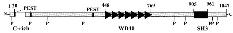
The mouseAhi-1gene product contains an SH3 motif and seven WD40 repeats. Computer-assisted analysis of the pre-dicted amino acid sequence of the Ahi-1 cDNA product re-vealed a number of significant structural features (Fig. 3B). In particular, the Ahi-1 cDNA encodes a region of ca. 50 aa
residues (aa 905 to 961) with significant sequence homology to the Src homology 3 (SH3) domain (25 to 35% identity [40 to 50% similarity]).
Several regions of the predicted Ahi-1 protein are also pro-line-rich (Fig. 3B) and contain nine potential SH3-binding sites (PxxP) at positions 9, 98, 182, 352, 803, 858, 976, 985, and 1001; the numbers here refer to the first proline residue in the PxxP consensus sequences present in high-affinity SH3 ligands (7, 43, 44, 56, 64).
Interestingly, another region of the Ahi-1 protein upstream of the SH3 motif and spanning⬃320 aa residues (aa 448 to 769) exhibits significant sequence homology to the WD40 re-peats (33). Seven WD40 rere-peats were identified in the pre-dicted Ahi-1 polypeptide sequence at positions 448 to 490, 493 to 532, 537 to 576, 583 to 622, 641 to 678, 684 to 721, and 724 to 769. Each WD40 repeat consists of stretch of ca. 40 aa in which tryptophan and aspartic acid residues and certain other amino acids are highly conserved, a finding consistent with the range of 23 to 41 observed in other members of this class of proteins (33).
In addition, computer analysis with the ScanProsite tool revealed the presence of many potential phosphorylation sites: among them, 21 protein kinase C phosphorylation sites, 3 cyclic AMP (cAMP)- and -cGMP-dependent protein kinase phosphorylation sites (23), and 26 potential casein kinase II phosphorylation sites. Also, two potential tyrosine kinase phos-phorylation sites are present (residue 71 [KLKEQLTY] and residue 941 [KDNEDWWY]) with sequences matching opti-mally the consensus sequence of tyrosine kinases [R/K-x-(2,3)D/E-x-(2,3)Y] (8) (Fig. 3A). Tyrosine phosphorylation has been shown to play an important role in several proteins in-volved in malignant transformation and signal transduction (57). The amino-terminal region of Ahi-1 contains an acid-rich domain (Fig. 3A). Such a domain is found in quite different proteins, such as nucleolines, calcium-binding proteins, and transcription factors (40). Finally, three potential PEST se-quences (at residues 19 to 30, 100 to 118, and 417 to 432) (46), three potential nuclear localization signal (aa 62 to 94, 134 to 151, and 825 to 868) (10), and four potential glycosylation sites (at positions 310, 536, 821, and 851) were found in the Ahi-1 protein. A summary of the structural domains present in the Ahi-1 protein is depicted in Fig. 3B.
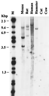
Conservation of theAhi-1gene in evolution.To determine whether theAhi-1gene has been conserved throughout evolu-tion, we searched for homologous sequences in other species. Mouse, rat, hamster, cat, cow, and human DNA samples were digested withEcoRI and hybridized at high stringency with
Ahi-1 cDNA clone-B probe. Specific DNA fragments were
detected in all of these species (Fig. 4), indicating a significant conservation of these sequences among mammals. Compari-son of the mouse, rat, and human sequences now available (see below) confirmed the high conservation of this gene. However, no gene coding for a protein containing WD40 repeats and a SH3 domain was found in the sequences ofDrosophila
mela-nogasterorCaenorhabditis elegans. This suggests that theAhi-1
gene may have an important function in mammals.
Identification of different isoforms of mouse and human Ahi-1 cDNA.A homology search of the GenBank databases with our mouse and ratAhi-1cDNA sequences identified three distinct human cDNA sequences (AL136797, AK024085, and
on November 8, 2019 by guest
http://jvi.asm.org/
FIG. 3. Nucleotide sequence of mouseAhi-1cDNA. (A) The nucleotide sequence of theAhi-1cDNA and the deduced amino acid sequence of the Ahi-1 protein are shown. Amino acids are given in the single-letter code. The conserved SH3 domain is underlined with a dashed line. The WD40 repeats are underlined. (B) Structure of the putative Ahi-1 protein. A summary of structural motifs present in the Ahi-1 protein is shown, including: one SH3 domain (black box), seven WD40 repeats (‹), an acidic-rich domain (“C-rich”) (䊐), nine proline-rich motifs (P), and three
PEST sequences (■).
9050 JIANG ET AL. J. VIROL.
on November 8, 2019 by guest
http://jvi.asm.org/
AK000076) and two mouse EST sequences (BG297436 and BB615071) that show high homology. Comparisons of these clones are schematically presented in Fig. 5A. The human Ahi-1 homolog clone AL136797 (5,277 bp) contains the entire coding region, whereas clones AK024085 (2,922 bp) and AK000076 (2,075 bp) appear to represent shorter isoforms. The alignment of the human and mouse Ahi-1 cDNA se-quences showed a high degree of homology and, interestingly, some major differences at both the 5⬘and the 3⬘ ends. Two novel sequences are present in the AK000076 and AK024085 cDNA clones downstream of the nucleotides coding for the last WD40 repeat (exon 17) and for the SH3 motif (exon 22), respectively. These two novel sequences, missing in the mouse
Ahi-1 cDNA and in the human AL136797 clone, represent
genuine exons and both contain an in-frame termination codon. We have confirmed the presence of the novel se-quences in exon 24 present in the AK00076 clone by RT-PCR on RNA from Jurkat cells (data not shown). Moreover, these results are supported by the presence of the similar early ter-mination codon in two mouse EST clones (BB615071 and BG 297436) found in GenBank. Furthermore, a similar sequence containing an early termination codon after the SH3 motif was found in one of our partial cDNA clones (clone i) isolated from the brain cDNA library.
In addition, at the 5⬘ end of the coding sequence of the human AL136797 cDNA, there are 420 additional nucleotides downstream of the ATG which showed no homology with the mouseAhi-1 cDNA. Furthermore, we identified and cloned novel extra untranslated 5⬘sequences of humanAhi-1. Using RT-PCR on RNA from human Jurkat cells with the primers 910 and 899, we were able to amplify, clone, and sequence three PCR products (⬃0.8, 0.9, and 1.0 kb) corresponding to three transcribed exons, designated exons 1 (68 bp), 2 (62 bp), and 3 (86 bp) (Fig. 5C). These exonic sequences are not
trans-lated and are spliced differently, thus leading to three isoforms of human Ahi-1 5⬘-untranslated region (Fig. 5B and C). There is no homology between these human 5⬘-untranslated se-quences and the mouse untranslatedAhi-1sequences.
The amino acid sequences predicted from these cDNA se-quences were also analyzed. The human Ahi-1 protein contains an additional 140 aa at its N terminus that are not present in the mouse Ahi-1 protein. This region harbors a potential coiled-coil domain, as revealed by the SMART program. The two other shorter human isoform proteins contain an initiation codon but are likely to be missing some sequences at the N terminus. Two of the human AHI-1 proteins (AL136797 and AK024085) contain the SH3 and WD40 domains found in the mouse homolog. Within this WD40/SH3 region, the amino acid sequences of human and mouse Ahi-1 are highly con-served (⬎82% identity and 90% similarity). However, the sixth WD40 repeat of the human proteins was more divergent from that of the mouse. Furthermore, the AK000076 isoform lacks the SH3 domain. In addition, this protein contains a novel amino sequence at the C terminus not present in either the mouse or in the other human clones. This novel sequence is encoded by a single exon (exon 24) present in the human genome (see below). Together, these results show thatAhi-1gene expression can be modulated by alternative splicing. Interestingly, the spliced variants lacking the SH3 domain (AK000076 and BB615071) would be expected to encode truncated forms of the protein with most likely a distinct function.
Mapping of theAhi-1provirus insertion sites at the 3ⴕend of the gene.The orientation of the proviruses relative to that of
theAhi-1gene and the localization of the coding exons of the
gene were performed by Southern blot, PCR, and sequence analyses (Fig. 1B). Moreover, the locations and sequences of these mouse exons were confirmed by data obtained from the database (see below and Fig. 6). These analyses showed that most of the proviruses had integrated at the 3⬘end of theAhi-1 gene in two clusters and in an inverse transcriptional orienta-tion relative to that of theAhi-1gene. In the more proximal cluster, at least one provirus (T3465) was inserted within the last intron (Fig. 1B), whereas the others were integrated within the last noncoding exon or right downstream of this last exon. The second distal cluster of provirus insertion was ca. 16 kbp downstream of the last exon of theAhi-1gene (Fig. 1A). Only in one tumor cell line (L48) did integration occur outside the main clusters in intron 22 (Fig. 1A).
Genomic organization of the mouse and human Ahi-1 genes.
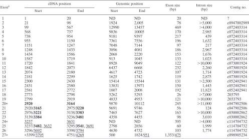
[image:6.587.109.213.73.274.2]Based on the BLAST N and BLAST X homology programs, the organization of the mouse and human Ahi-1 genes was deduced from the comparison of all of the mouse and human Ahi-1 cDNA isoform sequences available from the GenBank Databases. The results of these analyses are shown in Tables 1 and 2. This analysis revealed that theAhi-1gene spans more than 200 kb in a region of the human chromosome 6 and a minimum of 100 kbp (distributed on at least nine contigs) on the mouse chromosome 10 (Fig. 6 and Tables 1 and 2). The mouse or humanAhi-1gene contains at least 27 and 33 exons, respectively. The introns separating exons are between 200 bp and 35 kbp long. The exons range in size from 26 to 560 bp, excluding exons 33 (human) and 27 (mouse), which constitute a large 3⬘-untranslated region. The exonic sequences are iden-tical to the human and mouse Ahi-1 cDNA sequences with the
FIG. 4. Interspecies conservation of theAhi-1gene. DNAs (20g) from various species were digested withEcoRI and analyzed by South-ern blot with the ratAhi-1cDNA fragment B as a probe. Hybridization and washing were done as described in Materials and Methods. Lanes:1, mouse; 2, rat; 3, human; 4, hamster; 5, cat; 6, cow; M,HindIII/ EcoRI-digestedDNA.
on November 8, 2019 by guest
http://jvi.asm.org/
exception of a few nucleotides. The seven WD40 repeats are distributed in seven exons: each of the putative WD40 repeats is encoded mainly by a single exon (exons 11 to 17 for mice and exons 14 to 20 for humans). Exon 26 (human) or 22 (mouse) encodes the SH3 domain.
Differential splicing of the mouse and human Ahi-1 exons.
[image:7.587.115.465.79.523.2]The availability of the genomic organization of theAhi-1gene (Fig. 6) and of different forms of its encoded cDNA isoforms (Fig. 5) allowed an analysis of alternative exon usage. The full-length mouseAhi-1cDNA (isoform I) that was cloned in
FIG. 5. Structure of the mouse and humanAhi-1cDNA isoforms. (A) Schematic alignment of mouse and humanAhi-1cDNA clones. The numbers refer to the positions of the nucleotides. The open reading frames are indicated by thick bars, boxes, and triangles, and the untranslated sequences are indicated by thin lines. In-frame stop codons are indicated by asterisks. The polyadenlyation signals are shown for some cDNAs. All strippled bars represent alternative exons: exons 24 and 20 in mouse and exons 24 and 28 in humanAhi-1cDNA. Note that the 5⬘end (nt 1 to 402;䊐) and the 3⬘end (nt 3190 to the end) sequences of the full-length mouse Ahi-1 are more divergent from the corresponding human Ahi-1 sequences. Also note that the 5⬘end sequences (nt 271 to 604; aa 19 to 604; shaded bar) of human Ahi-1 are not present in mouse Ahi-1. The presence of exon 24 (containing a termination codon) in human Ahi-1 cDNA has been confirmed by RT-PCR analysis on a Jurkat cell RNA with primer 897 within the region spanning exons 23 and 24 and primer 898 in exon 24. Similarly, the presence of exon 20 (containing a termination codon) in the mouseAhi-1gene was confirmed by RT-PCR on brain RNA with 5⬘primer 264 in exon 19 and 3⬘primer 1312 in exon 24. Note that the 5⬘-untranslated sequences (nt 1 to 250) of the human Ahi-1 identified in our laboratory have been added to the nucleotides listed in the GenBank for each EST clone (AL136797, AK000076, and AK024085). (B) Nucleotide sequence of the 5⬘-untranslated humanAhi-1. RT-PCR from Jurkat cells was performed with the primers 910 and 899 located, respectively, in exons 1 and 4. The PCR products were cloned and sequenced. The putative ATG translation initiation codon is underlined. Vertical arrrows indicate intron-exon boundaries. Uppercase letters, coding sequences; lowercase letters, untranslated nucleotides. (C) Genomic organization of the 5⬘-untranslated region of human Ahi-1. Boxes indicate exons. The lines between the boxes represent introns. Translation initiation codon is indicated over exon 4.
9052 JIANG ET AL. J. VIROL.
on November 8, 2019 by guest
http://jvi.asm.org/
our laboratory consists of 25 exons (i.e., exons 1 to 27, exclud-ing exons 20 and 24) with an in-frame termination codon present in exon 27. The EST clone BB615071 (isoform II) lacking the SH3 domain appears to contain 26 exons (i.e., exons 1 to 27, excluding exon 24) with a termination codon in
exon 20. The EST clone BG297436 (isoform III) seems to contain 26 exons (i.e., exons 1 to 27, excluding exon 20), with a termination codon in exon 24.
[image:8.587.126.457.66.533.2]The full-length human Ahi-1 clone AL136797 (isoform I) consists of 29 exons (i.e., exons 1 to 33, excluding exons 24, 28,
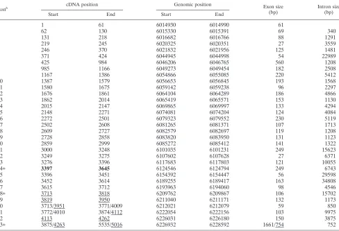
FIG. 6. Genomic organization of the mouseAhi-1gene and mapping of Mis-2 integration sites. (A) Physical map and schematic representation of exons 1 to 27 of the mouseAhi-1gene identified from 11 nonoverlapping combined contigs (A to K) drawn from the preliminary assembly of the mouse WGS reads based on an 15 October 2001 freeze at the Sanger Center and Whitehead Institute genome projects data bank. The contigs were as follows: B, c094702989 (7,308 bp); C, c072403314 (15,675 bp); D, c013001924 (9,046 bp); E, c052402941 (16,394 bp); F, 203795 (5,800 bp); G, c038402794 (11,198 bp); H, c047902506 (15,808 bp); I, c030002862 (8,235 bp); J, c114704732 (4,962 bp); and K, c098905234 (1,959 bp). The 5⬘ and 3⬘end ofAhi-1and c-mybgenes are indicated. The Ahi-1 and Mis-2 provirus integration sites are indicated. Black boxes represent coding exons, and the white box represents an untranslated exon. Strippled boxes are the rat DNA fragments (Fr 1 to 5) previously subcloned (62) around the Mis-2 provirus integrations. The exons coding for the SH3 and WD40 domains are also shown. Symbols: M, initiation codon; asterisks, termination codons. (B and C) Mapping and genomic organization of the proviruses integrated at theMis-2locus in tumors 1213-2 (B) and 1213-19 (C). Note the large deletion of the provirus up to the end of theenvgene, with the remaining 3⬘endenvand LTR in tumor 1213-2. Also note the perfect direct repeats (D.R.; 228 bp) flanking the deleted provirus. Black boxes represent exons 16 and 17, and the line represents the intron sequences. (D) Sequences of the intron-exon 14 and 15 junctions in rat DNA. Uppercase and lowercase letters: exon and intron sequences, respectively.
on November 8, 2019 by guest
http://jvi.asm.org/
29, and 32). The first three exons were cloned in our laboratory by RT-PCR from Jurkat cells (Fig. 5). The termination codon is located in exon 33. The clone AK000076 (isoform II) lacking the SH3 domain appears to be made of 24 exons (i.e., exons 1 to 24) and contain an in-frame stop codon in exon 24. The clone AK024085 (isoform III) consists of 32 exons (i.e., exons 1 to 33, excluding exon 24) with a termination codon present in exon 28.
Mapping of the Mis-2 provirus insertion sites within the 16th intron of Ahi-1.We have previously identified Mis-2 as a common provirus insertion site in Moloney MuLV-induced rat thymomas (62) and found that it maps to mouse chromosome 10. Long-range restriction enzyme mapping analysis revealed
thatMis-2mapped at a distance of ca. 160 kbp of theAhi-1
insertion site (62). Considering the length of the human and mouseAhi-1 genes, we predicted that the Mis-2integration sites would map within theAhi-1 gene itself. To test this hy-pothesis, we sequenced the rat genomic region adjacent to
Mis-2integration sites in tumor 1213-2 and 1213-19 (Fr 1 to 5,
Fig. 6A) (62). This analysis confirmed the site of the integra-tion and revealed a high degree of homology of the ratAhi-1 sequences with those of the mouseAhi-1around exons 14, 15, and 16 (contig D, Fig. 6). These results, combined with our previous mapping of theMis-2provirus integration sites, indi-cated that theMis-2locus corresponds to insertions within the intron 16 of theAhi-1gene.
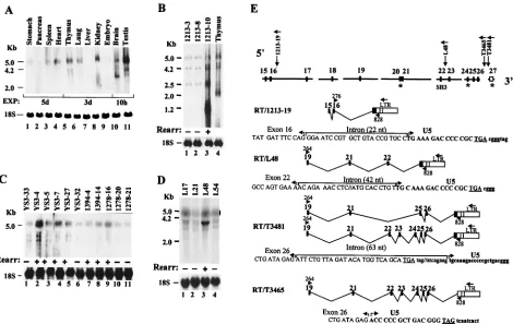
Expression of the Ahi-1 gene in rodent tissues. Northern blot analysis was performed on RNA extracted from various tissues of adult rat and mouse. This analysis revealed the pres-ence of Ahi-1 transcripts hybridizing with a probe derived from the 5⬘ end of mouse Ahi-1 cDNA (probe E1.3) (Fig. 2B) in several tissues (Fig. 7A). Ahi-1 expression was much higher in the brain and testis than in other organs but was especially low in the liver. In addition, various Ahi-1 RNA species were present in different tissues, suggesting different splicing vari-ants: a major hybridizing species of 5 kb in several mouse tissues tested and a less intensely hybridizing RNA of 3.5 kb in brain, 4.2 and 3.5 kb in testis, and 2 kb in embryos (Fig. 7A). In the rat thymus (Fig. 7B, lane 4) and in the brain and testis (data not shown), two major species of 5 and 2 kb could be detected with a 3⬘end ratAhi-1cDNA clone A (Fig. 2A) as a probe. In rat spleen, shorter (1.2 and 0.7 kb) and more abun-dant RNA species were detected (data not shown). These results indicated that theAhi-1 gene expresses several RNA species, a finding consistent with the various spliced variants cDNA identified (Fig. 5).
Expression of Ahi-1 in tumors with provirus insertions.To determine whether provirus integration at the Mis-2 or the
Ahi-1locus led to deregulation of theAhi-1 gene expression,
we first examinedAhi-1expression in T- and B-cell tumors by Northern blot analysis with a 3⬘end rat cDNA probe (probe B) or 5⬘ end mouse cDNA probe (probe E1.3) (Fig. 2),
respec-TABLE 1. Exon-intron boundaries of the mouseAhi-1genea
Exonb cDNA position Genomic position Exon size
(bp) Intron size(bp) Contig no.
Start End Start End
1 1 20 ND ND 20 ND ?
2 21 98 1924 2,005 78 ⬎5,000 c0947002989
3 99 567 12990 13457 469 ⬎4,000 c072403314
4 568 737 9836 10005 170 2,985 c072403314
5 738 954 9181 9397 217 439 c072403314
6 955 1150 7361 7556 196 1,632 c072403314
7 1151 1247 7048 7144 97 217 c072403314
8 1248 1433 3896 4081 186 2,967 c072403314
9 1434 1586 2068 2220 153 1,676 c072403314
10 1587 1719 913 1045 133 1,023 c072403314
11 1720 1841 8928 9049 122 ⬎10,000 c073001924
12 1842 2073 6437 6668 232 2,260 c073001924
13 2074 2180 4617 4723 107 1,714 c073001924
14 2181 2299 1625 1742 119 2,875 c073001924
15 2300 2430 15414 15544 131 ⬎2,500 c052402941
16 2431 2580 13831 13971 150 1,443 c052402941
17 2581 2772 1807 2008 192 11,823 c052402941
18 2773 2798 5262 5293 26 ⬎7,000 203795
19 2799 2919 4833 4953 121 ⬎10,000 2033793
20ⴱ 2920 3164 9870 10112 245 ⬎11,000 c047902506
21 2920/3165 2975/3220 9691 9746 56 124 c047902506
22 2976/3221 3138/3383 7465 7627 163 ⬎10,000 c030002862
23 3139/3384 3236/3481 4358 4455 98 3,010 c030002862
24ⴱ 3237 3631 ND ND 395 ⬎4,000 c114704732
25 3237/3482, 3632 3295/3541, 3691 2798 2856 60 1,999 c114704732
26 3296/3692 3398/3794 4630 4732 103 1,774 c114704732
27ⴱ ⴱ3399/3795 4751/4269 500 1824/951 1,353/475 c098905234
aThe full-length mouseAhi-1cDNA (isoform I) consists of 25 exons, from 1 to 27, excluding exons 20 and 24; EST clone BB615071 (isoform II) appears to contain
24 exons, from 1 to 25, excluding exon 24; EST clone BG297436 and clone I (isoform III) may consist of 26 exons, from 1 to 27, excluding exon 20. The sizes of exons 20 and 24 are approximately determined. ND, not determined; ?, contigs containing exon 1 were not available in the Genbank database; boldfacing, cDNA position in isoform II; underlining, cDNA position in isoform III.
bⴱ, Exons 20, 24, and 27 contain the termination codon in isoforms II, III, and I, respectively.
9054 JIANG ET AL. J. VIROL.
on November 8, 2019 by guest
http://jvi.asm.org/
[image:9.587.43.540.84.363.2]tively. Many tumors showing rearrangement in a very small percentage of the tumor cells [as judged by the intensity of the rearrangedAhi-1or Mis-2 fragments relative to the germ line fragment (41, 62)] were not suitable for this analysis. There-fore, only tumors harboring a significant proportion of cells showing rearrangement were tested.
In the RNA of Moloney MuLV-induced rat tumors 1213-2 and 1213-19 rearranged inMis-2, four majorAhi-1transcripts (5, 4.2, 2.5, and 1.2 kb) were detected. This pattern of expres-sion was the same in rearranged and nonrearranged tumors, but it was distinct from that of the thymus where the 1.2- or 2.5-kb transcripts were, respectively, not detected or less abun-dant, whereas another 2-kb species was abundant (data not shown). In the rat T-cell thymoma (i.e., 1213-10) which had a provirus inserted in theAhi-1locus, the levels of several RNA species (especially the 1.2-kb form) were higher than those found in unrearranged tumors (Fig. 7B). Analysis of Ahi 1 expression in A-MuLV-induced pre-B cell tumors or in T-cell thymomas from Moloney MuLV-infected MMTVD/myc Tg
mice, which had a rearrangedAhi-1 locus, showed a modest increase ofAhi-15-kb transcript in some, but not all, of these tumors, compared to levels detected in unrearranged tumors (data not shown). Interestingly, some of the tumors also
showed higher levels of shorter Ahi-1 transcripts (⬃4.2, 4, and 3.5 kb) (Fig. 7C). The levels of the shorter 4.2-kbAhi-1 tran-scripts were especially high in the L48 cell line (Fig. 7D, lane 3). Therefore, Northern analysis revealed that the integration of a provirus at the 3⬘ end or in exon 16 of the Ahi-1gene appears to affect only modestly the levels of its transcription in some tumors but significantly in two of the tested tumors.
Since the provirus integration within intron 16 of Ahi-1
(Mis-2 locus) would be expected to cause truncation of the
[image:10.587.51.539.85.418.2]gene, as we previously reported for similar intronic provirus integrations within theNotch-1 gene (16), RT-PCR analysis was also performed to detect such truncated transcripts. For identification of transcripts originating at the 5⬘ end of the gene and terminating at the site of provirus integration, we used primer 276 within exon 16 with primer 828 (i.e., 1213-9) or primer 110 (i.e., 1213-2) within the LTR. This analysis indeed revealed the presence of truncated Ahi-1 transcripts deleted of their half 3⬘end and fused to viral LTR sequences, with the generation of a translation termination codon in tu-mor 1213-19 RNA (Fig. 7E) but not in tutu-mor 1213-2 RNA (data not shown). In tumor 1213-19 RNA, exon 16 is properly spliced and the provirus insertion has forced the utilization of a cryptic splice acceptor site in the Ahi-1 intron, 22 nt just
TABLE 2. Exon-Intron boundaries of the humanAhi-1genea
Exonb cDNA position Genomic position Exon size
(bp) Intron size(bp)
Start End Start End
1 1 61 6014930 6014990 61
2 62 130 6015330 6015391 69 340
3 131 218 6016682 6016766 88 1291
4 219 245 6020325 6020351 27 3559
5 246 370 6021832 6021956 125 1481
6 371 424 6044945 6044998 54 22989
7 425 984 6046206 6046765 560 1208
8 985 1166 6049273 6049454 182 2508
9 1167 1386 6054866 6055085 220 5412
10 1387 1579 6056653 6056845 193 1568
11 1580 1675 6059142 6059238 96 2297
12 1676 1861 6064104 6064289 186 4866
13 1862 2014 6065419 6065571 153 1130
14 2015 2147 6069865 6069997 133 4294
15 2148 2271 6074081 6074204 124 4084
16 2272 2501 6079323 6079552 230 5119
17 2502 2608 6081265 6081371 107 1713
18 2609 2727 6082579 6082697 119 1208
19 2728 2858 6083820 6083950 131 1123
20 2859 2999 6085272 6085412 141 1322
21 3000 3248 6101035 6101231 249 15623
22 3249 3275 6107602 6107628 27 6371
23 3276 3396 6117683 6117803 121 10055
24ⴱ 3397 3645 6124546 6124794 249 6743
25 3396 3451 6154392 6154447 56 29598
26 3452 3614 6189255 6189417 163 34808
27 3615 3712 6193963 6194060 98 4546
28ⴱ 3713 3818 6209762 6209867 106 15702
29 3819 3950 6211040 6211171 132 1173
30 3713/3951 3771/4009 6212021 6212079 59 850
31 3772/4010 3874/4112 6222054 6222156 103 9975
32 4113 4262 6226031 6226180 150 3875
33ⴱ 3875/4263 5535/5016 6226932 6228592 1661/754 752
aThe full-length humanAhi-1(isoform I) consists of 29 exons, from 1 to 33, excluding exons 24, 28, 29, and 32; clone AK000076 (isoform II) seems to contain 24
exons from 1 to 24; clone AK02485 (isoform III) seems to contain 32 exons from 1 to 33, excluding exon 24. All exons are located on the same contig (NT_025741). Boldfacing, cDNA position in isoform II; underlining, cDNA position in isoform III.
bⴱ, Exons 24, 28, and 33 contain the termination codon in isoforms II, III, and I, respectively.
on November 8, 2019 by guest
http://jvi.asm.org/
upstream of the LTR. In tumor 1213-2 DNA, the LTR is inserted in the same transcriptional orientation as that ofAhi-1 and may work as a promoter. RT-PCR amplification with a U5 LTR primer 878 and primers 277 and 265 in exon 17 or 22 did not reveal such 5⬘ viral/Ahi-1 fused transcripts originating in the LTR (data not shown), suggesting that this promoter is silent.
Similar analysis with RNA from tumors having provirus in-tegration at the 3⬘ end of the gene (Ahi-1 locus) was also carried out. In tumors T3465 and T3481, this experiment showed Ahi-1/LTR fused transcripts with the proper utiliza-tion of the exon 26 splice donor site and use of a cryptic splice acceptor site in the viral U5 region (T3465) or in intronic sequences upstream of the LTR (T3481), thus splicing out exon 27 (Fig. 7E). These transcripts would be expected to encode a full-length protein. In the same tumor T3481, an-other interesting truncated Ahi-1/LTR splice variant transcript species was generated by splicing out exons 20, 22, 23, 24, and 27. These transcripts would be expected to encode truncated Ahi-1 proteins deleted of most of their C terminus, notably of their SH3 domain. In tumor L48, this analysis with primer pairs
in exon 19 and in LTR detected Ahi-1/LTR fused transcripts deleted of exons 23 to 27, with the use of a cryptic splice acceptor site in the sequences of intron 22. These transcripts would be expected to encode truncated Ahi-1 proteins deleted of their C terminus but still harboring the SH3 domain. To-gether, these results show that insertion of provirus within the
Ahi-1gene leads to the generation of distinct species of
trun-cated RNA.
DISCUSSION
TheAhi-1gene targeted by provirus insertional mutations encodes a modular protein.We have identified a novel gene
(Ahi-1) mutated by provirus insertions in MuLV-induced
[image:11.587.60.530.74.371.2]tu-mors at two distinct sites, designated Ahi-1 and Mis-2. The 3⬘-end Ahi-1 provirus insertion site comprises two clusters: the more proximal cluster, with proviruses inserted within the last introns, the last noncoding exon, or within sequences just downstream of the last exon, and the distal cluster located⬃16 kbp downstream of the last noncoding exon. Most of these Ahi-1 provirus insertions are located outside the coding region
FIG. 7. Expression of theAhi-1gene in normal tissues and tumors: (A) RNAs (20g) from various organs (lanes 1 to 11, as indicated) of a C57BL/6 mouse were screened by Northern blot analysis withAhi-1cDNA clone-E1.3 as a probe. Exposure time (EXP) is indicated in hours (h) or days (d). (B) RNAs (20g) from Moloney MuLV-induced rat thymomas 1213-3, 1213-8, and 1213-10 (lanes 1 to 3) and rat thymus (lane 4) were hybridized with the32P-labeled rat cDNA probe-B. (C and D) Northern blot analysis of RNAs (20g) from A-MuLV-induced pre-B cell
tumors (lanes 1 to 11) (C) or from cell lines established from thymomas of MMTVD/myc Tg mice infected with Moloney MuLV (D). Hybridization
was with the mouseAhi-1cDNA probe E1.3. (E) Analysis of truncatedAhi-1transcripts. RT-PCR amplification was performed on tumor RNAs (5g) from various tumors showingAhi-1rearrangement. RNAs were reversed transcribed in the presence of random hexamers. The cDNAs were then amplified by PCR with the indicated primers specific for theAhi-1sequences and the viral LTR. The amplified fragments were separated on 1% agarose gels, purified, cloned, and sequenced. In panels A to D, the blots presented were rehybridized with a 18S ribosomal probe (bottom). In panels B to D, the presence (⫹) or absence (⫺) of proviral rearrangement (Rearr) in tumor DNAs is indicated.
9056 JIANG ET AL. J. VIROL.
on November 8, 2019 by guest
http://jvi.asm.org/
ofAhi-1. Surprisingly, all of the mapped proviruses were found to be in an inverse transcriptional orientation relative to that of
theAhi-1gene itself. The second provirus insertion site, Mis-2,
is intronic (within the 16th intron) and is located at a large distance (⬃120 kbp) from the Ahi-1 insertion site. At this Mis-2 site, the proviruses are integrated both in the sense and antisense transcriptional orientation relative to that of the
Ahi-1gene. Therefore, it appears that Ahi-1is the gene
tar-geted by these insertional mutational events.
The structure of the predicted protein encoded by theAhi-1 gene is particularly interesting, containing an SH3 domain, WD40 repeats and SH3-binding sites, all known to mediate specific protein-protein interactions. SH3 domains are known to bind to proline-rich motifs (24, 37, 52). Among the best-studied functionally important SH3 interactions is the complex formed between the Grb2/Sem5 SH3 domain and the GDP-GTP exchange protein Sos, which forms a link in a signal transduction pathway that functionally ties tyrosine kinase re-ceptors to Ras (5, 11, 28, 30, 35, 54). Although the functional significance of the presence of a SH3 domain within Ahi-1 is not known at the present time, this protein clearly represents a novel member of the SH3-containing protein family.
The second most evident motif of the Ahi-1 protein is a WD40-repeat domain. Several proteins containing WD40 re-peats have been identified (27, 33, 55, 58). These proteins are associated with multiple and diverse aspects of cellular metab-olism, including signal transduction, regulation of cytoskeletal assembly, cell cycle regulation, RNA processing, programmed cell death, and gene regulation (33). In general, their functions appear to be regulatory in nature. Structural models suggest that the WD40-repeat-containing proteins are endowed with the potential for interactions with other proteins, a finding consistent with the fact that many such proteins are compo-nents of multiprotein complexes (33, 34). The Ahi-1 protein represents a novel member of this family. To our knowledge, Ahi-1 is the only protein reported to date harboring both WD40 repeats and a SH3 motif.
The third important series of motifs predicted in the Ahi-1 amino acid sequences are nine putative SH3-binding sites. Short (8 to 10 aa), proline-rich, and structurally conserved motifs were initially found to bind specifically to the SH3 domain of Abl (7, 43). These SH3-binding sites are present in a variety of proteins and appear to form two distinct classes: a class I motif containing a conserved N-terminal arginine and a class II motif containing a conserved C-terminal arginine (14, 29). The SH3-binding sites of Ahi-1 could mediate its interac-tion with other SH3-containing protein(s). One of these sites may even bind the SH3 domain of Ahi-1 itself, intramolecu-larly. Intramolecular interaction of the Src SH3 domain has been reported (12).
Finally, Ahi-1 harbors other motifs of interest, two potential tyrosine phosphorylation sites, one within the SH3 domain. Phosphotyrosine motifs are known to bind specifically to SH2 domains of proteins (24, 52). If the Ahi-1 protein can be phosphorylated at these sites by a tyrosine kinase, this phos-phorylation may allow it to recruit and bind to other SH2-containing protein(s).
Therefore, Ahi-1 appears to be a modular protein contain-ing several putative motifs that have been shown to be present in several signaling molecules and to mediate protein-protein
interactions. It is tempting to suggest that Ahi-1 may play a role in signal transduction. Because of the high number of these putative motifs mediating protein-protein interactions, Ahi-1 may be a docking site or scaffold protein recruiting a number of other signaling molecules and modulating and in-tegrating their action.
Ahi-1 provirus insertional mutations generate truncated forms of Ahi-1 RNA. The Ahi-1 gene mutations that were analyzed lead to the generation of various forms of transcripts: some fused to viral sequences and some deleted of their 3⬘end. This was expected with the provirus inserted within intron 16
(Mis-2locus), and such molecular rearrangement represents a
mechanism very similar to the one we described previously with proviruses inserted within theNotch1gene (16, 20). How-ever, the generation of similar truncated transcripts in tumors harboring provirus integrated at the 3⬘ end of the gene was more surprising. Some of these 3⬘-end insertional mutations seem to alter the normal splicing machinery and to force an alternative splicing within viral or intronic sequences close to them, thus deleting exons coding for the C terminus of Ahi-1. The molecular requirements for such unusual consequences of provirus integrations at the 3⬘ end of a gene remain obscure but may involve the opposite transcriptional orientation of the inserted proviruses relative to that of theAhi-1gene. Indeed, all of the 27 proviruses inserted within theAhi-1 locus (also recently designated Epi1) are in the same direction (Fig. 1) (2, 41), and we show here that this is in opposite transcriptional orientation relative to that ofAhi-1. Future work on how such insertional mutants may affect the splicing machinery should be informative. Interestingly, some of the truncated transcripts generated in tumors harboring Mis-2 or Ahi-1 provirus inser-tions appear to have a similar coding capacity, both having the capacity to code for Ahi-1 proteins truncated of some C-ter-minal sequences, including of their SH3 domain.
How is theAhi-1gene mutation involved in tumor develop-ment?Genetic evidence suggests that provirus insertional mu-tation of the Ahi-1 gene contributes to tumor formation in different cell lineages. The Ahi-1 locus was identified as a common provirus integration site initially in Abelson (v-abl -induced) pre-B lymphomas (41) and later in c-myc induced T-cell lymphomas of the MMTVD/myc Tg mice (Fig. 1),
sug-gesting that it may collaborate with these oncogenes. This locus has also been found by others to be rearranged by provirus insertion in other types of tumors, namely, in pre B-cell tumors of E/myc Tg mice (18, 61) and in acute myeloid leukemia in Nf1heterozygous mice (2). Similarly, theMis-2locus was iden-tified as a common provirus insertion site in Moloney MuLV-induced rat T-cell lymphomas (thymomas) (62). The fact that these provirus insertional mutations are not random and were identified in a relatively high proportion of tumors (⬃3% for Mis-2,⬃15% for Ahi-1 [Fig. 1] [41], and 44% for Epi1 [2]) suggests that they have been selected during the oncogenic process and that they may be involved in this process. This notion is reinforced by the several examples of common pro-virus insertional sites that have been found to activate protoon-cogenes in nondefective retrovirus-induced tumors (26, 39, 47, 60).
Molecular evidence also suggests that mutation of theAhi-1 gene may be involved in tumor formation. Our analysis of RNA in tumors harboring mutations in theAhi-1gene suggests
on November 8, 2019 by guest
http://jvi.asm.org/
that the mechanism by which these insertional mutations may enhance the oncogenic potential of theAhi-1gene is through the generation of truncated forms of theAhi-1RNA with the capacity to code for truncated Ahi-1 proteins. The levels of these truncated RNA are not very high in theAhi-1rearranged tumors analyzed but are specific to these tumors and are not found in nonrearranged tumors. These are likely to be involved in the transformation process. The deletion of sequences cod-ing for the C terminus, includcod-ing for the SH3 domain in some truncatedAhi-1RNA found in tumors is likely to significantly affect the interaction of Ahi-1 with other proteins, possibly converting this molecule to a dominant-negative mutant. Al-ternatively, truncated Ahi-1 proteins may represent gain-of-function mutants, if the SH3 domain binds intramolecularly, as reported for other SH3-containing molecules (12), or bind to an inhibitor. In nonreceptor tyrosine kinases, such as the proto-oncogene c-srcor c-abl, deletion or mutation of the SH3 domain generally leads to the oncogenic activation of their tyrosine kinase, suggesting that the SH3 domain binds to an inhibitor of the kinase activity (9, 15, 21, 32, 38, 53, 59, 63). Thus, the SH3 deletion in Ahi-1 may uncover some domains and allow novel protein interactions or may prevent the bind-ing of an inhibitor. The fact thatAhi-1cDNA with the poten-tial to encode very similar truncated proteins as the ones de-tected in tumors have been isolated in libraries from normal tissues, suggests that truncated Ahi-1 proteins may not be oncogenic in each tissue. Rather the inappropriate or higher expression of truncated Ahi-1 proteins in some specific cell types may contribute to the transformation process.
Because we were unable to detect truncated transcripts nor Ahi-1/viral fused transcripts in someAhi-1rearranged tumors, it is also quite possible that theAhi-1gene may be involved in oncogenesis by mechanism(s) not yet uncovered and distinct from the generation of truncated gene products. In addition, we cannot exclude the possibility that some proviruses inserted into theAhi-1locus may act by other, as-yet-uncharacterized processes or may affect the expression of other genes sur-rounding Ahi-1 at a distance. This latter mechanism has re-cently been proposed for other provirus insertion sites up-stream of c-myb(19).
It will be interesting to investigate how the Ahi-1 gene, apparently targeted by these proviruses and coding for a pro-tein that shows all of the characteristics of a modular signaling molecule involved in protein-protein interactions, participate in oncogenesis and what is its normal function.
ACKNOWLEDGMENTS
X.J, Z.H, and M.K. contributed equally to this study.
This work was supported by grants from the Medical Research Council of Canada and the National Cancer Institute of Canada to P.J. X.J. was the recipient of a fellowship from McGill University and from Hydro-Quebec.
We thank Alan J. Buckler for providing the pSPL1 vector. We thank Benoıˆt Laganie`re and Patrick Couture for excellent technical assis-tance. We are also grateful to Rita Gingras for preparing the manu-script.
REFERENCES
1.Abelson, H. T., and L. S. Rabstein.1970. Lymphosarcoma: virus-induced
thymic-independent disease in mice. Cancer Res.30:2213–2222.
2.Blaydes, S. M., S. C. Kogan, B. T. Truong, D. J. Gilbert, N. A. Jenkins, N. G. Copeland, D. A. Largaespada, and C. I. Brannan.2001. Retroviral
integra-tion at the Epi1 locus cooperates with Nf1 gene loss in the progression to
acute myeloid leukemia. J. Virol.75:9427–9434.
3.Boss, M., M. Greaves, and N. Teich. 1979. Abelson virus-transformed
haematopoietic cell lines with pre-B-cell characteristics. Nature278:551–553.
4.Buckler, A. J., D. D. Chang, S. L. Graw, J. D. Brook, D. A. Haber, P. A. Sharp, and D. E. Housman.1991. Exon amplification: a strategy to isolate mammalian genes based on RNA splicing. Proc. Natl. Acad. Sci. USA
88:4005–4009.
5.Chardin, P., J. H. Camonis, N. W. Gale, L. van Aelst, J. Schlessinger, M. H. Wigler, and D. Bar-Sagi.1993. Human Sos1: a guanine nucleotide exchange
factor for Ras that binds to GRB2. Science260:1338–1343.
6.Chomczynski, P., and N. Sacchi.1987. Single-step method of RNA isolation by acid guanidinium thiocyanate-phenol-chloroform extraction. Anal.
Bio-chem.162:156–159.
7.Cicchetti, P., B. J. Mayer, G. Thiel, and D. Baltimore.1992. Identification of a protein that binds to the SH3 region of Abl and is similar to Bcr and
GAP-rho. Science257:803–806.
8.Cooper, J. A., F. S. Esch, S. S. Taylor, and T. Hunter.1984. Phosphorylation sites in enolase and lactate dehydrogenase utilized by tyrosine protein
ki-nases in vivo and in vitro. J. Biol. Chem.259:7835–7841.
9.Dai, Z., and A. M. Pendergast.1995. Abi-2, a novel SH3-containing protein, interacts with the c-Abl tyrosine kinase and modulates c-Abl transforming
activity. Genes Dev.9:2569–2582.
10.Dingwall, C., and R. A. Laskey.1991. Nuclear targeting sequences: a
con-sensus? Trends Biochem. Sci.16:478–481.
11.Egan, S. E., B. W. Giddings, M. W. Brooks, L. Buday, A. M. Sizeland, and R. A. Weinberg.1993. Association of Sos Ras exchange protein with Grb2 is implicated in tyrosine kinase signal transduction and transformation. Nature
363:45–51.
12.Erpel, T., G. Superti-Furga, and S. A. Courtneidge.1995. Mutational anal-ysis of the Src SH3 domain: the same residues of the ligand binding surface
are important for intra- and intermolecular interactions. EMBO J.14:963–
975.
13.Feinberg, A. P., and B. Vogelstein.1983. A technique for radiolabeling DNA restriction endonuclease fragments to high specific activity. Anal. Biochem.
132:6–13.
14.Feng, S., J. K. Chen, H. Yu, J. A. Simon, and S. L. Schreiber.1994. Two binding orientations for peptides to the Src SH3 domain: development of a
general model for SH3-ligand interactions. Science266:1241–1247.
15.Franz, W. M., P. Berger, and J. Y. Wang.1989. Deletion of an N-terminal regulatory domain of the c-abl tyrosine kinase activates its oncogenic
poten-tial. EMBO J.8:137–147.
16.Girard, L., Z. Hanna, N. Beaulieu, C. D. Hoemann, C. Simard, C. A. Kozak, and P. Jolicoeur.1996. Frequent provirus insertional mutagenesis of Notch1
in thymomas of MMTVD/myc transgenic mice suggests a collaboration of
c-myc and Notch1 for oncogenesis. Genes Dev.10:1930–1944.
17.Green, P. L., D. A. Kaeler, and R. Risser.1987. Clonal dominance and progression in Abelson murine leukemia virus lymphomagenesis. J. Virol.
61:2192–2197.
18.Haupt, Y., W. S. Alexander, G. Barri, S. P. Klinken, and J. M. Adams.1991. Novel zinc finger gene implicated as myc collaborator by retrovirally
accel-erated lymphomagenesis in E-myc transgenic mice. Cell65:753–763.
19.Haviernik, P.2002. Linkage on chromosome 10 of several murine retroviral
integration loci associated with leukaemia. J. Gen. Virol.83:819–827.
20.Hoemann, C. D., N. Beaulieu, L. Girard, N. Rebai, and P. Jolicoeur.2000.
Two distinctNotch1mutant alleles are involved in the induction of T-cell
leukemia in c-myctransgenic mice. Mol. Cell. Biol.20:3831–3842.
21.Jackson, P., and D. Baltimore.1989. N-terminal mutations activate the
leukemogenic potential of the myristoylated form of c-abl. EMBO J.8:449–
456.
22.Jiang, X., L. Villeneuve, C. Turmel, C. A. Kozak, and P. Jolicoeur.1994. The Myb and Ahi-1 genes are physically very closely linked on mouse
chromo-some 10. Mammalian Genome5:142–148.
23.Kemp, B. E., and R. B. Pearson.1990. Protein kinase recognition sequence
motifs. Trends Biochem. Sci.15:342–346.
24.Koch, C. A., D. Anderson, M. F. Moran, C. Ellis, and T. Pawson.1991. SH2 and SH3 domains: elements that control interactions of cytoplasmic
signal-ing proteins. Science252:668–674.
25.Kozak, M.1986. Point mutations define a sequence flanking the aug initiator
codon that modulates translation by eukaryotic ribosomes. Cell44:283–292.
26.Kung, H. J., C. Boerkoel, and T. H. Carter.1991. Retroviral mutagenesis of cellular oncogenes: a review with insights into the mechanisms of insertional
activation. Curr. Top. Microbiol. Immunol.171:1–25.
27.Li, D., and R. Roberts.2000. WD-repeat proteins:structure characteristics-,biological function, and their involvment in human diseases. Cell. Mol. Life
Sci.58:2085–2097.
28.Li, N., A. Batzer, R. Daly, V. Yajnik, E. Skolnik, P. Chardin, D. Bar-Sagi, B. Margolis, and J. Schlessinger.1993. Guanine-nucleotide-releasing factor hSos1 binds to Grb2 and links receptor tyrosine kinases to Ras signaling.
Nature363:85–88.
29.Lim, W. A., F. M. Richards, and R. O. Fox.1994. Structural determinants of
9058 JIANG ET AL. J. VIROL.