methyltransferase SET7/9
.
White Rose Research Online URL for this paper:
http://eprints.whiterose.ac.uk/111/
Article:
Xiao, B., Jing, C., Wilson, J.R. et al. (7 more authors) (2003) Structure and catalytic
mechanism of the human histone methyltransferase SET7/9. Nature, 421 (6923). pp.
652-656. ISSN 0028-0836
https://doi.org/10.1038/nature01378
[email protected]
https://eprints.whiterose.ac.uk/
Reuse
Unless indicated otherwise, fulltext items are protected by copyright with all rights reserved. The copyright
exception in section 29 of the Copyright, Designs and Patents Act 1988 allows the making of a single copy
solely for the purpose of non-commercial research or private study within the limits of fair dealing. The
publisher or other rights-holder may allow further reproduction and re-use of this version - refer to the White
Rose Research Online record for this item. Where records identify the publisher as the copyright holder,
users can verify any specific terms of use on the publisher’s website.
Takedown
If you consider content in White Rose Research Online to be in breach of UK law, please notify us by
28. Ruban, A. V., Lee, P. J., Wentworth, M., Young, A. J. & Horton, P. Determination of the stoichiometry and strength of binding of xanthophylls to the photosystem II light harvesting complexes.J. Biol. Chem.274,10458–10465 (1999).
29. Laemmli, U. K. Cleavage of structural proteins during the assembly of the head of bacteriophage T4.
Nature227,680–685 (1970).
30. Andersson, J., Walters, R. G., Horton, P. & Jansson, S. Antisense inhibition of the photosynthetic antenna proteins CP29 and CP26: implications for the mechanism of protective energy dissipation.
Plant Cell13,1193–1204 (2001).
AcknowledgementsWe wish to thank R. Walters for discussions. This work was supported by the UK Biotechnology and Biological Sciences Research Council, the UK Joint Infrastructure Fund, the Netherlands Foundation for Scientific Research (NWO) through the Foundation for Life and Earth Sciences (ALW), and the Swedish Research Council for Environment, Agricultural Sciences and Spatial Planning and the Foundation for Strategic Research.
Competing interests statementThe authors declare that they have no competing financial interests.
[image:2.612.303.554.306.631.2]Correspondenceand requests for materials should be addressed to P.H. (e-mail: [email protected]).
...
Structure and catalytic mechanism of
the human histone methyltransferase
SET7/9
Bing Xiao*, Chun Jing*, Jonathan R. Wilson*, Philip A. Walker*, Nishi Vasisht*, Geoff Kelly*, Steven Howell*, Ian A. Taylor*, G. Michael Blackburn†& Steven J. Gamblin*
*Structural Biology Group, National Institute for Medical Research, Mill Hill, London NW7 1AA, UK
†Department of Chemistry, Krebs Institute, University of Sheffield, Sheffield S3 7HF, UK
...
Acetylation1,2, phosphorylation3and methylation4of the
amino-terminal tails of histones are thought to be involved in the regulation of chromatin structure and function5–7. With just
one exception8,9, the enzymes identified in the methylation of
specific lysine residues on histones (histone methyltransferases) belong to the SET family10. The high-resolution crystal structure
of a ternary complex of human SET7/9 with a histone peptide and cofactor reveals that the peptide substrate and cofactor bind on opposite surfaces of the enzyme. The target lysine accesses the active site of the enzyme and the S-adenosyl-L-methionine
(AdoMet) cofactor by inserting its side chain into a narrow channel that runs through the enzyme, connecting the two surfaces. Here we show from the structure and from solution studies that SET7/9, unlike most other SET proteins, is exclu-sively a mono-methylase. The structure indicates the molecular basis of the specificity of the enzyme for the histone target, and allows us to propose a model for the methylation reaction that accounts for the role of many of the residues that are invariant across the SET family.
Many SET proteins have now been characterized biochemically and several have been the subject of X-ray structure analysis: SET7/9 from human11 and its complex with the product S-adenosyl-L
-homocysteine (AdoHcy)12; Dim-5 fromNeurospora crassa6; Rubisco
large subunit methyltransferase (LSMT) from pea with AdoHcy13;
and Clr4 fromSchizosaccharomyces pombe14. SET proteins can be
classified according to the lysine residues that they target on histones H3, H4 and H2A4, and it is apparent that methylation at
these different sites gives rise to distinct biological effects. An additional level of complexity is that lysine residues may be mono-, di- or tri-methylated and that these distinct species lead
to different signalling events. For example, inSaccharomyces cere-visiae, although di-methylation of Lys 4 on histone H3 is present at both active and inactive euchromatic genes, tri-methylation is linked exclusively to active genes15.
NMR studies (see Supplementary Information) indicated that a histone peptide containing mono-methylated Lys 4 was better ordered in complex with SET7/9 than unmodified peptide. We therefore used the products of the normal histone methyltransferase (HMT) reaction for crystallization experiments (methylated lysine peptide and AdoHcy). In our previous studies11we obtained useful
SET7/9 crystals only from constructs lacking the small carboxy-terminal segment, which is, nevertheless, essential for the catalytic activity of the enzyme. Here, we have used a catalytically active construct that contains the complete C-terminal segment. We obtained well-ordered crystals of the ternary complex of SET7/9 that diffracted to at least 1.7 A˚ spacing, and the structure was readily solved by molecular replacement. The C-terminal segment, the AdoHcy cofactor and most of the substrate peptide are well defined in the electron density maps, as are all the important residues around the active site. The overall structure of the ternary complex
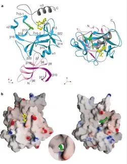
of SET7/9 is shown in Fig. 1 (see Supplementary Information for crystallographic statistics).
The most notable feature of the catalytic structure is that the AdoHcy and the peptide substrate are located on opposite sides of the SET domain and that there is a narrow channel passing through the enzyme that connects the peptide and cofactor binding surfaces. The target lysine residue of the substrate (Lys 4) is inserted into this channel so that its amine can access the methyl donor (AdoMet; see Fig. 1b). The packing of the C-terminal segment against the SET domain is required to form the lysine access channel, which explains
why this feature has not been observed in previous HMTase structures (although it was noted in the context of Rubisco LSMT, as was the presence of a molecule of HEPES in the channel)13. The
[image:3.612.307.557.193.654.2]C-terminal segment (residues 345–366) is organized into two structural features. Residues 337–349, belonging mainly to the SET domain, form an approximate b-hairpin structure that protrudes at a right angle to the surface of the enzyme. This is followed by three residues that accommodate a sharp bend in the polypeptide chain before the final stretch of the protein that adopts ana-helical conformation (Fig. 1a). Tyr 335 and Tyr 337, located
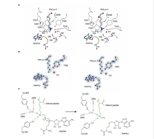
Figure 2Active site of SET7/9.a, Stereographic representation of selected active-site residues together with methylated Lys 4 (MeLys 4), AdoHcy and several main-chain carbonyl groups oriented towards the lysine amine.b, Electron density (2Fo2Fc) covering part of the active site; the view is in the same orientation as ina.c, Schematic diagram of the reaction. The structure shows that the lysine has been stripped of all solvent molecules except for the one water used as a hydrogen-bond acceptor to orient the amino group. This desolvation will lower the pKaof the lysine amino group and also enhance its nucleophilicity. At the same time, the local orientation of the dipoles of main-chain carbonyl groups towards the nitrogen will stabilize the developing positive charge on
[image:3.612.51.562.197.660.2]just before the C segment, are both important for the formation of the lysine access channel. The arrangement of theb-hairpin is such that it stabilizes the conformation of these two tyrosine residues while also contributing to one of the sides of the groove into which the peptide binds. The second side of the peptide-binding groove is made up by residues 255–268 (includingb-17). Thea-helix at the end of the C-terminal segment packs against b-19, particularly Phe 299 (located just beyond the conserved NHS signature motif)16,
and makes hydrophobic packing interactions with the AdoHcy cofactor through Trp 352.
From the high-resolution structure presented here, and from the previous co-crystal structures of Rubsico LSMT/AdoHcy13 and
SET7/AdoHcy12, it is apparent that in our earlier studies the
orientation of AdoMet, which we modelled into a low-resolution difference map, was incorrect. The mode of cofactor binding in our present structure is essentially identical to that described pre-viously12,13. In this alignment, the methyl group to be transferred
from the AdoMet to the amine is pointing into the lysine-binding channel (see Fig. 2a). At the peptide-binding site, the channel surface is largely made up by the side chains of Leu 267 and Tyr residues 305, 335 and 337 (Fig. 2a). The alkyl component of the lysine side-chain therefore inhabits a hydrophobic environment. The other end of the channel, where it opens onto the cofactor-binding surface, is dominated by four tyrosine OH groups (Tyr 245, 305, 335, 337) and five main-chain carbonyl groups, all approxi-mately oriented towards the lysine amine group. There are also several well-defined water molecules in the vicinity of the active site, although only one is close to the lysine amine. The structure, with its catalytic and binding surfaces, explains the role of a number of invariant residues whose mutation to alanine has been shown to abolish HMTase activity without significantly affecting the affinity for substrate or cofactor binding. The aromatic ring of Tyr 335 forms a significant part of the binding cavity for the alkyl portion of the Lys 4PEPresidue (histone peptide substrate) while its side-chain
oxygen makes a single hydrogen bond to the main-chain carbonyl of residue 295. The importance of the correct positioning of this tyrosine residue is underscored by the observation that His 297 (of the NHS signature) acts to stabilize its conformation by acting as a hydrogen-bond acceptor to its main-chain amide (Fig. 2a).
The electron density for the methyl Lys 4PEPis very well defined
and clearly shows the location of the single methyl group (Fig. 2b). Examination of the active site also reveals that the lysine amine 30 min
[image:4.612.38.285.229.663.2]60 min
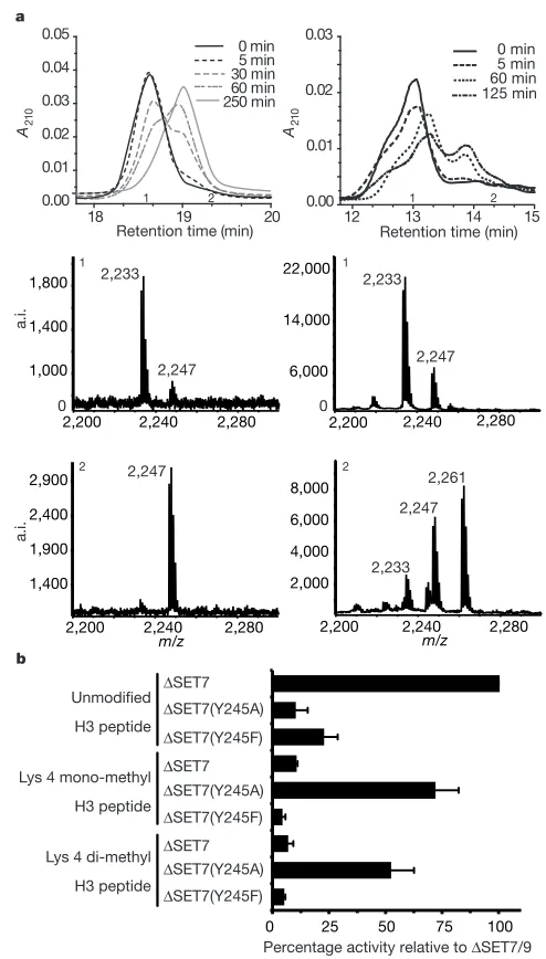
[image:4.612.312.552.416.702.2]Figure 3Activity of SET7/9.a, Analysis of methylation reaction products. The left-hand panels are for SET7/9; the right-hand panels are for G9a. The upper panel shows methylation of an H3 peptide at various time intervals as resolved by high-performance liquid chromatography (HPLC). The middle and lower panels show representative matrix-assisted laser desorption/ionization–time-of-flight (MALDI–TOF) analysis of fractions collected from the HPLC. Examples are from fraction points 1 and 2, respectively.A210, absorbance at 210 nm; a.i., arbitrary intensity.b, Histone methyltransferase assay of SET7/9 and the point mutations Y245A and Y245F, with H3 peptide as substrate either unmodified, mono- or di-methylated at Lys 4.
Figure 4Peptide interactions with SET7/9.a, Schematic representation of interactions made by the histone H3 peptide in complex with SET7/9. The positions of residues beyond theþ1 position are not shown because they are affected by lattice contacts.
donates hydrogen bonds to both the invariant Tyr 245 and to a tightly bound water molecule (W1). We are confident that the lysine is acting as a hydrogen-bond donor in both cases because of the nature of the other hydrogen bonds made by Tyr 245 and W1 (Fig. 2a). The orientation of the lysine amine group is such that the amine-methyl bond is aligned towards the sulphur atom of the AdoHcy. Moreover, it is directed at sulphur along the tetrahedral vector corresponding to the (S)-location of the methyl group in AdoMet17. In other words, the geometry is exactly that expected for
the trajectory of the methylation reaction and this feature will provide an important entropic contribution towards enzyme-mediated catalysis (Fig. 2c).
Our previous data11suggested that SET7/9 could not use
mono-methyl Lys 4 peptide as a substrate for further mono-methylation. Thus we have further analysed the methylation reaction. Reaction mixtures were set up with unmodified peptide and AdoMet, and conditions found where the reaction was driven essentially to completion. As shown in Fig. 3a, high-performance liquid chromatography (HPLC) separation of the reaction products resulted in a peak that eluted at a different position to the unmodified peptide, and matrix-assisted laser desorption/ionization (MALDI) analysis of this material revealed only mono-methylated peptide. Taken with our previous data, these results support the conclusion that SET7/9 catalyses the addition of a single methyl group to its target lysine. The reason for SET7/9 acting as a mono-methyltransferase is apparent from our crystal structure. The peptide used for crystal-lization was synthesized using mono-methylated lysine at position 4. In the crystal structure there is well-defined electron density for this methyl group in just one position. The arrangement of the two hydrogen-bond acceptor groups (for the lysine amine) provides an immediate explanation for the lack of rotation about the CE–NZ bond; Tyr 245 and W1 not only make favourable interactions that stabilize the observed rotamer, but they also preclude, on steric grounds, a methyl group in either of the two other positions that would orient a lone pair of electrons on the lysine nitrogen towards the sulphur of the AdoMet. Thus the structure shows that the arrangement of protein side chains and, indirectly, water molecules at the active site of SET7/9 is such that it can only catalyse the addition of a single methyl group to the lysine amine.
Inspection of the sequence of other SET proteins4near the active
site suggests why many of these enzymes catalyse di- or tri-methylation of their target lysines. Only Tyr 245 and Tyr 335 are considered invariant across the SET family, many other residues are highly variable. So, for example, Tyr 305 is substituted for a valine residue in SUV39H1 (a tri-methylase) and a proline in G9a (a di-methylase). Both of these substitutions would seem likely to produce a cavity at the active site that could accommodate a methyl substituent on the lysine amine. We have previously reported that the Y245A mutation in SET7/9 leads to a marked reduction in HMTase activity11. We now show that although this mutant has
greatly reduced activity with respect to an unmodified lysine substrate, it has substantial activity if assayed with mono- or with di-methylated Lys 4 substrate (Fig. 3b). Thus, Y245A is able to convert mono-methylated substrate to tri-methylated product. The structure readily provides a rationale for these observations. The chemistry of methyl transfer dictates that for di- and then for tri-substitution to occur, the existing methyl groups on the lysine would first have to be positioned approximately either at the OH of Tyr 245 or W1 sites, and finally in both of these positions. Removal of the aromatic ring of Tyr 245 creates a site for the first attached methyl group, thus enabling a second addition. The fact that the mutant enzyme can generate tri-methylated lysine further implies that the Y245A mutation also disrupts the local structure, so that W1 is displaced, allowing di-methylated lysine to bind appropri-ately for a third methylation.
The histone peptide binds in a largely extended conformation into a shallow groove, as shown in Fig. 1b. The binding is mediated
by a network of hydrogen bonds and salt bridges, involving both the main chain and side chains of the peptide (Fig. 4a). The target Lys 4PEPresidue is located approximately at the centre of the defined
peptide, and the interactions of non-conserved SET7/9 residues with the peptide seems to account for the enzyme’s specificity. Arg(22)PEP (residues are numbered relative to the target lysine)
forms a hydrogen bond with the main-chain carbonyl of residue 258, such that the alkyl component of its side chain packs against the face of Trp 260. Thr(21)PEPforms a hydrogen bond with Ser 268,
and Gln(þ1)PEP forms a hydrogen bond with the side chain of
Lys 317. Arg(22)PEPtherefore seems to have a particularly
import-ant role in mediating substrate specificity. Moreover, inspection of the various histone target sequences (Fig. 4b) shows that only the Lys 4 site of histone H3 contains a basic residue in the22 position. The other histone target sites all have a basic residue either at the21 orþ1 position. Both activity measurements11and peptide affinity
measurements, using a peptide in which Arg(22)PEPis mutated to
an alanine (data not shown), support the hypothesis that this arginine residue contributes significantly to peptide binding to SET7/9. It seems that the structure of the ternary complex of SET7/9 presented here will provide a good model for assessing the probable substrate specificity of other SET proteins from their
primary sequence. A
Methods
Protein constructs
DSET7/9 (residues 52–366) and G9a (621–1000) were expressed as glutathione
S-transferase (GST) fusions in pGEX 6P1 inEscherichia coliBL21. The GST was removed by overnight treatment with PreScission Protease (Amersham) before gel filtration. Preparation ofDSET7/9 in D2O for NMR studies resulted in a series of N-terminal degradation products that remained catalytically active. Subsequently,DDSET7/9 (108–366) was prepared as above and found to be stable for growth in D2O, and was consequently used for further NMR and crystallography experiments. Site-directed alanine mutations were introduced using the Stratagene Quikchange mutagenesis kit, and mutations were confirmed by DNA sequencing and electrospray mass spectrometry. Synthetic peptides were prepared by W. Mawby, and AdoHcy was obtained from Fluka.
Crystallography
Protein stock solution was prepared at 100 mg ml21in 50 mM Tris, pH 7.0, 100 mM NaCl, and then incubated with a twofold molar excess of mono-methylated Lys 4 ten-residue peptide and AdoHcy. Crystals were grown by vapour diffusion at room temperature as hanging drops. Drops were prepared by mixing equal volumes of protein complex with reservoir solution containing 0.1 M Tris, pH 7.8, and 22% PEG 3350. Crystals were first transferred into mother liquor augmented with an additional 5% PEG 400, before plunging into liquid nitrogen. Data were collected from flash-cooled crystals at 100K on a Raxis-II detector mounted on a Rigaku RU200 generator. Diffraction data were integrated and scaled using DENZO and SCALEPACK18. The structure was solved by molecular replacement using our previous model (1H3I.brk) with AMORE. Subsequent refinement was carried out using REFMAC519and manual model building in O20.
NMR
NMR spectra were recorded at 258C on Varian Inova spectrometers operating at1H frequencies of 600 MHz and 800 MHz. Protein samples (approximately 0.5 mM) were prepared in 50 mM Tris-HCl, 0.5 mM Tris(2-carboxyethyl)phosphine hydrochloride, 10% D2O, pH 6.5.
HMTase activity measurements
The methyltransferase activity of SET7/9 and mutant constructs described in the text were determined in a reaction volume of 20ml containing 3mM AdoMet supplemented with [methyl-3H]AdoMet (4
mCi) (Amersham) and 750 nM purified methylase in reaction buffer (50 mM Tris, pH 8.5, 100 mM NaCl, 1 mM EDTA, 1 mM dithiothreitol (DTT)) with 50mM histone peptide. After incubation at 378C for 60 min the reaction was vacuum-blotted onto membrane (Hybond-C; Amersham), washed, and the activity was measured by scintillation counting.
Analytical analysis of histone methylation
Received 22 November; accepted 23 December 2002; doi:10.1038/nature01378. Published online 22 January 2003.
1. Grunstein, M. Histone acetylation in chromatin structure and transcription.Nature389,349–352 (1997).
2. Turner, B. M. Histone acetylation and an epigenetic code.Bioessays22,836–845 (2000). 3. Berger, S. L. Histone modifications in transcriptional regulation.Curr. Opin. Genet. Dev.12,142–148
(2002).
4. Lachner, M. & Jenuwein, T. The many faces of histone lysine methylation.Curr. Opin. Cell Biol.14,
286–298 (2002).
5. Strahl, B. D. & Allis, C. D. The language of covalent histone modifications.Nature403,41–45 (2000).
6. Zhang, X.et al.Structure of the neurospora SET domain protein DIM-5, a histone H3 lysine methyltransferase.Cell111,117–127 (2002).
7. Spotswood, H. T. & Turner, B. M. An increasingly complex code.J. Clin. Invest.110,577–582 (2002).
8. Feng, Q.et al.Methylation of H3-lysine 79 is mediated by a new family of HMTases without a SET domain.Curr. Biol.12,1052–1058 (2002).
9. van Leeuwen, F., Gafken, P. R. & Gottschling, D. E. Dit1p modulates silencing in yeast by methylation of the nucleosome core.Cell109,745–756 (2002).
10. Jenuwein, T. Re-SET-ting heterochromatin by histone methyltransferases.Trends Cell Biol.11,
266–273 (2001).
11. Wilson, J. R.et al.Crystal structure and functional analysis of the histone methyltransferase SET7/9.
Cell111,105–115 (2002).
12. Jacobs, S. A.et al.The active site of the SET domain is constructed on a knot.Nature Struct. Biol.9,
833–838 (2002).
13. Trievel, R. C., Beach, B. M., Dirk, L. M. A., Houtz, R. L. & Hurley, J. H. Structure and catalytic mechanism of a SET domain protein methyltransferase.Cell111,91–103 (2002).
14. Min, J., Zhang, X., Cheng, X., Grewal, S. I. & Xu, R. M. Structure of the SET domain histone lysine methyltransferase Clr4.Nature Struct. Biol.9,828–832 (2002).
15. Santos-Rosa, H.et al.Active genes are tri-methylated at K4 of histone H3.Nature419,407–411 (2002).
16. Rea, S.et al.Regulation of chromatin structure by site-specific histone H3 methyltransferases.Nature
406,593–599 (2000).
17. Hofmann, J. L. Chromatographic analysis of the chiral and covalent instability ofS-adenosyl-L -methionine.Biochemistry25,4444–4449 (1986).
18. Otwinowski, Z. & Minor, W. inData Collection and Processing(eds Sawyer, L., Isaacs, N. & Bailey, S.) 556–562 (SERC Daresbury Laboratory, Warrington, 1993).
19. CCP4. The CCP4 suite: programs for protein crystallography.Acta Crystallogr. D50,760–763 (1994).
20. Jones, T. A., Zhou, J. Y., Cowan, S. W. & Kjeldgaard, M. Improved methods for building protein models in electron density maps and the location of errors in these models.Acta Crystallogr. A47,
110–119 (1991).
Supplementary Informationaccompanies the paper onNature’s website (çhttp://www.nature.com/nature).
AcknowledgementsWe are grateful to G. Dodson and S. Smerdon for critical reading of the manuscript, and to Y. Shinkai and Y. Tanaka for the gift of the G9a clone. NMR spectra were recorded at the MRC Biomedical NMR Centre.
Competing interests statementThe authors declare that they have no competing financial interests.
Correspondenceand requests for materials should be addressed to S.J.G.
(e-mail: [email protected]). Coordinates for the SET7/9 ternary complex have been deposited with the Protein Data Bank under accession code 1o9s.
...
corrigendum
Contemporary fisherian
life-history evolution in
small salmonid populations
Mikko T. Koskinen, Thrond O. Haugen & Craig R. Primmer
Nature419, 826–830 (2002).
... The neutrality test equation applied in this Letter ðF¼ ðNej2GBÞ=ðh2j2GWtÞÞ was incorrect: the correct equation is F¼ ðNej2GBÞ=ðj2GWtÞ: Consequently, the numbers in the right three
[image:6.612.41.292.79.429.2]columns of the original Table 1 are wrong (corrected below). In addition, some variance estimates in Table 1 and some values of Fig. 2 were reported incorrectly (the entire correct Table 1 and the correct Fig. 2 are available from the authors). These errors do not affect our conclusion that the populations evolved predominantly as a result of natural selection. We thank W. G. Hill for bringing
these errors to our attention. A
Table 1
Trait Pairwise comparison F1,1 P Ne(sign)
...
Length at termination (mm) Les–Ht 37.7 *** 2.5
Les–Aur 0.78 0.38 272
Ht–Aur 8.63 ** 17.7
Yolk-sac volume (mm3) Les–Ht 29.0 *** 3.21
Les–Aur 59.2 *** 3.59
Ht–Aur 3.17 0.07 48.3
Growth rate (mm/DD) Les–Ht 20.5 *** 4.55
Les–Aur 6.31 ** 33.7
Ht–Aur 41.5 *** 3.68
Survival (%) Les–Ht 5.90 * 15.8
Les–Aur 3.13 0.08 68.0
Ht–Aur 1.20 0.27 128
Incubation time (days) Les–Ht 4.02 * 23.1
Les–Aur 1.56£1027 .0.99 1.36£109
Ht–Aur 7.82 ** 19.5
Swim-up length (mm) Les–Ht 0.26 0.61 363
Les–Aur 0.04 0.84 5.79£103
Ht–Aur 2.73£1028 .0.99 5.60
£109
Hatching length (mm) Les–Ht 1.46£1028 .0.99 6.38£109
Les–Aur 0.78 0.38 274
Ht–Aur 2.16£1028 .0.99 7.07
£109