Carboxylesterases
A thesis submitted for the degree of Doctor of Philosophy of The
Australian National University
Davis Henry Hopkins
November 2018
i
Declaration
I declare that the work in this thesis is my own except where it is explicitly stated in the text before each chapter. The majority of the work in this thesis was conducted at the Australian National University under the supervision of Associate Professor Colin Jackson. Some experimental work was conducted at the Commonwealth Scientific and Industrial Research Organization at Black Mountain under the supervision of Dr. John Oakeshott. To the best of my knowledge, the work presented in this thesis has not been submitted as part of any other degrees.
Davis H. Hopkins
ii
Acknowledgements
I would like to acknowledge the Australian Government for their support through a Research Training Program Scholarship, and the Australian National University and Research School of Chemistry for their support through the Alan Sargeson Merit Scholarship and the Research School of Chemistry Scholarship.
First and foremost, I would like to thank my supervisor Associate Professor Colin Jackson. It’s been a long journey but your open personality, willingness to listen and candid advice have been of immeasurable help. I’ll remember fondly the comedically awkward group meetings, as we searched for the right format, and the cunning bartering over board games. I was lucky to have you as a supervisor.
I would also like to thank everyone who I collaborated with, particularly Dr. John Oakeshott, who took me in when I needed a boost, and exposed me to the distinct CSIRO life. Your wisdom, patience and soft-spoken humor were greatly appreciated. To Dr. Rahul Rane, your phylogenetic skills made a complex task simple. To Dr. Chris Coppin and Dr. Faisal Younus, your quick banter, readiness to chat and prompt advice made my time at CSIRO that much nicer.
The Jackson lab has gone through many changes over the years but has always been filled with intriguing, animated people. I am truly grateful for the fun, laughter and absurdity I got to share with many of you. It has been a great place to work and play.
To my family and friends, I owe a huge thank you. From the healthy distractions of frisbee with friends to the warmth and comfort of fun with family, I sincerely appreciate it all.
iii
Abstract
Insect carboxylesterases (CBEs) have proven to be a highly adaptable family of enzymes that has undergone extensive functional diversification and sequence divergence over a short span of evolutionary time. This makes these enzymes ideal examples to explore the evolutionary processes that lead to the unique functions of enzymes. In this thesis I present two such examples.
The first example is addressed in chapters 2 and 3: the evolution of insecticide resistance CBEs. These enzymes are implicated in the most common forms of insecticide resistance, a global issue that threatens both our agricultural productivity and health. While a great deal of work has gone into the identification of insecticide resistance CBEs, there has been little molecular characterization of these enzymes. This is vital to better understand how they function and to allow target-based inhibitor design to combat the resistance they provide. In chapter 2, I describe my attempts to express a large range of insecticide resistance CBEs in Eschericia coli. This is a critical first step in the large-scale expression required for crystallization and full characterization. I identified five insecticide resistance CBEs with sufficient expression for crystallization trials. In chapter 3, I describe the crystallization and characterization of one of these CBEs, Cqestβ21, which is the most common insecticide resistance CBE in the important disease vector, Culex quinquefasciatus. Cqestβ21 is the first insecticide sequestration CBE to be structurally characterized. Its structure demonstrates a high similarity to the insecticide target, acetylcholinesterase. Sequence similarity networks of all insect CBEs demonstrated that insecticide resistance CBEs share a level of similarity. This was further emphasized through a structural comparison between Cqestβ21 and other insect CBEs. Kinetic characterization of Cqestβ21 supported its role in organophosphate resistance via sequestration. Finally, a comparison between Cqestβ21 and its naturally occurring isoforms suggests target-based inhibitor design may have broad applicability.
iv
process. The distinct regulation and substrate specificities of these enzymes also provides a unique opportunity to explore the interaction of both structural and regulatory changes in neofunctionalization. A phylogenetic analysis shows that JHEs have been the template to many distinct functional groups of enzymes. Biochemical comparison reveals sufficient promiscuity in the D. melanogaster JHE (DmJHE) to have immediate utility as an ODE. Homology modelling and comparison with known structures of insect JHEs and ODEs revealed similarities and differences that distinguish these groups and suggests key structural changes that explain this example of neofunctionalization.
Finally, in chapter 5, I discuss the significance of my research and the insights that these two examples provide to the process of enzyme evolution. The first, the insecticide resistance CBEs provide a critical example of the early stages of enzyme evolution whereby a promiscuous activity results in a novel function. The comparisons drawn between Cqestβ21 and LcαE7, an insecticide resistance CBE from Lucilia
v
Table of Contents
Declaration ... i
Acknowledgements ... ii
Abstract ... iii
Table of Contents ... v
Abbreviations ... vii
Chapter 1. General Introduction ... 1
1.1. Overview ... 2
1.2. CBE’s diverse roles and functions ... 2
1.3. Classification of CBEs ... 2
1.4. α/β hydrolase fold ... 8
1.5. The catalytic mechanism of CBEs ... 9
1.6. Enzyme evolution ... 10
1.7. Evolution of the CBE multigene family ... 14
1.8. Focus of thesis ... 16
Chapter 2. Expression of Insecticide Resistance Carboxylesterases ... 18
2.1. Introduction ... 19
2.1.1. Insecticides ... 19
2.1.2. OP insecticides ... 20
2.1.3. Insecticide resistance mechanisms ... 21
2.1.4. CBE-mediated metabolic resistance ... 22
2.1.5. Insect CBE expression ... 23
2.1.6. E. coli-based expression of eukaryotic enzymes ... 23
2.2. Preface ... 24
2.3. Materials and methods ... 25
2.3.1. Literature review and protein expression and crystallization prediction tools ... 25
2.3.2. Cloning ... 25
2.3.3. Protein expression ... 25
2.3.4. Protein lysis, separation and purification ... 26
vi
2.4.1. Identification of candidate genes ... 26
2.4.2. Computational predictions of CBE expression and crystallization ... 27
2.4.3. Expression trials ... 29
2.4.4. Large-scale expression trials of insoluble proteins ... 43
2.5. Discussion... 43
2.6. Further research ... 44
Chapter 3. The First Structural Characterization of an Insecticide Sequestering Carboxylesterase, Cqestβ21, from Culex quinquefasciatus ... 46
3.1. Journal article overview ... 47
3.2 Statement of contribution ... 50
Chapter 4. The Evolution of a Juvenile Hormone Esterase Duplication into an Odorant Degrading Enzyme in Drosophila melanogaster ... 73
4.1. Journal article overview ... 74
4.2. Statement of contribution ... 77
Chapter 5. General Discussion ... 105
5.1. Insights into the structure, function and evolution of insect CBEs ... 106
5.1.1 The structure and function of insecticide resistance CBEs ... 106
5.1.2. The evolution of insecticide resistance CBEs ... 107
5.1.3. The structure and function of insect ODEs and JHEs ... 107
5.1.4. The neofunctionalization of enzyme duplicates ... 108
5.2. Future directions ... 109
5.2.1. Insecticide resistance CBEs ... 109
5.2.2. DmJHE and DmJHEdup ... 109
vii
Abbreviations
AaB1 Ac-CCE AChE AgB2 BdB1 CBE CcαE7 CpCE-1 Cqestα21 Cqestβ21 DmEST6 DmJHE DmJHEdup EC Est-A Est-B Est-C gor Hax001D IUBMB JH JHE MpE4 MpFE4 Nl-EST1 ODE OP RmEST9 RMSD Seq ID SP SSN S-tag T7 RNAP trxBAedes aegypti B1
Anisopteromalus calandrae CCE Acetylcholinesterase
Anopheles gambiae B2
Bactrocera dorsalis B1 Carboxylesterase
Ceratitis capitataαE7 Cydia pomonella CE-1
Culex quinquefasciatusestα21
Culex quinquefasciatusestβ21
Drosophila melanogaster Esterase 6
Drosophila melanogaster juvenile hormone esterase
Drosophila melanogaster juvenile hormone esterase duplication Enzyme commission
Esterases-A Esterases-B Esterases-C
Glutaredoxin reductase
Helicoverpa armigera 001D
International Union of Biochemistry and Molecular Biology Juvenile hormone
Juvenile hormone esterase
Myzus persicae E4
Myzus persicae FE4
Nilaparvata lugens EST1 Odorant degrading enzyme Organophosphate
Rhipicephalus microplus EST9 Root mean square deviation Sequence identity
Synthetic pyrethroid
Sequence similarity network Solubility tag
1
2
1.1. Overview
This section will introduce carboxylesterases (CBEs) highlighting their general catalytic mechanism and the important roles they play in biology. I will also discuss their current classification with a particular focus on insect CBEs. Commonalities in their structural and catalytic features will be presented before briefly introducing the process of enzyme evolution and how it relates to insect CBEs.
1.2. CBE’s diverse roles and functions
[image:10.595.162.438.540.597.2]CBEs are enzymes that catalyze the hydrolysis of a carboxyl ester through the addition of water, thereby converting the ester into an alcohol and a carboxylic acid (Figure 1.1) (1). They are widely distributed in all forms of life and are critical in a large range of biological processes including hormone regulation, neurotransmission, digestion and xenobiotic metabolism (2–5). Much of the research on these enzymes has focused on their ability to degrade xenobiotics, whether this be in humans, relating to drug activation and degradation, or in insects and bacteria, relating to the evolution of pesticide resistance (6–10). The latter presents an interesting case where enzymes have evolved over a relatively short period of time providing a useful example of enzyme evolution that gives insight into the process of natural evolution and how we can better engineer enzymes (11–14).
Figure 1.1. CBEs catalyze the hydrolysis of carboxyl esters through the addition of water converting them to free alcohol and carboxylate molecules.
1.3. Classification of CBEs
3
sequences obtained through genomic and metagenomic sequencing. There have been many attempts to create an ideal classification system for CBEs but as our interest and understanding of these enzymes has increased faults in each have emerged (15). This has resulted in many different systems being simultaneously used based on both convenience and relevance to individual studies and fields. The advantages and disadvantages of each will be discussed below.
Inhibition classification- In 1953, Aldridge proposed one of the first systems of classification that relied solely on the esterase’s interaction with insecticidal organophosphate esters (OPs) (16, 17). This classification divided esterases into three groups: esterases A (Est-A), capable of hydrolyzing OPs; esterases B (Est-B), inhibited by OPs; and esterases C (Est-C), which do not interact with OPs (16). These groups were seen to be too broad due to a majority of esterases falling into the Est-B category even though they had differing functions (18). An expansion of this system was later proposed that used a range of inhibitors: sulfhydryl reagents (mainly p-chloromercuribenzoate), OPs (paraoxon, amongst others) and carbamates (exclusively eserine) (19). This separated the esterases into four distinct classes:
1. Arylesterases, which preferentially hydrolyze aromatic esters and are only inhibited by sulfhydryl reagents.
2. CBEs, which preferentially hydrolyze aliphatic esters and are exclusively inhibited by OPs.
3. Cholinesterases, which react with choline esters at a higher rate than both aliphatic and aromatic esters and are inhibited by both OPs and carbamates. 4. Acetylesterases, which preferentially hydrolyze aromatic esters but are not
inhibited by any of the tested inhibitors (19).
Classification based on inhibition was able to distinguish esterases into a small number of groups relevant to the enzymatic interaction with insecticides but as our interest in this group of enzymes progressed more detailed forms of classification were necessary to better distinguish and understand the diverse roles of this complex family.
4
naming enzymes (20). The number of enzymes being discovered was rapidly increasing and due to the lack of a naming system inconsistencies were a major issue (21). In 1961, the first report was presented establishing the foundations for the enzyme commissions nomenclature of enzymes that are still used today (21). This was based on the principle that enzymes should have names indicating the reactions they catalyze. The role of updating and maintaining the enzyme nomenclature has been taken over by various committees resulting in a progressively more comprehensive set of recommendations and supplements for the suggested enzyme nomenclature (22).
The Enzyme Committee classification system classifies enzymes using a series of four code numbers separated by points and preceded by the letters EC (Enzyme Commission):
i) The first number divides enzymes into 6 classes based on their type of reaction (e.g. EC3 refers to enzymes that hydrolyze bonds using water)
ii) The second number defines the nature of the chemical bond acted upon (e.g.
EC3.1 indicates reaction with ester bonds)
iii) The third number relates to the broader nature of the substrate (e.g. EC3.1.1 indicates reaction with carboxylic ester bonds)
iv) The fourth number refers to a specific class of substrate (e.g. EC3.1.1.8 indicates reaction with an acylcholine ester)
5
combined with the rapid generation time of insects, allows CBEs to quickly adapt and evolve activities with different substrates (11, 27).
Time of discovery, substrate and electrophoretic mobility criterion– Many species-specific nomenclatures have emerged based on easily identifiable, enzymatic features including a combination of time of discovery, substrate preference and protein gel mobility. Some aphid CBEs (E1-E7) are named solely based on their mobility after native electrophoresis (28). Drosophila CBEs and isozymes have generally been classified by both their electrophoretic mobility and their preferential hydrolysis of the
artificial substrates α- and β-naphthyl acetate (α-esterases and β-esterases, respectively) (29, 30). Culex CBEs use the criteria of Drosophila and extend it. First the substrate preference and electrophoretic mobility is indicated as in Drosophila (e.g. Estβ2) but then, once the CBEs are characterized at the nucleotide level, a superscript number is added allowing a distinction between CBEs that differ at the nucleotide level but not electrophoretically (e.g.Estβ21and Estβ22) (31, 32). The convenience of this nomenclature is clear as in theory a CBE can be identified through a few simple tests. Unfortunately, each test has a number of problems in its use as an identifier. For example, a significant difference in nucleotide sequence is possible without affecting electrophoretic mobility and is thus hidden by using this technique (15). Also due to the generally broad substrate range of CBEs, the preferential activity against only two substrates is a poor indicator of their native substrates and can be misleading (e.g. an
α-esterase and β-esterase may act on similar native substrates) (32, 33).
Phylogenetic criterion- As more CBEs have been characterized, and genomic data for a large number of species has become available, attempts at classifying CBEs through the use of phylogenetic analysis has become more common (30, 34). There have been several attempts to classify insect CBEs phylogenetically and, as each new insect genome is identified, refinements to the phylogeny are suggested. One of the first comprehensive classifications was suggested by Oakeshott et al. (2005) and used sequences from the genomes of Drosophila melanogaster and Anopheles gambiae
and various characterized CBEs from other insects (30). This divided CBEs into fourteen clades (A-N) based on largely monophyletic groups and named based on function. These clades were grouped into three broad classes based on function:
6
functions. As more phylogenies have been proposed the number and nature of the clades has changed, yet the classes have persisted.
In 2010, the most comprehensive phylogeny produced so far was made using the genomes of seven insect species from the orders Diptera, Hymenoptera and Coleoptera, which resulted in a slight re-working of the definitions for the clades,
particularly those in the dietary/detoxification class (35). This was further expanded through the addition of CBEs from the genomes of Bombus terrestris and Bombus impatiens in 2015, yet the clades were unchanged (Figure 1.2) (36). A description of this phylogenetic classification follows.
The dietary/detoxification class contains three clades: clade A, hymenopteran xenobiotic metabolizing enzymes; clade B, α-esterase type enzymes, generally microsomal; and clade C, unknown functions. These CBEs generally have broad activities making their classification difficult. A number of other phylogenies, utilizing Lepidoptera and Hemiptera, suggest greater complexity exists in this class and that more clades may be required to fully describe the diversity of CBEs (37–40).
The hormone/semiochemical processing class contains four clades: clade G, lepidopteran-type JHEs; clade D, integument esterases; clade E, secreted β -esterases; and clade F, dipteran-type JHEs. The functional roles of these clades are better defined and are thus more consistently supported with the exception of clade D and clade E. Previous phylogenies have suggested that these clades may contain multiple monophyletic groups (39, 41). As more insect CBEs are characterized further distinction within these broader clades may be determined.
7
Figure 1.2. A diagrammatic representation of the phylogenetic clades proposed by Oakeshott et al. (2010) colored based on their suggested functional classes. Branches with percentage bootstrap confidence values above 50% are marked at nodes by a red dot (35).
8
1.4.
α/β hydrolase fold
CBEs belong to the carboxyl/cholinesterase gene family (Pfam PF00135) within the α/β hydrolase fold superfamily (42). The α/β hydrolase fold was determined in 1992
through the significant similarities found between the structures of five hydrolytic enzymes with varying functions: dienelactone hydrolase, haloalkane dehalogenase, wheat serine carboxypeptidase II, AChE and a lipase (43). In 1995, the ESTHER database was created to gather biochemical, pharmacological and structural data for
annotated genes and protein sequences with an α/β hydrolase fold (44). To date, at least 489 different proteins have been structurally determined with at least one structure present in 114 distinct, functional families (44). This superfamily contains proteins with a wide variety of catalytic functions including proteases, lipases, CBEs, peroxidases, dehalogenases, as well as a number of non-catalytic functions including gliotactins, neurotactins, neuroligins and more (44, 45). While members of the superfamily adopt the same fold they can share very little sequence identity demonstrating the plasticity in this fold and exemplifying its evolvability (46).
The canonical α/β hydrolase fold provides a stable scaffold for a wide variety of
functions and is composed of a mostly parallel, eight stranded β sheet, surrounded on each side by a total of six α helices (Figure 1.3) (45). For the catalytic members of this superfamily the catalytic residues are closely associated with this core fold and are thus highly conserved (45). These most often consist of a nucleophile (serine, cysteine
or aspartic acid) positioned at a sharp turn after β-strand 5, an acidic residue
positioned after β-strand 7 and an absolutely conserved histidine residue positioned
9
catalytic residues and widely vary between enzymes in the superfamily (45). For CBEs, the substrate binding site is often defined by two subdomains that stabilize the esters acyl and alcohol groups into distinct pockets, positioning the carbonyl group for ideal interaction with the catalytic residues and stabilization of the transition state by the oxyanion hole (45).
Figure 1.3. The secondary structural topology map of the canonical α/β hydrolase fold (45). β-strands are numbered and shown in red and α-helices are lettered and shown in yellow. The catalytic residues are represented by dots and labelled.
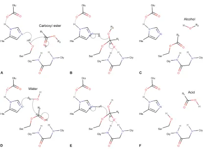
1.5. The catalytic mechanism of CBEs
The catalytic mechanism of CBEs is shared amongst other members of the α/β
10
reaction (Figure 1.4D-F) (47–52). The water molecule is thought to either be activated by the histidine and acid residues of the catalytic triad or other acidic residues in the active site (47–52).
Figure 1.4. The mechanism of a CBE reacting with a carboxyl ester. (A) The catalytic serine is activated by the coordinated histidine to react with the carboxyl ester resulting in a tetrahedral intermediate (B), which is stabilized by the glycine residues of the oxyanion hole. This collapses releasing the alcohol group, regenerating the histidine and resulting in an acyl-enzyme intermediate (C). (D) A water molecule is activated by the catalytic histidine to react with the acyl-enzyme intermediate resulting in a new tetrahedral intermediate (E). This collapses releasing the acid group from the catalytic serine and regenerating the catalytic triad (F).
1.6. Enzyme evolution
[image:18.595.93.509.154.459.2]11
promiscuity is key to the evolution of enzymes (55). He argued that the earliest forms of life had a small number of genes encoding enzymes with broad specificities allowing function with a wider range of substrates. Duplication of these genes would have created redundancies in their encoded enzyme’s activity allowing the accumulation of mutations to some of the duplicated enzymes that specialized them towards specific reactions. The increased catalytic efficiency of these enzymes would improve organismal fitness and thus be selected for. This process of duplication, mutation and selection has formed the basis for our understanding of enzyme evolution (56).
There are a number of different ways to describe the ability of certain enzymes to react with multiple substrates, all of which have implications for the evolution of new enzymes. Broad-specificity refers to enzymes that have evolved to transform a range of substrates with similar efficiency. This differs to substrate ambiguity, which refers to enzymes capable of reacting with substrates they would not normally encounter but that possess similar structures to their native substrate. Enzyme promiscuity refers to an enzyme displaying activity that it did not evolve for and that is not a part of the organism’s normal physiology (54, 57–59).
The ability of certain enzymes to catalyze multiple reactions must be inherent in their structure. This can be due to a number of features in the active site including the existence of different subsites, which could result in a non-specific binding pocket that provides a broad-specificity but low catalytic efficiency with any one substrate (54). Multiple reactions can also be made possible through conformational diversity, whereby the inherent plasticity in an enzyme’s structure enables it to adopt alternate conformations that favor promiscuous activity (56, 60, 61). This is highlighted by Campbell et al. in their study of the laboratory evolution of a Pseudomonas diminuta
phosphotriesterase to an arylesterase (62). In this, they demonstrated that bi-functional intermediates were surpassed through mutations that favored conformations related to the arylesterase activity. Interestingly, one such bi-functional
intermediate has recently been observed in the naturally occurring αE7, an insecticide
12
As there is no ‘fossil record’ of ancestral proteins, our understanding of the mechanisms and mutational pathways that led to today’s enzymes rely on comparisons of phylogenetically related enzymes, experiments that computationally can infer ancestral proteins or through laboratory directed evolution, studies that mimic the process of evolution over shorter periods of time (64). Phylogenetic studies are complicated by the fact that enzymes with distinct functions that are closely related, phylogenetically, may differ from 30% to 80% in sequence with an unknown proportion of these changes relating to “neutral drift” rather than change of function mutations (54). Thus, directed evolution experiments have formed the major basis for our understanding of the mechanisms behind enzyme evolution (56).
13
Figure 1.5. The two extremes of the mutational pathways possible for an enzyme to develop a new function. The weak negative trade-off pathway (the convex route) involves first a gradual shift from the specialist enzyme in the original function (white) with large increases in the new function. It then transitions through a generalist enzyme (red) that is equally capable of both the original and new function before a dramatic decrease in the original function to result in a specialist in the novel one (black). The strong negative trade-off pathway (the concave route) transitions through an enzyme weak in both and likely requires a gene duplication to maintain the original function while a duplicate enzyme evolves the new one. Adapted from references (54, 68).
14
1. No epistasis - where the addition of each mutation alone has the same magnitude of effect upon their combination.
2. Magnitude epistasis – where the combination of each mutation results in an increase of effect greater than each alone.
3. Sign epistasis – where one mutation is deleterious alone but when combined with an advantageous mutation also provides an improvement
4. Reciprocal sign epistasis – where both mutations are deleterious alone but when combined have an advantageous effect.
This effect is important to consider when tracing the evolution of novel enzyme functions(14, 77).
1.7. Evolution of the CBE multigene family
Due to the diversity of biological esters and their importance in life there appears to have been a great deal of diversification of esterases in a number of protein fold superfamilies(12, 15, 80, 81). Early evolution in prokaryotes required the capability to hydrolyze thio-, phospho- and carboxyl esters (43). As organisms grew more complex and required more processing of biological esters, esterases diversified accordingly. Eukaryotes developed a range of novel hormonal, neuronal and metabolic characteristics many of which utilized esters and thus required further control over the metabolism of these molecules (82, 83). Interestingly, while esterases in other
superfamilies played a role in this, the enzymes from the α/β hydrolase fold
superfamily proliferated and diversified far more, resulting in a number of new multigene families associated with distinct roles (84, 85). The CBE gene family was one of these, which has continued to diversify to accommodate the increasing biological complexity (86).
15
before the prokaryote/eukaryote split and that three of the key splits in the phylogeny
coincide with the three major divisions of the prokaryota (86). In this phylogeny there are a number of shared groups across the different classes of the Metazoa, in general, these include specialist proteins such as cholinesterases. Other major radiations clearly occur after the separation of the major classes of the Metazoa and have resulted in class specific groups (86).
One of the key features revealed by their phylogeny was the rapid evolution of paralogues in insect CBEs with paralogues within sub-lineages that separated in the last 50 million years having as little as 60% sequence identity (86). This demonstrates the ability for this gene family to tolerate rapid sequence changes, enabling them to adopt novel functions and explaining the ongoing diversification of this family (86). The phylogeny also demonstrated that CBEs with the highest identity were generally physically co-located on chromosomes (86). This confirms that local amplification through gene duplication has played a critical role in the evolution of this gene family and explains the increased copy number of CBEs in insects (25, 26). Specific
examples of this are the α-cluster of CBEs within the higher Diptera in which a varying number of CBEs are present in different insect organisms but all diversification can be traced through robust phylogeny to a series of single gene duplication events (91, 92). These duplication events would have allowed individual genes to accumulate mutations through both weak and strong negative trade-offs and may explain the fast
rates of change observed. Interestingly, CBE genes within the α-cluster of Drosophila
were found to be lost quickly during cluster evolution due to nonsynonymous mutations that occur with a frequency equal to or greater than synonymous mutations (92, 93). This demonstrates that qualitative shifts in function can occur through widespread changes in protein sequence over short periods of evolutionary time.
The widespread use of insecticides and selection for insecticide resistance has provided a unique example of the early stages of enzyme evolution in insect CBEs. Two of the major types of insecticides, carbamates and OPs, target the CBE, AChE.
16
1.8. Focus of thesis
Our interest in insecticide resistance CBEs has resulted in many studies focusing on their expression within insects and in identifying potential insecticide degrading mutations. Surprisingly, there are relatively few examples of these CBEs being biochemically characterized and even fewer that have been structurally characterized. Such studies are key in better understanding the interaction of CBEs with insecticides. In the second chapter of this thesis I will further introduce insecticide resistance CBEs and detail my attempts to express a number of insecticide resistance CBEs, from a diverse range of insect orders, in Escherichia coli. This is a critical first step in both biochemical and structural characterization and has enabled two new insecticide resistance CBE structures to be solved. The third chapter contains a first authored journal article focusing on one of these insecticide resistance CBEs, Cqestβ21, from the disease vector, Culex quinquefasciatus. This CBE is both structurally and biochemically characterized providing a molecular-level understanding of the sequestration mechanism shared by the majority of insecticide resistance CBEs. The relationship between all insecticide resistance CBEs is investigated through a sequence similarity network of all insect CBEs. This reveals that the insect CBEs are a more diverse family than previously thought, with a number of new subfamilies suggested and functionally annotated.
17
18
19
2.1. Introduction
2.1.1. Insecticides
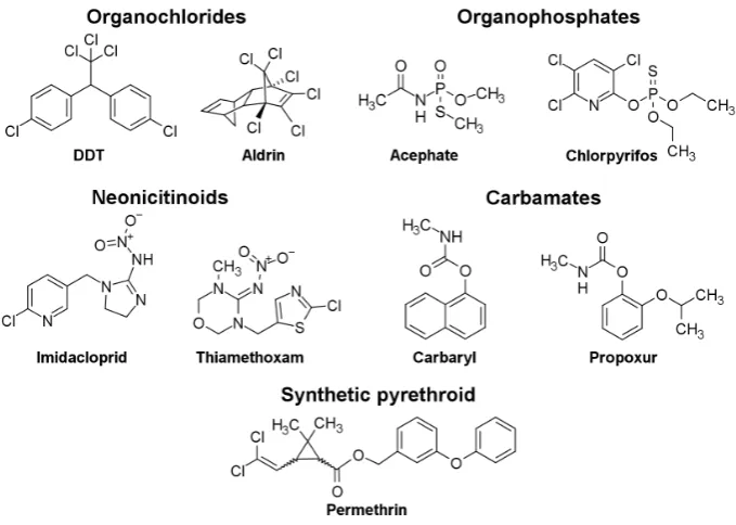
Insecticides are critical in both maintaining sufficient agricultural productivity and fighting insect-borne diseases (15, 97–99). However, the prevalence of insecticide use over the past 60 years has produced an excessive pressure on insects to evolve resistance (11, 95, 100, 101). Thus, the number of insect species identified with resistance has been rapidly increasing, reaching greater than 580 species in 2015 (102). To combat this, a large amount of research has gone into the development of novel insecticides (103–106). So far, insecticides utilizing greater than 25 modes of action and including at least 55 different chemical classes have been produced (102). The five major chemical classes are organochlorines, carbamates, synthetic pyrethroids (SPs), neonicotinoids, and OPs (Figure 2.1) (103, 107, 108). These major classes all target the insect nervous system, disrupting its normal function by interacting with one of the following targets: voltage-gated sodium channels; AChE; and nicotinic acetylcholine receptors (103, 107, 108).
[image:27.595.131.471.455.697.2]20
2.1.2. OP insecticides
OP insecticides are among the most widely used classes of insecticide (102, 109, 110). They (along with carbamates) target AChE, which is expressed in neuromuscular junctions and chemical synapses and hydrolyzes the neurotransmitter, acetylcholine, terminating signal transduction(111–114). This process is essential for the normal physiology of insects and is thus critical for their survival (115–117). The reaction between AChE and OPs results in a covalent linkage between the OP and AChE, which irreversibly inhibits AChE resulting in insect paralysis and eventual death (115–117).
OPs can be described as a group of phosphoric acid ester compounds with three ester bonds (Figure 2.2). The first ester bond generally contains a leaving group which consists of an electron withdrawing group that promotes its release during the first step of the reaction between the OP and the catalytic serine of AChE (Figure 2.3). The other two ester bonds generally consist of short alkyl side chains (o-methyl or o-ethyl) (Figure 2.2). The OP double bonded oxygen is often replaced by a sulfur atom in commercial synthesis, resulting in a thion form of the OP (Figure 2.2) (118). During uptake in insects, native P450 monooxygenases convert this sulfur into an oxygen resulting in the oxon form of the OPs, which are generally stronger inhibitors of AChE (118–120).
Figure 2.2. The template for an OP structure where X is either an oxygen or sulfur atom, R1 and R2 are either o-methyl or o-ethyl alkyl side chains and R3 is an electron withdrawing leaving group.
21
(121–125). The reaction can then proceed via two pathways: a water molecule may hydrolyze the complex reforming the catalytic serine; or through a process called aging, a water molecule may undergo nucleophilic substitution with one of the phosphoryl esters of the complex resulting in an irreversibly inhibited complex (Figure 2.3) (30, 114). Due to the stability and steric constraints imposed by the tetrahedral serine-OP complex each pathway proceeds very slowly rendering the enzyme catalytically inactive (121–125).
Figure 2.3. The basic reaction mechanism between AChE and an OP showing the two possible pathways from an Enzyme-OP complex.
2.1.3. Insecticide resistance mechanisms
22
the insecticide before they can reach their target (35, 137, 138). There are three major classes of detoxification enzyme utilized in insecticide resistance: mixed-function oxidases, glutathione S-transferases, and CBEs (94, 126, 139–143).While all have been shown to play significant roles in different organisms, CBEs are the most widespread class of detoxification enzyme and are the most closely associated with the most common forms of insecticides: OPs, carbamates, and to a lesser extent, SPs (144–148).
2.1.4. CBE-mediated metabolic resistance
CBE-mediated metabolic resistance has been identified in many insect species across a large number of insect orders(15, 40, 149, 150). The most common form is through insecticide sequestration, which involves weak promiscuous activity allowing a slow hydrolysis of the insecticide that results in an essentially stoichiometric, covalent sequestration of the insecticide (10, 127, 151, 152). For this mechanism to be effective it requires overexpression of the insecticide resistance CBE to accommodate the insecticide. This mechanism is sometimes referred to as the quantitative resistance mechanism due to its reliance on this overexpression, which is commonly achieved through tandem amplifications of the insecticide resistance CBE genes (96, 153, 154). Aphids and culicine mosquitos are the most well studied organisms that utilize this mechanism, where tandem amplification has been shown to generate up to 200 or more copies of the resistance gene (25, 155–158).
23
related to L. cuprina, such as Musca domestica and Cochliomyia hominivorax (137, 161, 165–167). It is thought that the fitness cost associated with this mutation has precluded its occurrence in other insect species(96, 168, 169). In L. cuprina, this cost is compensated by a mutation at another locus with an as of yet unknown function that rescues the fitness of L. cuprina and is widespread in L. cuprina populations (170).
2.1.5. Insect CBE expression
Our ability to understand how insecticide resistance CBEs function has been limited by the techniques that can be used to study them(30). The majority of effort has been spent in identifying insecticide resistance CBEs (10, 96, 144). This has been predominantly through transcriptome sequencing and RT-PCR (38, 128, 141, 171). This provides us with the sequence of CBE candidates and an association with insecticide resistance. While this is a critical first step, few studies have extended our understanding of these candidate CBEs through further characterization (109, 142, 157, 172). Those that have may rely on purification through complex separations using whole or partial insect body homogenates, which results in relatively small concentrations of the candidate CBE (4, 173–175). This enables basic biochemical characterization but is insufficient for structural characterization, which is key for both a molecular understanding of the insecticide resistance mechanism and for targeted-inhibitor design. For this, heterologous expression using E. coli expression systems is preferable due to its rapid growth and high expression of recombinant proteins (176– 178).
2.1.6. E. coli-based expression of eukaryotic enzymes
24
for the function and stability of many proteins, especially those that are secreted (184). The lack of these systems can result in improper protein folding and the formation of insoluble aggregates or inclusion bodies (176, 185, 186). To combat this, specialized
E. coli strains have been developed that have altered proteomes, eliminating detrimental proteins and adding beneficial ones, to promote the proper folding of eukaryotic proteins and thus expression in E. coli(182, 185, 187–189).
In this chapter, we utilized three E. coli strains to improve insect CBE expression: BL21(DE3) competent cells (NEB); Shuffle T7 Express Competent cells (NEB); and Origami B(DE3)pLysS competent cells (Novagen). BL21(DE3) cells are modified to be deficient in proteases (Lon and OmpT), resistant to phage T1 and to express a chromosomal copy of T7 RNA polymerase (T7 RNAP) allowing T7 expression, which is inducible by IPTG (190). The Origami B(DE3)pLysS contains the same modifications as BL21(DE3) cells in addition to: mutations in trxB (thioredoxin reductase) and gor (glutaredoxin reductase), which reduces their action and promotes disulfide bond formation; and the addition of T7 lysozyme, which suppresses T7 RNA polymerase prior to induction (190). Shuffle T7 Express cells have the largest number of modifications: the trxB and gor genes are deleted, promoting disulfide bond formation; a chromosomal copy of T7 RNAP is expressed; cells are deficient in Lon and OmpT; and a chromosomal copy of the disulfide bond isomerase, DsbC, is constitutively expressed, which promotes the correction of mis-oxidized proteins and acts as a chaperone for the proper folding of enzymes (189).
2.2. Preface
25
conditions for large-scale expression. This is a critical first step in determining the structure of these insecticide resistance CBEs, which is essential to enhance our molecular understanding of insecticide resistance.
2.3. Materials and methods
2.3.1. Literature review and protein expression and crystallization prediction tools
Insecticide resistance CBE sequences were identified through a literature review. Signal peptides were detected for removal by the SignalP 4.1 server (191). Potential disulfide bonds were predicted using the DiANNA 1.1 web server (192). Potential solubility in E. coli was predicted using PROSO II (193). The protein crystallizability was predicted for each CBE using the XtalPred-RF web server (194).
2.3.2. Cloning
All insecticide resistance CBE amino acid sequences were obtained from UniProtKB or GenBank (195, 196). All DNA sequences were optimized for expression in E. coli
and synthesized by Integrated DNA Technologies (IDT, USA) with N-terminal His-tags, TEV cleavage sites and N- and C-terminal sequences that overlap with the pETMCSIII vector to allow Gibson assembly (NEB) (197). DNA fragments were cloned into the pETMCSIII vector using Gibson assembly (197). Successful cloning was confirmed through DNA sequencing at the Biomolecular Resource Facility, Australian National University, Australia. Mutations, such as the addition of an S-tag, were introduced using overlapping primers and Gibson assembly (197). Plasmids for Hax42, Hax43 and Hax46 were provided by CSIRO Land and Water, Australia.
2.3.3. Protein expression
26
temperatures until induction at an OD600 of 0.4 - 0.8 using 0-1 mM IPTG. All cultures were then grown at room temperature for either 24 or 48 hours. In large-scale expression all conditions were maintained except 1 L of LB was used.
2.3.4. Protein lysis, separation and purification
For small-scale expression, cells were pelleted by centrifugation and resuspended in a BugBuster® solution (Merck, USA) containing resuspension buffer (20 mM HEPES pH 7.5, 150 mM NaCl) and turbonucease (Sigma, USA) and incubated at room temperature for 20 minutes. A sample of this ‘whole cell’ soluble and insoluble mixture was taken for protein separation before the lysed cells were pelleted and the soluble layer was removed for protein separation. In some cases, the insoluble pelleted fraction was then once again resuspended in BugBuster® solution and a sample of this insoluble fraction was taken for protein separation. Proteins were separated by SDS-PAGE using precast ExpressPlus 4 to 20% SDS-PAGE gels (GenScript) and stained using Coomassie brilliant blue (Sigma, USA) for visualization. For large-scale expression, cells were pelleted and resuspended in resuspension buffer before lysis by either sonication or French press. Cell debris was pelleted by centrifugation and the soluble fraction was applied to a HisTrap FF column (GE Healthcare). Bound protein was eluted using buffer B (20 mM HEPES pH 7.5, 150 mM NaCl, 300 mM imidazole).
2.4. Results
2.4.1. Identification of candidate genes
27
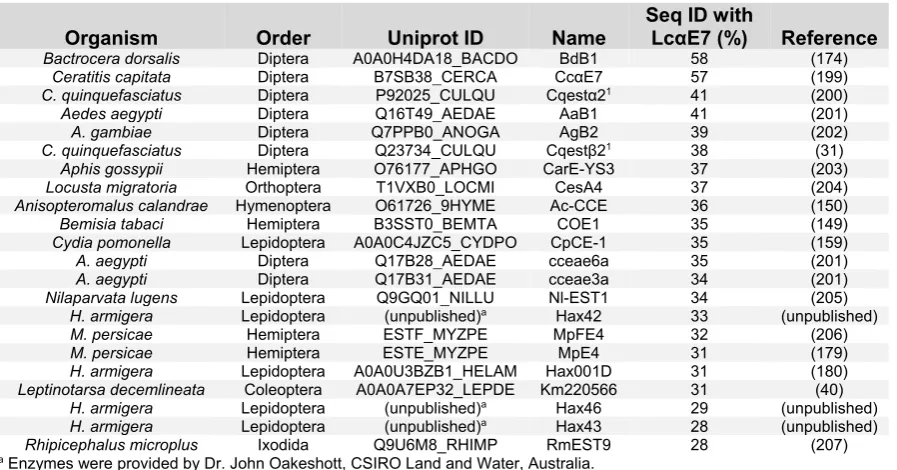
[image:35.595.73.522.212.447.2]of the most important caterpillar pests of cotton in many countries (198). In total, I identified 23 insecticide resistance CBE candidates for further analysis and potential expression and crystallization using E. coli. Within this set of proteins, there was a decreasing level of amino acid identity to the best studied catalytic insecticide resistance CBE, LcαE7 from L. cuprina (Table 2.1).
Table 2.1. A list of insecticide resistance CBEs identified for future analysis.
Organism Order Uniprot ID Name Seq ID with LcαE7 (%) Reference Bactrocera dorsalis Diptera A0A0H4DA18_BACDO BdB1 58 (174)
Ceratitis capitata Diptera B7SB38_CERCA CcαE7 57 (199) C. quinquefasciatus Diptera P92025_CULQU Cqestα21 41 (200)
Aedes aegypti Diptera Q16T49_AEDAE AaB1 41 (201)
A. gambiae Diptera Q7PPB0_ANOGA AgB2 39 (202)
C. quinquefasciatus Diptera Q23734_CULQU Cqestβ21 38 (31)
Aphis gossypii Hemiptera O76177_APHGO CarE-YS3 37 (203) Locusta migratoria Orthoptera T1VXB0_LOCMI CesA4 37 (204) Anisopteromalus calandrae Hymenoptera O61726_9HYME Ac-CCE 36 (150)
Bemisia tabaci Hemiptera B3SST0_BEMTA COE1 35 (149)
Cydia pomonella Lepidoptera A0A0C4JZC5_CYDPO CpCE-1 35 (159)
A. aegypti Diptera Q17B28_AEDAE cceae6a 35 (201)
A. aegypti Diptera Q17B31_AEDAE cceae3a 34 (201)
Nilaparvata lugens Lepidoptera Q9GQ01_NILLU Nl-EST1 34 (205) H. armigera Lepidoptera (unpublished)a Hax42 33 (unpublished)
M. persicae Hemiptera ESTF_MYZPE MpFE4 32 (206)
M. persicae Hemiptera ESTE_MYZPE MpE4 31 (179)
H. armigera Lepidoptera A0A0U3BZB1_HELAM Hax001D 31 (180) Leptinotarsa decemlineata Coleoptera A0A0A7EP32_LEPDE Km220566 31 (40)
H. armigera Lepidoptera (unpublished)a Hax46 29 (unpublished)
H. armigera Lepidoptera (unpublished)a Hax43 28 (unpublished)
Rhipicephalus microplus Ixodida Q9U6M8_RHIMP RmEST9 28 (207)
a Enzymes were provided by Dr. John Oakeshott, CSIRO Land and Water, Australia.
The CBE Cqestβ21 from C. quinquefasciatus was of particular interest due to three key factors: (i) it has been suggested to act through sequestration; (ii) a closely related CBE, Cqestβ1, has been expressed in E. coli; and (iii) the enzyme was shown to be unaffected by the equivalent to the G137D OP hydrolase gain-in-function mutation found in LcαE7 (31, 33, 173, 208).
2.4.2. Computational predictions of CBE expression and crystallization
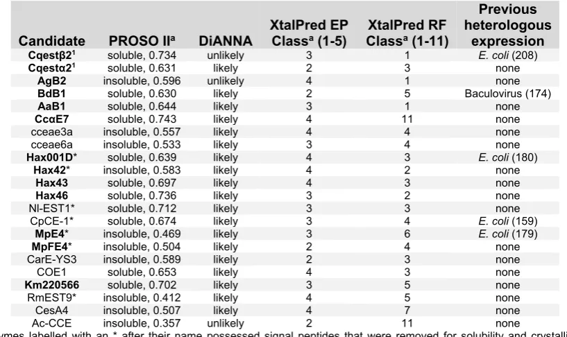
28
[image:36.595.92.501.413.656.2]expression data with the exception of MpE4, which is predicted to be insoluble (159, 179, 180, 208). As disulfide bonds are fairly common in insect enzymes and can create issues with expression in E. coli, I utilized the disulfide bond prediction tool, DiANNA (192). This determined that 20 of the 23 candidates were likely to have disulfide bonds (Table 2.2). Due to the presence of specialized E. coli strains for the expression of proteins with disulfide bonds, this was not used as a criterion to disregard a candidate for expression(177). To determine the likelihood of crystallization I used the XtalPred web server (194). This uses two methods to predict “crystallizability”: the Expert Pool method (EP class); and the Random Forest Classifier (RF class) (194). EP class uses eight protein features to generate a score that bins the candidate into one of five crystallization classes where 1 is the most likely to crystallize (194, 209). RF class uses additional features to generate the score and uses eleven classes where 1 is the most likely to crystallize (194, 210). Five candidates were found to be in the top two EP class and eleven in the top three RF class (Table 2.2).
Table 2.2. A summary of the results from a range of computational prediction tools used to determine the suitability for each CBE candidate for further research.
Candidate PROSO IIa DiANNA XtalPred EP Classa (1-5) ClassXtalPred RF a (1-11)
Previous heterologous
expression
Cqestβ21 soluble, 0.734 unlikely 3 1 E. coli (208) Cqestα21 soluble, 0.631 likely 2 3 none
AgB2 insoluble, 0.596 unlikely 4 1 none
BdB1 soluble, 0.630 likely 2 5 Baculovirus (174)
AaB1 soluble, 0.644 likely 3 1 none
CcαE7 soluble, 0.743 likely 4 11 none
cceae3a insoluble, 0.557 likely 4 4 none
cceae6a insoluble, 0.533 likely 3 4 none
Hax001D* soluble, 0.639 likely 4 3 E. coli (180)
Hax42* insoluble, 0.583 likely 4 2 none
Hax43 soluble, 0.697 likely 4 3 none
Hax46 soluble, 0.736 likely 3 2 none
Nl-EST1* soluble, 0.712 likely 3 3 none
CpCE-1* soluble, 0.674 likely 3 4 E. coli (159)
MpE4* insoluble, 0.469 likely 3 6 E. coli (179)
MpFE4* insoluble, 0.504 likely 2 4 none
CarE-YS3 insoluble, 0.589 likely 2 3 none
COE1 soluble, 0.653 likely 4 3 none
Km220566 soluble, 0.702 likely 3 5 none
RmEST9* insoluble, 0.412 likely 4 5 none
CesA4 insoluble, 0.507 likely 4 7 none
Ac-CCE insoluble, 0.357 unlikely 2 11 none
a Enzymes labelled with an * after their name possessed signal peptides that were removed for solubility and crystallization
predictions. Enzymes that were selected for expression trials are shown in bold.
29
include candidates from a range of insect orders taking the significance of each insect pest into consideration.
2.4.3. Expression trials
Each selected insecticide resistance CBE candidate was optimized and cloned into the pETMCSIII vector for expression in E. coli. Previous studies had confirmed expression of the H. armigera CBE, Hax001D in BL21(DE3) cells using a combination of a His-tag and a solubility tag (S-tag), which was incorporated in its sequence (180). Candidates were first expressed in BL21(DE3) competent cells and if no clear soluble expression was detected they were expressed in both Origami B(DE3)pLysS competent cells and Shuffle T7 Express Competent cells. I incorporated an S-tag to a number of candidates without clear soluble expression to test its effect. The results of each candidate are summarized below:
Expression of the C. quinquefasciatus CBE, Cqestβ21
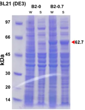
[image:37.595.228.371.447.631.2]While there is no soluble expression present for Cqestβ21 in BL21(DE3) cells without induction, a large amount is present upon induction with IPTG (Figure 2.1).
30
Expression of the C. quinquefasciatus CBE, Cqestα21
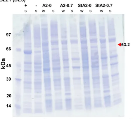
[image:38.595.185.413.204.407.2]There is no clear insoluble or soluble expression of Cqestα21 or Cqestα21 with an S-tag in BL21(DE3) cells (Figure 2.2) or in Origami B(DE3)pLysS cells (Figure 2.3). There is a small amount of insoluble expression of both Cqestα21 and Cqestα21 with an S-tag in Shuffle T7 Express cells (Figure 2.4).
31
[image:39.595.191.405.393.579.2]Figure 2.3. Expression trial of Cqestα21 and S-tagged Cqestα21 using Origami B(DE3)pLysS E. coli competent cells without induction or with induction using 0.7 mM IPTG: “-” refers to a negative control using empty vector; “A2-0” refers to Cqestα21 expression without induction and “A2-0.7” refers to its expression with induction; "StA2-0" refers to S-tagged Cqestα21 without induction and “StA2-0.7” refers to its expression with induction; “w” refers to the whole cell fraction; and “s” refers to the soluble fraction. Target protein size is indicated by the red arrow and labelled.
32
Expression of the A. aedes CBE, AaB1
There is a moderate amount of soluble expression of AaB1 without induction and a large amount with induction in BL21(DE3) cells (Figure 2.5).
Expression of the A. gambiae CBE, AgB2
There is a moderate amount of soluble expression of AgB2 without induction and a large amount with induction in BL21(DE3) cells (Figure 2.5).
Expression of the B. dorsalis CBE, BdB1
[image:40.595.161.435.349.528.2]There is no clear soluble or insoluble expression of BdB1 without induction but strong insoluble and low levels of soluble expression with induction in BL21(DE3) cells (Figure 2.5).
33
Expression of the C. capitata CBE, CcαE7
[image:41.595.219.371.181.360.2]In BL21(DE3) cells there is no clear expression of CcαE7 without induction, however, with induction there are moderate levels of insoluble and soluble expression (Figure 2.6).
Figure 2.6. Expression trial of CcαE7 using BL21(DE3) E. coli competent cells and varied IPTG concentrations for induction: “+” refers to a positive control using LcαE7; “-” refers to a negative control using empty vector; “Cc-0” refers to CcαE7 expression without induction; “Cc-0.7” refers to its expression with induction using 0.7 mM IPTG; “w” refers to the whole cell fraction; and “s” refers to the soluble fraction. Target protein size is indicated by the red arrow and labelled.
Expression of the M. persicae CBE, MpE4
34
Figure 2.7. Expression trials of MpE4 and S-tagged MpE4 using BL21(DE3) E. coli competent cells and varied IPTG concentrations for induction: 0” refers to MpE4 expression without induction, “E4-0.1”, “E4-0.5” and “E4-1” refer to its expression with induction using 0.1 mM, 0.5 mM and 1 mM IPTG, respectively; “-” refers to a negative control using empty vector; “StE4-0” refers to S-tagged MpE4 expression without induction and “StE4-0.7” refers to its expression with induction using 0.7 mM IPTG; “w” refers to the whole cell fraction; and “s” refers to the soluble fraction. Target protein size is indicated by the red arrow and labelled.
Figure 2.8. Expression trials of MpE4 using Origami B(DE3)pLysS and Shuffle T7 Express E. coli
[image:42.595.108.493.420.602.2]35
Figure 2.9. Expression trial of S-tagged MpE4 using Shuffle T7 Express E. coli competent cells, Origami B(DE3)pLysS E. coli competent cells and varied IPTG concentrations for induction: “-” refers to a negative control using empty vector; “StE4-0” refers to S-tagged MpE4 expression without induction and “StE4-0.7” refers to its expression with induction using 0.7 mM IPTG; “I” refers to the insoluble fraction; and “s” refers to the soluble fraction. “a” indicates that expression was extended for 48 hours after induction. Target protein size is indicated by the red arrow and labelled.
Expression of the M. persicae CBE, MpFE4
36
Figure 2.10. An expression trial of MpFE4 and an expression trial of S-tagged MpFE4 using BL21(DE3)
[image:44.595.102.490.449.629.2]E. coli competent cells and varied IPTG concentrations for induction: “-” refers to negative controls using empty vector; “FE4-0” refers to MpFE4 expression without induction; “FE4-0.2” and “FE4-0.7” refer to MpFE4 expression with induction using 0.2 mM and 0.7 mM IPTG, respectively; “StFE4-0” refers to S-tagged MpFE4 expression without induction and “StFE4-0.7” refers to its expression with induction using 0.7 mM IPTG; “w” refers to the whole cell fraction; and “s” refers to the soluble fraction. Four irrelevant protein samples on both gels were removed between ladder and sample “FE4-0, w” and samples “-, s” and “StFE4-0, w”, respectively. Target protein size is indicated by the red arrow and labelled.
Figure 2.11. Expression trials of MpFE4 using Origami B(DE3)pLysS and Shuffle T7 Express E. coli
37
Figure 2.12. Expression trial of S-tagged MpFE4 using Shuffle T7 Express E. coli competent cells, Origami B(DE3)pLysS E. coli competent cells and varied IPTG concentrations for induction: “-” refers to a negative control using empty vector; “StFE4-0” refers to S-tagged MpFE4 expression without induction and “StFE4-0.7” refers to its expression with induction using 0.7 mM IPTG; “I” refers to the insoluble fraction; and “s” refers to the soluble fraction. “a” indicates that expression was extended for 48 hours after induction. Target protein size is indicated by the red arrow and labelled.
Expression of the L. decemlineata CBE, Km220566
38
Figure 2.13. Expression trials of Km220566 using BL21(DE3), Origami B(DE3)pLysS and Shuffle T7 Express E. coli competent cells with varied IPTG concentrations for induction: “+” refers to a positive
control using LcαE7; “-” refers to a negative control using empty vector; “Km-0” refers to Km220566 expression without induction and “Km-0.7” refers to its expression with induction using 0.7 mM IPTG; “w” refers to the whole cell fraction; and “s” refers to the soluble fraction. Target protein size is indicated by the red arrow and labelled.
Expression of the H. armigera CBE, Hax001D
39
Figure 2.14. Expression trials of Hax001D using BL21(DE3), Origami B(DE3)pLysS and Shuffle T7 Express E. coli competent cells with varied IPTG concentrations for induction: “-” refers to a negative control using empty vector; “H1D-0” refers to Hax001D expression without induction and “H1D-0.7” refers to its expression with induction using 0.7 mM IPTG; “w” refers to the whole cell fraction; and “s” refers to the soluble fraction. Target protein size is indicated by the red arrow and labelled.
Expression of the H. armigera CBE, Hax42
In BL21(DE3) cells there is no clear difference between the soluble expression of Hax42 and empty vector, indicating no soluble expression of Hax42 (Figure 2.15). However, there is strong insoluble expression in induced BL21(DE3) cells (Figure 2.15). Similarly, induction resulted in the production of moderate levels of insoluble expression with Origami B(DE3)pLysS cells but no clear soluble expression (Figure 2.16). While there is no soluble expression in Shuffle T7 Express cells, there is an increase in the levels of insoluble expression in the uninduced sample (Figure 2.17).
Expression of the H. armigera CBE, Hax43
40
Expression of the H. armigera CBE, Hax46
[image:48.595.143.456.220.422.2]There is no clear difference between the empty vector soluble expression and soluble or insoluble expression of Hax46, with or without induction, in BL21(DE3) cells (Figure 2.15) Origami B(DE3) cells (Figure 2.16) or Shuffle T7 Express cells (Figure 2.17). This suggests that there is neither soluble nor insoluble expression for Hax46 in any of the conditions tested.
41
Figure 2.16. Expression trial of Hax42, Hax43 and Hax46 using Origami B(DE3)pLysS E. coli
[image:49.595.157.455.451.633.2]competent cells without induction or with induction using 0.7 mM IPTG: “+” refers to a positive control using DmEST6-1; “-” refers to a negative control using empty vector; “H42-0” refers to Hax42 expression without induction and “H42-0.7” refers to its expression with induction; “H43-0” refers to Hax43 expression without induction and “H43-0.7” refers to its expression with induction; “H46-0” refers to Hax46 expression without induction and “H46-0.7” refers to its expression with induction; “w” refers to the whole cell fraction; and “s” refers to the soluble fraction. Target protein sizes are indicated by the red arrow and labelled by size and name.
42
[image:50.595.93.510.242.698.2]In summary, the expression trials identified five insecticide resistance CBEs that display some level of soluble expression (BdB1, CcαE7, Cqestβ21, AaB1 and AgB2) and three with expression ideal for utilization in crystallization trials (Cqestβ21, AaB1 and AgB2) (Table 2.3).
Table 2.3. A summary of the results for CBE expression. +IPTG indicates the addition of 0.7 mM IPTG to cell cultures at OD600 0.4 – 0.8 whereas -IPTG indicates no addition. Strength of protein bands were decided qualitatively and are indicated by intensity of color where green indicates soluble expression and blue indicates insoluble expression.
Solubility
fraction BL21(DE3)
Origami
B(DE3)pLysS Shuffle T7 Express
-IPTG +IPTG -IPTG +IPTG -IPTG +IPTG
Cqestβ21 insoluble - low
soluble - strong AaB1 insoluble soluble mediumlow stronglow
AgB2 insoluble soluble mediumlow stronglow
BdB1 insoluble soluble -- stronglow
CcαE7 insoluble soluble -- mediummedium
Cqestα21 insoluble - - - - low low
soluble - - -
-Cqestα21
w S-tag insoluble soluble -- -- low- low- medium- medium
-MpE4 insoluble soluble -- strong- -- -- -- -
-MpE4
w S-tag insoluble soluble strong- medium- low- low- medium- medium -MpFE4 insoluble soluble -- strong- -- -- -- medium -MpFE4
w S-tag insoluble soluble medium- medium- -- -- strong- strong -Km220566 insoluble soluble low- low - -- -- -- -
-Hax001D insoluble soluble -- -- -- -- -- -
-Hax42 insoluble soluble -- strong- -- medium- medium- -
-Hax43 insoluble soluble -- strong- -- -- medium- medium
-43
2.4.4. Large-scale expression trials of insoluble proteins
There were four insecticide resistance CBE candidates that showed strong insoluble expression but no soluble expression: MpE4, MpFE4, Hax42 and Hax43. For each candidate I attempted a large-scale expression using 1 L of LB in the same growth conditions. As different protein extraction techniques can affect the amount of soluble protein, I tried both French press and sonication techniques opposed to BugBuster, which was used for the small-scale expressions (211). Purification using affinity chromatography did not yield any soluble protein (data not shown).
2.5. Discussion
44
Previous studies have shown successful heterologous expression for both MpE4 and Hax001D using E. coli expression systems. However, I was unable to detect any soluble expression for either in my expression trials (Table 2.3) (179, 180). There are a number of differences that may have affected each enzyme’s expression in my trials. One key difference is the vector used for expression: for the expression of Hax001D they tried a number of vectors (pE1, pET32a and pET30a) but only found decent expression and purity with pET30a (180); and for the expression of MpE4 they used the pET28b vector (179). While pET28b, pET30a and pETMCSIII, which I used, are all high expression, T7 promoter vectors from the pET series, as shown with Hax001D, different vectors, even similar ones, can have a large impact on expression (178, 180). The growth medium used with Hax001D was also supplemented with casein hydrolysate, which may have also improved its expression (180).
The majority of insecticide resistance CBEs that I tested were found to only have insoluble expression (Table 2.3). Apart from Cqestα21 these CBEs all share a
sequence identity of less than 34% with LcαE7 and were all predicted to possess
disulfide bonds (Table 2.1, Table 2.2). While this does not preclude expression in E. coli it can make it more difficult. It is also possible that these CBEs require post translational modifications, such as glycosylation, acylation, phosphorylation and acetylation, or folding machinery not present in the E. coli strains I used to generate soluble protein (181, 188). This is a common feature of eukaryotic enzymes that complicates expression in E. coli (181, 188, 212). Interestingly, while the addition of an S-tag to enzymes did not promote greater soluble expression, it did result in a greater production of insoluble protein in the same conditions. This suggests it may have increased the incorrectly folding proteins resistance to degradation and thus the propensity for protein aggregation into inclusion bodies.
2.6. Further research
45
the structural limitations in insecticide resistance CBEs related to the G137D mutation in LcαE7 and to determine the crystal structure of AgB2 (unpublished).
46
Chapter 3. The First Structural Characterization of an
Insecticide Sequestering Carboxylesterase, Cqest
β
2
1, from
47
3.1. Journal article overview
As explained in the previous chapter, insecticide resistance is an ever-growing issue that endangers both our health and agricultural productivity (15, 97–99). The increased occurrence of resistance has encouraged extensive insecticide design, however, resistance remains an issue (103–106). Thus, new strategies for insecticide application and targets for insecticides need to be discovered to better combat resistance. While the number of insect species with resistance has been progressively growing, the number of resistance mechanisms remain relatively small (25, 126–128). By better understanding these mechanisms we will be able to determine new targets and formulate new strategies to combat insecticide resistance that may be broadly applicable.
One of the limiting factors in our understanding of insecticide resistance mechanisms has been the lack of molecular structures of the enzymes involved. Even in the most common insecticide resistance mechanism, CBE-mediated metabolic resistance, which relies on CBEs to either sequester or hydrolyze the insecticide before it reaches its target, only one enzyme’s structure has been determined (5). This enzyme, LcαE7, acts by catalytically detoxifying insecticides and is thus an example of a qualitative resistance mechanism (5, 63, 161–163). Since its discovery this mechanism has been found in very few species, predominantly from the higher Diptera (161, 166, 167, 216). While the quantitative mechanism, insecticide sequestration, is more common and widespread, there was no enzyme structure determined until the work described in this chapter.
The southern house mosquito, C. quinquefasciatus¸ is not only an important vector of a range of filarial diseases including Japanese encephalitis, West Nile virus and Lymphatic filariasis but also utilizes CBE-mediated insecticide sequestration to provide resistance to a wide range of insecticides (4, 217, 218). In this chapter, we describe the expression and utilization of lysine methylation to crystallize and solve the structure of Cqestβ21, one of the most common insecticide resistance CBEs in C.
48
To confirm that Cqestβ21 functions as an insecticide sequestration CBE with OPs and carbamates we conducted a range of inhibition assays between Cqestβ21 and relevant insecticides. Stopped-flow kinetic analysis demonstrated that the reaction with OPs proceeds via a rapid binding event with high affinity that results in an essentially irreversible, covalent intermediate. This agrees with previous studies and explains its action in insecticide sequestration (33). While its interaction was strong with OP insecticides, it demonstrated a weaker interaction with the carbamate tested suggesting a minor role in carbamate resistance.
To better understand the relationship between Cqestβ21, insecticide resistance CBEs and other insect CBEs we utilized a novel technique called sequence similarity networks (SSNs) (220, 221). This utilizes all-by-all BLAST rather than multiple sequence alignment allowing much larger sets of sequences to be analyzed than phylogenetic trees (thousands opposed to hundreds) (222). While this technique can generate groups of related sequences, it does not present any information on the evolutionary history of enzymes (222). Due to the increased sequence coverage, the risk of stochastic error present with phylogenies is reduced (223). The SSNs demonstrated that, irrespective of insect species, CBEs associated with insecticide resistance share a level of similarity that sets them apart from other insect CBEs. The SSNs also had the added benefit of revealing greater complexity to the insect CBE family suggesting further phylogenetic work may be required to improve classification.