SERCA directs cell migration and branching across species and germ layers
Danielle V. Bowera,b,i,*, Nick Lansdalef, Sonia Navarrob,e, Thai V. Truonga,d, Dan J. Bowerg, Neil C. Featherstonef, Marilyn G. Connellf, Denise Al-Alamb, Mark R. Freyb, Le A. Trinha,d, G. Esteban Fernandezb, David Warburtonb, Scott E. Frasera,c, Daimark Bennettf, Edwin C. Jesudasona,b,h
aDivision of Biological Sciences, California Institute of Technology, USA bThe Saban Research Institute, Children’s Hospital Los Angeles, USA
cBiological Sciences and Biomedical Engineering, University of Southern California, Los Angeles, USA
dBiological Sciences and Molecular and Computational Biology, Translational Imaging Center, University of Southern California, Los Angeles, USA
eCraniofacial Biology, Herman Ostrow School of Dentistry, University of Southern California
fDepartment of Biochemistry & Centre for Cell Imaging, Institute of Integrative Biology, University of Liverpool, UK
gInstitute of Geophysics, Department of Earth Sciences, ETH Zürich, Zürich, Switzerland hNHS Lothian, Edinburgh, UK
iDepartment of Diagnostic, Interventional and Pediatric Radiology, Inselspital, Bern University Hospital, University of Bern, Bern, Switzerland.
*Correspondence to: Danielle V. Bower, PhD, MD [email protected]
Summary Statement: Dynamic imaging of living embryos demonstrates that SERCA is a conserved regulator that controls cell migration and branching across species and germ layers, and PKC activation can rescue SERCA inhibition.
Abstract:
Branching morphogenesis underlies organogenesis in vertebrates and invertebrates, yet
is incompletely understood. Here, we show that the sarco-endoplasmic reticulum Ca2+ reuptake pump (SERCA) directs budding across germ layers and species. Clonal
knockdown demonstrated a cell-autonomous role for SERCA in Drosophila air sac
budding. Live imaging of Drosophila tracheogenesis revealed elevated Ca2+ levels in migratory tip cells as they form branches. SERCA blockade abolished this Ca2+
differential, aborting both cell migration and new branching. Activating protein kinase
C (PKC) rescued Ca2+ in tip cells and restored cell migration and branching. Likewise, inhibiting SERCA abolished mammalian epithelial budding, PKC activation rescued
budding, while morphogens did not. Mesoderm (zebrafish angiogenesis) and ectoderm
(Drosophila nervous system) behaved similarly, suggesting a conserved requirement for
Introduction:
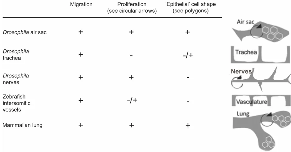
Branching morphogenesis through repetitive budding offers a powerful means to build complex structures without the information costs of separately encoding each branch (Hogan 1999); however, such efficiencies must be balanced carefully, as they also facilitate the pathological branching seen in tumor angiogenesis and proliferative retinopathy. This mandates a mechanistic understanding of iterative budding and its regulation, yet bud iteration has not been explained in terms of its fundamental cellular behaviors, such as cell shape change, migration, and proliferation (Fig. S1). Shape change allows single cells to branch (e.g. axons) or create basic tubes by self-canalizing or fusing (e.g. Drosophila trachea); migration permits cells to rearrange themselves to form tubal networks (e.g. zebrafish intersomitic vasculature, Drosophila trachea); proliferation permits the scaling up needed to form more extensive branched structures in larger organisms (e.g. human lung) (Affolter et al., 2009). A conserved ‘master routine’ (Metzger et al., 2008) that directs the timing and implementation of specialized branching sub-modules would permit the evolution of complex specialized branching structures while preserving a robust regulatory foundation.
In animals, growth factors have been proposed to play key roles, acting as morphogens that direct repetitive budding and integrate broader influences such as oxygen (Jarecki et al., 1999). Tissue-specific growth factor “morphogen clocks” have been proposed to explain the stereotypic pattern of budding (Metzger et al., 2008, Scott et al., 2010). However, extensive investigations of growth factors have yet to define a master program governing branch iteration. We adopted an alternative approach, based on two lines of reasoning. First, the cell behaviors used for budding (shape change, migration and proliferation) each have
morphogens, suggesting that the conserved programs controlling budding are unlikely to rely upon morphogens. Secondly, multicellular morphogenesis requires a robust balance between reliability of signal transmission and flexibility to modulate the signal. Morphogen clocks may be suboptimal for achieving this balance given the substantial variation in gene expression that can exist even between identical adjacent cells (Elowitz et al., 2002). In contrast, cellular Ca2+ signaling has been shown by modeling and empirical studies to offer both signal reliability and flexibility in the face of variable protein expression (Abell et al., 2011). Furthermore, Ca2+ cycling can regulate budding, whether unicellular or multicellular, in fungi and plants (Torralba and Heath, 2001, Trewavas and Knight, 1994). In animals, repetitive Ca2+ waves occur in varied aspects of development, including during organogenesis of the mammalian lung. Live imaging with Ca2+ sensitive fluorophores shows periodic propagating Ca2+ waves in normally developing vertebrate lungs. Additionally, these waves are abnormal during the reduced branching in hypoplastic lungs (Featherstone et al., 2005, Featherstone et al., 2006).
Given the diverse settings in which Ca2+ waves appear correlated with budding and branching, we have tested their causal roles. Repetitive Ca2+ waves depend critically on SERCAs (sarco-endoplasmic reticulum Ca2+ reuptake pumps). These are the P-type ATPases that return cytosolic Ca2+ to the endoplasmic reticulum, and regulate cardiac periodicity and contractility (Wu et al., 1995, Sanyal et al., 2006). Lung Ca2+ waves require SERCA and are abolished by the specific inhibitor, cyclopiazonic acid (CPA) (Featherstone et al., 2005, Seidler et al., 1989). We hypothesized that SERCA controls Ca2+ activity to regulate the ‘spatial periodicity’ of branching, and thus may serve as a conserved central organizer of
and mammalian lung. The results demonstrate that SERCA controls repetitive budding by establishing asymmetric Ca2+ levels at branch sites to direct cell migration, and that key morphogens (FGF, EGF) require SERCA for them to operate optimally.
Results
Budding requires SERCA cell-autonomously for normal epithelial migration and
proliferation
RNAi knockdown of serca in the budding Drosophila air sac epithelium was used to examine its functions in vivo. The single serca gene in Drosophila makes RNAi knockdown simpler than in the mammalian lung, which has three serca genes (Klämbt et al., 1992). Serca mRNA expression and protein function were diminished in the air sac by the first instar larval stage (Fig. S2 A, B). Larval air sacs showed absent or severely stunted buds (Fig. 1A, Fig. S2 C) and reduced proliferation. The expression of escargot, a migration-related transcription factor (Tanaka-Matakatsu et al., 1996) normally expressed in cells of the distal air sac, instead was expressed within cells positioned in the proximal air sac (Fig. 1B, arrows). This suggests that cell differentiation proceeded normally, and the cells which should populate the air sac tip still expressed escargot, however, they failed to migrate distally and instead were retained within the air sac stalk.
A cell-autonomous requirement for epithelial expression of serca was demonstrated by generating labeled, random ‘flip-out’ clones in which serca is absent, by using heat-shock induced FLP-recombinase (Harrison and Perrimon, 1993). Air sac development was
for loss of epithelial serca. Only 12% of serca RNAi cells reached the air sac tip (distal third) compared to 40% of GFP-labelled wild-type epithelial cells; conversely, 52% of serca RNAi cells remained within the proximal third (the stalk) of the air sac, compared to 15% of control ‘flipped’ cells (Fig. 1D). The clones of serca RNAi cells contained fewer cells than
wild type clones (Fig. 1E). Rates of apoptosis were negligible in both cases (Fig. S3). This does not exclude the possibility that apoptosis could have occurred at an earlier stage, however, these results suggest that serca deficiency disrupts budding principally via cell-autonomous defects in epithelial migration and proliferation, which remain uncompensated by adjacent wild type cells.
Budding requires SERCA to control cell migration, irrespective of proliferation
Branching of the Drosophila trachea proceeds without cell proliferation (Samakovlis et al., 1996), and thus serves as a useful model system to study effects of cell migration on branching independently of cell proliferation. RNAi knockdown is ineffective at early embryonic stages, and until late stages, stores of maternal protein result in normal levels of SERCA and of intracellular Ca2+ (Fig. S4). Cyclopiazonic acid (CPA) (Seidler et al., 1989) inhibition of SERCA protein function disrupted budding, resulting in breaks in the tracheal network that were reversible on washout (Fig. 2A-C, Fig. S5). Ca2+-dependent PKCs enhance SERCA function (Usachev et al., 2006). PKC activation using the agonist, phorbol myristate acetate (PMA), rescued the budding defects induced by SERCA inhibition (Fig. 2D, E, Fig. S5). No cell death was detected to account for these observed gaps (Fig. S5 F).
hours (Movie 1). Airway cells converged, from adjacent segments of the embryo, reducing their starting separation by nearly 60%, despite the underlying increase in spacing between tracheal segments (Fig. 2F, H, I). This convergent migration failed when SERCA was
inhibited; instead, as the embryo grew, the separation of neighboring cells increased by about 5% (Fig. 2G, H, I, Movie 2).
SERCA directs cell migration at the budding tip by keeping Ca2+ levels higher in the lead
cell
Live Ca2+ imaging during Drosophila tracheogenesis was performed using the GCaMP3 Ca2+ indicator, expressed exclusively in tracheal cells (Btl:GCaMP3) (Fig. 3A, B). Two-photon light-sheet microscopy was used to visualize the complete tracheal network on one side of the living embryo in 4D, with a time resolution of 3 seconds (Truong et al., 2011). During the formation of the lateral trunk, the lead cells that migrate to fuse with counterparts from neighboring segments (“leaders”) showed high levels of Ca2+ (Fig. 3C). The imaging
revealed a lower level of Ca2+ in those cells trailing just behind them (“trailers”), resulting in a Ca2+ differential between leaders and trailers. Figure 3 shows the cells and a graph of the quantified Ca2+ level intensities in leader (blue) and trailer (magenta) cells. See Movies 3, 9 to visualize the Ca2+ intensity levels of tracheal cells in the live embryos over time.
(Movie 4). These findings are consistent with the cell-autonomous nature of SERCA activity, whereby the cells that take up minimal SERCA inhibitor continue to migrate normally, and only those cells that are sufficiently inhibited fail to migrate. PMA not only rescued cell migration and budding from the effects of SERCA blockade, but also normalized overall Ca2+ levels, and re-instated the Ca2+ differential between leader and trailer cells (Fig. 3F, G, Fig. S6, Movies 5, 11).
SERCA controls non-endodermal budding, again by regulating cell migration
Parallel studies of other systems corroborate the findings in the tracheal branching.
Stereotyped neural branching in Drosophila (Klämbt et al., 1991)was disrupted by SERCA inhibition with CPA. The parallel longitudinal tracts of the wild type nerve cord (Fig. S7 A) become disordered, with discontinuities and aberrant midline crossing following SERCA inhibition (Fig. S7 B). Peripheral nerves are also disorganized and sometimes absent. Washout of CPA mostly corrects the structure of the central nerve cord, except that the longitudinal tracts are slightly more widely spaced than in controls (Fig. S7 C). Neural branching was corrected by activation of PKC by co-treatment with PMA (Fig. S7 D). Furthermore, the aberrations resulting from SERCA inhibition with CPA again support a cell-autonomous role for SERCA activity (Fig. S7 E). Within individual embryos, some portions of the nerve cord and peripheral nerve projections are disrupted (Fig. S7 E, red arrowheads), while adjacent segments can be generally normal (white arrowheads).
SERCA regulates budding in tissues from all three germ layers—ectoderm, mesoderm, and endoderm.
SERCA regulates cell migration to control the onset and rate of bud iteration in mammals
In both rat and mouse embryo lung explants, 20 μM CPA completely suspended branching for the duration of the 3-day treatment. Following washout of CPA after either 1 or 2 days, branching resumed, and the next scheduled branch emerged (Fig. 6A). Lower doses of CPA (4, 10 μM) showed dose-dependent effects on lung explant branching rate (Fig. 6B). Parallel dose-dependent changes were observed in the frequency of airway Ca2+ waves (Fig. 6C) and rates of cell proliferation (Fig. 6D). SERCA inhibition altered the levels of intrinsic lung morphogens: SHH, FGF10, FGF9 and mSpry2 (Fig. 6E). Thus, SERCA activity controls the onset and rate of lung budding in mammals and affects proliferation and the expression of pulmonary morphogens.
SERCA blockade perturbed epithelial migration assayed in culture. Migration closes
standardized wounds in a mammalian epithelial monolayer (IEC6 cells), without reliance on cell proliferation or branching. SERCA blockade with CPA slowed wound closure in a dose-dependent manner (Fig. 6F) and abolished EGF-stimulated migration (relative migration for controls+EGF: 135 + 21.6 vs EGF+CPA: 100 + 10; p = 0.04; n=3).
In mammalian airway epithelium, loss of epithelial SERCA function inhibited budding. Epithelial tip explants from embryonic mouse lung, isolated in culture, bud independently of regulators of budding in the surrounding mesenchyme, such as nerves and vessels (Jesudason et al., 2005, Englund et al., 1999, Del Moral et al., 2006, Bower et al., 2014). SERCA
6G). The PKC inhibitor, Bisindolylmaleimide IHydrochloride abolished this PKC-mediated rescue (Fig. 6G); PKC inhibition alone did not reprise the CPA phenotype (Fig. S8 A). SERCA inhibition halted branching independently of Ca2+-dependent mechanotransduction (Malek and Izumo, 1996, Mammoto and Ingber, 2010, Frey et al., 2004): inhibitors of ROCK, PKC, PLC and Rac1 neither reproduced nor alleviated the CPA phenotype (Fig. S8). Thus, PKC-regulatedSERCA is specifically required in the mammalian airway epithelium for budding.
SERCA controls budding without inducing major changes in cell shape
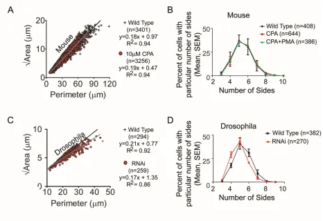
Comparison of cell geometries between wild type and SERCA-inhibited mouse airway epithelium revealed similar linear relationships between ‘area’ and ‘perimeter’ and no differences in cell sidedness (Fig. S9 A, B). Likewise, there was no change in cell shape between wild type and serca RNAi cells in the Drosophila air sac (Fig. S9 C, D). Together, these data show that SERCA provides conserved control of budding through Ca2+-directed cell migration, rather than by regulating proliferation or cell shape.
Discussion
SERCA has wide-ranging regulatory functions, ranging from roles in periodic contractility in muscle to ER stress and protein folding (Sanyal et al., 2006, Caspersen et al., 2000). Our findings reveal a new function for SERCA, as a conserved controller of iterative budding. The initiation of new buds and encoding of the timing of formation of these buds has been proposed to be controlled by growth factor morphogens (Metzger et al., 2008, Morrisey and Hogan, 2010). Specifically, FGF10 acting on airway epithelial FGFR2b (in mammals) or Branchless acting on Breathless (in Drosophila) are required for proper branching of
1992, Min et al., 1998). Unidentified morphogens have also been proposed to act as a ‘branching clock’ that work with FGF signaling to coordinate the branching program
(Metzger et al., 2008). In contrast to the morphogen hypothesis that unidentified growth factor morphogens serve as the “clock” to direct the timing of branching, we show here that SERCA is a central organizer that directs the onset and rate of budding. Morphogens must operate upstream of SERCA, because SERCA blockade stalls the branching program, while supply of exogenous morphogens (e.g. FGFs) is insufficient to overcome this blockade. Thus, we propose that SERCA must integrate inputs from morphogens like FGF and establishes a differential in Ca2+ levels at branching tips to indicate the timing for directed cell migration and branch formation.
This novel role of SERCA as a central organizer of branching seems highly conserved, as branching in both invertebrates and vertebrates, as well as tissues from all germ layers, requires SERCA. In all these systems, branch iteration rate is determined by the level of SERCA function; these effects are mediated by controlling cell migration. SERCA’s effects are not mediated by altering cell shape, and do not require alterations in proliferation. Our live Ca2+ imaging in Drosophila reveals that SERCA directs cell migration at branch points by establishing a local Ca2+ differential, where the Ca2+ level is higher in the leading cell that migrates to form a new branch. The cells trailing behind it maintain comparatively lower Ca2+ levels. Loss of this local Ca2+ differential halts migration and branching. Reinstatement of this local Ca2+ differential, whether by lifting of SERCA blockade or by PKC activation, restores cell migration and branching.
cells migrate and fuse to form their branched network. These propagating Ca2+ waves have been predicted by computational modeling (Kang and Othmer, 2007), yet they do not appear to be important for branch iteration, raising the question as to their function. A recent publication on tracheal tube anastomosis did not implicate these whole-cell Ca2+ impulses in membrane fusion (Caviglia et al., 2016). The increase in frequency of these impulses upon fusion of cells from adjacent segments suggests they may be a response to cell-cell contact, which could in turn modulate cell membrane machinery. Similar Ca2+ impulses have been described in other cell types, such as in fungi following contact with a pathogen (Kim et al., 2012). The remarkable similarity of these Ca2+ impulses from animals to fungi suggests that they are highly conserved and may have been adapted by evolution to suit each specific cellular environment. The function of these impulses may, alternatively, relate to
maintenance or elongation of the branched network that has formed. Indeed, in mammalian lung, periodic Ca2+ waves course through airway smooth muscle, inducing waves of
contractility. These waves are thought to mechano-regulate branching morphogenesis, whereby abolishing the Ca2+ waves impairs airway growth and elongation (Jesudason et al., 2005).
Our results consistently demonstrate that SERCA instructs budding across germ layers, tissue types, and species, suggesting that the role of SERCA may be more broadly generalizable. A conserved regulator simplifies our understanding of how a vast array of branched tissues could arise from one platform, and specialize based on local morphogen inputs. Thus, our findings may unite disparate observations of Ca2+ signaling involvement in other types of branching, such as axonal pathfinding, plant gravitotropism, angiogenesis, and endothelial wound healing (Usachev et al., 2006, Urbina et al., 2006, Evangelista et al., 2012). A
mechanisms. Regarding the lung, the significance of reduced epithelial SERCA function has been highlighted in human and animal studies of asthma (Cantero-Recasens et al., 2010, Mahn et al., 2009) as well as in other burgeoning diseases such as cystic fibrosis (Ahmad et al., 2009), lung fibrosis (Lawson et al., 2011) and lung cancer (Korosec et al., 2006). Our study suggests that these oft intractable pulmonary challenges may feature SERCA-related lesions of cell migration. Examples include airway remodeling in asthma, alveolar
Materials and Methods
Ethics Statement
Protocols complied with NIH Guide for the Care and Use of Laboratory Animals. Mouse and rat protocols were approved by the Institutional Animal Care and Use Committee at Children’s
Hospital Los Angeles (IACUC protocol #252) or with UK Home Office License (Animal Scientific Procedures Act 1986). The zebrafish protocol was approved by the Caltech Institutional Animal Care and Use Committee (1227-09).
RNA extraction
Drosophila embryos and lung explants snap frozen. RNA was extracted using RNeasy Mini
Kit according to Qiagen’s handbook. The concentration of RNA was determined at 260 nm
using a NanoDrop ND-1000 spectrophotometer. The A260/A280 ratio was assessed for RNA purity.
Quantitative RT-PCR
CCCTGACATGGTGTCCAATC), Spry2 (F: TTGTGGTTTGCAGTGAGAGG; R: TCTTCGCCTAGGAGTGTTGG) and Vegf (F: ATGTGACAAGCCAAGGCGGTG; R: TGGCGATTTAGCAGCCAGATA). The MX3000P® Multiplex Quantitative QPCR System (Agilent) was used for all reactions and MxPro software for analysis. Transcripts were quantified using the relative standard curve method. Real-time qRT-PCR efficiency was determined by analysis of serial dilutions of a pool of cDNA sample. All reactions were run in duplicate or triplicate; mRNA expression per gene was normalized to 18S (F: TCCGATAACGAACGAGACTC; R: CAGGGACTTAATCAACGCAA).
Drosophila stocks and crosses
Stocks were obtained from the Bloomington Drosophila Stock Centre (Indiana University, USA), the Vienna Drosophila RNAi Centre (Institute of Molecular Biotechnology and Research Institute of Molecular Pathology, Vienna, Austria) and kindly provided by Dirk Bohmann’s laboratory (University of Rochester, USA). Fly stocks were maintained at 25°C
with a 12 hour light/dark photo cycle, on Drosophila yeast/glucose medium. Spatially restricted gene silencing of serca in the air sac was achieved using the Drosophila GAL4-UAS system and RNAi constructs (Brand and Perrimon, 1993, Dietzl et al., 2007): w*; btl-GAL4,
UAS-Act5C. GFP was crossed to the RNAi line w1118; P{GD436}v4474 in order to specifically
a temperature sensitive GAL80 line (w*; btl-GAL4, UAS-Act5C:GFP; tubGAL80ts), to circumvent early lethality. A similar experiment was carried out using btl-GAL4, UAS-GFP,
tubGAL80ts, esg-LacZ to assess escargot expression.
hsFlp; act>y>GAL4, UAS-GFP; btl-mRFPmoe was similarly crossed to w1118;
P{GD436}v4474 or w1118 to create GFP-labelled serca loss-of-function or control flip-out
clones. In this experiment the tracheal system was labelled with RFP.
Collection of Drosophila embryos and larvae
Embryos were collected on apple juice agar plates smeared with yeast paste. A pre-lay collection helped synchronize subsequent collections which were carried out over 1-4 hours. Embryos were aged on agar plates at 25°C to reach the desired stage. They were removed by washing with distilled water through a fine mesh sieve (Sefar Nitex 120 µM mesh, Sefar Ltd., Bury, UK). The chorion was removed by placing the sieve into thin household bleach for 1-2 min. The bleach was drained off and embryos were washed with distilled water. Embryos were removed with a damp paintbrush and dissociated for RNA extraction or Ca2+ imaging. For larval collection, embryos were collected over 4 – 8 hours and allowed to develop to the required stage at 25°C.
Immunohistochemistry of Drosophila imaginal discs
al., 1997, Wang et al., 2010). Four 20-minute washes were performed with PBST before incubation with secondary antibody. Alexa Fluor® (1:500, Invitrogen) secondary antibody was incubated for 2 hours at 25°C. We washed four x 20 minute washes in PBST and then a final wash of 20 minutes in PBS to remove detergent. Wing discs were mounted on microscopy slides with VECTASHIELD® Mounting Medium (Vector Laboratories). Slides were kept dark at 4°C to reduce fluorophore fading.
Fluorescent Thapsigargin staining of Drosophila embryos.
Embryos were fixed in heptane and 5% PFA in PBS for 15 minutes at room temperature. The PFA was removed and 100% methanol added, and the embryos were shaken to remove the vitelline membrane. The heptane was aspirated and embryos were rehydrated to PBS, then blocked for 2x 30 minutes in PBS + 0.05% TritonX-100 + 0.5% BSA. Embryos were incubated in 5 µM red-fluorescent BODIPY® TR-X thapsigargin (Invitrogen) for 2 hours at 25°C, then washed in PBST for 2 x 30 seconds, 2 x 5 minutes, and 2 x 30 minutes. Embryos were mounted on microscopy slides with VECTASHIELD® Mounting Medium (Vector Laboratories, Peterborough, UK) using coverslip spacers and sealed with clear nail polish.
Generation of flip-out and mitotic clones
serca loss-of-function ‘flip-out’ clones were produced using the heat-shock induced
Larval micro-dissection
Larvae were washed and then dissected in ice cold PBS. Larvae were transected at the abdomen and inverted over forceps. The wing disc was dissected from the thoracic trachea and transferred to cold PBS.
Imaging Ca2+levels in dissociated Drosophila embryos
Embryos were collected and dechorionated as above. 100-200 embryos were placed in a micro-centrifuge tube containing 800 µL of GIBCO™ Schneider’s Drosophila medium (Invitrogen) and dissociated using a sterile pestle. The suspension was centrifuged at 40 G for 5 minutes and the process repeated. The cell suspension was diluted to 1200 µL with medium. 300 µL of this was loaded into each chamber of a 4-well Lab-Tek™ II Chambered Coverglass (Nunc, Thermo Fisher Scientific, Rochester, NY, USA) pre-coated with Poly-L-Lysine (Sigma-Aldrich) for 1 hour and washed with sterile water. Cells were loaded using 1 µM Fluo-4 (Invitrogen) for 1 hour. Confocal microscopy was performed as described below. When required, thapsigargin (Sigma-Aldrich) was added to give a concentration of 2 µM. For real-time Ca2+ release experiments, cells were imaged with a 20X objective on a Zeiss LSM 710 (Carl Zeiss Ltd, Hertfordshire, UK) microscope using maximum scan speed without averaging.
Air sac microscopy and image processing
maximally at 607 and 565 nm, respectively. The pinhole was set at ~1 AU. When z-stacks were taken, we used the slice thickness specified by the software for 1 AU (usually 0.5 – 2 µm). Images were captured using Zen 2010 (Zeiss) software, exported in Tagged Image File Format and edited in Adobe Photoshop CS5 (Adobe Systems Europe Ltd, Maidenhead, UK). When z-stacks were produced, images are presented as single slices, unless a projection is specified. When a 3-dimensional (3D) projection was required, stacks were rendered using Zen 2010 software (Zeiss). If image brightness was altered for publication, this change was standardized across groups to retain comparability.
Embryo preparation for Drosophila embryonic tracheal and nerve studies
Drosophila embryos were collected for 3 hours from wild type or w; Btl:Gal4,
UAS-dsRed-nuclear localization signal, UAS-actinGFP flies. Embryos were dechorionated in 50% bleach
For wholemount preparation, when embryos reached stages 15-16, they were transferred to glass vials and treated with heptane and 5% PFA in PBS for 15 minutes at room temperature to fix. The PFA was removed and 100% methanol added, and the embryos were shaken to remove the vitelline membrane. The heptane was aspirated and embryos were rehydrated to PBS and transferred to PBT (PBS + 0.05% TritonX-100 + 0.1% BSA).
For antibody staining, embryos were blocked for 1 hour at room temperature in 5% normal goat serum (NGS) then incubated with primary antibodies 2A12 (1:2) (Developmental Studies Hybridoma Bank), rabbit anti-GFP (1:1000) (Abcam AB290) and 1D4 (1:3) (Developmental Studies Hybridoma Bank) in PBT + 2% NGS overnight at 4°C. Embryos were washed 6 times for 30 minutes at room temperature in PBT then blocked for 20 minutes in 5% NGS. Goat anti-mouse IgG1, goat anti-mouse IgM, goat anti-rabbit secondary antibodies (Invitrogen) were used at 1:500 in PBT + 2% NGS overnight at 4°C. Embryos were washed 6 times for 30 minutes at room temperature and transferred to PBT/14% glycerol then mounted on slides.
Fillet preparations were performed as described (Lee et al., 2009). Confocal and two-photon tiled z-stacks were collected with a Zeiss LSM 510 meta microscope with a 2 µm pinhole (for confocal) and 1.5 µm z-step interval. Images were assembled using Fiji stitching plugins (Preibisch et al., 2009) and viewed in 3D using Imaris software (Bitplane).
Severity of tracheal phenotypes for each treatment group was scored as follows: a “severe” tracheal phenotype was defined by the presence of breaks, missing sections, grossly abnormal structure, and in the case of the washout samples, the complete formation of a supernumerary “lateral” trunk. A “moderate” phenotype was classified as those with slightly abnormal
Dynamic imaging of live Drosophila embryos and tracking of lateral trunk cell migration
Drosophila embryos from the transgenic line w; Btl:Gal4, UAS-dsRed-NLS, UAS-actinGFP
were collected and permeablized as described above. Embryos were treated with DMSO (control) or 20 µM CPA and screened for fluorescence around stage 12-13. Fluorescent embryos were mounted lateral side down on glass coverslips with heptane glue, or in coverslip-bottomed dishes in 1% agarose molds. The latter were covered with 1% low-melting point agarose and incubation medium and imaged through the coverslip with water immersion fluid. We used the LD-C-Apochromat 40X/1.1W Korr UV-VIS-IR objective on a Zeiss LSM 510 meta or Zeiss LSM 5 Exciter confocal microscope with 488 nm and either 543 or 561 nm lasers. Z-stacks of 318x318x64 µm were collected with 0.62x0.62x2 µm3 voxel size, every 3 to 3.5 minutes.
Datasets were compiled and registered using Imaris 7.6 (Bitplane). Individual cells destined to migrate into the lateral tracheal trunk were manually tracked from stage 14 to 16. Positions of the cells over the timecourse were exported to Matlab. For pairs of tracheal cells in adjacent segments, the direction of travel of one cell relative to the other was calculated, and a vector was plotted for each pair to compare the convergence or divergence of all pairs from each treatment together. The separation between pairs of cells that should migrate together to form the trunk was similarly measured to determine their convergence. Movies were made using Imaris 8.4, ImageJ, and FFmpeg.
Light-sheet imaging of Ca2+dynamics in live Drosophila embryos, data processing and
analysis
w;Btl:Gal4, UAS-GCamP3 embryos expressing the GCamP3 Ca2+ indicator in tracheal cells
as described previously. When the embryos reached approximately stage 13 by gut development, they were aligned in a row on agarose, lateral side down, and gently touched to a heptane glue-coated glass cylinder to mount them to the cylinder with one lateral side exposed. The glass cylinder was quickly mounted into the sealed, fluid-filled sample chamber of the two-photon light-sheet microscope (Truong et al., 2011). Light-sheet microscopy illuminates a single z-slice at a time, minimizing phototoxicity, which is further reduced by using infrared excitation. The image of the entire x-y plane in focus can be captured simultaneously with a camera since there is no out-of-focus excitation. This affords extremely high time resolution. The bi-directionally scanning light-sheet on this microscope also ensures even illumination at each end of the x-y plane. The span of the axial (z) imaging depth was set to capture the entire tracheal network on the side of the embryo facing the collection objective.
Embryos were imaged from approximately stage 13 to stage 16 using 940 nm illumination at the same laser power and exposure time and with the same camera detection gain across all samples. The z-slice thickness was 1.5 µm. The x-y resolution of the image was 0.8 µm/px. An entire z-stack capturing the tracheal network on one side of the embryo was collected in less than 3 seconds, permitting 3 second time resolution between timepoints with resting time for the embryo between each scan. Embryos showed no sign of phototoxicity, and several hatched during the course of the imaging. For embryos that had been treated with CPA or CPA + PMA, the same concentration of drug was added to the water in the sample chamber to maintain the drug treatment while imaging.
Autofluorescence from the embryo surface resulted in a ‘shell’ when images were
first and last columns with data in each row. For each individual image, a mask was uniquely created to eliminate the ‘shell’, employing an R loess smoothing function to obtain a smooth
boundary on the inside of the ‘shell’.
Masked images were reviewed using ImageJ to find the z-slices with lateral trunk cells. Matlab was used to generate a summative projection of each timepoint z-stack that included the lateral trunk but minimized contributions from gut autofluorescence. Other projections were also made to include more of the tracheal network, albeit with more gut as well. The summed images of the lateral trunk were imported into ImageJ for analysis.
The frequency of Ca2+ spikes was determined by counting impulses after summing to include the complete tracheal network on the half of the embryo that was imaged. Spike duration was likewise determined by counting the number of timepoints across which an individual impulse lasted. The error in pulse duration is +/- 3 seconds. Data were plotted with Matlab.
Zebrafish intersomitic vessel studies
Transgenic zebrafish with the VEGF receptor promoter driving eGFP expression (Tg(kdrl:eGFP)) express cytoplasmic eGFP in endothelial cells. These fish were crossed with wild type fish and embryos collected and incubated until 21 hours post-fertilization (hpf). eGFP positive embryos were sorted and dechorionated. Embryos were incubated with cyclopiazonic acid (CPA) or DMSO from 22 hpf until 28 hpf in 1.5 mL of egg water. For the washout study, embryos were incubated in 10 µM or 20 µM CPA for 2 hours, rinsed 3 times in egg water and incubated for 4 hours without drug. After 6 hours of incubation, embryos were rinsed 3 times in egg water and fixed overnight at 4°C in 4% PFA. Embryos were mounted in agarose molds and imaged with a Zeiss LSM 510 meta microscope. Z-stacks were collected using: 488 nm laser excitation, 2 µm pinhole, 2 µm z-step interval. Z-stacks were assembled, and Imaris software (Bitplane) was used to generate 3D images and measure vessel dimensions in 3D.
Lung cultures
(Jesudason et al., 2005). At the end, lung cultures were homogenized for RNA extraction or prepared for histology. Mitotic cells were labelled with Anti-phospho-Histone H3 Ser10 staining (Brand and Perrimon, 1993). Epithelial tip cultures were performed as described, but without enzymatic digestion (Bellusci et al., 1997). Mechanotransduction inhibitors used included: PKCi (Bisindolylmaleimide I Hydrochloride, #203290 Calbiochem), PLCi (L108 Edelfosin, #BML-L108, Enzo Life Science), ROCKi (Y26732, #Y0503, Sigma), RACi (NSC 23766, #553502, Millipore).
Epithelial migration assay
Confluent IEC-6 intestinal epithelial cell (ATCC CRL-1592) monolayers were treated with 0, 1, 2, or 10 μM CPA after wounding with a rotating silicone tip (Frey et al., 2004). The latter CPA dose was tested ± 10ng/ml EGF. Wound closure rates were determined by time-lapse microscopy.
Cell shape analyses
Lung epithelial tips were fixed in 4% formaldehyde (w/v) solution and stored at -20˚C in 70% ethanol. F-actin fluorescence staining was performed by permeabilizing with 0.5% Triton X-100 in PBS for 10 min at room temperature, and then staining with Rhodamine phalloidin (Molecular Probes R415, 5 units/ml) and DAPI (10 ng/µl) in PBS containing 1% BSA overnight at 4˚C. Confocal z-stacks were acquired with an LSM 700 confocal system mounted
Fiji ImageJ software (Schindelin et al., 2012). Images were processed with a median filter of radius 2.0 pixels to smooth while preserving edges, then with an unsharp mask with a radius of 1.0 pixels and weight of 0.9 to enhance phalloidin staining. The images were binarized according to local thresholding by the Sauvola method (Sauvola and Pietikainen, 2000) with a radius of 15 pixels to handle staining variations. The binary images were subjected to Watershed segmentation (Vincent and Soille, 1991) to separate joined cells and then area and perimeter were measured with Analyze Particles. Measured objects were compared with original images to omit non-cellular objects. We used hand-counting for sidedness as described (Gibson et al., 2006).
Statistical Analyses
Data were analyzed using SPSS Statistics 18.0 (IBM®). Sample sizes were calculated with Cohen’s d tables or Mead’s resource equation. Fisher’s exact or Chi-squared testing was used
Acknowledgments
We thank Sean McCann (SM) for his assistance with the migration and shedding assays, and Kai Zinn and Hyung-Kook Lee for advice on the Drosophila embryo studies.
Competing Interests
The authors declare that there are no competing interests.
Author Contributions
ECJ conceived the overall study. DVB and ECJ wrote the manuscript. All authors contributed to redrafting the manuscript. DVB, NL and SN enumerated the methods and with ECJ realized the figures. DVB produced the movies.
ECJ and DB conceived the genetic studies in Drosophila air sac. NL and DB designed these studies with ECJ. They were performed by NL.
ECJ conceived the studies of periodic branching in Drosophila and zebrafish embryos. Experiments were designed by DVB, DB, LAT, and ECJ with SEF and DW. They were performed by DVB.
DVB conceived the live imaging studies with ECJ. DVB conducted the live imaging in
Drosophila with help from TVT. DVB performed the cell tracking studies, and
processed the live imaging data with help from DJB. DVB analyzed the live imaging data.
ECJ conceived the tip culture, mechanotransduction pathway, and geometry studies. They were designed with SN, DA, MRF, DW and GEF and performed by SN, DA and GEF.
MRF and ECJ conceived the mammalian wound closure studies. They were designed by MRF and performed by the MRF lab.
Funding
DVB was funded by the National Institutes of Health NHLBI NRSA 1F30 HL110723,
National Institutes of Health FaceBase grant #U01 DE020063 and the Pasadena Guild Endowment of Children’s Hospital Los Angeles.
NL was funded by the Royal College of Surgeons of England and The Wellcome Trust, UK.
SN was funded by a HURM graduate student supplement to the National Institutes of Health NIGMS R01GM096195 (DW as co-PI) and DE training grant T90 DE021982.
NCF was funded by The Royal College of Surgeons of England and the Medical Research Council, UK.
MRF is funded by National Institutes of Health award R03DK090295, and a Senior Research Award from the Crohn’s and Colitis Foundation of America.
DW was funded by the National Institutes of Health HL44060, 44977, 60231 and 12268, California Institute for Regenerative Medicine Training Grant TG2-01168 and Shared Laboratory Grant CL1-00507, the Garland Foundation, the Webb Foundation, the Pasadena Guild Endowment of Children’s Hospital Los Angeles and the St Andrew’s
References
Abell, E., Ahrends, R., Bandara, S., Park, B.O., and Teruel, M.N. (2011). Parallel adaptive feedback enhances reliability of the Ca2+ signaling system. Proc Natl Acad Sci U S A
108, 14485-14490.
Affolter, M., Zeller, R., and Caussinus, E. (2009). Tissue remodeling through branching morphogenesis. Nature Reviews Molecular Cell Biology10, 831-842.
Ahmad, S., Ahmad, A., Dremina, E.S., Sharov, V.S., Guo, X., Jones, T.N., Loader, J.E., Tatreau, J.R., Perraud, A.L., Schöneich, C. et al. (2009). Bcl-2 suppresses
sarcoplasmic/endoplasmic reticulum Ca2+-ATPase expression in cystic fibrosis airways.
Am J Respir Crit Care Med179, 816-826.
Arbabian, A., Brouland, J.P., Gélébart, P., Kovàcs, T., Bobe, R., Enouf, J., Papp, B. (2011). Endoplasmic reticulum calcium pumps and cancer. Biofactors37, 139-149.
Bellusci, S., Grindley, J., Emoto, H., Itoh, N., and Hogan, B.L. (1997). Fibroblast growth factor 10 (FGF10) and branching morphogenesis in the embryonic mouse lung.
Development124, 4867-4878.
Bower, D.V., Lee, H.K., Lansford, R., Zinn, K., Warburton, D., Fraser, S.E., and Jesudason, E.C. (2014). Airway branching has conserved needs for local parasympathetic innervation but not neurotransmission. BMC Biol12, 92.
Brand, A.H. and Perrimon, N. (1993). Targeted gene-expression as a means of altering cell fates and generating dominant phenotypes. Development118, 401-415.
Caspersen, C., Pedersen, P.S., and Treiman, M. (2000). The sarco/endoplasmic reticulum calcium-ATPase 2b is an endoplasmic reticulum stress-inducible protein. J Biol Chem
275, 22363-22372.
Caviglia, S., Brankatschk, M., Fischer, E.J., Eaton, S., and Luschnig, S. (2016).
Staccato/Unc-13-4 controls secretory lysosome-mediated lumen fusion during epithelial tube anastomosis. Nature Cell Biol.18, 727-739.
Childs, S., Chen, J.N., Garrity, D.M., and Fishman, M.C. (2002). Patterning of angiogenesis in the zebrafish embryo. Development129, 973-982.
Del Moral, P.M., Sala, F.G., Tefft, D., Shi, W., Keshet, E., Bellusci, S., and Warburton, D. (2006). VEGF-A signaling through Flk-1 is a critical facilitator of early embryonic lung epithelial to endothelial crosstalk and branching morphogenesis. Dev Biol290, 177-188. Dietzl, G., Chen, D., Schnorrer, F., Su, K.C., Barinova, Y., Fellner, M., Gasser, B., Kinsey,
K., Oppel, S., Scheiblauer, S. et al. (2007). A genome-wide transgenic RNAi library for conditional gene inactivation in Drosophila. Nature448, 151-6.
Duffy, J.B., Harrison, D.A., and Perrimon, N. (1998). Identifying loci required for follicular patterning using directed mosaics. Development125, 2263-71.
Elowitz, M.B., Levine, A.J., Siggia, E.D., and Swain, P.S. (2002). Stochastic gene expression in a single cell. Science297, 1183-1186.
Englund, C., Uv, A.E., Canter, R., Mathies, L.D., Krasnow, M.A., and Samakovlis, C.
(1999). adrift, a novel bnl-induced Drosophila gene, required for tracheal pathfinding into the CNS. Development126, 1505-1514.
Featherstone, N.C., Jesudason, E.C., Connell, M.G., Fernig, D.G., Wray, S., Losty, P.D., and Burdyga, T.V. (2005). Spontaneous propagating calcium waves underpin airway
peristalsis in embryonic rat lung. Am J Respir Cell Mol Biol33, 153-160.
Featherstone, N.C., Connell, M.G., Fernig, D.G., Wray, S., Burdyga, T.V., Losty, P.D., and Jesudason, E.C. (2006). Airway smooth muscle dysfunction precedes teratogenic
congenital diaphragmatic hernia and may contribute to hypoplastic lung morphogenesis.
Am J Respir Cell Mol Biol35, 571-578.
Flores-Peredo, L., Rodriguez, G., Zarain-Herzberg, A. (2017). Induction of cell differentiation activates transcription of the Sarco/Endoplasmic reticulum calcium-ATPase 3 gene (ATP2A3) in gastric and colon cancer cells. Mol Carcinog, 56, 735-750. Frey, M.R., Golovin, A., and Polk, D.B. (2004). Epidermal growth factor-stimulated
intestinal epithelial cell migration requires Src family kinase-dependent p38 MAPK signaling. J Biol Chem279, 44513-21.
Gibson, M.C., Patel, A.B., Nagpal, R., and Perrimon, N. (2006). The emergence of geometric order in proliferating metazoan epithelia. Nature442, 1038-1041.
Glazer, L, and Shilo, B.Z. (1991). The Drosophila FGF-R homolog is expressed in the
embryonic tracheal system and appears to be required for directed tracheal cell extension.
Genes Dev5, 697-705.
Harrison, D.A. and Perrimon, N. (1993). Simple and efficient generation of marked clones in Drosophila. Curr Biol3, 424-433.
Hogan, B.L.M. (1999). Morphogenesis. Cell96, 225-33.
Ikeya, T. and Hayashi, S. (1999). Interplay of Notch and FGF signaling restricts cell fate and MAPK activation in the Drosophila trachea. Development126, 4455-4463.
Jarecki, J., Johnson, E., and Krasnow, M.A. (1999). Oxygen regulation of airway branching in Drosophila is mediated by branchless FGF. Cell99, 211-220.
Jesudason, E.C., Connell, M.G., Fernig, D.G., Lloyd, D.A., and Losty, P.D. (2000). Early lung malformations in congenital diaphragmatic hernia. J Pediatr Surg35, 124-127. Jesudason, E.C., Smith, N.P., Connell, M.G., Spiller, D.G., White, M.R., Fernig, D.G., and
Losty, P.D. (2005). Developing rat lung has a sided pacemaker region for morphogenesis-related airway peristalsis. Am J Respir Cell Mol Biol32, 118-127.
Kang, M. and Othmer, H.G. (2007). The variety of cytosolic calcium responses and possible roles of PLC and PKC. Phys Biol4(4):325-343.
Kim, H.S., Czymmek, K.J., Patel, A., Modla, S., Nohe, A., Duncan, R., Gilroy, S., and Kang, S. (2012).Expression of the Cameleon calcium biosensor in fungi reveals distinct Ca2+ signatures associated with polarized growth, development, and pathogenesis. Fungal
Genet. Biol.49(8), 589-601.
Klämbt, C., Jacobs, J.R., and Goodman, C.S. (1991). The midline of the Drosophila central-nervous- system - a model for the genetic-analysis of cell fate, cell-migration, and growth cone guidance. Cell64, 801-815.
Klämbt, C., Glazer, L., and Shilo, B. (1992). breathless, a Drosophila FGF receptor homolog, is essential for migration of tracheal and specific midline glial cells. Genes Dev6, 1668-1678.
Lacabaratz-Porret, C., Launay, S., Corvazier, E., Bredoux, R., Papp, B., Enouf, J. (2000). Biogenesis of endoplasmic reticulum proteins involved in Ca2+ signalling during megakaryocytic differentiation: an in vitro study. Biochem J. 350, 723-734.
Launay, S., Gianni, M., Kovàcs, T., Bredoux, R., Bruel, A., Gélébart, P., Zassadowski, F., Chomienne, C., Enouf, J., and Papp, B. (1999). Lineage-specific modulation of calcium pump expression during myeloid differentiation. Blood 93, 4395-4405.
Lawson, W.E., Cheng, D.S., Degryse, A.L., Tanjore, H., Polosukhin, V.V., Xu, X.C., Newcomb, D.C., Jones, B.R., Roldan, J., Lane, K.B. et al.. (2011). Endoplasmic
reticulum stress enhances fibrotic remodeling in the lungs. Proc Natl Acad Sci USA108, 10562-10567.
Lee, H-K.P., Wright, A.P., and Zinn, K. (2009). Live dissection of Drosophila embryos: streamlined methods for screening mutant collections by antibody staining. J Vis Exp: JoVE 34, 1647.
Mahn, K., Hirst, S.J., Ying, S., Holt, M.R., Lavender, P., Ojo, O.O., Siew, L., Simcock, D.E., McVicker, C.G., Kanabar, V. et al. (2009). Diminished sarco/endoplasmic reticulum Ca2+ ATPase (SERCA) expression contributes to airway remodelling in bronchial asthma. Proc Natl Acad Sci USA106, 10775-10780.
Malek, A.M. and Izumo, S. (1996). Mechanism of endothelial cell shape change and cytoskeletal remodeling in response to fluid shear stress. J Cell Sci109, 713-726.
Mammoto, T., and Ingber, D.E. (2010). Mechanical control of tissue and organ development.
Development137, 1407-1420.
Metzger, R.J., Klein, O.D., Martin, G.R., and Krasnow, M.A. (2008). The branching programme of mouse lung development. Nature453, 745-750.
exhibits striking functional similarity to Drosophila branchless. Genes Dev12, 3156-3161.
Morrisey, E.E. and Hogan, B.L.M. (2010). Preparing for the first breath: Genetic and cellular mechanisms in lung development. Dev Cell18, 8-23.
Papp, B. and Brouland, J.P. (2011). Altered endoplasmic reticulum calcium pump expression during breast tumorigenesis. Breast Cancer5,163-174.
Papp, B., Brouland, J.P., Arbabian, A., Gélébart, P., Kovàcs, T., Bobe, R., Enouf, J., Varin-Blank, N., and Apáti, A. (2012). Endoplasmic reticulum calcium pumps and cancer cell differentiation. Biomolecules2, 165-186.
Preibisch, S., Saalfeld, S., and Tomancak, P. (2009). Globally optimal stitching of tiled 3D microscopic image acquisitions. Bioinformatics25, 1463-1465.
Rand, M.D., Kearney, A.L., Dao, J., and Clason, T. (2010). Permeabilization of Drosophila embryos for introduction of small molecules. Insect Biochem Mol Biol40, 792-804. Samakovlis, C., Hacohen, N., Manning, G., Sutherland, D.C., Guillemin, K., and Krasnow,
M.A. (1996). Development of the Drosophila tracheal system occurs by a series of morphologically distinct but genetically coupled branching events. Development122, 1395-1407.
Sanyal, S., Jennings, T., Dowse, H., and Ramaswami, M. (2006). Conditional mutations in SERCA, the sarco-endoplasmic reticulum Ca2+-ATPase, alter heart rate and rhythmicity in Drosophila. J Comp Physiol [B] 176, 253-263.
Sato, M. and Kornberg, T. (2002). FGF is an essential mitogen and chemoattractant for the air sacs of the Drosophila tracheal system. Dev Cell3, 195-207.
Sauvola, J. and Pietikainen, M. (2000). Adaptive document image binarization. Pattern
Schindelin, J., Arganda-Carreras, I., Frise, E., Kaynig, V., Longair, M., Pietzsch, T., Preibisch, S., Rueden, C., Saalfeld, S., Schmid, B. et al. (2012). Fiji: an open-source platform for biological-image analysis. Nature Methods9, 676-82.
Scott, C.L., Walker, D.J., Cwiklinski, E., Tait, C., Tee, A.R., and Land, S.C. (2010). Control of HIF-1{alpha} and vascular signaling in fetal lung involves cross talk between
mTORC1 and the FGF-10/FGFR2b/Spry2 airway branching periodicity clock. Am J
Physiol Lung Cell Mol Physiol299, L455-471.
Seidler, N.W., Jona, I., Vegh, M., and Martonosi, A. (1989). Cyclopiazonic acid is a specific inhibitor of the Ca-2+-ATPase of sarcoplasmic-reticulum. J Biol Chem264, 17816-23. Strecker, T.R., McGhee, S., Shih, S., and Ham, D. (1994). Permeabilization, staining and
culture of living Drosophilaembryos. Biotechnic & Histochem69, 25-30.
Struhl, G. and Basler, K. (1993). Organizing activity of wingless protein in Drosophila. Cell
72, 527-540.
Tanaka-Matakatsu, M., Uemura, T., Oda, H., Takeichi, M., and Hayashi, S. (1996).
Cadherin-mediated cell adhesion and cell motility in Drosophila trachea regulated by the transcription factor Escargot. Development122, 3697-705.
Torralba, S. and Heath, I.B. (2001). Cytoskeletal and Ca2+ regulation of hyphal tip growth and initiation. Curr Top Dev Biol 51, 135-87.
Trewavas, A. and Knight, M. (1994). Mechanical signaling, calcium and plant form. Plant
Mol Biol26, 1329-1341.
Truong, T.V., Supatto, W., Koos, D.S., Choi, J.M., and Fraser, S.E. (2011). Deep and fast live imaging with two-photon scanned light-sheet microscopy. Nat Methods8 (9):757-760.
Usachev, Y.M., Marsh, A.J., Johanns, T.M., Lemke, M.M., and Thayer, S.A. (2006). Activation of protein kinase C in sensory neurons accelerates Ca2+ uptake into the endoplasmic reticulum. J Neurosci26, 311-318.
Vincent, L., and Soille, P. (1991). Watersheds in digital spaces - an efficient algorithm based on immersion simulations. IEEE Transactions on Pattern Analysis and Machine
Intelligence13, 583-598.
Wang, Q., Uhlirova, M., and Bohmann, D. (2010). Spatial restriction of FGF signaling by a matrix metalloprotease controls branching morphogenesis. Dev Cell18, 157-64.
Wu, K.D., Lee, W.S., Wey, J., Bungard, D., and Lytton, J. (1995). Localization and
quantification of endoplasmic-reticulum Ca2+-ATPase isoform transcripts. Am J Physiol
Fig. 1. SERCA inhibition disrupts Drosophila air sac via cell-autonomous defects in
epithelial migration and proliferation
(A) serca RNAi disrupts airway morphogenesis. Wildtype air sac (left), with GFP-labelled
respiratory epithelium. serca RNAi, driven by breathless in respiratory epithelium, results in a stunted (right) or absent air sac. Bar = 25 μm. (Also see Fig. S2).
(B) serca RNAi alters the position of cells expressing distal marker, escargot (red), which
normally are positioned only at the air sac tip (top panels). In serca RNAi mutants (bottom),
escargot-expressing cells are retained also in the malformed air sac stalk (arrows, right).
breathless-dependent GFP expression identifies the wildtype air sac (top row, left) and
abnormal serca RNAi epithelial air sac (bottom, left). Bar = 20 μm.
(C) serca-deficient cells are necessary within the air sac to disrupt budding. FLP-recombinase
to generate serca RNAi clones (green) shows that if no such clones are induced in the air sac it forms normally (top, epithelium in red), whereas when flipped cells populate the air sac, it fails to develop properly (bottom). Bar = 25 μm.
(D) Flip-out serca RNAi cells have a cell-autonomous migration defect. Plot shows proportions of flipped cells localized to the tip, middle and stalk thirds of the air sac for control and
serca RNAi clones. serca RNAi cells are relatively excluded from the air sac tip and
restricted more to the stalk.
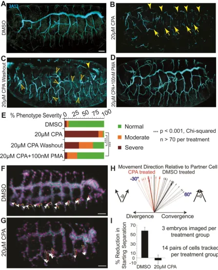
Fig. 2. SERCA regulates cell migration to control budding, even in the absence of
proliferation
[image:41.595.72.520.72.628.2]Whole mount Drosophila embryos at stage 15-16 are viewed from the lateral aspect, anterior left, and the 2A12 antibody stains for tracheal lumen protein following the indicated treatments. (A) DMSO-treated controls displaying orderly tracheal branches. (B) 20 µM CPA disrupts branching, resulting in gaps in the dorsal trunk (arrowheads) and subsidiary branches (arrows). (C) CPA washout at stage 12 results in fewer breaks (solid arrowhead) and undulating branches with extended sprouts (open arrowheads). (D) PMA with CPA rescues tracheal budding defects. Occasionally, an embryo treated with CPA+PMA or one from which CPA was washed out exhibits a phenotype of excess tracheal cell migration (see Movie 8). (E) Severity of phenotypes was scored for each treatment. Both washout and PMA rescue vs CPA alone significantly reduce the proportions of embryos in higher severity groups (p<0.001; Chi-squared; n>70 per treatment).
(F-I)Live imaging shows SERCA is necessary for airway cells to converge during completion
of Drosophila tracheogenesis.
(F-G) The trajectories (white arrows) and yellow-to-red migration paths are shown for individual cells that form the tracheal lateral trunk from stage 14 to early 16. The displacements shown represent cell movement over 100 minutes. (F) Cells from adjacent segments in DMSO-treated wildtype embryos converge. (G) During SERCA blockade, tracheal cells lack active migration and slightly diverge as the embryo develops.
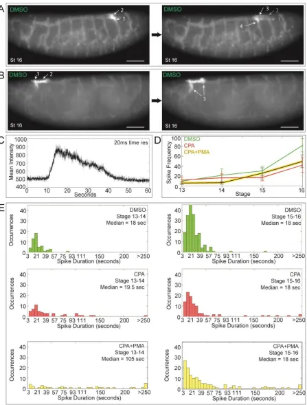
Fig. 3. SERCA regulates cell migration and budding by maintaining higher Ca2+ levels in “leader” versus “trailer” cells
(A-B) Btl:GCamp3 embryos treated with DMSO, CPA, or CPA+PMA were imaged between stage 13 and 16, and images were reconstructed to analyze the Ca2+ levels in tracheal cells. Shown are 3D reconstructed images of stage 13 embryos treated with (A) DMSO or (B) CPA (arrow indicates discontinuous trunk). Insets mark segments tracked in (C) and (D). Scale bars = 50 µm.
(C-D,F) Inset images of tracked cells from DMSO- (C), CPA- (D), and CPA+PMA-treated (F) embryos. “Leader” cells (blue arrow) that migrate from adjacent segments and fuse to form the lateral trunk and “trailer” cells (magenta arrow) behind them were tracked over
the timecourse and their Ca2+ levels quantified and plotted below the panel of images. Vertical lines mark the timepoints corresponding to each image. (C) In controls, there is a Ca2+ level differential whereby “leader” cells have consistently higher Ca2+ levels than “trailers”, particularly early when the cells are migrating. After fusion, “leaders”
periodically display surges of Ca2+ (black arrows; also green spikes in E and spikes in top panel of G). (D) In CPA-treated embryos, migration is lost so the lateral trunk remains discontinuous, and “leaders” have lower Ca2+ levels than “trailers” during the time they should be migrating. Thus the Ca2+ level differential is reversed. (F) CPA+PMA co-treatment reinstates the higher Ca2+ level in “leaders” and rescues migration.
(E,G) For each treatment, the ratio of intensities of ten pairs of cells (leader/trailer) was plotted. (E) The DMSO ratios (green) average >1. SERCA inhibition (red, ratio <1) inverts this.
Fig. 4. Live Ca2+ imaging shows that impulses propagate the Drosophila tracheal network
Btl:GCamp3 embryos were imaged in 3D + time (3 sec time resolution) from stage 13 to
(A-B) Timelapse imaging of two control embryos at stage 16 reveal Ca2+ pulses propagating through electrically coupled cells once the tracheal network has fused, such as between adjacent transverse connectives via the dorsal trunk (A), or bidirectionally (B) (follow arrows in numbered sequence). The timepoints shown are (A) 21 and (B) 9 seconds apart. Scale bars = 50 µm.
(C) At 20 ms time resolution, a typical Ca2+ spike shows a fast upstroke and slower decay. (D) The mean Ca2+ spiking frequency and SEM for control, CPA-treated, and
CPA+PMA-treated embryos at each stage are plotted (n > 3). Spike frequency increases with embryo age. Compared to controls, the frequency of Ca2+ impulses at later stages is diminished by SERCA blockade, even in the presence of the PKC activator PMA.
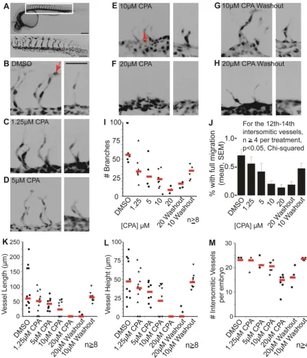
Fig. 5.SERCA function controls the rates of mesodermal migration and budding
(B-H) Paired confocal images show 3D reconstructions of the 14th-15th and 20th intersomitic vessels from embryos treated with (B) DMSO, (C) 1.25 µM CPA, (D) 5 µM CPA, (E) 10 µM CPA, (F) 20 µM CPA (G) 10 µM CPA washed out after 2 h, and (H) 20 µM CPA washed out after 2 h. Varying CPA dose allows dose-dependent reduction in vascular budding until at 20 μM, budding is reversibly suspended. Budding resumes after CPA washout. Scale bar = 50 µm for all images.
(I-J) Plots show, for each treatment, the collective number of branches (red lines = medians) on vessels 13-16 which form in the middle of the treatment (I) and the proportion of branches with cell nuclei at tip positions (J): compare the positions of nuclei (arrowed) in B vs E. In a parallel dose-dependent manner, CPA reduces branch numbers (I) and distal migration of endothelial cells (J) with resumption of branching and migration upon CPA washout.
Fig. 6. SERCA function controls the onset and rate of lung branching
(A) SERCA function dictates the onset of new buds. Plot of lung bud count vs time in culture for E13 rat lung explants shows the budding rate in controls (no CPA), lack of budding with 20 μM CPA, and resumption of budding when CPA is removed.
(B) SERCA function titrates budding rate. Bud count plotted against days in culture. The normal accretion of buds is shown in the absence of CPA (0 μM). Escalating the CPA dose controls the budding rate. At 20 μM, branching is arrested.
(C) The frequency of airway peristaltic waves decreases with escalating CPA dose, with statistical significance between each treatment group (p<0.05). Median & interquartile range (IQR) are plotted, n>10 for each treatment.
(D) Proliferation of lung epithelial and mesenchymal cells decreases with escalating CPA dose, with statistical significance between treatments for each cell type, except 10 µM and 20 µM are equivalent (p<0.05). Median & IQR of PH3 positive nuclei are plotted, n>24 for each treatment.
(E) SERCA blockade is associated with downregulation of lung morphogens SHH, FGF10, and SMMHC (smooth muscle myosin heavy chain), and significant upregulation of SPRY2, FGF9 and VEGF (qRT-PCR).
(F) SERCA inhibition impairs epithelial cell migration. Plot of percentage closure at 7 h (mean+/-SEM) of a standardized wound in a confluent monolayer of IEC-6 intestinal epithelial cells treated with 0, 1, 2, or 10 μM CPA. Wound closure is significantly reduced by 10 μM CPA (p<0.05).
Supplementary Figures
Fig. S1. Cell shape change, migration, and proliferation are unicellular behaviors adopted
for multicellular branching
Multicellular tissues across species employ different combinations of cell shape change, cell
migration, and proliferation to undergo branching morphogenesis. For each branching
structure in the table, + indicates the presence of a specific behavior and – indicates its
absence.
Fig. S2. serca RNAi disrupts Drosophila larval air sac budding after depleting serca mRNA
and Ca2+ stores
(A) Each configuration of da-GAL4 driven serca RNAi reduced serca mRNA levels relative to
18S when compared to wild type (w1118).
(B) Cells were dissociated from stage 16 wild type and da> serca RNAi Drosophila embryos.
(C) The air sacs are visualized by btl-driven GFP expression. Compared to the control ‘leaf and
Compared to wild type Drosophila cells (left), RNAi cells (right) have deficient SERCA
protein function by late embryogenesis featuring a subnormal rise in intracellular Ca2+
(measured by Ca2+ indicator fluorescence intensity) upon thapsigargin challenge.
stalk’ type structure, btl-GAL4 driven serca RNAi +/- dicer induced a range of phenotypes,
from no budding to very stunted and dysmorphic buds without the typical stalk. Combining
RNAi constructs (4474 and 107446) to enhance the knockdown further increased the frequency
of severely deranged air sac morphogenesis (Bar = 25 µm). Bottom panels are three
representative examples.
Fig. S3. serca RNAi does not affect apoptosis rate in larval air sac primordia
Confocal micrographs show third instar larval imaginal wing discs, with tracheal system in
green (actin-GFP) and apoptotic cells labeled by cleaved Caspase-3 in magenta. Scale bars =
20 µm.
(A) Wild-type control air sac primordia.
(B) Mutant serca RNAi air sac primordia, arrow indicates one apoptotic cell outside the air sac.
(C) Positive control showing Caspase-3 positive cells in the wing disc border.
(D) Table shows number of air sac primordia imaged for each genotype, and total number of
cleaved Caspase-3 positive cells detected within the air sacs across all samples: no apoptotic
cells were seen in wild type air sacs, and only a single apoptotic cell was seen in one of 22
mutant air sacs.
7
Fig. S4. RNAi is not sufficient to disrupt SERCA protein levels or function during early
Drosophila embryogenesis
(A)qRT-PCR of stage 11 wild type embryos (w1118) or embryos ubiquitously expressing RNAi
constructs (4474, 107446, 4474+dicer2, or 4474+107446) using the da-Gal4 driver to knock
down serca does not deplete serca mRNA relative to 18S mRNA. (Error bars indicate +/- 1
SEM.)
(B)In da-Gal4 X sercaRNAi4474 / CyO-twi>GFP embryos, when GFP is not expressed, the RNA
construct against serca is ubiquitously expressed. Confocal micrographs show ubiquitous
expression of SERCA protein (red, fluorescent thapsigargin) in controls (left side) and with
RNAi construct expression (right side), at stage 11.
(C)Cells were dissociated from stage 11 wild type and da> serca RNAi Drosophila embryos.
Compared to wild type Drosophila cells (left), RNAi cells (right) have normal SERCA
protein function (mean +/- SD) with a normal rise in intracellular Ca2+ (measured by Ca2+
indicator fluorescence intensity) upon thapsigargin challenge.
Fig. S5 SERCA inhibition with CPA creates physical gaps in tracheal trunks, but these are
not due to cell death, and tracheal structure is rescued by PKC activation
Tracheal cells are genetically labeled by Breathless (Btl) driving eGFP fused to actin. The dorsal
trunk is outlined with dotted yellow lines in each panel. Scale bar = 50 µm throughout.
(A)Control embryos treated with DMSO.
(B)Incubation with SERCA inhibitor CPA results in physical gaps in the dorsal trunk where
tracheal cells are absent (arrowhead) and tenuous cytoplasmic connections between segments
(arrow).
(C)Removal of CPA at stage 12 and further incubation without the inhibitor results in fewer
gaps (solid arrowhead) but exuberant sprouting (open arrowheads) and undulating branches.
Sometimes an extranumerary lateral trunk partially or completely forms (arrows).
(D) Treatment with PKC activator PMA alone results in normal tracheal structure.
(E)Co-treatment with SERCA inhibitor and PKC activator rescues tracheal structure.
(F)In live embryos, fluorescently labeled nuclei of tracheal cells were tracked in controls treated
with DMSO, and in CPA-treated embryos at segments where a continuous lateral trunk failed
to form. From stages 14-16 during the time the lateral trunk should have formed, there was
no tracheal cell death observed in either CPA- or DMSO-treated embryos. All labeled nuclei
remained present throughout; at affected segments in the CPA-treated embryos, the cells
simply did not migrate to form the trunk.
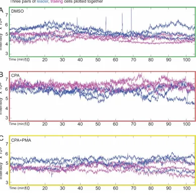
Fig. S6. SERCA blockade with CPA elevates overall Ca2+ level of Drosophila tracheal cells
(A-C) For DMSO-treated (A), CPA-treated (B), and CPA+PMA-treated (C) embryos, three
representative pairs of leader (blue) and trailer (magenta) cells are plotted together. Ca2+ level
is consistently higher in leader cells in A and C, while this differential is reversed in
CPA-treated embryos (B). Also, the overall Ca2+ levels of the tracheal cells in the CPA-treated
embryos are higher than in the DMSO- and CPA+PMA-treated embryos, consistent with
blockade of the SERCA pump. This is alleviated by co-treatment with PKC-activator PMA