Sequence-Dependent Regulatory Genome in
E. coli.
Thesis by
Nathan Maurice Belliveau
In Partial Fulfillment of the Requirements for the Degree of
Doctor of Philosophy in Bioengineering
CALIFORNIA INSTITUTE OF TECHNOLOGY Pasadena, California
2018
© 2018
Nathan Maurice Belliveau ORCID: 0000-0002-1536-1963
ACKNOWLEDGEMENTS
It is no question that I owe my wonderful time at Caltech to Professor Rob Phillips. His breadth of interest and never-ending enthusiasm about the science have been a constant source of inspiration for me. I also believe his constant questioning of assumptions and skepticism throughout my journey here have led to more interesting results and made me a better researcher. I hope more students continue to stumble upon the group as I did.
I would especially like to extend my gratitude to the members of the group. The socialist experiment that is Chapter 2 was truly a team effort and I enjoyed working with Manuel, Tal, Griffin, and Stephanie throughout the project. The positive atmosphere within the group has made my many hours stuck in the lab more manageable. Manuel, Griffin, and Soichi in particular were constant sounding boards to my many random thoughts and questions, and I appreciate their willingness to listen and provide feedback. In addition, Justin Kinney, who is at Cold Spring Harbor, has provided expert guidance in the Sort-Seq efforts of Chapter 3 and 4, and I have appreciated the counter perspectives that he was able to offer throughout that work.
I also had the opportunity to join Rob and other members in the group on many of his teaching engagements around the world and I have been happy to tag along whenever I could. I’m deeply indebted to Heun Jin Lee, who taught me everything I know about optics. While I’m sure I’ve only managed to absorb but a small fraction of his wealth of knowledge. I have done my best to pass that knowledge on accurately in the courses that I’ve been involved in.
ABSTRACT
Transcriptional regulation of gene expression is one of the most ubiquitous processes in biology. But while the catalog of bacterial genomes continues to expand rapidly, we remain ignorant about how almost all of the genes in these genomes are regulated. Given a gene, we would like to know the transcription factors that regulate them, how strongly they bind to the DNA, and how they interact with RNA polymerase and other external signals to control gene expression.
The theoretical framework of statistical thermodynamics provides us with a useful way to quantitatively describe the different mechanisms of regulation. One of the important ways genes are regulated is through external signals. To that end, we begin by presenting a general theory of allosteric transcriptional regulation using a statistical mechanical formulation of the Monod-Wyman-Changeux model. Allostery is central to many biological processes, and in the context of gene regulation, it describes a transcription factor’s conformational changes that modulate activity in response to external signals. We rigorously test this model using the ubiquitous simple repression motif with the transcription factor LacI inEscherichia coli. Our model not only accurately captures the allosteric response of these strains but also enables us to derive analytic expressions for key phenotypic properties such as the available dynamic range in gene expression.
We then move to consider the consequences for gene expression of the regulatory sequences themselves. Understanding how regulatory sequence maps to function remains a difficult problem in biology. Here we apply a massively parallel reporter assay, Sort-Seq, to build models that describe the sequence-dependent binding energies of transcription factors and RNA polymerase to DNA. By coupling such models to our thermodynamic models of regulation, we construct a genotype to phenotype map that predicts gene expression as a function of regulatory sequence. Here we demonstrate this approach by designing roughly 30 mutant LacI binding site sequences, and accurately predict expected levels of gene expression as a function of these sequences. We also show how such regulatory sequences can be designed to optimize the inducible response of LacI in the context of the allosteric simple repression motif considered above.
PUBLISHED CONTENT AND CONTRIBUTIONS
Belliveau, N. M., Barnes, S. L., Ireland, W. T., Jones, D. L., Sweredoski, M. J., Moradian, A., Hess, S., Kinney, J. B., and Phillips, R. (2017). A systematic approach for dissecting the molecular mechanisms of transcriptional regulation in bacteria.bioRxiv. DOI: https://doi.org/10.1101/239335.
N. M. B. helped conceive of the project, performed experiments, analyzed results, prepared data, and co-wrote the manuscript.
Razo-Mejia, M., Barnes, S. L., Belliveau, N. M., Chure, G., Einav, T., Lewis, M., and Phillips, R. (2017). Tuning transcriptional regulation through signaling: A predic-tive theory of allosteric induction.bioRxiv. DOI: https://doi.org/10.1101/111013. M.R.M., S.L.B., N.M.B., G.C., T.E. contributed equally to the work.
TABLE OF CONTENTS
Acknowledgements . . . iii
Abstract . . . iv
Published Content and Contributions . . . vi
Table of Contents . . . vii
List of Illustrations . . . ix
List of Tables . . . xiii
Chapter I: Introduction . . . 1
1.1 Transcription and transcriptional regulation . . . 3
1.2 Thermodynamic models . . . 8
1.3 Allostery . . . 10
1.4 Status of regulatory knowledge inE. coli . . . 13
1.5 SI: Candidate genes with growth-dependent differential expression . 20 References . . . 27
Chapter II: Tuning transcriptional regulation through signaling: A predictive theory of allosteric induction . . . 33
2.1 Introduction . . . 33
2.2 Results . . . 37
2.3 Discussion . . . 54
2.4 Methods . . . 60
2.5 SI: Inferring allosteric parameters from previous data . . . 65
2.6 SI: Induction of simple repression with multiple promoters or com-petitor sites . . . 70
2.7 SI: Flow cytometry . . . 77
2.8 SI: Single-cell microscopy . . . 81
2.9 SI: Fold-change sensitivity analysis . . . 88
2.10 SI: Alternate characterizations of induction . . . 91
2.11 SI: Global fit of all parameters . . . 95
2.12 SI: Applicability of theory to the Oid operator sequence . . . 103
2.13 SI: Comparison of parameter estimation and fold-change predictions across strains . . . 106
2.14 SI: Properties of induction titration curves . . . 110
2.15 SI: Applications to other regulatory architectures . . . 113
2.16 SI:E. coliprimer and strain list . . . 115
2.17 SI: Effect of chromosomal occupancy by other transcription factors on NN Sand the formulation of fold change . . . 118
References . . . 125
Chapter III: Characterization of the sequence-dependent occupancy of LacI. . 131
3.1 Introduction . . . 131
3.3 Discussion . . . 148
3.4 Methods . . . 149
3.5 SI: Summary of designed O1 binding site mutant results . . . 152
References . . . 153
Chapter IV: A systematic approach for dissecting the molecular mechanisms of transcriptional regulation in bacteria . . . 158
4.1 Introduction . . . 158
4.2 Results . . . 160
4.3 Discussion . . . 176
4.4 Methods . . . 177
4.5 SI: Characterization of library diversity and sorting sensitivity. . . 185
4.6 SI: Generation of sequence logos . . . 189
4.7 SI: Statistical mechanical model of the DNA affinity chromatography approach . . . 193
4.8 SI: DNA affinity chromatography and mass spectrometry experimen-tation and analysis . . . 196
4.9 SI: Identification of unannotated promoters inE. coliwhose expression appears to be regulated . . . 200
4.10 SI: Selection of the mutagenesis window for promoter dissection by Sort-Seq . . . 201
4.11 SI: Additional data from Sort-Seq experiments on theyebG,purT, xylE, anddgoRpromoters . . . 204
References . . . 212
Appendix A: Extended details on Sort-Seq data analysis. . . 220
LIST OF ILLUSTRATIONS
Number Page
1.1 Central dogma of molecular biology . . . 4
1.2 Transcription of bacterial genes by RNA polymerase . . . 5
1.3 Summary of known transcription factor binding sites of theE. coli genome. . . 6
1.4 Thelacoperon . . . 8
1.5 States and weights for the simple repression motif. . . 10
1.6 Examples of allosteric regulation inE. coli . . . 12
1.7 States and weights for the simple repression motif. . . 13
1.8 Identification of operons inE. coliwith and without regulatory annotation 14 1.9 Analysis of Schmidtet al.census study inE. coli . . . 15
1.10 Analysis of expression variability in Schmidtet al.census study across 22 growth conditions . . . 16
1.11 Identification of unannotated genes that are sensitive to particular growth conditions in the Schmidtet al. census study . . . 17
1.12 E. colinetwork map . . . 19
2.1 Transcription regulation architectures involving an allosteric repressor 37 2.2 States and weights for the simple repression motif . . . 40
2.3 Understanding the modular components of induction . . . 43
2.4 An experimental pipeline for high-throughput fold-change measurements 45 2.5 Predicting induction profiles for different biological control parameters 47 2.6 Comparison of predictions against measured and inferred data . . . . 49
2.7 Predictions and experimental measurements of key properties of induction profiles . . . 51
2.8 Fold-change data from a broad collection of different strains collapse onto a single master curve . . . 54
2.9 Multiple sets of parameters yield identical fold-change responses . . 66
2.10 Fold-change of multiple identical genes . . . 70
2.11 Induction with variable Rand multiple specific binding sites . . . 73
2.12 Induction with variable specific sites and fixed R. . . 74
2.14 Phenotypic properties of induction with multiple specific binding sites 76 2.15 Phenotypic properties of induction with a single specific site and
multiple competitor sites . . . 77 2.16 Plate arrangements for flow cytometry . . . 79 2.17 Representative unsupervised gating contours . . . 80 2.18 Comparison of experimental methods to determine the fold-change . 81 2.19 Experimental workflow for single-cell microscopy . . . 83 2.20 Correction for uneven illumination . . . 84 2.21 Segmentation of single bacterial cells . . . 86 2.22 Comparison of measured fold-change between flow cytometry and
single-cell microscopy . . . 87 2.23 Determining how sensitive the fold-change values are to the fit values
of the dissociation constants . . . 90 2.24 Hill function and MWC analysis of each induction profile . . . 93 2.25 Parameter values for the Hill equation fit to each individual titration . 94 2.26 A thermodynamic model coupled with a Hill analysis can characterize
induction . . . 95 2.27 Global fit of dissociation constants, repressor copy numbers and
binding energies . . . 100 2.28 Key properties of induction profiles as predicted with a global fit
using all available data . . . 101 2.29 Predictions of fold-change for strains with an Oid binding sequence
versus experimental measurements with different repressor copy numbers104 2.30 Comparison of fold-change predictions based on binding energies
from Garcia and Phillips and those inferred from this work . . . 105 2.31 O1 strain fold-change predictions based on strain-specific parameter
estimation ofKAandKI . . . 107 2.32 O2 strain fold-change predictions based on strain-specific parameter
estimation ofKAandKI . . . 108 2.33 O3 strain fold-change predictions based on strain-specific parameter
estimation ofKAandKI . . . 109 2.34 Dependence of leakiness, saturation, and dynamic range on the
operator binding energy and repressor copy number . . . 111 2.35 EC50and effective Hill coefficient depend strongly on repressor copy
2.36 Representative fold-change predictions for allosteric corepression and
activation. . . 115
2.37 Total cellular protein mass and DNA binding protein copy numbers inE. coliacross 22 growth conditions . . . 119
2.38 States and Weights for simple repression with pool of non-specific DNA binding proteins . . . 122
2.39 Percent of proteins that are DNA binding proteins inE. coliacross 22 growth conditions . . . 124
3.1 Process flow for using Sort-Seq to obtain energy matrices . . . 135
3.2 States and weights for the simple repression motif. . . 136
3.3 Inference of LacI energy matrices . . . 138
3.4 Energy matrices for the naturallacoperators from Sort-Seq data . . . 141
3.5 Fold-change data reflects expected values from predicted fold-change curves . . . 143
3.6 Energy matrix predictions can be used to design precise phenotypic responses . . . 146
3.7 Point mutations to LacI DNA-binding domain cause subtle changes to sequence specificity . . . 147
4.1 Summary of transcriptional regulatory knowledge inE. coli . . . 160
4.2 Overview of approach to characterize transcriptional regulatory DNA, using Sort-Seq and mass spectrometry . . . 162
4.3 Sort-Seq identifies the regulatory landscape of thelac,rel, andmar promoters . . . 166
4.4 DNA affinity purification and identification of LacI and RelBE by mass spectrometry using known target binding sites . . . 168
4.5 Sort-Seq distinguishes directional regulatory features and uncovers the regulatory architecture of thepurT promoter . . . 171
4.6 Sort-Seq identifies a set of activator binding sites that drive expression of RNAP at thexylEpromoter . . . 173
4.7 ThedgoRKADTpromoter is induced in the presence of D-galactonate due to loss of repression by DgoR and activation by CRP . . . 175
4.8 Analysis of the library mutation spectrum and effect of Sort-Seq sorting conditions . . . 188
4.9 Comparison between Sort-Seq and genomic-based sequence logos . . 193
4.11 Identification of unannotated genes with potential regulation and distribution of known transcription factor binding sites inE. coli. . . 203 4.12 Extended analysis of theyebG,purT, andxylEpromoters . . . 206 4.13 Extended analysis of thedgoRpromoter . . . 211
LIST OF TABLES
Number Page
1.1 Candidate unannotated genes with increased expression . . . 20 1.2 Candidate unannotated genes with decreased expression . . . 26 2.1 Instrument settings for data collection using the Miltenyi Biotec
MACSQuant flow cytometer . . . 78 2.2 Key model parameters for induction of an allosteric repressor . . . . 99 2.3 Global fit of all parameter values using the entire data set . . . 102 2.4 Primers used in this work . . . 116 2.5 E. colistrains used in this work . . . 117 3.1 Summary of designed O1 mutant binding site sequences and the
C h a p t e r 1
INTRODUCTION
Since the technological developments that led us to the first draft of the human genome in 2001 (Lander et al., 2001), our catalog of sequenced genomes has expanded at an incredible pace (Loman and Pallen, 2015; Land et al., 2015). While this global effort took 13 years and almost 3 billion dollars, today we could accomplish it for about 1,000 dollars and in roughly a day (Levy and Myers, 2016). As further illustration of this pace, between 2010 and 2014 a bird species from each major avian clades was sequenced (Jarvis et al., 2014), and larger scale intiatives such as the G10K project to sequence species in every vertebrate genus are well under way (Koepfli et al., 2015).
The advent of so-called next-generation sequencing technologies (and now third-generation technologies that offer much longer read lengths) has revolutionized how we perform research in biology (Goodwin et al., 2016). It has led to the development of over one hundred sequencing-based methods (Pachter, 2013) to study almost any aspect of biology, from chromosomal structure (e.g., Hi-C-Seq (Belton et al., 2012)), transcriptional regulation (e.g., Sort-Seq (Kinney et al., 2010) and ChIP-Seq (Park, 2009)) and RNA structure (e.g. SHAPE-ChIP-Seq (Loughrey et al., 2014)) to name but only a few of these techniques. Many of these approaches have become important tools to biophysicists due to their ability to provide a quantitative measure of biological function. It is also having a pervasive impact on society that is only beginning to be realized; in the form of personalized medicines that may significantly improve the effectiveness of current treatment options for many diseases (Rabbani et al., 2016).
of regulatory mechanisms involved.
At the core of this dissertation is an aim to develop quantitative descriptions of the mechanisms of transcriptional regulation that will allow us to describe and predict the input-output responses in gene expression across a bacterial genome. InChapter 1we begin by first reviewing the processes associated with gene expression (i.e., the central dogma of molecular biology) and regulation. We then describe how ideas from statistical mechanics can be used to quantitatively describe the regulatory mechanisms of transcription. We end with a discussion of the current state of regulatory knowledge inE. colito highlight our still limited understanding of the regulatory code which motivate the efforts of Chapter 4.
InChapter 2we extend previous theoretical and experimental efforts of regulation to account for the phenomenon of allostery. Allosteric proteins respond to changes in their environment by binding to ligands or other effector molecules and are central to most metabolic and signal-transduction pathways (Fenton, 2008; Motlagh et al., 2014). While it is common to apply phenomenological models such as Hill functions to describe this response, such models consist of lumped parameters that are a conceptual dead-end (Kuhlman et al., 2007). Instead, we develop a general quantitative framework with which to describe allosteric transcriptional regulation with the Monod-Wyman-Changeux (MWC) model of allostery (Monod et al., 1965), that instead more accurately recognizes that proteins as existing in two of more structural states that drive this allosteric response. We use the model to produce a set of predictions that we then test experimentally in the context of Lac repressor (LacI) and the simple repression motif (Bintu et al., 2005a).
We then move to consider the consequence of the regulatory sequences themselves on gene expression. In order to connect regulatory sequence to biophysical mechanisms of regulation across theE. coligenome, we propose applying a massively parallel reporter assay, Sort-Seq (Kinney et al., 2010), to characterize the regulatory DNA. This approach will allow us to identify transcription factor binding sites and enable the quantitative dissection of individual promoters with base pair resolution. In
such as dynamic range, saturation, and leakiness (Martins and Swain, 2011).
Finally, with the preceding chapters demonstrating the type of quantitative rigor we intend to apply to regulation more broadly across a bacterial genome, inChapter 4we then develop a systematic approach to quantitatively decipher the regulation of any promoter more generally. Here we first apply Sort-Seq across different bacterial promoters to uncover the functional binding sites where transcription factors bind to regulate gene expression. Using DNA affinity chromatography and mass spectrometry we then identify the transcription factors that bind these sites, and apply information-theoretic modeling to infer energy matrix models of binding by each transcription factor. We demonstrate the validity of the approach by first applying it to the well-characterized promoters oflacZYA,relBE, andmarRAB. We then apply it to uncover the regulatory architectures for the promoters ofpurT,xylE, anddgoRKADT, whose regulation was previously unknown.
1.1 Transcription and transcriptional regulation
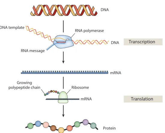
The central dogma of molecular biology describes the order in which genetic information flows from genomic DNA to produce proteins that will then perform biological functions (Figure 1.1) (Crick, 1970). The process begins when RNA polymerasetranscribesa gene’s coding sequence on the DNA into a single-stranded mRNA template. A ribosome thentranslatesthe gene coded on the mRNA into a protein that consists of a polypeptide of amino acids. While the nucleotides on the DNA map directly to those on mRNA (A→A, C→C, G→G, T→U), each amino acid that makes up a protein will map to triplet sets of nucleotides on the mRNA known as codons.
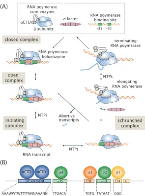
To begin transcription, RNA polymerase must first bind the DNA in the upstream region that precedes a gene, known as the gene’s promoter. Figure 1.2 shows a general schematic of the transcription process. During growth in nutrient-rich conditions, the core RNA polymerase enzyme (which consists of five subunits,
ββ0α
2ω) recognizes particular DNA binding sites by forming a complex with the primary sigma factor, RpoD (also written asσ70orσD), which prefers the consensus -35 and -10 sequence TTGACA(N)nTATAAT (where (N)nis a spacer sequence, with
Translation
DNA
mRNA
mRNA RNA polymerase
DNA DNA template
RNA message
Ribosome Growing
polypeptide chain
Protein
[image:17.612.171.446.64.286.2]Transcription
Figure 1.1: Central dogma of molecular biology. The pathway from DNA to protein. When a gene is on, RNA polymerase translocates along the DNA and transcribes the coding sequence into an mRNA template. This mRNA template is then read by the ribosome, which produces a polypeptide chain by stringing together amino acids according to triplet sets of nucleotides known as codons specified by the mRNA. Obtained from reference (Phillips, 2015).
enhanced by the presence of A/T rich sequences called ‘UP elements,’ that are found upstream of the -35 site (see Figure 1.2B). This is due to recognition by the flexibly tetheredα-subunit C-terminal domain (αCTD) of the core RNA polymerase enzyme (Browning and Busby, 2016; Murakami and Darst, 2003). Upon binding to the DNA, RNA polymerase will then proceed through several well-characterized steps to separate the two strands of DNA and begin transcription. Here, the polymerase first transitions from a closed complex with the DNA into a stable open complex where transcription can then begin to generate the mRNA template (Browning and Busby, 2016; Murakami and Darst, 2003).
NTPs
NTPs
NTPs Abortive transcripts
αCTD αCTD σ3 σ2 σ1
Ext
UP element –10 Dis +1
AAAWWTWTTTTNNNAAANN TTGACA TGTG TATAAT GGG
(A)
(B)
4 3 2 1
–35 –10
RNA poymerase binding site RNA poymerase
core enzyme
αCTD
β subunits
σ factor
closed complex
open complex
initiating complex
RNA transcript
RNA poymerase holoenzyme
terminating RNA poymerase
elongating RNA poymerase
schrunched complex
σ4
[image:18.612.154.449.68.465.2]–35
position relative to transcription start site number of binding sites overlapping at a base pair
Figure 1.3: Summary of known transcription factor binding sites of the E. coligenome. The locations of all annotated transcription factor binding sites on RegulonDB were used to generate a histogram of their locations on the genome. Each binding site is listed relative to a transcription start sites for the promoter where it binds. Figure was adapted from reference (Rydenfelt et al., 2014).
under certain physiological conditions (Gur et al., 2011). Despite this variety of control mechanisms that exist, regulation at the level of transcription is arguably the among the dominant ways in which cells across all domains of life regulate their expression. Here, cells decide when a gene is ‘switched’ on or off in large part by proteins called transcription factors that bind the DNA and modulate the activity of RNA polymerase at each promoter (Browning and Busby, 2016).
If we take a survey of the location of binding sites where transcription factors bind the genomic DNA, we find them distributed near the transcription start site where RNA polymerase begins transcription (see Figure 1.3). The transcription factors that bind these sites can be categorized as either repressors (preventing transcription) or activators (enhancing transcription) (Seshasayee et al., 2011). It is interesting to note however that even LacI, the canonical example of a repressor, can also be converted into an activator (Labow et al., 1990), so this categorization is somewhat fluid and may depend on context.
lac(Boedicker et al., 2013b; Boedicker et al., 2013a),gal(Mandal et al., 1990), and araC(Martin et al., 1986; Schleif, 2010) inE. coli.
Activators generally bind upstream of RNA polymerase, where they enhance tran-scription through interaction with theαCTD domain of RNA polymerase (class I activation), or directly with the sigma factor (class II activation) (Lee et al., 2012). It is interesting to note that if the activator binding site is slid along the DNA just upstream of the RNA polymerase binding site, a periodic pattern is observed in the extent of activation (experimentally shown using synthetic constructs (Ushida and Aiba, 1990; Gaston et al., 1990)). By noting that a full turn of the DNA helix requires about 10.5 base pairs, this is explained by the need for both activator and RNA polymerase to share the same face of the DNA and serves to highlight the physical mechanism underlying this process.
lacZ
lacZYA promoter
LacI-allolactose
CRP RNAP
lactose
E. coli
LacY
lacY lacA
LacZ
allolactose
repressor site
activator site
RNAP site
[image:21.612.178.437.71.340.2]coding gene
Figure 1.4: Thelacoperon. The promoter for thelacoperon drives expression of lacZ,lacY, andlacA. Transcription by RNA polymerase is regulated: a) repression by LacI which binds at three binding sites (O1 and O2 shown; O3 is within the lacZgene), and b) activation by CRP, which binds upstream of RNA polymerase. After translation, the LacZ protein forms a homotetramer that catalyzes cleavage of lactose to glucose and galactose (and lactose into allolactose). The LacY protein is a membrane protein that allows intake of lactose from the cell’s environment. The functional role of LacA is not well known. In the absence of allolactose, the LacI tetramer strongly represses transcription. In the presence of allolactose, LacI is allosterically induced and no longer binds strongly to the LacI binding sites, and transcription can be enhanced by CRP. Note that binding sizes and coding regions are not shown to scale.
1.2 Thermodynamic models
1982). This might seem a little absurd since cells are definitely out of equilibrium. However, these models have been quite successful in making quantitative predictions about gene regulation. Indeed, due to a separation of times scales for different biological processes, a quasi- equilibrium treatment of regulation is generally valid. In particular, the relevant interactions, such as binding by transcription factors to the DNA, occurs with fast on/off rates relative to the rate of transcription and translation (Moran et al., 2010).
We can derive such models of gene expression by first enumerating all possible states of a promoter and their corresponding statistical weights (Bintu et al., 2005a). Here we briefly consider the simple repression architecture, which we will be used extensively in Chapters 2 and 3. As shown in Figure 1.5, the promoter can be empty, occupied by RNA polymerase, or occupied by a repressor. In addition to the specific binding sites at the promoter, we have assumed that there are NN S non-specific binding sites elsewhere (i.e., on parts of the genome outside the simple repression architecture) where the RNA polymerase or the repressor can bind. Our model explicitly ignores the complexity of the distribution of non-specific binding affinities across the genome, and makes the assumption that a single parameter can capture the energy difference between our binding site of interest and the average non-specific site in the genome background. Thus,∆εPrepresents the energy difference between the specific and non-specific binding for RNA polymerase to the DNA. Likewise, ∆εRrepresents the difference in specific and non-specific binding energies for the repressor.
We can now calculate the probability that RNA polymerase is bound to the promoter
pbound, which is given by
pbound =
P NN Se
−β∆εP 1+ NR
N Se
−β∆εR + P NN Se
−β∆εP, (1.1)
with β= k1
BT, wherekB is the Boltzmann constant andT is the temperature of the system. Prepresents the RNA polymerase copy number per cell, whileRrepresents the copy number of repressor.
E. coli
state statistical weight description
RNA polymerase bound
P NNS e
− β∆εP
active repressor
bound NRA
NS e
− β∆εR
empty promoter 1
RNA polymerase non-specific
background DNA
bacterial promoter
repressor
Figure 1.5: States and weights for the simple repression motif. There arePRNA polymerase (blue) and a R repressors (red) per cell that compete for binding to a promoter of interest. The difference in energy between a repressor bound to the promoter of interest versus another non-specific positions elsewhere on the DNA equals∆εRin the active state and∆εRI in the inactive state; thePRNA polymerase have a corresponding energy difference ∆εP relative to non-specific binding on the DNA. NN S represents the number of non-specific binding sites for both RNA polymerase and repressor.
1.3 Allostery
Allosteric control is a molecular mechanism in which a conformational change occurs at one site on a protein in response to binding by a ligand or effector molecule at another distinct site of that same protein (Fenton, 2008; Motlagh et al., 2014). Allostery is central to most metabolic and signal-transduction pathways and examples can be found in a wide variety of cellular processes that include ligand-gated ion channels (Auerbach, 2012), enzymatic reactions (Einav et al., 2016; Velyvis et al., 2007), chemotaxis (Keymer et al., 2006), quorum sensing (Swem et al., 2008), and G-protein coupled receptors (Canals et al., 2012). While first described more than 50 years ago to account for the feedback inhibition that was apparent in the activity of certain enzymes of metabolic pathways (Changeux, 1961; Gerhart and Pardee, 1961; Gerhart and Pardee, 1962; Monod and Jacob, 1961), it continues to be an important area of study (Fenton, 2008). In particular, quantitative models describing the molecular mechanisms that provide this action at a distance remain elusive for most known cases of allostery.
DNA. The elucidation of thelacoperon was only just completed when the ideas of allostery were begining to take a more concrete form, and those ideas brought better insight into the mechanism of repression by LacI (and theλrepressor in phageλ) (Monod et al., 1963; Changeux, 2013). Here we provide several examples of allosteric gene regulation, and then in Chapter 2 develop an analytical framework for allosteric transcriptional regulation in bacteria through the Monod-Wyman-Changeux (MWC) model of allostery (Monod et al., 1965). While not the focus of Chapters 3 and 4, allostery appears to underlie how several newly identified regulatory architectures respond and modify their regulatory state.
Proteins are capable of being allosteric due to a probability of being in multiple structural conformations. When viewed through the MWC model, binding of an effector is seen to shift the protein’s allosteric equilibrium toward another state or conformation available to the protein. In the example of LacI, the repressor binds the DNA and represses expression from thelacoperon when cells are grown in the absence of lactose. However, in the presence of lactose, cells produce the metabolite allolactose from lactose (shown in Figure 1.4) that binds to LacI and ‘induce’ the repressor such that it no longer favors binding to its DNA binding site (Lewis, 2011) (see Figure 1.6A). The activator of the lac operon (and many other genes across theE. coligenome), CRP, is also allosteric. Here the cellular concentration of the nucleotide cyclic AMP influences the conformational state of CRP that enables it to bind DNA and regulate transcription (Schultz et al., 1991; Sharma et al., 2009)
strongly repressed
increasing Doc/Phd ratio
Phd
Doc
Doc induces structured domain in Phd ‘‘Intrinsically disordered domain’’
strongly repressed (B)
(A)
[image:25.612.187.425.70.313.2]allolactose LacI
Figure 1.6: Examples of allosteric regulation inE. coli. (A) Allosteric induction of LacI occurs when allolactose, or the synthetic compound, isopropyl β -D-1-thiogalactopyranoside (IPTG) is present and binds to ligand binding sites on the LacI protein. Each dimer has two binding sites. (B) Doc/Phd toxin–antitoxin system and regulation through conditional coopertivity. In the absence of antitoxin PhD protein, the toxin Doc causes cell arrest due to disruption of translation by ribosomes. As the ratio of antitoxin to toxin increases, the antitoxin binds the toxin and disables its toxicity. The antitoxin also acts as a repressor it provides autoregulation of its promoter. As a toxin-antitoxin complex, it binds more strongly to the DNA. Due to steric hindrance from an intrinsically disordered domain on the antitoxin, strong repression is only expected when the toxin/antitoxin is not too high or low, and two antitoxin dimers are able to bind the DNA. PhD is shown as a homodimer (red) and Doc is depicted as the blue ligand.
the tandem repressor binding sites.
active
state statistical weight
inactive
state satistical weight
c KA
2
c KA
c KA
1 + Kc
A 2
e− β ∆ εAI c KI
2
e− β ∆ εAI c KI
e− β ∆ εAI c KI
e− β ∆ εAI 1 + c KI
2 e− β ∆ εAI 1
Figure 1.7: States and weights for the simple repression motif. A repressor has an active conformation (red, left column) and an inactive conformation (purple, right column), with the energy difference between these two states given by∆εAI. The inducer (blue circle) at concentrationcis capable of binding to the repressor with dissociation constantsKAin the active state andKI in the inactive state. The eight states for a dimer with two inducer binding sites are shown along with the sums of the active and inactive states.
As another example, the repressor MarR, which represses themarRABpromoter, has been found to have four inducer binding sites (Wilkinson and Grove, 2006).
1.4 Status of regulatory knowledge inE. coli
Much of the insight we have on transcriptional regulation relies on careful and extensive work of a few model regulatory systems (Daber et al., 2011; Kuhlman et al., 2007; Buchler et al., 2003; Vilar and Leibler, 2003; Ackers et al., 1982). The Phillips group has relied on much of these efforts and used components of thelac operon to develop and test models of gene regulation (Garcia and Phillips, 2011; Garcia et al., 2011; Brewster et al., 2012; Boedicker et al., 2013b; Boedicker et al., 2013a; Brewster et al., 2014). While impressive advances in molecular biology have made it possible to map thousands of gene interactions and create genetic networks for a variety of organisms, they still leave us with a regulatory landscape that is qualitative in description. Here we take stock of what is known about regulation inE. coli. As we will find, we still remain ignorant to how most genes across the genome are regulated, and this prevents any attempt to begin to write down the types of quantitative models considered so far. This inability motivates much of the work of Chapter 4.
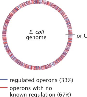
Figure 1.8: Identification of operons in E. coli with and without regulatory annotation. The plot identifies the genomic location of different operons with annotated TF binding sites (blue), and those lacking regulatory descriptions (red). The identification of regulated operons was performed using data from RegulonDB (Gama-Castro et al., 2016), which are based on manually curated experimental and computational data. All operons listed in the database were considered, where an operon was assumed to be regulated if it had at least one transcription factor binding site associated with it (lists of operons and transcription factor binding sites are available on the RegulonDB ’Download’ page, http://regulondb.ccg.unam.mx).
from the database of RegulonDB (Gama-Castro et al., 2016), which lists all the known regulatory features in this organism. Using this database, Figure 1.8 identifies the positions of each operon on theE. coligenome and whether it contains annotated transcription factor binding sites (blue) or not (red).
(A)
GalE copy
number / cell DgoD copy number / cell
(B)
gluc ose
xyloseaceta te
galac tose
glycer
ol
gluc ose
xyloseaceta te
galac tose
glycer
ol
Figure 1.9: Analysis of Schmidt et al. census study in E. coli. (A) Here we show the protein copy numbers per cell for GalE across several carbon sources. Expression was sensitive to the presence of galactose which is consistent with its known regulation (with about 5000 copies per cell, versus about 500 for most other growth conditions). (B) DgoD was also found to be sensitive to the presence of galactose as the carbon source. The copy number was measured to be 675 copies per cell when cells were grown in galactose, and 15 copies per cell or less in all other conditions considered. For both (A) and (B), values are shown for growth in M9 minimal media, with glucose, xylose, acetate, galactose, and glycerol as carbon sources and obtained from Schmidtet al., 2016.
et al. measured the copy number per cell of more than 2,300 proteins (about 55% of theE. coliproteome) across conditions that included different carbon sources, temperature, pH, growth phase, media, and growth in chemostats.
As a confirmation that the data of Schmidtet al. could identify regulated operons, we find that the GalE protein shows significantly higher expression when cells were grown in galactose (Figure 1.9A). GalE is involved in galactose catabolism, and its expression is known to increase due to loss of repression of thegalEpromoter when cells are grown in galactose (Irani et al., 1983; Semsey et al., 2007). Among promoters without any known regulation, we show the expression of DgoD in Figure 1.9B in several carbon sources. Cells grown in galactose showed much higher expression, with about 675 copies per cell, compared to at most 15 copies per cell across the other growth conditions. This is only one of many examples where a protein showed a large differential expression level across growth conditions and suggests that RegulonDB is incomplete.
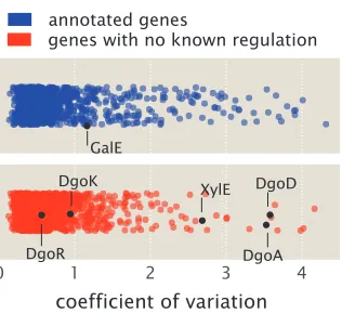
annotated genes
genes with no known regulation
coefficient of variation
XylE DgoD
DgoA DgoK
[image:29.612.229.387.67.212.2]DgoR GalE
Figure 1.10: Analysis of expression variability in Schmidt et al.census study across 22 growth conditions. Coefficient of variation is calculated (standard deviation divided by mean copy number) across the 22 growth conditions for each protein measured in Schmidtet al., 2016. Proteins are identified as either having regulatory annotation (blue) or not (red) using the annotations in RegulonDB (Gama-Castro et al., 2016). GalE is noted among the annotated genes and provides a reference as a gene that is known to be regulated and be perturbed in this study, as shown in Figure 1.9(A). Among the unannotated genes, those assocaited with the promoters ofpurT,xylE, anddgoRKADTare noted and are investigated in Chapter 4.
should be among those that exhibit a large change in copy number in one or a few growth conditions.
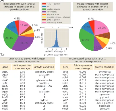
Lastly, this data represents a valuable resource to identify specific candidate genes for further regulatory investigation. In addition to calculating the coefficient of variation above, we can directly identify genes that are likely subject to transcriptional regulation and the growth condition necessary to perturb them. Here we do so by calculating the fold-change in expression for each protein relative to its average expression across all 22 growth conditions and summarize the analysis in Figure 1.11. Interestingly, among the highest fold-change values calculated, a substantial fraction are due to growth in a chemostat and in different carbon sources (Figure 1.11A, left plot). This contrasts with the lowest fold-changes values found, which are dominated by growth in different carbon sources or growth in stationary phase (Figure 1.11A, right plot). The highest and lowest fold-changes that were calculated from the data are summarized in Figure 1.11B and in Supplemental Section 1.5. Among these candidates, we consider the promoters ofpurT,xylE, anddgoRKADT in Chapter 4, where we use Sort-Seq to demonstrate that they are indeed under regulation at the transcriptional level.
measurements with largest increase in expression in a
growth condition
measurements with largest decrease in expression in a
growth condition
gene fold expressionover average
yqeG dgoA fdoI dusC yjiM amiC fdoH dgoD ygiQ fdoG xylE ymdF sdaB ykgE growth condition 22.0 22.0 22.0 22.0 22.0 19.3 18.4 18.3 18.0 17.5 17.1 16.3 16.0 14.9 stationary phase galactose LB glycerol + AA
fructose glycerol + AA
LB galactose LB LB xylose stationary phase LB LB (A) (B)
unannotated genes with largest increase in expression
gene fold expressionover average
sgcB yeeD ybhA yqcA yebO ylaC ymdF yqcC sgcB ybeY ydiZ syd sgcB ymdF growth condition 0.005 0.007 0.007 0.012 0.012 0.012 0.014 0.017 0.019 0.019 0.019 0.021 0.022 0.023 chemostat stationary phase stationary phase stationary phase stationary phase stationary phase stationary phase stationary phase pyruvate glycerol stationary phase
42C + glucose Succinate
pyruvate unannotated genes with largest
decrease in expression
[image:30.612.105.509.78.466.2]ln fold change in protein expression
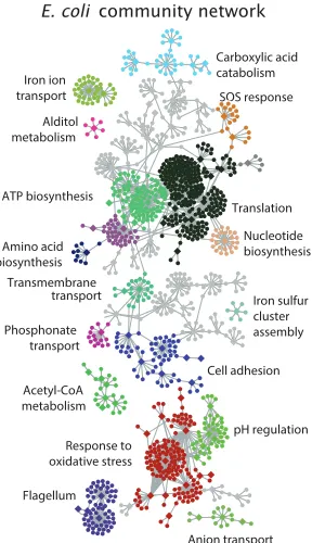
of genes (Gama-Castro et al., 2016). At the level of the genome-wide regulon, we can also consider how sets of genes are controlled (i.e., gene network maps). An example of this is shown in Figure 1.12, which shows an inferred genetic network for E. coli. This represents the average mapping from a variety of inference approaches, using about 800 microarray datasets across about 500 growth conditions (Marbach et al., 2012). The map is quite enlightening; for example, they were able to identify clusters of physiologically associated genes, some of which had no prior known function. However, the authors note the accuracy of their network map at about 50%, which they checked in two ways. The first is by comparing their network map to the manually curated RegulonDB database that is the gold-standard for regulatory knowledge inE. coli. Here they find that the model appears to be consistent with RegulonDB about 50% of the time. To their credit, RegulonDB is likely incomplete and perhaps they have identified new regulatory interactions. However, the authors also tested 53 target gene predictions across five transcription factors, and only about half the time they found experimental support for the predicted regulatory connection.
SOS response
Amino acid biosynthesis
Acetyl-CoA metabolism Iron ion transport
Transmembrane transport
Flagellum ATP biosynthesis
Response to oxidative stress
Carboxylic acid catabolism
Phosphonate transport Alditol metabolism
Anion transport Cell adhesion
Iron sulfur cluster assembly
pH regulation Translation
Nucleotide biosynthesis O
[image:32.612.230.374.221.471.2]SSS E. coli community network
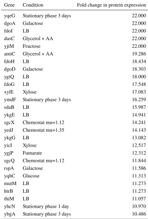
1.5 Supplemental Information: Candidate genes with growth-dependent
dif-ferential expression
Gene Condition Fold change in protein expression
yqeG Stationary phase 3 days 22.000
dgoA Galactose 22.000
fdoI LB 22.000
dusC Glycerol + AA 22.000
yjiM Fructose 22.000
amiC Glycerol + AA 19.286
fdoH LB 18.434
dgoD Galactose 18.303
ygiQ LB 18.000
fdoG LB 17.548
xylE Xylose 17.083
ymdF Stationary phase 3 days 16.259
sdaB LB 15.987
ykgE LB 14.941
sgcX Chemostat mu=1.12 14.241
yedJ Chemostat mu=1.35 14.143
ykgG LB 13.082
yicI Xylose 12.517
ygjP Fumarate 12.312
sgcQ Chemostat mu=1.12 11.844
rspA Galactose 11.586
yqhC Glucose 11.313
mutM LB 11.273
htrB LB 11.273
thiM LB 11.057
yhcN Stationary phase 1 day 10.970
[image:33.612.160.448.112.538.2]ybgA Stationary phase 3 days 10.486
Table 1.1: Candidate unannotated genes with increased expression.
Fold change in protein expression was calculated relative to the average
protein expression in the data of Schmidtet al. (2016). Proteins and
Gene Condition Fold change in protein expression
sdaB Stationary phase 1 day 0.100
yidE Glucose 0.100
maa pH6 glucose 0.100
sgcB pH6 glucose 0.099
rssA Stationary phase 1 day 0.099
yffB Galactose 0.099
djlA Stationary phase 1 day 0.099
rihC pH6 glucose 0.098
yedW pH6 glucose 0.098
dgoK Fumarate 0.098
yjiX pH6 glucose 0.098
yagE Stationary phase 3 days 0.098
rssA Chemostat mu=1.21 0.097
rph pH6 glucose 0.097
yddM 42C glucose 0.097
sgcX pH6 glucose 0.097
ycfS Galactose 0.096
yidE Osmotic-stress glucose 0.096
yqaB Stationary phase 3 days 0.096
cspB Glucose 0.095
psuG pH6 glucose 0.095
yjiX Acetate 0.094
dgoD Stationary phase 3 days 0.094
ybeY Acetate 0.094
maa Osmotic-stress glucose 0.093
hspQ pH6 glucose 0.093
ybgK Stationary phase 3 days 0.093
eutM Stationary phase 3 days 0.093
ybaQ Glycerol + AA 0.093
ugd pH6 glucose 0.093
cueR pH6 glucose 0.092
yagI Stationary phase 3 days 0.092
yphF LB 0.092
yidE pH6 glucose 0.092
yehX Stationary phase 1 day 0.092
yfhA Stationary phase 1 day 0.092
fdoH Pyruvate 0.092
cspB 42C glucose 0.092
rsmF Stationary phase 3 days 0.091
glsA Glycerol 0.091
yfgJ Stationary phase 1 day 0.091
yigA Glycerol + AA 0.090
acpH Stationary phase 1 day 0.090
wecG Stationary phase 3 days 0.090
ygdR Glycerol + AA 0.090
sapF Pyruvate 0.089
ytfL Stationary phase 1 day 0.089
eutM Fructose 0.088
hda Chemostat mu=1.5 0.088
yagI Chemostat mu=1.21 0.088
chpS LB 0.088
djlA Acetate 0.088
yfeC Stationary phase 3 days 0.088
ymgD pH6 glucose 0.088
ygfJ Mannose 0.088
cspB LB 0.087
tatE Stationary phase 1 day 0.087
ynfL 42C glucose 0.087
ybiJ Chemostat mu=1.12 0.086
fxsA Glucose 0.086
ybeD Stationary phase 1 day 0.086
fdoG Stationary phase 1 day 0.085
yfeC Stationary phase 1 day 0.085
fdoH Chemostat mu=1.5 0.085
ymdF Chemostat mu=1.21 0.084
ybeY Glycerol + AA 0.084
ygdR Glucose 0.083
cutC Stationary phase 1 day 0.083
yjhH Fumarate 0.082
psuG 42C glucose 0.082
fdoH Glucosamine 0.082
fxsA 42C glucose 0.082
kefF Stationary phase 3 days 0.082
fdoH Glycerol + AA 0.081
rph Stationary phase 3 days 0.082
ade 42C glucose 0.081
ytfQ pH6 glucose 0.081
ybiJ LB 0.081
cobU Stationary phase 1 day 0.081
dosC 42C glucose 0.081
ygfJ Xylose 0.080
ygdR 42C glucose 0.080
ybiJ Chemostat mu=1.21 0.078
fdoH Xylose 0.078
glsA Pyruvate 0.078
glsA Pyruvate 0.078
yhcN Glucosamine 0.078
yfeY 42C glucose 0.078
sgcB Osmotic-stress glucose 0.077
ydcH Glycerol + AA 0.077
yheV Stationary phase 3 days 0.077
ygfJ Fructose 0.077
mltC Glycerol + AA 0.076
truA Chemostat mu=1.12 0.076
ybeY LB 0.075
ybcL LB 0.075
uppS Stationary phase 3 days 0.075
ygfJ Osmotic-stress glucose 0.074
yajI Chemostat mu=1.21 0.074
cspB Stationary phase 3 days 0.074
ycfS Xylose 0.074
dgoD Pyruvate 0.074
yfiC Stationary phase 1 day 0.073
ybeY Pyruvate 0.073
yfiC Chemostat mu=1.21 0.072
ybdF Chemostat mu=1.21 0.072
ycaP LB 0.071
ygfJ pH6 glucose 0.072
yhhK pH6 glucose 0.071
psuG Xylose 0.071
cspB Chemostat mu=1.21 0.071
mltC Acetate 0.070
pptA Stationary phase 3 days 0.070
yciU Stationary phase 1 day 0.070
hslR Glycerol + AA 0.069
tusD 42C glucose 0.069
ilvG pH6 glucose 0.069
ycfS 42C glucose 0.069
yjhH 42C glucose 0.068
yodC Osmotic-stress glucose 0.068
yjiX 42C glucose 0.068
ybeD Stationary phase 3 days 0.068
maa Stationary phase 3 days 0.067
ygiD Chemostat mu=1.21 0.067
yjhH pH6 glucose 0.066
yedW LB 0.065
ygdR Xylose 0.064
mltC Pyruvate 0.064
greB LB 0.063
pyrB Glycerol + AA 0.063
ygdR Stationary phase 1 day 0.062
ymdF Glycerol 0.062
ymdF Chemostat mu=1.12 0.062
yfjG Chemostat mu=1.12 0.062
eutM Xylose 0.062
ybeY Fumarate 0.061
ymdF pH6 glucose 0.061
mltC Fumarate 0.061
priC Glycerol + AA 0.061
dicA Fructose 0.061
yidE LB 0.061
ybcL Osmotic-stress glucose 0.059
nudI Chemostat mu=1.12 0.059
ydiZ LB 0.059
fdoH Stationary phase 3 days 0.059
yfeD Chemostat mu=1.12 0.059
yjdI Galactose 0.058
dgoD Osmotic-stress glucose 0.058
ydcH Glucosamine 0.058
yjiX Chemostat mu=1.12 0.058
yceA Stationary phase 1 day 0.058
mltC Glycerol 0.058
yebO Stationary phase 1 day 0.058
mltC Glucosamine 0.057
ilvG Stationary phase 1 day 0.056
ygfJ Glycerol + AA 0.056
tdk Stationary phase 3 days 0.055
ybaV Pyruvate 0.054
rpmJ Glucose 0.054
pyrB LB 0.054
ydcH Glucose 0.054
ymdF Chemostat mu=1.5 0.053
ymgD Chemostat mu=1.12 0.052
yjjG Stationary phase 3 days 0.052
holD Xylose 0.051
yacG Fructose 0.051
ynfL Chemostat mu=1.5 0.050
ybaQ Stationary phase 3 days 0.050
cobU pH6 glucose 0.050
ybeY Glucose 0.050
yeeD 42C glucose 0.050
ydcH pH6 glucose 0.049
ybaQ Stationary phase 1 day 0.048
ugd Osmotic-stress glucose 0.048
mdfA pH6 glucose 0.048
yjiX Galactose 0.048
djlA Glycerol + AA 0.047
rnhB Stationary phase 3 days 0.047
cspB Osmotic-stress glucose 0.046
ydcI Stationary phase 1 day 0.046
poxA Chemostat mu=1.12 0.045
dedD Stationary phase 3 days 0.044
nudI Pyruvate 0.043
ycaP Glycerol + AA 0.042
yfeY Stationary phase 3 days 0.042
rfaC Stationary phase 1 day 0.041
ybhA Stationary phase 1 day 0.041
ypfH Succinate 0.041
yfeY Stationary phase 1 day 0.041
yjiX Succinate 0.041
yffB Glucosamine 0.040
eutM Galactose 0.040
yjiX Chemostat mu=1.5 0.040
hicB Glycerol + AA 0.039
ydcI Stationary phase 3 days 0.039
yddM Stationary phase 3 days 0.039
hicB LB 0.039
yjiX Glycerol 0.039
ybiJ Glycerol + AA 0.038
ybcL pH6 glucose 0.038
fdoH Fructose 0.037
cspG Chemostat mu=1.35 0.037
yigP Stationary phase 1 day 0.037
ymdF Osmotic-stress glucose 0.036
fdoH Osmotic-stress glucose 0.036
djlA Glucosamine 0.036
ybiJ Galactose 0.036
ymgD Chemostat mu=1.5 0.035
nudI Osmotic-stress glucose 0.035
ymdF LB 0.035
ybiJ Chemostat mu=1.35 0.034
djlA Glucose 0.033
yqcC Stationary phase 1 day 0.033
ymdF Succinate 0.033
maa Stationary phase 1 day 0.032
mltC LB 0.032
rnhB pH6 glucose 0.028
ymdF 42C glucose 0.027
yacG Stationary phase 3 days 0.027
ymdF Galactose 0.025
ylaC Stationary phase 1 day 0.024
yddM pH6 glucose 0.023
ymdF Pyruvate 0.023
sgcB Succinate 0.022
syd 42C glucose 0.021
ydiZ Stationary phase 1 day 0.019
ybeY Glycerol 0.019
sgcB Pyruvate 0.019
yqcC Stationary phase 3 days 0.017
ymdF Stationary phase 1 day 0.014
ylaC Stationary phase 3 days 0.012
yebO Stationary phase 3 days 0.012
yqcA Stationary phase 3 days 0.012
ybhA Stationary phase 3 days 0.007
yeeD Stationary phase 1 day 0.007
yeeD Stationary phase 3 days 0.007
sgcB Chemostat mu=1.35 0.005
Table 1.2: Candidate unannotated genes with decreased expression.
Fold change in protein expression was calculated relative to the average
protein expression in the data of Schmidtet al. (2016). Proteins and
References
Ackers, G. K., Johnson, A. D., and Shea, M. A. (1982). Quantitative model for gene regulation by lambda phage repressor.Proceedings of the National Academy of Sciences79.4, pp. 1129–33.
Auerbach, A. (2012). Thinking in Cycles: MWC is a Good Model for Acetylcholine Receptor-Channels.The Journal of Physiology590.1, pp. 93–8.
Belton, J.-M., McCord, R. P., Gibcus, J. H., Naumova, N., Zhan, Y., and Dekker, J. (2012). Hi–C: A comprehensive technique to capture the conformation of genomes. Methods58.3, pp. 268–276.
Bintu, L., Buchler, N. E., Garcia, H. G., Gerland, U., Hwa, T., Kondev, J., and Phillips, R. (2005a). Transcriptional regulation by the numbers: models.Current Opinion in Genetics and Development15.2, pp. 116–124.
Bintu, L., Buchler, N. E., Garcia, H. G., Gerland, U., Hwa, T., Kondev, J., Kuhlman, T., and Phillips, R. (2005b). Transcriptional regulation by the numbers: applications. Current Opinion in Genetics and Development15.2, pp. 125–135.
Boedicker, J. Q., Garcia, H. G., Johnson, S., and Phillips, R. (2013a). DNA sequence-dependent mechanics and protein-assisted bending in repressor-mediated loop formation.Physical Biology10.6, p. 066005.
Boedicker, J. Q., Garcia, H. G., and Phillips, R. (2013b). Theoretical and Experimental Dissection of DNA Loop-Mediated Repression.Physical Review Letters 110.1, p. 018101.
Brewster, R. C., Jones, D. L., and Phillips, R. (2012). Tuning Promoter Strength through RNA Polymerase Binding Site Design inEscherichia coli.PLoS Compu-tational Biology8.12.
Brewster, R. C., Weinert, F. M., Garcia, H. G., Song, D., Rydenfelt, M., and Phillips, R. (2014). The transcription factor titration effect dictates level of gene expression. Cell156.6, pp. 1312–1323.
Browning, D. F. and Busby, S. J. W. (2016). Local and global regulation of tran-scription initiation in bacteria. Nature Reviews Microbiology 14.10, pp. 638– 650.
Buchler, N. E., Gerland, U., and Hwa, T. (2003). On schemes of combinatorial transcription logic. Proceedings of the National Academy of Sciences 100.9, pp. 5136–41.
Busby, S. and Ebright, R. H. (1999). Transcription activation by catabolite activator protein (CAP).Journal of Molecular Biology293.2, pp. 973–979.
Changeux, J.-P. (2013). The Origins of Allostery: From Personal Memories to Material for the Future.Journal of Molecular Biology425.9, pp. 1396–1406. Changeux, J.-P. (1961). The Feedback Control Mechanism of Biosynthetic
L-Threonine Deaminase by L-Isoleucine.Cold Spring Harbor Symposia on Quanti-tative Biology26.1, pp. 313–318.
Cipriano, M. J., Novichkov, P. N., Kazakov, A. E., Rodionov, D. A., Arkin, A. P., Gelfand, M. S., and Dubchak, I. (2013). RegTransBase – a database of regula-tory sequences and interactions based on literature: a resource for investigating transcriptional regulation in prokaryotes.BMC Genomics14.1, pp. 213–221. Cournac, A. and Plumbridge, J. (2013). DNA Looping in Prokaryotes: Experimental
and Theoretical Approaches.Journal of Bacteriology195.6, pp. 1109–1119. Crick, F. (1970). Central Dogma of Molecular Biology.Nature227.5258, pp. 561–
563.
Daber, R., Sochor, M. A., and Lewis, M. (2011). Thermodynamic analysis of mutant lac repressors.Journal of Molecular Biology409.1, pp. 76–87.
Einav, T., Mazutis, L., and Phillips, R. (2016). Statistical Mechanics of Allosteric Enzymes. EN.The Journal of Physical Chemistry B121 (15).
Feklístov, A., Sharon, B. D., Darst, S. A., and Gross, C. A. (2014). Bacterial Sigma Factors: A Historical, Structural, and Genomic Perspective.Annual Review of Microbiology68.1, pp. 357–376.
Fenton, A. W. (2008). Allostery: an illustrated definition for the ‘second secret of life’.Trends in Biochemical Sciences33.9, pp. 420–425.
Galperin, M. Y. and Koonin, E. V. (2010). From complete genome sequence to “complete“ understanding?Trends in Biotechnology28.8, pp. 398–406.
Gama-Castro, S. et al. (2016). RegulonDB version 9.0: high-level integration of gene regulation, coexpression, motif clustering and beyond.Nucleic Acids Research 44.D1, pp. D133–D143.
Garcia, H. G. and Phillips, R. (2011). Quantitative dissection of the simple repression input-output function.Proceedings of the National Academy of Sciences108.29, pp. 12173–8.
Garcia, H. G., Grayson, P., Han, L., Inamdar, M., Kondev, J., Nelson, P. C., Phillips, R., Widom, J., and Wiggins, P. A. (2007). Biological consequences of tightly bent DNA: The other life of a macromolecular celebrity.Biopolymers85.2, pp. 115– 130.
Garcia-Pino, A., Balasubramanian, S., Wyns, L., Gazit, E., De Greve, H., Magnuson, R. D., Charlier, D., Nuland, N. A. J. van, and Loris, R. (2010). Allostery and Intrinsic Disorder Mediate Transcription Regulation by Conditional Cooperativity. Cell142.1, pp. 101–111.
Garcia-Pino, A., De Gieter, S., Talavera, A., De Greve, H., Efremov, R. G., and Loris, R. (2016). An intrinsically disordered entropic switch determines allostery in Phd–Doc regulation.Nature Chemical Biology12.7, pp. 490–496.
Gaston, K., Bell, A., Kolb, A., Buc, H., and Busby, S. (1990). Stringent spacing requirements for transcription activation by CRP.Cell62.4, pp. 733–743.
Gerhart, J. C. and Pardee, A. B. (1962). The Enzymology of Control by Feedback Inhibition.Journal of Biological Chemistry237.3, pp. 891–896.
Gerhart, J. C. and Pardee, A. B. (1961). Separation of feedback inhibition from activity of aspartate transcarbamylase (ATCase). In:Fed. Proc. Vol. 20, p. 224. Goodwin, S., McPherson, J. D., and McCombie, W. R. (2016). Coming of age: ten
years of next-generation sequencing technologies.Nature Reviews Genetics17.6, pp. 333–351.
Gruber, T. M. and Gross, C. A. (2003). Multiple Sigma Subunits and the Partitioning of Bacterial Transcription Space.Annual Review of Microbiology57.1, pp. 441– 466.
Gur, E., Biran, D., and Ron, E. Z. (2011). Regulated proteolysis in Gram-negative bacteria - how and when?Nature Reviews Microbiology9.12, pp. 839–848. Irani, M. H., Orosz, L., and Adhya, S. (1983). A control element within a structural
gene: Thegaloperon ofEscherichia coli.Cell32.3, pp. 783–788. Jacob, F. (2011). The Birth of the Operon.Science332.6031, pp. 767–767.
Jacob, F. and Monod, J. (1961). Genetic regulatory mechanisms in the synthesis of proteins.Journal of Molecular Biology3.3, pp. 318–356.
Jarvis, E. D. et al. (2014). Whole-genome analyses resolve early branches in the tree of life of modern birds.Science346.6215, pp. 1320–1331.
Jishage, M. and Ishihama, A. (1995). Regulation of RNA polymerase sigma subunit synthesis inEscherichia coli: intracellular levels of sigma 70 and sigma 38.Journal of Bacteriology177.23, pp. 6832–6835.
Jishage, M., Iwata, A., Ueda, S., and Ishihama, A. (1996). Regulation of RNA polymerase sigma subunit synthesis inEscherichia coli: intracellular levels of four species of sigma subunit under various growth conditions.Journal of Bacteriology 178.18, pp. 5447–5451.
Kılıç, S., White, E. R., Sagitova, D. M., Cornish, J. P., and Erill, I. (2013). CollecTF: a database of experimentally validated transcription factor-binding sites in Bacteria. Nucleic Acids Research42.D1, pp. D156–D160.
Kinney, J. B., Murugan, A., Callan, C. G., and Cox, E. C. (2010). Using deep se-quencing to characterize the biophysical mechanism of a transcriptional regulatory sequence.Proceedings of the National Academy of Sciences107.20, 9158–9163. Koepfli, K.-P., Paten, B., O’Brien, S. J., and Scientists, t. G.K.C. o. (2015). The Genome 10K Project: A Way Forward.Annual Review of Animal Biosciences3.1, pp. 57–111.
Kuhlman, T., Zhang, Z., Saier, M. H., and Hwa, T. (2007). Combinatorial tran-scriptional control of the lactose operon ofEscherichia coli.Proceedings of the National Academy of Sciences104.14, pp. 6043–6048.
Labow, M. A., Baim, S. B., Shenk, T., and Levine, A. J. (1990). Conversion of the lac repressor into an allosterically regulated transcriptional activator for mammalian cells.Molecular and Cellular Biology10.7, pp. 3343–3356.
Land, M. et al. (2015). Insights from 20 years of bacterial genome sequencing. Functional & Integrative Genomics15.2, pp. 141–161.
Lander, E. S. et al. (2001). Initial sequencing and analysis of the human genome. Nature409.6822, pp. 860–921.
Lee, D. J., Minchin, S. D., and Busby, S. J. W. (2012). Activating Transcription in Bacteria.Annual Review of Microbiology66.1, pp. 125–152.
Levy, S. E. and Myers, R. M. (2016). Advancements in Next-Generation Sequencing. Annual Review of Genomics and Human Genetics17.1, pp. 95–115.
Lewis, M. (2011). A tale of two repressors – a historical perspective.Journal of Molecular Biology409.1, pp. 14–27.
Loman, N. J. and Pallen, M. J. (2015). Twenty years of bacterial genome sequencing. Nature Reviews Microbiology13.12, pp. 787–794.
Loomis, W. F. and Magasanik, B. (1967). The catabolite repression gene of the Lac operon inEscherichia coli.Journal of Molecular Biology23.3, pp. 487–494. Loughrey, D., Watters, K. E., Settle, A. H., and Lucks, J. B. (2014). SHAPE-Seq 2.0:
systematic optimization and extension of high-throughput chemical probing of RNA secondary structure with next generation sequencing.Nucleic Acids Research, pp. 1–10.
Maeda, H. (2000). Competition among sevenEscherichia colisigma subunits: relative binding affinities to the core RNA polymerase. Nucleic Acids Research28.18, pp. 3497–3503.
Marbach, D. et al. (2012). Wisdom of crowds for robust gene network inference. Nature Methods9.8, pp. 796–804.
Martin, K., Huo, L., and Schleif, R. F. (1986). The DNA loop model forararepression: AraC protein occupies the proposed loop sites in vivo and repression-negative mutations lie in these same sites.Proceedings of the National Academy of Sciences 83.11, pp. 3654–3658.
Martins, B. M. C. and Swain, P. S. (2011). Trade-Offs and constraints in allosteric sensing.PLoS Computational Biology7.11, pp. 1–13.
Monod, J., Wyman, J., and Changeux, J.-P. (1965). On the nature of allosteric transitions: A plausible model.Journal of Molecular Biology12.1, pp. 88–118. Monod, J. and Jacob, F. (1961). General Conclusions - Teleonomic Mechanisms in
Cellular Metabolism, Growth, and Differentiation.Cold Spring Harbor Symposia on Quantitative Biology26, pp. 389–401.
Monod, J., Changeux, J.-P., and Jacob, F. (1963). Allosteric proteins and cellular control systems.Journal of Molecular Biology6.4, pp. 306–329.
Moran, U., Phillips, R., and Milo, R. (2010). SnapShot: Key Numbers in Biology. Cell141.7, 1262–1262.e1.
Motlagh, H. N., Wrabl, J. O., Li, J., and Hilser, V. J. (2014). The ensemble nature of allostery.Nature508.7496, pp. 331–339.
Münch, R., Hiller, K., Barg, H., Heldt, D., Linz, S., Wingender, E., and Jahn, D. (2003). PRODORIC: prokaryotic database of gene regulation. Nucleic Acids Research31.1, pp. 266–269.
Murakami, K. S. and Darst, S. A. (2003). Bacterial RNA polymerases: the wholo story.Current Opinion in Structural Biology13.1, pp. 31–39.
Oehler, S., Eismann, E. R., Krämer, H., and Müller-Hill, B. (1990). The three operators of the lac operon cooperate in repression. The EMBO Journal 9.4, pp. 973–979.
Pachter, L. (2013). *Seq. https://liorpachter.wordpress.com/seq/. Blog.
Park, P. J. (2009). ChIP-seq: advantages and challenges of a maturing technology. Nature Reviews Genetics10.10, pp. 669–680.
Phillips, R. (2015). Napoleon Is in Equilibrium.dx.doi.org6.1, pp. 85–111.
Rabbani, B., Nakaoka, H., Akhondzadeh, S., Tekin, M., and Mahdieh, N. (2016). Next generation sequencing: implications in personalized medicine and pharma-cogenomics.Mol. BioSyst.12.6, pp. 1818–1830.
Salis, H. M., Mirsky, E. A., and Voigt, C. A. (2009). Automated design of synthetic ribosome binding sites to control protein expression.Nature Biotechnology27.10, pp. 946–950.
Schleif, R. (2010). AraC protein, regulation of the l-arabinose operon inEscherichia coli, and the light switch mechanism of AraC action.FEMS Microbiology Reviews 34.5, pp. 779–796.
Schmidt, A. et al. (2016). The quantitative and condition-dependentEscherichia coli proteome.Nature Biotechnology34 (1), pp. 104–111.
Schultz, S, Shields, G, and Steitz, T (1991). Crystal structure of a CAP-DNA complex: the DNA is bent by 90 degrees.Science253.5023, pp. 1001–1007.
Semsey, S., Krishna, S., Sneppen, K., and Adhya, S. (2007). Signal integration in the galactose network ofEscherichia coli.Molecular Microbiology65.2, pp. 465–476. Seshasayee, A. S. N., Sivaraman, K., and Luscombe, N. M. (2011). An Overview of Prokaryotic Transcription Factors. In:A Handbook of Transcription Factors. Dordrecht: Springer, Dordrecht, pp. 7–23.
Sharma, H., Yu, S., Kong, J., Wang, J., and Steitz, T. A. (2009). Structure of apo-CAP reveals that large conformational changes are necessary for DNA binding. Proceedings of the National Academy of Sciences106.39, pp. 16604–16609. Swem, L. R., Swem, D. L., Wingreen, N. S., and Bassler, B. L. (2008). Deducing
Receptor Signaling Parameters from In Vivo Analysis: LuxN/AI-1 Quorum Sensing in Vibrio harveyi.Cell134.3, pp. 461–473.
Ushida, C. and Aiba, H. (1990). Helical phase dependent action of CRP: effect of the distance between the CRP site and the -35 region on promoter activity.Nucleic Acids Research18.21, pp. 6325–6330.
Van Assche, E., Van Puyvelde, S., Vanderleyden, J., and Steenackers, H. P. (2015). RNA-binding proteins involved in post-transcriptional regulation in bacteria. Frontiers in Microbiology6.300, p. 134.
Velyvis, A., Yang, Y. R., Schachman, H. K., and Kay, L. E. (2007). A solution NMR study showing that active site ligands and nucleotides directly perturb the allosteric equilibrium in aspartate transcarbamoylase.Proceedings of the National Academy of Sciences104.21, pp. 8815–20.
Vilar, J. M. G. and Leibler, S. (2003). DNA Looping and Physical Constraints on Transcription Regulation.Journal of Molecular Biology331.5, pp. 981–989. Weinert, F. M., Brewster, R. C., Rydenfelt, M., Phillips, R., and Kegel, W. K. (2014).
Scaling of gene expression with transcription-factor fugacity.Physical Review Letters113.25, pp. 1–5.
C h a p t e r 2
TUNING TRANSCRIPTIONAL REGULATION THROUGH
SIGNALING: A PREDICTIVE THEORY OF ALLOSTERIC
INDUCTION
A version of this chapter originally appeared as M. Razo-Mejia, S. L. Barnes, N. M. Belliveau, G. Chure, T. Einav, M. Lewis, R. Phillips (2017). Tuning transcriptional regulation through signaling: A predictive theory of allosteric induction. bioRxiv, 111013. http://doi.org/10.1101/111013. It is also in preparation for publication in a peer-reviewed journal. M.R.M., S.L.B., N.M.B., G.C., T.E. contributed equally to this work.
2.1 Introduction
Understanding how organisms sense and respond to changes in their environment has long been a central theme of biological inquiry. At the cellular level, this interaction is mediated by a diverse collection of molecular signaling pathways. A pervasive mechanism of signaling in these pathways is allosteric regulation, in which the binding of a ligand induces a conformational change in some target molecule, triggering a signaling cascade (Lindsley and Rutter, 2006). One of the most important examples of such signaling is offered by transcriptional regulation, where a transcription factor’s propensity to bind to DNA will be altered upon binding to an allosteric effector.
meaning that the underlying minimal set of parameters cannot be pinned down unequivocally. Without minimal models involving clear, specific parameters, such fits are primarily descriptive and can do little to predict the system response as parameters are varied. Furthermore, phenomenological fits with unclear parameters provide little prospect for predicting or understanding what molecular properties determine key phenotypic parameters such as leakiness, dynamic range,[EC50], and the effective Hill coefficient as discussed in Martins and Swain (2011) and Marzen et al. (2013) and illustrated in Fig. 2.1. In response to these concerns, we formulate a minimal Monod-Wyman-Changeux (MWC) model of transcription factor induction in conjunction with a corresponding thermodynamic model of repression. While some treatments of induction have used MWC models to predict transcriptional outputs (Daber et al., 2009; Daber et al., 2011; Sochor, 2014), there has been no systematic experimental test of how well such a model can predict the induction process over broad swathes of regulatory parameter space. To that end, we use the MWC model to make parameter-free predictions about how the induction response will be altered when transcription factor copy number and operator strength are systematically varied.