Int. J. Electrochem. Sci., 14 (2019) 1940 – 1953, doi: 10.20964/2019.02.07
International Journal of
ELECTROCHEMICAL
SCIENCE
www.electrochemsci.orgEffect of a Bacterial Consortium on Passivation Property of
2024 Aluminum Alloy
Daniel Ortega1,Claudia Alvarado1,Lisa Muñoz2, Hans Kohler3, Fabiola Pineda4, Maritza Páez2, Mamie Sancy4, Nelson D. Vejar1 and Jenny M. Blamey.2,3,*
1 Academia Politécnica Aeronáutica, Fuerza Aérea de Chile, Av. José Miguel Carrera 11087, El Bosque, Santiago, Chile.
2 Facultad de Química y Biología, Universidad de Santiago de Chile, Alameda 3363, Estación Central, Santiago, Chile
3 Fundación Biociencia, José Domingo Cañas 2280, Ñuñoa, Santiago, Chile.
4 Escuela de Construcción Civil, Facultad de Ingeniería, Pontificia Universidad Católica de Chile. Av. Vicuña Mackenna 4860, Santiago, Chile.
*E-mail: [email protected]
Received: 23 May 2018 / Accepted: 28 September 2018 / Published: 5 January 2019
A bacterial consortium was recollected from corrosion products formed on a vertical fin of a military helicopter exposed to Antarctic environment for 4 years. For the identification and characterization of the consortium, DNA sequencing was performed showing that the consortium was mainly composed by three strains. Two of the strains showed a close affiliation to Bacillus toyonensis and Staphylococcus aureus. However, the third strain studied did not show affiliation by 16S rRNA analysis to any genus so far described. Additionally, morphological and electrochemical studies of an aluminum 2024-T3 alloy prior and after exposure to a sterile and inoculated minimal salt medium at 4
C were carried out by using scanning electron microscope, open circuit potential and electrochemical impedance spectroscopy. The experimental results revealed that steady-state properties of the passive film were modified by the bacterial consortium and isolates.
Keywords: Antarctica, aluminum alloy, 2024-T3, consortium, Bacillus toyonensis, Staphylococcus aureus
1. INTRODUCTION
levels of radiation (UV) are high. This indicates there are other factors influencing the corrosion process. One of these determining factors may be the presence of microorganisms, which are adapted to the extreme environment present in Antarctica. In addition, anaerobic heterotrophic and aerobic bacteria as well as sulfate reducing bacteria, fungi and yeasts have been found in samples taken from corroded sites in different airplanes [1]. For instance, a microbial community was found in fuel tanks manufactured with AA2024-T3, containing jet propellant-8 (JP-8) [2]. Isolated cultures were closely related to the genus Bacillus and to the fungi Aureobasidium and Penicillium influencing the electrochemical results revealing corrosion of the metallic surface [3]. Investigations about corrosion of AA2024 in the presence of the hydrocarbon-degrading bacteria Bacillus cereus ACE4 (a Gram-positive bacteria) and Serratia marcescens ACE2 (a Gram-negative bacteria) expose to a fuel tank simulated environment showed pitting corrosion damage caused by B. cereus ACE4, being more vigorous compared to that from S. marcescens ACE2. This could be associated to the higher capacity of adhesion from B. cereus ACE4 due to cell surface hydrophobicity [4]. In addition, AA2024-T3 exposed to bacteria Bacillus subtilis in minimal salt medium (MSM) solution has also shown evidence of corrosion after one month of immersion in this medium, determined by electrochemical measurements [5].
In this work, we study a corrosion phenomenon detected in a vertical fin of a military helicopter exposed for 4 years to the environmental conditions present in the Antarctic continent. Samples of corrosion products were collected from this component. The corrosion products were analyzed by chemical and microbiological techniques. Cultures revealed the presence of three strains belonging to the genera Bacillus, Staphyloccocus and third one to an unknown genus not described in the data bases. Electrochemical measurements and morphological analysis of AA2024-T3 specimens exposed to sterile and inoculated media indicate that one of these isolated bacteria influenced the evolution of the passive film.
2. EXPERIMENTAL METHODS
2.1. Metallic Substrates
To carry out the experimental studies, the Chilean aerospace company ENAER provided testing aluminum alloy AA2024-T3 specimens, sized 100×100×2 mm, with nominal chemical composition: (wt.%) 92.400 Al, 4.900 Cu, 1.520 Mg, 0.169 Fe, 0.520 Mn, 0.080. The specimens were degreased using a trichloroethylene solution at 90°C for 5 min, followed by an anodizing process using Turco 4215-S solution at 54°C for 15 min with stirring. Later, washing was done with bi-distilled water at room temperature and a pickling cleaning surface treatment was performed by using the industrial reagent Smut Go N°4 deoxidizer (30–45 g/l of 10% HNO3)at room temperature for 10 min
with stirring. Washing was again done with bi-distilled water at room temperature. The chromic acid anodizing (CAA) process was carried out using an electrolyte with following containing: 50g/L of H2CrO4, 0.5 g/L of Na2SO4, 0.2 g/L of NaCl, and a current density of 5 mA/cm2 for 30 min at 35°C
2.2. Sample Collection and Bacterial Strains
Samples of corrosion products were obtained from a vertical fin of a military helicopter located in a Chilean Antarctic Air Base (marine environment). The corrosion products were stored in sterile plastic receptacles (Zelle, Germany). Samples were transported in a cooler packed with ice to maintain the integrity of the sample. Microorganisms present in the samples conforming a consortium were cultivated and then isolated by serial dilutions and solid media plating in Luria Bertani Broth medium (LB) composed of (per L): 10 g tryptone, 5 g yeast extract, and 10 g NaCl. The bacterial consortium and isolated strains were stored at −20 °C to ensure the purity and consistency of the microbial material through all subsequent experiments.
2.3. Microbiological and Biochemical characterization Gram stain
Gram stain reaction was carried out using a Merck gram stain kit (Cat # 111885), according to the manufacturers recommended protocol.
Scanning electron microscopy of microorganisms
For Scanning Electron Microscopy (SEM), cells were concentrated by centrifugation and then fixed 16 h in 3% V/V glutaraldehyde in phosphate buffer. They were filtered using a polyamide filter (Sartorius) with a porous size of 0.45 m. The fixed cells were dehydrated by sequential passage through increased concentrations of ethanol (50-100% V/V) in 10% increments, dried with liquid CO2 in a critical point dryer, and then coated with gold - palladium for 60s. Samples were visualized using an electronic microscope Hitachi TM3000 with a beam voltage of 15kVolts.
Molecular Bacterial identification
using Chromas Pro software (Technelysium Pty Ltd.). Strains were taxonomically classified using the Naïve Bayesian rRNA Classifier function from the RDP-Ribosome Database Project [10].
Screening of enzyme activities
Activity for several enzymes was studied using a semi-quantitative method for detection of enzymatic activities (Kit API 20E, BioMerieux) and analyzed using Bergey’s Manual of Bacteriology [11,12]. A pure broth culture was prepared and centrifuged at 13000 rpm to prepare a suspension which was later re-suspended on an isotonic medium, prior the detection of enzymatic activities. All assays were carried out at 15 ºC.
2.4. Development of biofilms and corrosion in an aerated inoculated medium
The metallic alloy AA2024-T3 plates were exposed to UVC germicidal light in a laminar flow hood to sterilize them for 15 min per side. The same procedure was implemented for the material used in all the experiments. For the electrochemical studies, an electrochemical cell was used containing media with the different inoculum. The sterile culture medium utilized was MSM, which contained (per L): 0.22 g of (NH4)2SO4, 1.20 g of KH2PO4, 0.23 g of MgSO4. 7H2O, 0.25 g of CaCl2, and 0.024 g of yeast extract [13]. In addition, 0.3 mL of strains B-A, B-C and B-B grown in LB were cultured in MSM at -4 °C (1/100 mL) overnight in order to mimic the Antarctic conditions. The culture was inoculated (108 CFU/mL) on the electrochemical cell containing an AA2024-T3 plate, as working electrode, at the bottom of the electrochemical cell. To maintain bacterial density near the steady state growth phase throughout the experiment, a semi-continuous mode of culture growth was employed. The cultures were maintained in the cell to observe the development of biofilm along with corrosion.
2.5 Electrochemical Measurements
The electrochemical measurements were registered with a VSP 200 Bi-potentiostat (Biologic, USA) at 4 ± 2 °C. The experiments were performed in a three-electrode electrochemical cell. The working electrode was an AA2024-T3 sample and the area for it was 21 cm2. Prior to perform the electrochemical tests the samples were sterilized with alcohol, washed with distilled water and exposed to UV light for 15 min. The same procedure was implemented for the all material used in the experiments. A large platinum grid was used as the counter electrode. The potentials were referenced to a KCl saturated calomel electrode (SCE).
bacterial density near the steady state growth phase throughout the experiment a semi-continuous culture growth was employed, i.e. 75% of the medium was drained and replaced with an equal amount of fresh sterile medium every 48h. As the cultures grew we observed the development of biofilm. For each sample studied, 1 mL of the respective bacterial strain was added to MSM media.
The measurements were performed in triplicate to ensure reproducibility and were tested different times of exposure for the bacterial strain to the metallic surface. Electrochemical impedance spectroscopy (EIS) measurements were obtained at open circuit potential as a function of immersion time, using a sinusoidal signal with 10 mV as amplitude over frequencies ranging from 100 kHz to 3 mHz.
2.6 Morphological analysis
Two different types of microscopes were used for morphological studies. First it was used a phase contrast microscope (Nikon Eclipse 80i). Pictures were taken from dilutions of the cultures under study from a 10-3 dilution. Then, a scanning electron microscope (JEOL, model JSM-6010LA) with a beam voltage of 14-15 kV was used to visualize the morphology of the AA2024-T3 surface expose to sterile and inoculated media. The microorganisms at a concentration of ~108 CFU/ml measured at 600 nm were inoculated on the coupons. The bacterial strain on the coupons were then fixed for 45min in a 2.5% glutaraldehyde solution at 4 °C. Later, the samples were dehydrated using an increasing ethanol concentration solutions (25, 50 and 75%) for 15 min [14].
The metal surface topography of the coupons and elemental composition were examined after removal of biofilms formed on the metallic surface using ethanol and an ultrasonic device (Elma, model S3) for 15 min, before subjecting the coupons to SEM-EDAX analyses.
3. RESULTS
3.1 Microbiological analysis
Figure 1. Optical Microscopic pictures of three microorganisms present in the biocorrosion consortium: (a) Bacillus toyonensis, (b) Bacillus not previously described (B-B) and (c)
Staphyloccocus aureus.
The Gram test identified microorganism A as Gram positive and microorganisms B and B-C as Gram negative. The 16S rDNA sequence analysis was used to further identify the bacterial strains. This analysis identified strain B-A as member of Bacillus genera, with a very close affiliation to Bacillus toyonensis. Similarly, strain B-C was identified as member of the genera Staphylococcus with a very close affiliation to Staphylococcus aureus. However, one of the microorganism cultivated, bacterial strain B-B, did not show any affiliation to already described genus in the literature, based on the 16S rDNA analysis. In this work, this strain is referred as B-B (see Table 1).
Table 1. Identification of the isolated microorganisms present in the 2024-T3 Antarctic microbial consortium.
Sample Microorganism Identity (%)
B-A Bacillus toyonensis BCT-7112 99%
B-B Unknown microorganism not identified
[image:6.596.58.544.72.200.2]B-C Staphyloccocus aureus subsp. aureus N315 99%
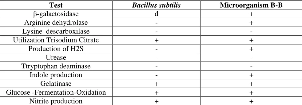
Table 2. Biochemical comparison of microorganism B-B and Bacillus subtilis using API 20E STREP.
Test Bacillus subtilis Microorganism B-B
β-galactosidase d +
Arginine dehydrolase - +
Lysine descarboxilase - -
Utilization Trisodium Citrate + +
Production of H2S - +
Urease - -
Ttryptophan deaminase - -
Indole production - +
Gelatinase + +
Glucose -Fermentation-Oxidation + +
Nitrite production + +
[image:6.596.49.548.461.521.2] [image:6.596.53.544.564.736.2]
Additionally, B-B bacterial strain was analyzed by API 20E STREP Kit to perform a biochemical characterization. Results were compared with information found in the Bergey’s Manual (Table 2). This microorganism showed a different enzymatic biochemical profile when compared with Bacillus subtilis (Table 2). After this analysis, results support the idea that this microorganism is a novel bacterial strain isolated from the Antarctic consortium and may play a role in the corrosion process.
3.2 Electrochemical behavior
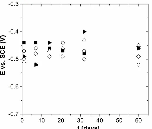
Figure 2 shows the variation of the open circuit potential (E) of AA2024-T3 after being exposed to sterile and inoculated MSM as a function of immersion time. As can be seen, in the sterile and inoculated MSM with the consortium, the open circuit potential value was slightly shifted to more negative values with immersion time, which might indicate a slight increase of the corrosion current. On the other hand, for the inoculated media with S. aureus, Bacillus not previously described (B-B) and B. toyonensis, the open circuit potential value was also slightly shifted with immersion time, but at both positive and negative values. For inoculated media with B. toyonensis after day 60 the open circuit potential value was shifted from -0.51 to -0.45 V vs SCE, that might indicate a slight decrease of the corrosion current.
Figure 2. Variation of open circuit potential values of AA2024-T3 after day 60 of exposure in MSM () sterile and inoculated with () consortium, () S. aureus, () B. toyonensis and () B-B
at 4 C. Electrode surface Area = 21 cm2 and E = EOCP.
[image:7.596.170.428.399.618.2]
associated to the chemical and biological reactions resulting from the biofilms formation, which may be associated to a CPE behavior, as it was described previously in Ref [2,7,15,16]. On the other hand, the impedance modulus of AA2024-T3 after day 21 of immersion in sterile medium was lower than the inoculated media at all frequency. Nevertheless, the impedance modulus of AA2024-T3 after day 60 of immersion in sterile medium was also the same that in inoculated media with S. aureus but greater than in inoculated media with B-B and B. toyonensis.
[image:8.596.153.446.191.692.2]
Note that the impedance modulus at low frequency range may be associated with the oxide resistance. Recently, Alvarado [2] showed a slight increase of the impedance modulus at 5 mHz (ǀZǀf=5 mHz) with time for AA2024-T3 after being exposed to a media containing jet JP-8 and a microbial
community recollected from corrosion products. As can be seen in Table 3, for all media the impedance modulus at 3 mHz (ǀZǀf=3 mHz) decreased with time, which might reveal a slight increase of
[image:9.596.175.420.471.616.2]the anodic current.
Table 3. Impedance parameters obtained of AA2024-T3 samples in sterile and inoculated media after day 21 and 60 by using numerical analysis.
System texp (days) ǀZǀf=3 mHz (Ωcm2) Q (Ω-1sαcm-2 )
MSM Sterile
21
7.4 x 10⁶ 0.85 16.0 x 10⁷
MSM Consortia 9.9 x 10⁶ 0.89 9.4 x 10⁷
MSM/ B. Toyonensis 17.6 x 10⁶ 0.96 8.1 x 10⁷
MSM /B 15.9 x 10⁶ 0.92 7.9 x 10⁷
MSM/ S. aureus 12.1 x 10⁶ 0.91 9.2 x 10⁷
MSM Sterile 0.1 x 10⁶ 0.89 9.2 x 10⁷
MSM Consortia 5.7 x 10⁶ 0.88 10.6 x 10⁷
MSM/ B. toyonensis 60 2.4 x 10⁶ 0.85 11.5 x 10⁷
MSM /B 7.1 x 10⁶ 0.92 9.8 x 10⁷
MSM/ S. aureus 0.1 x 10⁶ 0.89 8.9 x 10⁷
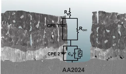
Figure 4. Schematic representation for coated 2024 alloy and corresponding equivalent circuit [16,17].
An equivalent circuit and schematic representation of alumina oxide film/2024 alloy interface is presented in Figure 4. In this case, Re is the electrolyte resistance. The oxide film is composed by an
the outer oxide film. Cdl represents the double layer capacitance and Zf is the Faradaic impedance, such as was described in Ref [4,18,19]. Note that the experimental data were perfectly fitted with the equivalent circuit and values of the parameters were extracted by graphic and numerical analysis. The CPE parameters are also showed in Table 3.
As it was described in Ref [19,20], from the impedance analysis the thickness of the oxide film formed on a metallic surface may be obtained by using the capacitance values. As shown Table 4, the thickness values are in agreement with those previously reported for passive layers by Alvarado [2] and Benoit [19]. In particular, the thickness of the oxide film increased slightly with time in sterile and inoculated MSM with the consortium, which is in agreement with the variation of the OCP with time. However, in inoculated media with B. toyonensis. B-B and S. aureus, the range of the thickness of the oxide film slightly decreased with time. This behavior might indicate that the anodic current slightly increased or that the oxide properties were modified.
Table 4. Impedance parameters obtained of AA2024-T3 samples in sterile and inoculated media after day 21 and 60 by using numerical analysis.
System texp (days) Thickness range (nm)
MSM Sterile
21
6.8 – 10.4
MSM Consortia 12.8 – 15.3
MSM/ B. toyonensis 11.9 – 13.6
MSM /B 10.8 – 13.6
MSM/ S. aureus 12.2 -14.9
MSM Sterile 10.4 – 13.2
MSM Consortia 9.9 – 12.9
MSM/ B. toyonensis 60 10.9 – 14.4
MSM /B 11.2 – 13.4
[image:10.596.42.550.597.679.2]MSM/ S. aureus 10.4 – 13.2
Table 5. Corrosion parameters obtained from polarization curves of AA2024-T3 samples in sterile and inoculated media after day 21.
texp / days System Ia at E = -0.395 V / A cm-2
21
MSM 3.26 x 10⁻⁷
MSM/B. toyonensis 2.97 x 10⁻⁶
MSM/B-B 4.84 x 10⁻⁷
MSM/S. aureus 2.13 x 10⁻⁶
[image:11.596.85.507.175.364.2]
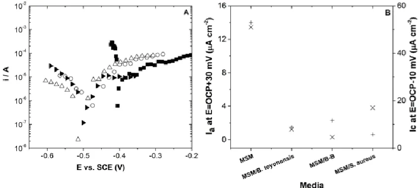
decreased in inoculated media. Nevertheless, if the anodic current is compared in the passive domain, for example at E = 0.395 vs. SCE (V), as shown Table 5, the anodic current increased from sterile to inoculated media for all isolated bacteria, which indicate that the oxide film properties might be modified by the biofilm formation.
Figure 5. Polarization curves of AA 2024-T3 after 21 day of exposure in MSM () sterile, ( ) S. aureus, () B-B and () B. toyonensis. Scan rate 0.1 mV s-1 and electrode surface Area = 21 cm2.
3.3 Morphological characterization of strains
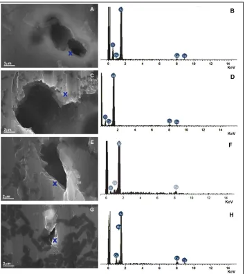
Figure 6. SEM/EDX analysis of AA2024-T3 alloy sample after to be exposed to (a,b) B. toyonensis, (c,d) S. aureus (e,f) consortium and (g,h) Bacillus not previously described (B-B).
4. CONCLUSIONS
The experimental electrochemical results revealed that when in contact with AA2024-T3 alloy, the microorganisms described above, and the consortium formed by them modify the electrochemical behavior of the aluminum alloy as a function of immersion time, changing the passive oxide film properties formed on the AA2024-T3 surface. This behavior may be attributed to the influence on the dissolution/formation reaction of the aluminum oxide layer by the presence of the microorganisms and their metabolic products, exerting an influence on the corrosion development process. However, no information has been found on the direct relationship between the interactions of these variables. Additional analysis and studies are needed in order to better understand the interaction of these microorganisms individually and as a consortium with the metallic surface.
ACKNOWLEDGEMENTS
The authors also would like to thank to the following agencies: AFOSR (Grant FA9550-14-1-0407), PIA-Conicyt (Grant ACT-1412), Fondecyt (Grant 1160604 and 11170419).
References
1. A. Hagenauer, R. Hilpert, T. Hack, Mater. Corros., 45 (1994) 355.
2. C. Alvarado, M. Sancy, J.M. Blamey, C. Galarce, A. Monsalve, F. Pineda, N. Vejar, M. Páez, Electrochim. Acta, 247 (2017) 71.
3. C. McNamara, T. Perry, R. Leard, K. Bearce, J. Dante, R. Mitchell, Biofouling, 21 (2005) 257. 4. A. Rajasekar, B. Anandkumar, S. Maruthamuthu, Y.P. Ting, P.K.S.M. Rahman, Appl.
Microbiol. Biotechnol., 85 (2010) 1175.
5. A. Monsalve, M. Páez, M. Toledano, A. Artigas, Y. Sepúlveda, N. Valencia, Eng. Mater. Struct., 30 (2007) 748.
6. A.N. Khramov, V.N. Balbyshev, N.N. Voevodin, M.S. Donley, Prog. Org. Coatings, 47 (2003) 207.
7. M. Sancy, A. Abarzúa, M.I. Azócar, J.M. Blamey, F. Boehmwald, G. Gómez, N. Vejar, M. Páez, J. Electroanal. Chem., 737 (2015) 212.
8. M.A. Tanner, C.L. Everett, W.J. Coleman, M.M. Yang, D.C. Youvan, Biotechnol. Alia., 8 (2000) 1.
9. Q. Wang, G.M. Garrity, J.M. Tiedje, J.R. Cole, Appl. Environ. Microbiol., 73 (2007) 5261. 10. A. Rajasekar, Y.P. Ting, Ind. Eng. Chem. Res,. 49 (2010) 6054.
11. E. Gruner, A. von Graevenitz, M. Altwegg, J. Microbiol. Methods, 16 (1992) 101. 12. A.C. Vieira, A.R. Ribeiro, L.A. Rocha, J.P. Celis, Wear. 261 (2006) 994.
13. B.H. Sen, B. Piskin, T. Demirci, B.H. Sen, Endod. Dent. Traumatol., 11 (1995) 6. 14. S. Baeza, N. Vejar, M. Gulppi, M. Azocar, F. Melo, A. Monsalve, J. Pérez-Donoso, C.C.
Vásquez, J. Pavez, J.H. Zagal, X. Zhou, G.E. Thompson, M.A. Páez, Corros. Sci., 67 (2013) 32. 15. A.K. Mishra, R. Balasubramaniam, Corros. Sci., 49 (2007) 1027.
16. S. V. Lamaka, M.L. Zheludkevich, K.A. Yasakau, M.F. Montemor, M.G.S. Ferreira, Electrochim. Acta., 52 (2007) 7231.
17. M.A. Páez, T.M. Foong, C.T. Ni, G.E. Thompson, K. Shimizu, H. Habazaki, P. Skeldon, G.C. Wood, Corros. Sci., 38 (1996) 59.
18. M.A. Paez, O. Bustos, G.E. Thompson, P. Skeldon, K. Shimizu, G.C. Wood, Film. 147 (2000) 1015.
Tribollet, V. Vivier, Electrochim. Acta. 201 (2016) 340.
20. M. E. Orazem, B. Tribollet, Electrochemical Impedance Spectroscopy. Wiley, (2017) New Jersey, USA.