Physiological Analysis of
Structural/Functional Features of
Neuronal Calcium Sensor-1
Thesis submitted in accordance with the
requirements of the University of Liverpool
for the degree of Doctor in Philosophy
By
Victoria Marie Martin
ii
Contents
Abstract ... vi
Publications ... vii
Acknowledgements ... viii
Abbreviations ... ix
Chapter 1: INTRODUCTION ... 1
1.1 Calcium Signalling Introduction ... 2
1.2 Ca2+ Sensor Proteins ... 4
1.2.1 Calmodulin ... 4
1.2.2 Ca2+ Sensor Proteins in Neurons ... 6
1.3 Neuronal Ca2+ Sensor Protein 1 ... 8
1.4 Voltage Gated Ca2+Channels ... 13
1.5 Caenorhabditis elegans as a Model Organism ... 15
1.6 C. elegans Nervous Systems... 16
1.6.1 Motor Neurons ... 20
1.6.2 Interneurons ... 20
1.6.3 Sensory Neurons ... 20
1.6.4 Ciliated Sensory Neurons ... 21
1.7 Thermosensation and Regulation ... 23
1.7.2 Thermotaxis Neuronal Circuits ... 26
1.7.3 Thermoavoidance ... 28
1.7.4 Temperature-Dependent Locomotion ... 29
1.8 C. elegans and Ca2+ Binding Proteins ... 31
1.9 Aims and Objectives ... 36
Chapter 2: METHODS ... 37
2.1 Materials and Methods for Characterisation of NCS-1 Protein and Analysis Target Peptide Binding ... 38
2.1.1 Reagents ... 38
2.1.2 Preparation of Plasmids for Protein Expression ... 39
2.1.2.1 pE-SUMOpro-NCS-1 ... 39
2.1.2.2 His-SUMO-P/Q-N and His-SUMO-P/Q-N2 ... 39
2.1.3. Protein Expression and Purification of Unlabeled NCS-1, GST-CaM and P/Q peptides ... 40
2.1.4. Protein Purification ... 41
iii
2.1.6 Purification by Hydrophobic Interaction and Size Exclusion Gel Chromatography .. 44
2.1.7 Mass spectrometry ... 45
2.1.8 Analysis of the P/Q-IQ peptide ... 45
2.1.8.1 Helical Prediction ... 45
2.1.8.2 Circular Dichroism ... 45
2.1.9 Pull-down Assay The analysis of the Ca2+-dependency of binding of GST-P/Q-L -NCS-1 ... 46
2.1.9.1 Binding Assay ... 46
2.1.9.2 Western Blot of Pull-Down Assay ... 47
2.1.10 Gel Filtration analysis ... 47
2.1.11 NMR Spectroscopy ... 48
2.1.11.1 CaM and P/Q-IQ interactions ... 48
2.1.11.2 NCS-1 and P/Q-IQ interactions ... 49
2.2 Materials and Methods –For analysis of NCS-1 function in C. elegans ... 50
2.2.1 Reagents ... 50
2.2.1.1 C. elegans Strains ... 50
2.2.1.2 Plasmids and C. elegans Sequences ... 51
2.2.1.3 Enzymes ... 52
2.2.2 C. elegans Husbandry ... 52
2.2.3 Preparation of Plasmids for Microinjection. ... 53
2.2.3.1 PCR and Restriction Digestion ... 53
2.2.3.2. Gateway Cassette Cloning ... 56
2.2.4 Microinjection ... 58
2.2.5 Imaging ... 60
2.2.6 C. elegans Protein Extraction and Western Blotting... 60
2.2.7 Behavioural Assays ... 61
2.2.7.1 Crawling locomotion Assays ... 61
2.2.7.2. Neurotransmission (Aldicarb Resistance) Assay ... 61
2.2.7.3 Temperature-Dependent Locomotion Assay ... 62
2.2. 8 Predicted Models of C. elegans NCS-1 Protein Structure ... 62
Chapter 3: RESULTS Characterisation of NCS-1 and Calmodulin Interactions with the P/Q-type (Cav2.1) Calcium Channel ... 63
3.1 Introduction Modulation of Cav2.1 by Ca2+ Sensor Proteins ... 64
iv
3.2 Results ... 68
3.2.1 Recombinant protein expression and purification ... 68
3.2.3 Analysis of NCS-1 and P/Q peptide interactions by Surface Plasmon Resonance. ... 76
3.2.4 Secondary structure characterization of the P/Q-IQ peptide ... 76
3.2.5 Gel filtration analysis of putative NCS-1 P/Q-IQ complex... 78
3.2.6 NMR Temperature Titration of NCS-1 and CaM ... 79
2.2.7 NMR analysis of CaM and P/Q-IQ peptide interaction ... 79
3.2.8 NMR analysis of NCS-1 and P/Q-IQ peptide interaction ... 85
3.3 Discussion ... 87
Chapter 4: RESULTS Investigation of the Structure/Function Relationship of NCS-1 in the Model Organism C. elegans ... 92
4.1 INTRODUCTION ... 93
4.2 C. elegans RESULTS ... 97
4.2.1 Phenotyping the ncs-1 (qa406) strain ... 97
4.2.1.1 Comparison of the Basic Reproductive and Alimentary Anatomy of ncs-1 Null and Wild-type C. elegans. ... 97
4.2.1.2. Comparison of the Crawling Locomotion of ncs-1 Null and Wild-Type C. elegans. ... 99
4.2.1.3. Comparison of Cholinergic Neurotransmission of ncs-1 Null, Wild-Type and ric-4 Mutant Control C. elegans. ... 100
4.2.1.4 Comparison of Temperature-Dependent Locomotion of ncs-1 Null and Wild-Type C. elegans. ... 101
4.2.2 Confirmation of Expression of Transgenic Genes by GFP Marker Expression ... 102
4.2.3 Temperature-Dependent Locomotion of the ncs-1 null C. elegans Rescued by Transgenic Expression of Wild-Type ncs-1 Expression. ... 103
4.2.3.1 Effect of Expression of Genomic Unspliced ncs-1 in the ncs-1 Null Strain on Temperature-Dependent Locomotion. ... 103
4.2.3.2 Effect of Expression of Synthetic Spliced ncs-1 in the ncs-1 Null Strain on Temperature-Dependent Locomotion. ... 104
4.2.4 Anti-Human NCS-1 Antibodies in Detection of Recombinant C. elegans NCS-1 .... 106
4.2.5 Effect of Expression of Unspliced ncs-1 in the Wild-Type and ncs-1 Null Strain on Temperature-Dependent Locomotion. ... 107
4.2.6. Effect of Expression of ncs-1 mutants in the ncs-1 Null Strain ... 108
v
4.2.6.3 Effect C-Terminal Tail Truncations of NCS-1 Protein on Temperature-dependent
Locomotionin C. elegans ... 112
4.2.7 Effect ncs-1 expression in Sensory and AIY Neurons on Temperature-dependent Locomotion ... 114
4.2.8 Effect of Knockdown of PIFK-1 on temperature-dependent locomotion ... 118
4.3 DISCUSSION ... 119
Chapter 5: DISSCUSION ... 126
5.1 Discussion ... 127
5.2 Conclusion ... 131
vi
Abstract
Calcium (Ca2+) signalling regulates many neuronal functions including neurotransmission, axonal growth and development. Neuronal calcium sensor-1 (NCS-1) has been shown to be involved in many of these processes. On Ca2+ binding, NCS-1 changes conformation and exposes a hydrophobic binding pocket. In yeast, NCS-1 binds to a PI4-kinase orthologue required for survival. In mammalian cells, NCS-1 is localised to the Golgi and plasma membranes and has been linked to multiple target proteins that have roles in neuronal signalling. NCS-1 has been shown to regulate the P/Q Ca2+ channel subunit Cav2.1; although no direct binding interaction has been identified between the proteins.
The Cav2.1 C-terminal tail contains two Ca2+-sensor binding regions, the IQ domain and
the calmodulin (CaM) binding domain (CBD). The first part of this study investigated NCS-1 or CaM and Cav2.1 interactions using biochemical and biophysical interactions.
Pull-down analysis found that NCS-1 binds to a Cav2.1 C-terminal peptide in a Ca2+
-dependent manner. Use of nuclear magnetic resonance spectroscopy also showed that the IQ domain of Cav2.1 bound to NCS-1 in the presence of Ca2+, though the NCS-1
vii
Publications
Work presented in this thesis has been published in part in the following paper:
viii
Acknowledgements
First and foremost, I would like to thank my supervisors Prof. Bob Burgoyne and Dr Lee Haynes. I would like to thank Bob for his excellent supervision, guidance and patience throughout my PhD, it has been a privilege to be his student. Thanks Lee for all his help in the lab especially in the early days with protein expression, molecular biology and guidance with protein science. Thanks to Dr Jeff Barclay for putting up with my never ending questions on worms. I owe massive thanks to Dr James Johnson for his microinjecting skills, his countless advice and patience, and to Dr Andy Herbert, I am grateful for his introduction to SPR it was definitely character building. Thank you ALL of Red block past and present including Alan, Matt, Pryank, Sudhanva, Joe, Joanna, Sarah and Helen. And I would like to say a special thank you to Hannah, Ciara, Martin (honorary Red block member), Leanne, Xi, Dayani, Mimi, Marie, Megan, Paul and Michela for their friendship, there have been really fantastic times but also tough times and without your help and encouragement I do not know how I would have got through them. Thank you to those in Lab C and the NMR centre for making me welcome during our collaboration and helping me with chromatography and NMR especially Prof. Lu-Yun Lian, Dr Marie Phelan and Sravan Pandalaneni. I would also like to thank my undergrad lectures at LJMU, without whose excellent teaching I would not have had the confidence and ability to do this in the first place. I would like to thank the Wellcome Trust Studentship Program for funding me and the work in this thesis.
I cannot thank my family enough for their never ending love, support and belief in me. I could not have got through it without you; my Dad, Clare, Gary, Kathy, Bette
ix
Abbreviations
1A Voltage gated P/Q Ca2+ channel forming subunit (aka Cav2.1)
Ach Acetylcholine ADF Amphid dual F ADL Amphid dual L
ADP Adenosine diphosphate AFD Amphid finger-like D AIA Amphid interneuron A AIB Amphid interneuron B AID Alpha interacting domain AIY Amphid interneuron Y AIZ Amphid interneuron Z ARF 1 ADP ribosylation factor 1 ASE Amphid single E
ASG Amphid single G ASH Amphid single H ASI Amphid single I ASJ Amphid single J ASK Amphid single K
AVK Amphid, NR, VC Interneuron K AWA Amphid winged A
x BAG Head Sensory Neuron BSA Bovine serum albumin
C. elegans Caenorhabditis elegans
[Ca2+]i Intracellular free calcium ion concentration Ca2+ Calcium ion
CaBPs Calcium Binding Proteins
CACNA1A (aka CACNL1A4) Voltage gated P/Q Ca2+ channel 1A/Cav2.1 subunit
gene
CaM Calmodulin
Cav Voltage gated Ca2+ channel (aka VGCC)
Cav2.1 Voltage gated P/Q Ca2+ channel forming subunit (aka 1A)
CBD Calmodulin binding domain
CCPNmr Collaborative Computing Project for NMR CD Circular dichroism
CDF Ca2+-dependent facilitation CDI Ca2+-dependent inactivation
CGC Caenorhabditis Genetics Centre
CMD-1 CaM (C. elegans orthologue) D2R Dopamine receptor 2
DAG Diacylglycerol
DC Dorsal cord
DNA Deoxyribonucleic acid DTT Dithiothreitol
xi EA-2 Episodic ataxia type-2
ECL Enhanced chemiluminescence EDTA Ethylenediaminetetraacetic acid
EF EF hand motif
ER Endoplasmic reticulum
FHM-1 Familial hemiplegic migraine type-1 FLP Head sensory neuron
Frq-1 Frequenin 1 (Neuronal calcium sensor-1, Budding yeast protein and
Drosophila)
Frq-2 Frequenin 2 (Neuronal calcium sensor-1 Drosophila orthologue)
GABA Gamma-aminobutyric acid
GCAPs Guanylyl cyclase-activating proteins GFP Green fluorescent protein
GRK-2 G-protein-coupled receptor kinase 2 GST Glutathione S-transferase
GTP Guanosine triphosphate
GTPase Guanosine triphosphate hydrolysing enzyme HCLP-1 Hippocalcin-like protein-1
HEPES Hydroxyethyl piperazineethanesulfonic acid His- 6X Histidine peptide tag
HPLC High-performance liquid chromatography HRP Horse radish peroxidise
xii IP3 Inositol 1,4,5,trisphosphate
IP3R inositol 1,4,5,trisphosphate receptors
IPTG Isopropylthio-β-galactoside IQ IQ-like motif
ITC Isothermal titration calorimetry
KChIPs Potassium channel interacting proteins Kv Potassium channel
MES Morpholino ethanesulfonic acid Mg2+ Magnesium ions
MOPS Morpholino-propanesulfonic acid Munc-18 Mammalian uncoordinated protein 18 MWCO Molecular weight cut off
NBRP National Bioresource Project
NCS Neuronal calcium sensor family proteins
NCS-1 Neuronal calcium sensor-1 (mammalian and C. elegans orthologue) Ncs-1 Neuronal calcium sensor-1 (Fission yeast orthologue)
NGM Nematode growth media NMR Nuclear magnetic resonance
NR Nerve ring
OD Optical density
osm-6 Osmotic avoidance abnormal-6
xiii PBS Phosphate buffered saline
PBST Phosphate buffered saline tween 20 PCR Polymerase chain reaction
PDB Protein database PEG Polyethylene glycol
PHA Phasmid A
PHB Phasmid B
PHC Phasmid C (tail neuron possibly in the phasmid)
PI4K III Phosphatidylinositol-4-kinase type 3 beta (Mammalian orthologue) PI4P Phosphatidylinositol-4-phosphate
Pik-1 Phosphatidylinositol-4-kinase 1 (Budding yeast orthologue) PKC-2 Protein kinase 2
pm1 Pharyngeal Muscle
PVC Venteral cord interneuron, tail gangian Rba-1 Rat brain alpha-1 (Rat Cav2.1 orthologue)
RIA Head, NR interneuron
ric-4 Resistance to inhibitors of cholinesterase RMG Head Motor/Interneuron G
S.E.M. Standard error of mean SDS Sodium dodecyl sulphate
SNARE Soluble NSF attachment protein receptor SPR Surface plasmon resonance
xiv SUMO Small ubiquitin-like modifier
TDL Temperature-dependent locomotion TEV Tobacco etch virus
TFE 2, 2, 2-trifluoroethanol
Tris Tris-hydroxymethyl-aminomethane tRNA Transfer RNA (Ribonucleic acid) ttx-3 Thermotaxis abnormal-3 ULP-1 Ubiquitin-like protease-1
UNC-18 Uncoordinated protein 18 (Munc-18 C. elegans orthologue) UNC-2 Uncoordinated protein 2 (Cav2.1 C. elegans orthologue)
VC Ventral nerve cord
VGCC Voltage gated Ca2+ channel (AKA Cav)
2 1.1 Calcium Signalling Introduction
Intracellular calcium signalling regulates cell growth, proliferation, cell death and also specialised cell functions (Berridge et al., 2000). In neurons, many specific activities are calcium-dependent including the stimulation of neurotransmission, neuronal development and neuroplasticity (Barclay et al., 2005, Berridge, 1998, Catterall and Few, 2008, Bito et al., 1997). Calcium acts as a messenger via changes in its free ion concentration and location inside the cell. Ca2+ is a large positively charged metal ion and transduces intracellular signals by binding to specialised proteins. Ca2+ can be neither created or destroyed, so must be regulated by being moved, buffered or sequestered. Resting intracellular free Ca2+ concentration ([Ca2+]i) is in the range 10–100 nM depending on the cell type and can be increased to 1000 nM or higher when cells are stimulated. Regulation of [Ca2+]i is essential for normal neuronal function. Unregulated [Ca2+]i leads to cell damage and toxicity and it has been suggested that the dysregulation of [Ca2+]i in neurons may be linked to neurodegenerative disorders such as Alzheimer’s and Parkinson’s disease (Mattson and Magnus, 2006, Berridge, 2010).
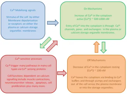
Ca2+ signalling networks have been divided into four mechanisms (Berridge et al., 2000) (Figure 1.1):
1. Mobilisation of Ca2+. When cells are excited a stimulus allows Ca2+ to enter the cell cytoplasm through Ca2+ channels by either crossing the plasma membrane or by release from Ca2+ storage organelles such as the endoplasmic reticulum (ER).
2. Activation of on-mechanisms. This allows the [Ca2+]i to rise to an active concentration. Ca2+ binds to channels, allowing further Ca2+ to continue to enter the cytoplasm and remain there.
3. Stimulation of Ca2+-dependent activities. Ca2+-binding proteins control the regulation of these functions.
3
Figure 1.1. Overview of the Ca2+ signalling network. A simple schematic showing the relationship between Ca2+ signalling mechanisms. Blue shows Ca2+ mobillising signals/stimuli. Green shows on-mechanisms which increase Ca2+ in the cytoplasm to active levels. Red shows Ca2+-sensitive processes which stimulate Ca2+-dependent function. Orange shows off-mechanisms which decrease the Ca2+ to resting levels.
[image:17.595.116.528.72.382.2]4 1.2 Ca2+ Sensor Proteins
Ca2+ sensor proteins can have several different roles in Ca2+ networks. They have a stimulatory role, regulating effector molecules, to switch Ca2+-sensitive processes on or off. For example, the Ca2+ sensor protein synaptotagmin-1 has specific Ca2+-binding domains. This binding to Ca2+ causes synaptotagamin-1 to coordinate interactions between soluble NSF attachment protein receptor (SNARE) proteins and membrane lipids involved in vesicle docking and membrane fusion, which is important for the regulation of neurotransmission. Ca2+ sensor proteins can have a Ca2+ signalling feedback role, modulating other proteins in Ca2+ signalling networks. For example, modulation of voltage gated Ca2+ channels, involved in both the on and off mechanisms, are regulated by several Ca2+ sensor proteins including calmodulin, Ca2+ binding protein-1 (CaBP-1) and visinin-like protein -2 (VILIP-2). This can have both a positive feedback effect, allowing Ca2+ into the cytoplasm or negative feedback effect by inhibiting the entry of Ca2+ into the cytoplasm (Nejatbakhsh and Feng, 2011).
1.2.1 Calmodulin
5
6
Aspartate, found at position 1 and aspartate or glutamate, found most often at position 12 of the motif, provide negatively charged oxygen for the electrostatic interactions between the protein and the cation.
Different EF-hands within CaM and other Ca2+ sensor proteins have different affinities for Ca2+, this is thought have a role in the regulation of Ca2+ sensor protein function and activity at different physiological [Ca2+]i. When Ca2+ binds to CaM, this causes a conformational change exposing a hydrophobic region of the protein that is important for mediating interactions with target proteins (Figure 1.2b) (Zhang et al., 1995).
1.2.2 Ca2+ Sensor Proteins in Neurons
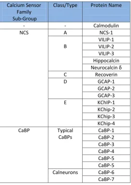
Neurons contain a number of CaM related Ca2+ sensor proteins. Two families of these proteins are the neuronal Ca2+ sensor proteins (NCS) and the Ca2+ binding proteins (CaBP), which each contain 4 EF-hands motifs (Table 1.1). The CaBPs show sequence homology to CaM and have been found only in vertebrates (McCue et al., 2010), whilst NCS proteins show approximately 20% homology with CaM and are found in early eukaryotes up to man. This array of Ca2+ sensing proteins may allow neurons to have greater versatility in Ca2+ signalling.
7
Table 1.1 Summary of Mammalian NCS-1 and CaBP Protein Families Calcium Sensor
Family Sub-Group
Class/Type Protein Name
- - Calmodulin
NCS A NCS-1
B VILIP-1 VILIP-2 VILIP-3 Hippocalcin Neurocalcin δ
C Recoverin
D GCAP-1
GCAP-2 GCAP-3
E KChIP-1
KChip-2 KChip-3 KChip-4 CaBP Typical
CaBPs CaBP-1 CaBP-2 CaBP-3 CaBP-4 CaBP-5 CaBP-5 Calneurons CaBP-6 CaBP-7
8
The Ca2+-myristoyl switch mechanism allows Ca2+-dependent localisation of several NCS proteins. When not bound to Ca2+, the myristoyl group is located within the hydrophobic core of the protein, allowing the NCS protein to be free in the cytoplasm. When the protein is Ca2+ bound, its conformation changes, this exposes the myristoyl group and tethers the protein to organelles or the plasma membrane. How the NCS proteins, are localised to specific membranes is not yet known but the specific membrane localisation of NCS proteins could allow them to have different regulatory roles in neurons and other cell types.
1.3 Neuronal Ca2+ Sensor Protein 1
Neuronal Ca2+ sensing protein 1 (NCS-1) is found in all eukaryotes, from yeast through to higher organisms including man. NCS-1 was first identified as frequenin in Drosophila melanogaster (Pongs et al., 1993). In mammals it has been shown to have a role in synaptic transmission, neuronal outgrowth and memory and learning (Saab et al., 2009, Génin et al., 2001, Pongs et al., 1993). It is found in neuronal and neuroendocrine mammalian cells (Weiss and Burgoyne, 2001) and has been observed to be localised to presynaptic terminals in both hippocampal and cerebral neurons (Jinno et al., 2002). Conservation of the NCS-1 sequence has been retained throughout the species. Fission yeast Ncs-1 and budding yeast Frq-1 orthologues have 60% similarity to mammalian NCS-1 (Lim et al., 2011). Like CaM, NCS-1 has been shown to regulate a large array of proteins (Burgoyne, 2007, Haynes et al., 2006).
9
PI4K IIIin a Ca2+-dependent manner (Zhao et al., 2001, Haynes et al., 2005). This enzyme is important in cell signaling as it regulates phosphoinositide levels. PI4K III produces phosphatidylinositol 4-phosphate (PI4P) which is involved in the localisation of specific cytosolic proteins to the Golgi membrane (Downes et al., 2005). NCS-1 and ADP ribosylation factor 1 (ARF 1), a GTPase, have both been shown to co-localise with PI4K III in mammalian cells and are both involved in PI4K III regulation and membrane traffic from the Golgi complex to the plasma membrane (Haynes et al., 2007, Haynes et al., 2005).
NCS-1 also interacts with interleukin-receptor 1 accessory protein- like 1 (IL1RAPL1) (Bahi et al., 2003). IL1RAPL is a plasma membrane protein, found in the brain and like NCS-1 it has been linked to neuronal development and exocytosis. During a genetic study of families who have autistic spectrum disorders, a NCS-1 (R102Q) mutation was found in an individual and mutations of IL1RAPL were found in a number of subjects (Piton et al., 2008). Structural studies have shown this mutation in NCS-1 does not prevent IL1RAPL1 binding but does have structural and functional effects on NCS-1 (Handley et al., 2010). NCS-1 has also been shown to associate with dopamine receptor 2 (D2R) and blocks the phosphorylation of the receptor by G-protein-coupled receptor kinase 2 (GRK-2) (Kabbani et al., 2002). This prevents the receptor being internalised and has been shown to promote memory and learning in mice (Saab et al., 2009).
NCS-1 regulates inositol 1,4,5,trisphosphate receptors (IP3R), which are Ca2+
channels situated mainly on the ER and release Ca2+ to the cytoplasm. NCS-1 has been shown to bind directly to the IP3R and to enhance channel opening in the
presence of IP3 (Schlecker et al., 2006). NCS-1 and IP3R interactions are thought to
10 1.3.1 NCS-1 Structure and Localisation
NCS-1 is a small globular protein of 190 amino acids and a molecular mass of approximately 22 kDa. It contains a core hydrophobic pocket which is exposed on binding to Ca2+ and forms a binding site for target proteins. Two tryptophan residues (W30 and W130 in human NCS-1) have been shown to move out of the core of the protein in the presence of Ca2+ (Aravind et al., 2008) and these may be essential amino acids for target protein interactions.
NCS-1, like CaM, contains 4 EF-hands; however, only EF-hands 2, 3 and 4 bind Ca2+. The Ca2+ affinity of EF-hand 2 is 10.0 M and for EF-hand 3 and 4 is 0.4 M (Ames et al., 2000). It has been shown that EF-hands 2 and 3 bind Mg2+ at resting [Ca2+ ]i (Aravind et al., 2008). NMR has revealed the structure of NCS-1 in the Mg2+ bound form has a different conformation than in the Apo-NCS-1 form, with the hydrophobic pocket tightly closed in the core. Mg2+ lowers the affinity of NCS-1 for Ca2+ and is thought to have a role in NCS-1 regulation, reducing non-specific binding of NCS-1 at resting Ca2+ levels (Aravind et al., 2008). The effects of Mg2+ on NCS-1 and its interactions with target proteins are beginning to be investigated. Mg2+ may stabilise NCS-1 structure by binding to EF-hand 3. Mg2+ may also compete with Ca2+ for EF-hand 2 binding which may disrupt target protein binding and inactivate NCS-1 at resting [Ca2+]i (Woll MP, 2011).
NCS-1 is N-terminally myristoylated and is localised to the Golgi complex and the plasma membrane (O'Callaghan et al., 2002). It was thought that all NCS-1 proteins have a constitutively exposed myristoyl tail and are always localised to membranes. In contrast, a recent study looking at Ncs-1 in S. pombe, discovered that this orthologue has a Ca2+ dependent-myristoyl switch, as seen in other classes of NCS proteins such as recoverin, suggesting this NCS-1 orthologue is not always tethered to the membrane (Lim et al., 2011).
11
while the second region binds to the C-terminal cleft, showing 1:1 stoichiometric ratio (Strahl et al., 2007, Lim et al., 2011). This binding is thought to change the conformation of the Pik-1 protein activating its catalytic site (Lim et al., 2011).
12
[image:26.595.120.524.71.610.2]13 1.4 Voltage Gated Ca2+Channels
Voltage gated Ca2+ channels (VGCC or Cav) are present in excitable cells. In neurons
they are localised at the presynaptic terminals close to the SNARE complex machinery (Augustine et al., 2003). The Cav channels are the link between
membrane depolarisation and neurotransmission. They open as the axon potential reaches the synapse and the membrane depolarises. Ca2+ enters the cell through the open channel increasing the [Ca2+]i (Catterall, 2000, Augustine et al., 2003).
Cav channels are heteromeric proteins consisting of 4-5 subunits, which are 1,
2, and sometimes an additional subunit. The 1 subunit of the Cav channel
consists of four transmembrane domains, I-IV, each consisting of six alpha helices. The domains span the plasma membrane and form the Ca2+ channel pore (Catterall, 2000). Cytoplasmic loop regions between the four domains and a C-terminal tail provide sites for interaction with regulatory molecules and other channel subunits. There are 10 known genes for 1 channel subunits. These genes may have different isoforms and splice variants, expressed in different cell types (Catterall, 2000). The type of 1 subunit the channel determines which group it belongs to. The 5 major groups of Cav channels were originally determined by the type of Ca2+
current they carried. L-type (Cav1s) channels, carry long lasting Ca2+ currents,
P/Q-type, N-type and R-type (Cav2s) channels carry non-long lasting currents and T-type
(Cav3s) channels carry transient currents (Catterall and Few, 2008).
The auxiliary subunit of the channel is an intracellular globular protein that regulates the channel function. The 1 I-II loop contains a motif called the interacting domain (AID), which binds to the subunit (Pragnell et al., 1994). This interaction is involved in voltage dependent inactivation of the channel. The 2 heterodimer consists of the transmembrane 2 subunit and the extracellular
subunit that are attached by disulphide bonds. This subunit also has a regulatory role. The function of a fifthsubunit, which is associated with some Cav channels, is
14 1.4.1 Cav 2.1 subunit of the P/Q channel
The Cav 2.1 (aka 1A) is the 1 subunit of the P/Q-type Ca2+ channel and is found
mainly in the central nervous system in mammals. It is located on the presynaptic terminal membrane in nanodomains that contain the exocytosis machinery essential for neurotransmission (Westenbroek et al., 1995, Taverna et al., 2004). The 1 subunit has orthologues in different organisms which include cacophony in
D. melanogaster and UNC-2 in C. elegans. The protein was first identified as a 2262 amino acid protein. A further 4 isoforms have been identified in humans rangeing from approximately 2200 to 2500 amino acids. The gene for the protein, CACNA1A (aka CACNL1A4) is located at chromosome 19p13 (Ophoff et al., 1996).
Mutations of CACNA1Ahave been linked to decreased Ca2+-dependent facilitation of channels in neurons (Adams et al., 2010) and have also been linked to disease. Mutations of the gene change Cav2.1 expression or function (Nejatbakhsh and Feng,
15
sites near the C-terminus. Modulation of the Cav2.1 subunit of the P/Q channel by Ca2+ sensor proteins will be discussed and investigated in Chapter 3.
1.5 Caenorhabditis elegans as a Model Organism
In nature, Caenorhabditis elegans (C. elegans) is a non-pathogenic nematode worm found in decaying vegetation in soil in temperate regions of the world, (Kiontke and Sudhaus, 2006). It was established as a model organism by Sidney Brenner and used to study the genetic basis of behaviour (Brenner, 1974). It was the first multi-cellular organism to have its whole genome sequenced (CSC, 1998). Many genes of interest from other species including humans have homologues in C. elegans
leading to many laboratories using the worm to study biological functions and diseases (Kaletta and Hengartner, 2006). In 2002, Brenner, Sulston and Horvitz won the Nobel Prize “for their discoveries concerning genetic regulation of organ development and programmed cell death'" which included work using the C. elegans model organism (Nobelprize.org., 2002, Sulston and Horvitz, 1977, Brenner, 1974, Ellis and Horvitz, 1986). Other prominent discoveries that have been made using C. elegans include, the work of Fire and Mello who won a Nobel prize for the use of RNA interference in the worm to knockdown genes of interest (Nobelprize.org., 2006, Fire et al., 1998). Martin Chalfie won a Nobel prize for his part in " the discovery and development of the green fluorescent protein, GFP"
whereby he expressed GFP in specific cells within living C. elegans (Nobelprize.org., 2008, Chalfie et al., 1994).
16
mutations into strains. The lineage of all 959 cells of the adult hermaphrodite worm have all been identified (Sulston and Horvitz, 1977).
C. elegans lacks the major complex organs found in humans, but they share similar molecular mechanisms which can be exploited to understand human physiology and diseases. For example, despite having no organs comparable to the kidney,
C. elegans have been used to investigate kidney function, development and polycystic kidney disease because genes and molecular pathways which are involved in male mating behaviour in the worm are homologous to those of the human kidney (Barr and Sternberg, 1999, Barr, 2005).
C. elegans have a short reproduction and developmental cycle, giving rise to hundreds of adults in a few days. The complete life cycle of C. elegans from fertilisation to death is approximately 20-30 days (Olsen et al., 2006) making
C. elegans ideal for high-throughput studies of not only developmental control mechanisms, which can take months in other models (Sulston et al., 1983), but also molecular pathways, disease models and drug targets. (Kaletta and Hengartner, 2006).
1.6 C. elegans Nervous Systems
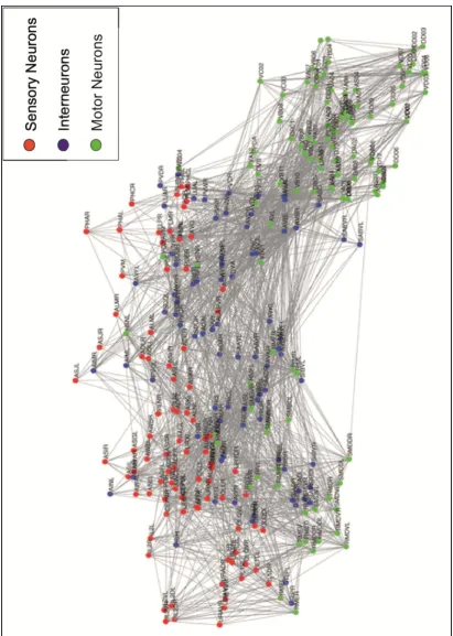
Hermaphrodite C. elegans have a simple nervous system which consists of 302 neurons compared to the complex nervous system in humans which contains billions of neurons (White et al., 1986). The function of the C. elegans nervous system is to detect its environment, process information and adapt behaviour allowing the worm to find food and amiable habitat and to an avoid harmful conditions. A wiring diagram for chemical synaptic connections for every neuron in hermaphrodite worm has been created (White et al., 1986, Varshney et al., 2011). No wiring diagram exists for the gap junctions, which allow electrical signalling between the neurons or modulation of neurons by neuropeptides released from contains sensory, interneurons and motor neurons like the human nervous system (Figure 1.4)
17
18
The nervous system of C. elegans contains genetic and molecular pathways similar to those in mammals and has been used to study many of the shared mechanisms including the SNARE complex machinery involved in neurotransmission (Barclay et al., 2012). For example, UNC-18 is a protein which has a role in SNARE complex formation. Mouse Munc-18 (the murine orthologue of UNC-18) has been expressed in the worm unc-18 null mutant where it completely rescued the defective neurotransmission function in the mutant (Gengyo-Ando et al., 1996). C. elegans
has also been used as a model for neurodegenerative diseases to screen for novel neuronal drug targets (Leung et al., 2008, Chen and Burgoyne, 2012, Johnson et al., 2010).
The worm nervous system is divided into two separate systems, the general nervous system which consists of 282 neurons and the pharyngeal nervous system which consists of 20 neurons and functions as a neuromuscular pump for feeding. (Altun et al., 2002-2012). The two nervous systems operate almost independently communicating via the RIP interneuron pair (Altun et al., 2002-2012).
19
20 1.6.1 Motor Neurons
Motor neurons control the motility of the alimentary canal and locomotion of the organism. After receiving a signal from the interneurons, three types of motor neurons in the VC and DC regulate the dorsal and ventral muscle wall cell function. Type A and B motor neurons are excitatory cholinergic neurons, which cause the muscle wall cell to contract (White et al., 1986). Type A neurons regulate backward motion and type B regulate forward motion (Chalfie et al., 1985). Type D motor neurons are inhibitory dorsal cord neurons which cause the muscle wall cells to relax (White et al., 1986) The characteristic rhythmic wave of C. elegans
locomotion is regulated by the coordinated relaxation and contraction of muscle wall cells at opposite sides of the worm (McIntire et al., 1993). Motor neurons can change locomotion by modulating the body wall muscle in response to a stimulus upstream in the neuronal circuit, which can include touch or other sensory stimulus. Changes in locomotion can include forward or backward movement, acceleration or turns (Chalfie et al., 1985).
1.6.2 Interneurons
In humans interneurons are located predominately in the central nervous system.
C. elegans interneurons are located predominately in the nerve ring and VC (Altun et al., 2002-2012) and perform a similar role to those of humans, forming connections in the neuronal network; they also receive input from sensory neurons, process the signal, modulate a decision and relay an output to the motor neurons.
1.6.3 Sensory Neurons
21
tail tip. The cell bodies of sensory neurons of sensory structures, such as the amphids, may form ganglions in the head and tail. Dendrites project from the cell body of the neuron and extend to the tip of the nose or tail surface (Figure 1.5). In the head, the axons of sensory neurons project from the cell body and can enter the nerve ring where they communicate with other neurons and pass on the sensory input (Sasakura and Mori, 2012).
1.6.4 Ciliated Sensory Neurons
Cilia are organelles which form projections from the cell surfaces, and contain microtubules as their major component. There are two groups of cilia; motile cilia which are required for movement or flow and primary cilia (non-motile) which are present on most types of mammalian cells and appear to have a role in cell signalling regulating homeostasis and development (Lee and Gleeson, 2010). Some specialised sensory neurons in humans are ciliated, these include photoreceptors in the eye, cochlear hair cells in the inner ear and olfactory sensory neurons in the nose, which detect external signals of light, sound or smell from the outside world via the cilia to the nervous system (Lee and Gleeson, 2010, Jenkins et al., 2009).
As with other types of sensory neurons, the majority of ciliated sensory neurons in
22
Table 1.2 Amphid and Phasmid Ciliated Sensory Neurons
Name Cilia Type External
exposed
Sensory Role
(From Wormatlas, Individual Neuron Database http://www.wormatlas.org)
ASE Single rod cilia Y Gustatory-chemosensory
ASG Single rod cilia Y Gustatory-chemosensory
ASH Single rod cilia Y Nociceptive osmo-, mechano-
and odour
ASI Single rod cilia Y Gustatory-chemosensory, thermosensory
ASJ Single rod cilia Y Sensory
ASK Single rod cilia Y Gustatory-chemosensory and pheromone sensory
ADF Dual rod cilia Y Chemosensory and oxygen sensory
ADL Dual rod cilia Y Chemosensory, odour sensory, pheromone sensory
and nociceptive
AWA Winged Cilia N Odour sensory
AWB Winged Cilia N Odour sensory
AWC Winged Cilia N Odour sensory and thermosensory
AFD Finger-like N Thermo (thermal nociception and thermotaxis) C02 sensory
PHA Single rod cilia Y Chemosensory
23
Figure 1.6 Ciliated Sensory Neurons of the Amphid and Phasmid at C. elegans Head and Tail.
Illustration of ciliated neurons (adapted from images shown in (Perkins et al., 1986) a) single and b) dual rod ciliated neurons are exposed to the external environment but c) winged and d) finger-like ciliated sensory neurons are embedded in sheath cells.
1.7 Thermosensation and Regulation
In the wild, C. elegans live in moderate soil temperatures between 16oC -26oC. The worm has the ability to detect changes in temperature (Schafer, 2012). Thermotaxis and thermoavoidance (Schafer, 2012, Wittenburg and Baumeister, 1999, Hedgecock and Russell, 1975) are the two distinct types of thermosensation in
24
and to avoid noxious conditions i.e. extreme high or low temperatures. Three reviews give a complete picture of the complexity and controversy surrounding the thermosensation field including neuronal circuits and signalling pathways involved in isothermal tracking and memory (Sasakura and Mori, 2012), on the negative temperature bias (Ma and Shen, 2012) and on thermoavoidance (Liu et al., 2012). This section of the introduction will give a brief summary on these behaviours.
1.7.1 Thermotaxis
Thermotaxis behaviour of C. elegans was identified in 1975. During thermotaxis, the worm migrates towards its cultivation temperature (Hedgecock and Russell, 1975). Thermotaxis, along with other sensory-regulated locomotion behaviours such as chemotaxis, is regulated by two strategies:- the gradual steering strategy and the biased random walk strategy (Sengupta and Samuel, 2009). The biased random walk strategy navigates the worm towards the favourable conditions it has detected which it does by increasing the length of its forward runs. If it encounters unfavourable conditions it will shorten a forward run by turning, thereby increasing the frequency of the turns until conditions are more favourable (Sengupta and Samuel, 2009). The gradual steering strategy works in tandem with the biased random walk and it enables the worm to travel up gradients and remain within favourable conditions (Sengupta and Samuel, 2009).
25
In the wild-type C. elegans the thermotaxis behaviour was shown to be modified by the cultivation temperature the worm had previously experienced, suggesting a function of memory and learning (Hedgecock and Russell, 1975). The worms display behavioural plasticity and are able to associate temperature with the availability of food. When cultivated at the standard temperature of 20oC, worms migrated towards 20oC and displayed isothermal tracking behaviour. When cultivated at a lower temperature of 16oC they migrated and tracked to 16oC and had the same response when cultivated at the higher temperature of 25oC. C. elegans can also use this memory to avoid temperatures that it predicts it will not find food, hence displaying learning behaviours. Thermotaxis behaviour has been further characterised by video tracking which analysed run duration time, turn frequency and run speed and swimming locomotion (Ryu and Samuel, 2002, Tsalik and Hobert, 2003, Sasakura and Mori, 2012).
26 1.7.2 Thermotaxis Neuronal Circuits
Three pairs of sensory neurons are involved in the thermotaxis circuit, the AFD neurons are the major thermotaxis sensory neurons, the AWC neurons have been shown to be involved in signalling during temperature change (Figure 1,7) (Biron et al., 2008, Kuhara et al., 2008) and the ASI neurons have also been shown to be involved in negative thermotaxis (Beverly et al., 2011). Using thermotaxis behavioural assays and neuronal Ca2+ imaging in sensory neuronal ablated animals, different combinations of these thermosensing neurons pairs appear to have single or combined responses under specific temperature conditions, such as cultivation temperature and gradient temperature, which enables the worm have normal thermotaxis bias (Beverly et al., 2011).
The molecular pathways in AWC, AFD, AIY, AIZ and RIA neurons have also been shown to be involved in isothermal tracking and memory (Sasakura and Mori, 2012, Mori et al., 2007, Kimata et al., 2012) (Figure 1.7).
A recent review hypothesised that the ASI sends thermosensory signals to the AIA and AIB interneurons (Figure 1.8) (Ma and Shen, 2012). ASI, AIA or AIB are not thought to be involved in isothermal tracking and memory as ablating the AIB neuron was shown to not affect isothermal tracking, while ablation of the AIY or the AIZ neurons did affect this behaviour (Mori and Ohshima, 1995, Ma and Shen, 2012).
AFD, AWC and ASI neurons are three different types of amphid sensory (finger-like, winged and single rod) ciliated neurons and also have been shown to have a role in other sensory responses. The AFD neurons sense CO2; the AWC neurons sense
27
28
Figure 1.8 The Hypothesised Negative Thermotaxis Neuronal Circuit. A wiring diagram showing proposed synaptic and gap junction neuronal conections. The diagram shows the excepted thermotaxis circuitry sensory and interneurons neurons AFD, AWC , AIY, AIZ and RIA. The ASI sensory neuron recently linked to negative thermotaxis and AIA and AIB hypothesized to also be involved. (Figure taken from (Ma and Shen, 2012)
1.7.3 Thermoavoidance
29
which is signalled in thermotaxis by synaptic neurotransmission (Kimura et al., 2004) (Figure 1.9). Thermoavoidance behaviour is not effected by altering cultivation temperature and shows no memory adaption (Wittenburg and Baumeister, 1999).
1.7.4 Temperature-Dependent Locomotion
Recently our group employed a temperature-dependent locomotion (TDL) assay for
30
31 1.8 C. elegans and Ca2+ Binding Proteins
C. elegans have a large number of proteins involved in Ca2+ signalling. Computational analysis performed on the C. elegans genome predicted Ca2+ binding properties for 209 proteins of which 170 posses EF hand motif proteins including calmodulin, NCS and proteins which have other functions such as forming ion channels which also contain the EF hand motif. The other 39 proteins contained different Ca2+ binding motifs such as the C2 domain (Kumar et al., 2012). C. elegans
have more than 100 candidate neuronal Ca2+ binding proteins, of which 65 contain EF hand motifs and no other functional domains (Hobert, 2013). They include the worm CaM orthologue (CMD-1) and eight other CaM related genes (Hobert, 2013). Previously three NCS proteins in the worm were identified NCS-1,2 and 3 (Decastro et al., 1995, Rajaram et al., 2000). Recent reanalysis of the C. elegans genome has identified four additional potential NCS proteins (NCS-4 to NCS-7) (Hobert, 2013). The nomenclature of the C. elegans NCS proteins suggests they are all NCS-1 Class A orthologues of mammalian NCS-1, however, only NCS-1 and NCS-3 genes have orthologues with NCS-1 showing the highest sequence identity of 75% to human NCS-1 (Table 1.3).
Table 1.3 C. elegans NCS Proteins
# Orthologue and % Sequence Identity of Ce- NCS to Human NCS were calculated using NCBI.BLASTp. *Functional EF hand motifs were predicted using Interpro.EMBL sequence functional analysis software using the SMART.EMBL database.
Ce NCS Human NCS
#
Sequence Identity
Ce NCS to Human NCS
#
Class Position number of
predicted functional EF
hand motifs *
NCS-1 NCS-1 75% A 2,3,4
NCS-2 HCLP-1 51% B 2,3,4
NCS-3 NCS-1 67% A 2,3,4
NCS-4 KChIP1, 2, 3, 4? ~35%? E 2, 4
NCS-5 - - - 4
NCS-6 - - - 0
32
A blast search of NCS-2 shows its closest human orthologue is NCS class B hippocalcin-like protein 1 (HCLP-1) showing 51% sequence identity. NCS-4 and NCS-7 share sequence identity with the human class E proteins KChIPs 1-4 (approximately 35% and 40% respectively). NCS-5 and NCS-6 do not appear to be orthologues of any class of human NCS proteins, although, they show a higher sequence identity to human KChIP2 than to any other human protein. As discussed early in this chapter, NCS proteins contain an inactive EF hand (motif 1), which is unable to bind Ca2+ and three functional EF hand motifs 2, 3, and 4. Analysis of all
C. elegans NCS sequences (wormbase) using functional analysis software (Interpro.EMBL) predicted that only NCS-1, 2, 3 and 7 proteins contain all three functional Ca2+ binding sites. Therefore it is questionable whether NCS-4, 5, 6 are functional NCS proteins. It could be that these proteins form a different subclass of NCS protein with divergent structures and functions; however considerable work will be needed to establish this.
Analysis of the worm ncs-3 null strain showed no obvious phenotype. It was only assessed, however, for a kinked motion behaviour and other behaviours have not been characterized (Rajaram et al., 2000). It was surmised that ncs-3 null showed no phenotype because there is redundancy with other NCS proteins, in particular NCS-1 to which it shows 80% homology (Rajaram et al., 2000). In worm Alzheimer’s disease model, in which amyloid aggregates build up in muscle cells and cause paralysis, knockdown of ncs-3 was shown to suppress the toxicity (Lopez, 2010). This not only suggests that NCS-3 is expressed in muscle cells but also that NCS proteins might have a role in control of amyloid plaque formation in Alzheimer’s disease and other amyloid pathologies (Lopez, 2010). Although localisation and function of NCS-2 have not been established, it appears to have an essential role in the worm, as knockout is lethal. The location of expression and functions of the other NCS proteins is yet to be determined.
33
muscle cell type by GFP reporter gene and validated by immunostaining (Table 1.4) (De Castro, 1997, Gomez et al., 2001).
Table 1.4. C. elegans Cell Types Expressing NCS-1
Cell
(De Castro, 1997,
Gomez et al., 2001)
Cell Type
Taken from (Altun et al.,
2002-2012)
Function
Taken from (Altun et al., 2002-2012)
ASE L/R Amphid Sensory Neuron Gustatory-chemosensory
ASG L/R Amphid Sensory Neuron Gustatory-chemosensory
ADF L/R Amphid Sensory Neuron Chemosensory and oxygen sensory
AWA L/R Amphid Sensory Neuron Odour sensory
AWB L/R Amphid Sensory Neuron Odour sensory
AWC L/R Amphid Sensory Neuron Odour sensory and
thermosensory-(thermotaxis)
AFD L/R Amphid Sensory Neuron Thermosensory- (thermal nociception and
thermotaxis) and C02 sensory
BAG L/R Head Ciliated Sensory Neuron
(Not part of amphid)
Nociception, C02
and oxygen sensory
PHA L/R Phasmid Sensory Neuron Chemosensory
PHB L/R Phasmid Sensory Neuron Chemosensory
AVK L/R Interneuron NR/VC Little known
AIY L/R Interneuron Head Amphid interneuron receives and
processes output from amphid neurons,
involved in regulating gustatory and
olfactory behaviour, thermotaxis,
memory, lifespan and stress responses.
RMG L/R Polymodal
Interneuron/Motorneuron
Head
Receives and processes signals from
sensory neurons, modules chemosensory
responses. Involved in Pheromone and
social behaviour.
34
Over-expression of NCS-1 driven by its endogenous promoter on a wild-type background showed improved memory compared to the wild-type worm based on the ability to associate the presence of food with a specific temperature, whereas the ncs-1 null (qa406) worm showed impaired memory formation (Gomez et al., 2001). The regulation of memory by NCS-1 was shown to be Ca2+ dependent, as expression of the NCS-1 loss of function protein, in which all 3 functional EF hands motifs (EF 2,3, and 4) are muted and unable to bind Ca2+, failed to rescue memory in the null worm (Gomez et al., 2001).
Using neuron-specific promoters to drive NCS-1 expression, the AIY neuron was shown to be the site where NCS-1 regulates memory and isothermal tracking (Gomez et al., 2001). Expression of NCS-1 in the AFD neuron failed to rescue this behaviour, suggesting that NCS-1 has an alternative role in this neuron (Gomez et al., 2001). AWC was not identified as being involved in temperature sensation at the time of this study; as a consequence the role of NCS-1 in this neuron was not investigated.
35
36 1.9 Aims and Objectives
Up until now no direct interaction between P/Q channels and NCS-1 has been described. During the first part of this PhD study biochemical and biophysical techniques were used to address the following aims.
To identify whether the P/Q channel interacts directly with NCS-1
To identify which domains of the P/Q channel interact with NCS-1
To characterise the interaction using surface plasmon resonance and nuclear magnetic resonance, to identify which amino acids of the NCS-1 binding pocket are important for this interaction.
Using C. elegans as a model organism, the objective of the second part of this study was to take the known structural and binding information for NCS-1 and apply it to a physiological system in order to address the following aims.
To investigate the behaviour of ncs-1 null strains expressing mutated NCS-1 and evaluate the molecular significance for function of key characteristics of the NCS-1 protein identified in structural studies
To determine in which neurons NCS-1 functions in relation to the neuronal circuit for temperature-dependent locomotion.
37
38
2.1 Materials and Methods for Characterisation of NCS-1 Protein and
Analysis Target Peptide Binding
2.1.1 Reagents
All P/Q Cav 2.1 subunit fragments were derived from rat Rba-1 (NP037050.2) and
other constructs from human CaM (CAA36839) or rat NCS-1 (NP077342.1).
The pEYFPN1-NCS-1 plasmid encoding NCS-1 (O'Callaghan et al., 2002), pGex-6P1 plasmid encoding GST-NCS-1 (Haynes et al., 2004) and pET-m11 plasmid encoding His tagged NCS-1 (Handley et al., 2010) were described previously. pGex-6P1 plasmids (GE Healthcare) encoding a GST fusion tag containing human CaM or rat P/Q-L inserts and pE-SUMOpro plasmids (LifeSensors) with a His-SUMO protein fusion tag encoding P/Q-XL and P/Q-CBD were a gift from Dr Haynes, University of Liverpool. The pOPINS plasmid with a His-SUMO tag encoding P/Q-L and a modified pET15b plasmid encoding rat CaM gene insert and a deletion to remove the His-tag coding region were a gift from Sravan Pandalaneni, University of Liverpool. The rat P/Q-IQ peptide defined in previous structural studies to contain the IQ domain (Kim et al., 2008), was synthesised and purified by HPLC (GenicBio Limited).
39
Laboratories) and the ligase enzyme used was T4 DNA ligase (New England Laboratories).
All recombinant proteins and peptides where analysed using SDS-PAGE electrophoresis using 15% polyacrylmide (v/v) gel and See Blue Plus 2 molecular mass markers (Invitrogen), all gels were stained with Coomassie blue to visualise the protein.
2.1.2 Preparation of Plasmids for Protein Expression
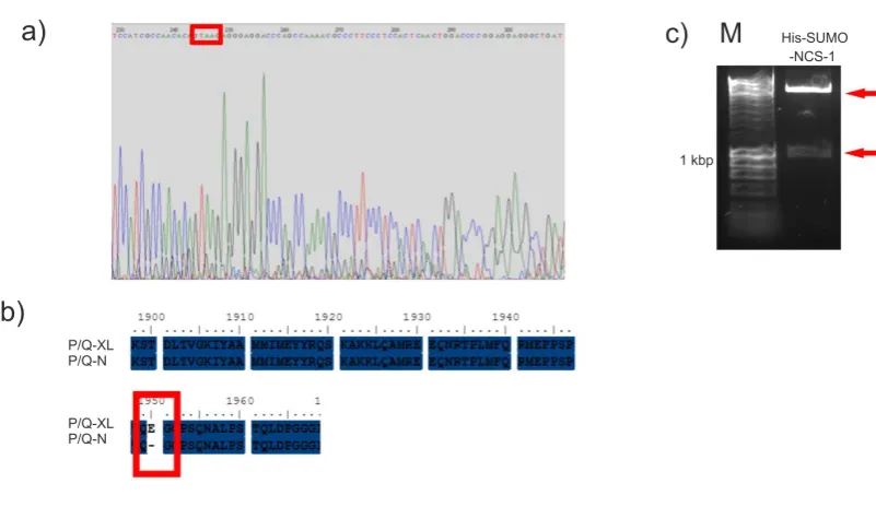
2.1.2.1 pE-SUMOpro-NCS-1
To clone NCS-1 into the pE-SUMOpro plasmid, the NCS-1 sequence was amplified from pEYFPN1-NCS-1 (O'Callaghan et al., 2002). PCR was performed using Phusion High Fidelity Polymerase (Finnzymes) for amplification of rat cDNA and following
the manufactures protocol. Briefly, 0.5g of each Primer (forward 5’-ATATCGTCTCAAGGTATGGGGAAATCCAACAGC-3 and reverse 5’-ATATCTCGAGTCATACCAGCCCGTCGTAGAG-3’) and 0.5g of template DNA was
required for PCR. The annealing temperature used was 69oC and extension time was 15 seconds. The NCS-1 PCR product was purified and digested with the restriction enzymes BsmBI and XhoI (NEB). pE-SUMOPro was digested with BsaI (which has a complementry overhang to BsmBI) and XhoI (NEB). The digested DNA was run on a gel and DNA bands were extracted. pE-SUMOPro and the NCS-1 insert were ligated using a 1:3 ratio, at room temperature for 20 minutes.
2.1.2.2 His-SUMO-P/Q-N and His-SUMO-P/Q-N2
To clone His-SUMO-P/Q-N and His-SUMO-P/Q-N2, the Stratagene QuikChange® Site-Directed Mutagenesis protocol was followed. The constructs were created by inserting a stop codon at the appropriate point in the longer His-SUMO-P/Q-XL template plasmid. Briefly, 0.5g of template plasmid and 125 ng of primers, for His-SUMO-P/Q-N (forward 5’-GGTCTCAAGGTAAGTCCACGGACCTGACATGGG-3’ and reverse 5’- CCCATGTCAGGTCCGTGGACTTACCTTGAGACC-3’) and for
40
5’- GGGTCCAGCTGAGTGGTTAGGGAAGGGCGTTTGGC-3) were used for PCR, using Pfu Turbo DNA polymerase (Stratagene). An annealing temperature used was 55oC; with an extension time of 13 minutes (double recommended time) was used. 16 cycles of PCR were required to insert the single stop codon by site directed mutagenesis. To remove the template plasmid, the sample was incubated with DpnI (Promega) restriction enzyme.
All cloned His-SUMO plasmids, were amplified using standard transformation protocols, into BIOBlue E. coli cells (Bioline) and grown in LB media (1 % w/v Tryptone, 0.5 % w/v yeast exact, 5.6 mM NaCl, pH 7.2), containing 30 g/ml of kanamycin. To ensure the insertion of the NCS-1 sequence into pE-SUMOpro, the amplified plasmid was digested with XbaI and XhoI (NEB). BsmBI could not be used as a diagnostic restriction enzyme as this site is removed from the pE-SUMOPro plasmid during cloning. The digested sample was analysed by DNA electrophoresis. All plasmids were amplified and purified using miniprep kits (NBS biologicals Ltd). All three newly cloned constructs were sent for DNA sequence verification (The sequencing service, Dundee). The resulting DNA sequences were translated to their amino acid sequence using ensembl software and aligned to the relevant protein sequences using Clustal W2 software (EMBL-EBI).
2.1.3. Protein Expression and Purification of Unlabeled NCS-1, GST-CaM and P/Q peptides
Standard transformation protocols were used to produce colonies of bacteria containing the relevant plasmid. Plasmids encoding for GST-CaM, GST-P/Q-L, His-SUMO-NCS-1, His-SUMO-P/Q-XL, His-SUMO-P/Q-N and His-SUMO-P/Q-L were transformed into BL21 (DE3) E. coli cells (Bioline). The plasmid encoding for P/Q-CBD was transformed into both BL21 (DE3) and Rosetta 2 (DE3) pLysS E. coli
41
BL21 cells transformed with pGEX-6P1 plasmids were grown media containing 100
g/ml ampicillian. BL21 cells transformed with pOPINS or pE-SUMO plasmids were grown in media containing 30 g/ml kanamycin. Rosetta 2 (DE3) pLysS cells transfected with pE-SUMO plasmid was grown in media containing 30 g/ml kanamycin and 34 g/ml chloramphenicol.
After overnight incubation, the starting culture was used to inoculate the growth media so that the starting optical density at wavelength of 600 nm (OD600) was less
than 0.1. Protein expression was induced when the OD600 reached 0.6-0.8 by
adding 1mM IPTG (Merck) then incubated at 37oC, 220 rpm for 3 hours.
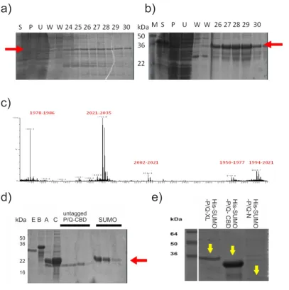
2.1.4. Protein Purification
Bacterial culture samples were centrifuged at 2500 x g for 15 minutes at 4oC. The pellets were resuspended in breaking buffer containing Complete Mini, cocktail protease inhibitor tablet (Roche). Cell expressing GST proteins were suspended in GST buffer (20 mM Tris.HCL, 50 mM NaCl, pH 7.4). Cells expressing His-SUMO-P/Q-L was suspended in Talon buffer (50 mM Na2HPO4, 300 mM NaCl, pH 7). Cells
expressing all other His-SUMO proteins were suspended in Histrap buffer (50 mM HEPES, 150 mM NaCl pH 7.5). All resuspended pellets were frozen at -80oC overnight. Pellets were defrosted quickly and the cells lysed using a Cell Disrupter One Shot (Constant systems Ltd) at 27 kPsi. 250 g of DNaseI (Sigma) was added to the Rosetta 2 (DE3) pLysS cell lysate and incubated for 15 minutes on ice. All protein preparations were ultracentrifuged at 100K x g for 1 hour at 4oC.
42
His-SUMO-P/Q-L was purified on cobalt affinity Talon resin (Clontech). The supernatant was incubated on the prewashed resin on a rotor for 1 hour at 4oC. The resin was then washed with Talon buffer and the recombinant protein eluted with Talon buffer containing 200 mM, 400 mM and 800 mM imidazole concentrations respectively.
GST-NCS-1 and GST-CaM protein were dialysed overnight in buffer (50 mM Tris.HCl, 150 mM NaCl, 1 mM EDTA and 1 mM DTT, pH 7). The protein was then bound to the glutathione cellulose resin. Precision protease, a gift from Dr Hannah McCue (University of Liverpool), was added to the resin and the sample incubated overnight at 4oC. The resin was centrifuged at 3000 xg for 5 minutes and the supernatant was retained as it contained the untagged recombinant protein. The cleaved GST remained on the resin along with the GST-tagged PreScission protease.
His-SUMO-P/Q-XL, His-SUMO-P/Q-CBD, His-SUMO-NCS-1 and His-SUMO required for SPR were purified using an ATKA (GE Healthcare) chromatography system. The first purification step used a nickel affinity, Histrap FF 5ml column (GE Healthcare). After the supernatant was loaded on to the column, it was washed with 2 column volumes of Histrap running buffer. The recombinant protein was eluted from the column using Histrap elution buffer (0.5 M Imidazole, 50 mM HEPES, 150 mM NaCl, pH 7.4), with a 0-100% elution gradient over 20 column volumes, the flow rate was 5 ml/min and fraction size was 5ml.
43
Untagged NCS-1, untagged P/Q-CBD, His-SUMO-P/Q-XL and His-SUMO-P/Q-CBD samples were concentrated using Vivaspin 20 columns (GE Healthcare) with a MWCO 3 or 10 kDa as appropriate. The samples were then diluted 1 in 6 with Mono Q buffer (20 mM HEPES, pH 7.0) to lower the concentration of salt from previous steps. The sample was then further concentrated to 2.5 ml. Untagged and tagged proteins where further purified using ion exchange chromatography. The sample was injected on to a Mono Q 5/5 1 ml column (GE Healthcare) at a flow rate of 1 ml/min. Elution buffer (20 mM Hepes, 1 M NaCl, pH 7.0) was run through the column at 0-100% gradient over 25 column volumes. His-SUMO-P/Q-CBD was concentrated and underwent a third purification step by Gel filtration. After concentrating, using a Vivaspin 20 (MWCO of 10 kDa), the sample was loaded on a to a HiLoad 26/60 Superdex 75 column 26/60 (GE Healthcare) for size exclusion chromatography. Running buffer (10 mM HEPES 150 mM NaCl, pH 7.4) was run though the column at a flow rate of 2ml/min and fractions were eluted at 5 ml volumes.
2.1.5 Expression of Unlabeled CaM, 15N Labelled NCS-1 and CaM.
pET-15b CaM or pET-m11 His-NCS-1 plasmids were transformed into BL21 (DE3) using standard protocols. Media for CaM expression contained 100 g/ml ampicillian and for NCS-1 expression contained 30 g/ml kanamycin. An LB starting culture of 5 ml was grown for 5 hours at 37 oC, with agitation at 220 rpm, then transferred to a starter culture of 2M9 minimal media (20 mM 15N labelled ammonium chloride, 20 mM glucose, 55 mM KH2PO4, 88 mM Na2HPO4, 1 mM
MgSO4, 136 M CaCl2 30 M) and incubated overnight at 37oC, 220 rpm. After
44
Complete Mini EDTA free Protease inhibitor tablets (Roche)) and frozen at -20oC overnight. The cells were lysed using a cell disrupter as above and 250 g DNaseI (Sigma) added. The lysate was centrifuged at 50,000 xg for 30 minutes at 4oC. The CaM preparations underwent an additional purification step, the supernatant was removed and it was heated to 65oC for 3 minutes. This heat step denatures protein impurities in the sample, centrifugation was repeated at 50,000 x g for 30 minutes at 4oC, and supernatant retained.
2.1.6 Purification by Hydrophobic Interaction and Size Exclusion Gel Chromatography
Supernatants for both CaM and His-NCS-1 were filtered (0.2m acrodisc). The
samples were loaded on a HiPrep 16/10 Phenyl FF High Sub column (GE Healthcare), using a flow rate of 2 ml/min. Then loading buffer (50 mM Tris.HCl,
500 mM NaCl, 5 mM CaCl, pH 7.5) was run over the column to saturate the column with the recombinant protein and remove unbound proteins. Saturation was indicated by decline of the load peak on the chromatogram. Wash buffer (50 mM Tris.HCl, 0.5 mM CaCl, pH 7.5) was ran over the column until the chromatogram UV units reached ~ 50 mUV to remove impurities. Elution buffer (50 mM Tris.HCl, 10 mM EDTA, pH 7.5) was used to remove CaM from the column and distilled water was used to remove NCS-1 from the column. A flow rate of 2 ml/min was used and protein fractions were eluted at 5ml volumes.
45
NaCl, 5 mM CaCl2 ,pH 6.5). CaM and untagged NCS-1 samples were injected on to
HiLoad 26/60 Superdex 75 column 26/60 (GE Healthcare) using the gel filtration buffer and conditions used above. Aliquots of fractions were flash frozen using liquid nitrogen and stored at – 80 oC.
2.1.7 Mass spectrometry
For identification, the untagged P/Q-CBD peptide band was extracted from an SDS PAGE gel stained with Coomassie blue. In-gel, tryptic digestion and mass spectrometry were performed by Mark Wilkinson and Mark Prescott, University of Liverpool as follows. Gel slices from SDS-PAGE were washed with 50% acetonitrile, 0.2 M ammonium bicarbonate pH 8.9 and then dried in a rotary evaporator. The slices were re-swollen in 0.2 M ammonium bicarbonate pH 7.8 containing trypsin and incubated at 25ºC overnight. Peptides were extracted from the gel slices with 60% acetonitrile, 0.1% TFA and then de-salted for MS using C18 ZipTips. MS analysis was performed on a MALDI-Tof instrument (Waters-Micromass). Peptides were mixed 1:1 with a saturated solution of alpha-cyano-4 hydroxycinnaminic acid in 50% acetonitrile/0.1% trifluoroacetic acid and analysed in the mass range 800 – 4000 Da
2.1.8 Analysis of the P/Q-IQ peptide
2.1.8.1 Helical Prediction
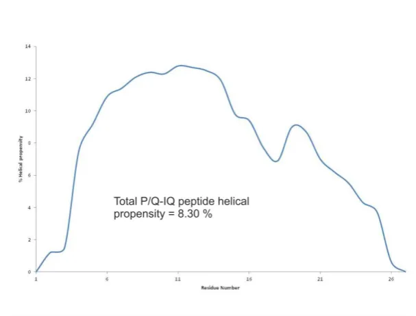
The helical prediction software AGADIR was used to calculate the helical content of the 27 residue P/Q-IQ peptide from its amino acid sequence TVGKIYAAMMIMEYYRQSKAKKLQAMR.
2.1.8.2 Circular Dichroism
46
25oC. Seven scans were performed for the peptide at each TFE titration. The CD signal was averaged and corrected for the buffer.
Measured ellipticity (CD mdeg) at 222 nm was converted to mean residue molar ellipticity [222], using the following equation:-
[222]= (100 x) /Cnl,
Where C is the protein concentration, n is the number of residues in the peptide and l is the path length in cm.
Percentage helicity was calculated using the following formula
([222]/[max222]) x100=% helicity
[max222]= -40,000 [1 - (2.5/n)],
Where -40,000 is the constant for infinite helicity at 0oC, 1 - (2.5/n) corrects for peptide length, n = number of amino acids in peptide.
2.1.9 Pull-down Assay The analysis of the Ca2+-dependency of binding of GST-P/Q-L -NCS-1
2.1.9.1 Binding Assay
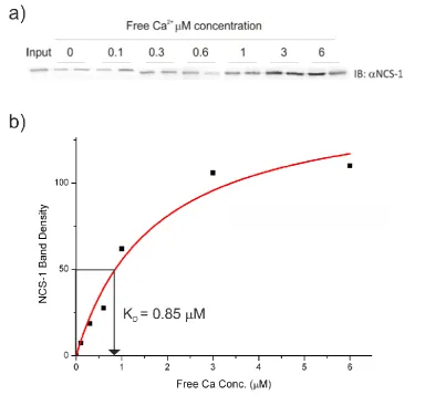
The concentrations of GST-P/Q-L and untagged NCS-1 was determined using densitometry by comparing the bands of recombinant protein to known BSA standards with Image J software. Samples were prepared in 25 mM Tris.HCl, 50 mM KCl, 5 mM EGTA, 5 mM NTA, 1 mM DDT, 2 mM Free Mg2+ pH 7.4. A titration of Ca2+ concentrations was performed adding CaCl2 to give 0, 0.1, 0.3, 0.6, 1, 3, 6 mM
47
tube and each of the following buffer conditions was run in duplicate. GST-P/Q-L was added to a final concentration of 1 M to tubes containing the appropriate Ca2+ buffer 0-6 mM. The tubes were incubated on a rocker on ice for 30 minutes. NCS-1 was added to a final concentration of 1 M and incubated for 1 hour. The resins were washed three times with the appropriate Ca2+ buffer. The tubes were centrifuged to remove excess wash buffer and boiled with 30 l SDS loading buffer for 5 minutes. The resin was centrifuged again and the SDS loading buffer was analysed using SDS-PAGE and Western blotting.
2.1.9.2 Western Blot of Pull-Down Assay
For Western blot analysis, protein bands were transferred from the gel to a nitrocellulose membrane using electroblotting. The membrane was then blocked using 3% (w/v) milk in PBS to prevent non-specific binding of antibodies. The membrane was then incubated with 1:1000rabbit polyclonal anti-human NCS-1 antibody (McFerran et al., 1998) in 3% (w/v) milk PBS, overnight on a rocker at 4oC (McFerran et al., 1998) . After washing three times with PBS and 0.05% (v/v) tween 20 (PBST) and rinsed, the membrane was then incubated with 1:400 dilution HRP conjugated goat anti-rabbit secondary antibody (Sigma) in 3 % (w/v) milk PBS, for 1 hour. The membrane was further washed and incubated with equal volumes of ECL reagents. The blot was visualised using the ChemiDoc System (Bio-Rad), using Chemi-Hi sensitivity setting. The image was further analysed by measuring the density of the bands using ImageJ software. The density was plotted against Ca2+ concentration and fitted against a hyperbolic model using Origin8 software.
2.1.10 Gel Filtration analysis
The mixture of NCS-1 and P/Q-IQ peptide, at concentrations of 100 M and 350 M respectively, containing a putative complex was suspended in a buffer (20 mM MES, 100 mM NaCl, and 5 mM CaCl2 buffer pH6.5), and analysed by size exclusion gel