Laboratory of Tumor Cell Biology, National Cancer Institute, Bethesda, Maryland 20892
Received 29 June 1994/Accepted 16 December 1994
Spontaneous revertants of the immune-selected variant HXB2thr582, which resists neutralization by certain
conformationally dependent antibodies specific for the CD4-binding site on gp120 (such as F105), appeared
after long-term culture in the absence of immune-selecting serum. Molecular analysis showed some of the
viruses in the revertant stock contained a simple back mutation, whereas others retained the Thr-582 codon
but contained a substitution of serine for phenylalanine in gp41 at position 673. Neutralization sensitivity to
the selecting serum and to F105 of infectious clones containing either the back mutation or the compensatory
mutation, HXB2thr582ser673, was confirmed. HXB2thr582-infected cells have a greater propensity for
syncy-tium formation and single cell killing than do either the parental HXB2 or the revertant HXB2thr582ser673.
This suggests that the revertant arose by selection in vitro for a less cytopathic virus. Our results link three
envelope regions shown to influence virus-cell fusion as well as neutralization by antibody: the CD4-binding
region, the leucine zipper domain, and a region hidden to antipeptide antibodies upon envelope
oligomeriza-tion. Taken together they illustrate the functional importance of the gp120-gp41 interaction and emphasize the
impact of the interplay between envelope regions on overall conformation and function and on recognition by
neutralizing antibodies.
Human immunodeficiency virus type 1 (HIV-1) exhibits
ex-tensive genetic heterogeneity, particularly in the envelope gene
(23, 63). The resulting amino acid sequence variability in the
viral surface glycoprotein, gp120, and transmembrane protein,
gp41, provides the basis for the elicitation of type-specific
an-tibodies. These in turn are able to exert immune-selective
pressure which can lead to the propagation of variants in vitro
and in vivo which have escaped immunologic control (1, 2, 31,
34, 46, 55). We previously described such an escape mutant
derived by using a human high-titered neutralizing serum in an
in vitro system to generate a neutralization-resistant variant of
an infectious molecular clone of HIV-1, HXB2 (46). The
mo-lecular basis for the observed neutralization resistance was
subsequently shown to be a point mutation in the env gene,
resulting in the substitution of a threonine for an alanine
res-idue at position 582 in the transmembrane protein (42).
Fur-ther analysis of the variant, termed HXB2thr582, showed that
the 582 region did not constitute a neutralizing epitope itself
nor did it seem to be part of a noncontiguous neutralizing
epitope. The results were best explained by a conformational
change in the envelope protein, resulting from the alanine to
threonine substitution and leading to alteration of a
neutral-izing epitope at another location (62). This hypothesis
subse-quently was confirmed by showing that HXB2thr582 is
resis-tant
to
neutralizing
antibodies
which
recognize
a
conformational epitope overlapping the CD4-binding region
on gp120 (26).
Following transfection of the molecular clone HXB2thr582
into H9 cells and long-term culturing in the absence of
im-mune-selecting serum, we have repeatedly observed its
rever-sion to neutralization sensitivity. Because the basis for its
orig-inal selection was a conformational change, we expected that
although restoration of the parental conformation and
neutral-izing phenotype could be due to a back mutation, it was equally
likely to result from a compensating change elsewhere within
the viral envelope. We therefore analyzed the revertant virus
population to determine the molecular basis for its altered
phenotype. In addition, in order to gain further insight
con-cerning the functional importance of regions highlighted by
our molecular studies and the nature of selection pressures,
other than immunologic, leading to the propagation of
partic-ular variants, we investigated the biologic basis for reversion of
HXB2thr582 to neutralization sensitivity.
(This work was carried out in partial fulfillment of the Ph.D.
requirements [for T. L. Stern] in the Graduate Genetics
Pro-gram at the George Washington University, Washington,
D.C.)
MATERIALS AND METHODS
Cells and viruses.The parental virus used in these studies was HXB2, an infectious molecular clone of HIV-1 (17). It is sensitive to neutralization by the original immune-selecting serum (46). As a neutralization-resistant variant we used the mutant HXB2thr582 created from HXB2 by site-directed mutagenesis (42). This mutant reproduces the base change and subsequent amino acid sub-stitution of alanine to threonine at position 582 which was originally selected in vitro in the presence of the neutralizing serum. pHXB2gpt, HXB2thr582, and HXB2thr582ser673 viral DNAs were transfected by electroporation into COS-1 cells, and progeny virus was transmitted 24 h later by coculturing into H9 cells for propagation (39). The virus-producing H9 cells were cultured in the absence of immune-selecting serum in RPMI 1640 containing 10% fetal calf serum, 1 mM glutamine, and penicillin and streptomycin. Cell-free virus preparations from the parental and mutant cultures were assayed periodically for neutralization resis-tance or sensitivity to the original selecting serum.
Antibodies.The original selecting human serum, possessing high titer and broadly reactive neutralizing antibody, was obtained from a healthy HIV-sero-positive individual and was used to determine neutralization sensitivity or resis-* Corresponding author. Mailing address: Laboratory of Tumor Cell
Biology, Building 37, Room 6A09, National Cancer Institute, National Institutes of Health, 37 Convent Dr. MSC 4255, Bethesda, MD 20892-4255. Phone: (301) 496-2114. Fax: (301) 496-8394.
1860
on November 9, 2019 by guest
http://jvi.asm.org/
tance of the revertant viral population and cloned variants. Normal human serum served as a control. The human monoclonal antibody F105 (40, 41), specific for a conformational determinant overlapping the CD4-binding region (53), was used as ascites fluid containing the antibody obtained from nude mice or as purified immunoglobulin G (IgG). Ascites fluid from mice inoculated with P33
63 hybridoma cells or purified human IgG was used as a control.
Immunologic assays.Neutralizing antibody assays were carried out as previ-ously described (43, 44), using fresh cell-free supernatant of the virus-producing H9 cultures as the source of virus. The sensitivity or resistance to neutralization of the viral strains was assessed by using 1:10 dilutions of the original selecting serum or normal human serum as control and serial dilutions of virus. Deter-mination of neutralization sensitivity or resistance was made at viral dilutions at which the kinetics of infection of each of the viruses in the presence of control serum were similar. Viral infection of the target H9 cells was determined by expression of p24 as monitored in an indirect immune fluorescence assay (IFA) with an HIV-1 p24-specific monoclonal antibody.
Antibody binding to H9 cells infected with HXB2, HXB2thr582, and HXB2thr582ser673 was assessed by FACScan analysis as previously described (45) by using a pooled human serum, purified IgG of the F105 monoclonal antibody, and human IgG as control.
Assessment of virologic properties.Production of infectious virus and viral proteins was determined by measuring p24, reverse transcriptase (RT), and gp120 levels of expression in supernatant media of infected cells. Levels of p24 and gp120 were quantitated with commercially available antigen capture kits (Coulter, Hialeah, Fla., and American Bio-Technologies, Cambridge, Mass., respectively). Virus particles were precipitated with polyethylene glycol from cell-free supernatant as previously described (39). RT activity was measured by using oligo(dT12-18)-poly(A) as template in the presence of Mg21as previously described (38). The percentage of infected cells was determined by IFA on cells fixed for 10 min at room temperature in 50% methanol–50% acetone, using an HIV-1 p24-specific monoclonal antibody.
Syncytium formation by virus-infected cells was determined with uninfected CEM-SS, SupT-1, and HUT 78 cells as targets. Twenty-five-microliter dilutions of infected cells, ranging from 13105to 53105/ml were mixed with 25ml of CEM-SS cells (106/ml) in a total volume of 100ml in half-well, flat-bottomed, 96-well microtiter plates. Syncytia were counted after overnight incubation at 378C. The number of syncytia per 103p24-expressing infected cells (determined by IFA) was calculated from the results of triplicate determinations.
Single-cell killing was assessed by infecting H9 cells with equivalent amounts of either HXB2, HXB2thr582, or HXB2thr582ser673 and culturing for 3 weeks. Total cell count and viability by trypan blue staining and percentage of cells expressing viral p24 by IFA were determined every 2 days, at which time the cultures were adjusted to 106viable cells per ml in fresh medium.
Amplification of DNA for direct sequencing.Cells (23107) producing the uncloned revertant virus were digested for 18 h at 508C with 100mg of proteinase K per ml in TNE (0.01 M Tris-HCl buffer [pH 9.0], 0.1 M NaCl, and 1 mM EDTA) containing 0.5% sodium dodecyl sulfate. Genomic DNA was extracted twice with phenol-chloroform-isoamyl alcohol (24:24:1) equilibrated to pH 9.0 and twice with chloroform-isoamyl alcohol (24:1). The DNA was precipitated with ethanol, washed twice with 80% ethanol, and resuspended in water.
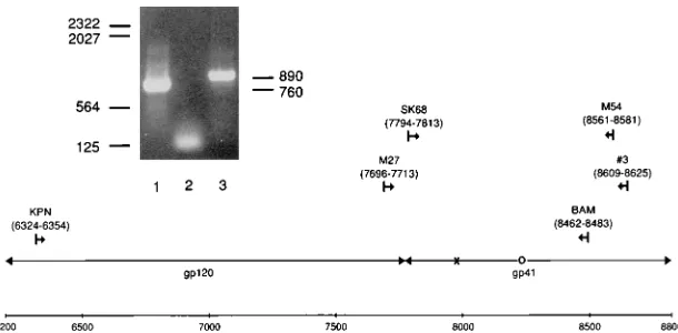
One microgram of genomic DNA was used as the template for amplification by PCR. Mixtures contained 0.5mg of each primer, 3.5 U of Taq DNA poly-merase, and 5% glycerol in addition to components specified by the Perkin-Elmer Cetus (Norwalk, Conn.) PCR kit instructions. Thirty amplification cycles were carried out, with 45 s at 948C for denaturation, 1.5 min at 508C for anneal-ing, and 2 min at 728C for extension. The primers used (M27 and no. 3) are specified in Fig. 1.
DNA from 15ml of the first amplification was separated electrophoretically on a 1% agarose gel. The appropriately sized DNA product (890 bp) was excised from the gel and freeze fractured by freezing overnight at 208C and subsequently thawing at 378C for 5 min. DNA was eluted following maceration of the agarose by suspending it in 400ml of TE (0.01 M Tris-HCl buffer [pH 7.4], 1 mM EDTA), transferring to a Millipore (Bedford, Mass.) ultrafree MC 0.45-mm-pore-size filter unit, and centrifuging at 12,000 rpm for 30 min. DNA in the flow through was concentrated by ethanol precipitation and resuspended in 10ml of sterile water.
One microliter of the purified first amplification product was subjected to a second round of amplification under the same PCR conditions as outlined above (without 5% glycerol) by using 0.5mg of nested sense and antisense primers (SK68 and M54) (Fig. 1). Fifteen microliters of the second amplification product was electrophoretically separated and purified as described above. The double-stranded DNA product, concentrated by ethanol precipitation, was dissolved directly in appropriate buffer for sequencing by one of two methods.
The32
P-end-labeled primer extension method was used in association with an altered Sequenase (version 2.0) protocol of the U.S. Biochemical Corp. (Cleve-land, Ohio) (32). The DNA product was dissolved in 12ml of32
P-end-labeled primer (2 ng/ml) to which 2ml of Sequenase reaction buffer was added. The DNA product and32
P-end-labeled primer mixture were denatured in boiling water for 2 min and then annealed by snap-cooling for 10 min in an ice-water bath. Components of the Sequenase protocol were added, and the procedure was carried out as described by the manufacturer. No labeling mix or35S-labeled dATP was required, because the primer was already labeled. Alternatively, the double-stranded cycling procedure of Applied Biosystems Inc. (Columbia, Md.) and Promega (Madison, Wis.) was followed by using the dyedeoxy terminator Taq sequencing kit and analyzed on a model 373A DNA sequencer according to the directions of the manufacturer. In either case, the sequencing reaction mixtures were electrophoresed on 8% sequencing gels, followed by autoradiog-raphy or laser-fluorescent scanning as appropriate.
Clonal analysis of revertant DNA.A single round of PCR amplification of revertant genomic DNA with a set of primers (KPN and BAM) (Fig. 1) encom-passing most of the envelope was carried out as described above. The compo-nents and the cycle parameters of the PCR were identical to the procedure described above. The DNA product was purified as described above, concen-trated by precipitation in ethanol, and dissolved directly in appropriate buffer for digestion by the restriction enzymes KpnI and BamHI (Boehringer Mannheim, Indianapolis, Ind.). One microgram of the Bluescript cloning vector (Stratagene, La Jolla, Calif.) was similarly digested, and the cut DNA product and cloning vector were electrophoretically separated and purified as described above. The DNA products from both were dissolved and combined in a total of 20ml, including 2ml of ligation buffer and 1 U of T4 DNA ligase (Boehringer Mann-heim). The ligation mix was incubated for 16 h at 108C. Two microliters of ligation mix was added to 100ml of thawed HB101-Max-competent cells accord-ing to the protocol of GIBCO-BRL (Gaithersburg, Md.) and incubated on wet ice for 1 h. The cells and ligation mix were incubated at 428C for 45 s and placed on ice for 2 min. Nine-hundred microliters of SOC medium (GIBCO-BRL) was added to the competent cell mix, and the cells were grown on a shaking water bath at 378C for 1 h. The transformed cells were grown on agar plates containing ampicillin (50mg/ml) for 16 h at 378C. The colonies were screened by miniprep-restriction digest analyses.
Positive minipreps were expanded, and plasmids (cloning vector plus ligated PCR DNA) were isolated and sequenced according to the Sequenase double-stranded DNA protocol (U.S. Biochemical Corp.).
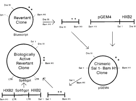
[image:2.612.154.459.70.220.2]Construction of biologically active clones.The two-step scheme used to con-struct infectious molecularly cloned viruses containing the original Thr-582 and FIG. 1. Location of primers used for amplification of revertant envelope DNA. Amino acid position 582 is marked by an ‘‘3’’ and position 673 is marked by an ‘‘O.’’ The insert shows the DNA products of the first (lane 3) and second (lane 1) PCR amplifications. The reaction of lane 2 contained no DNA. Migration of standard kilobase markers is indicated on the left, and the calculated size of DNA products is on the right.
on November 9, 2019 by guest
http://jvi.asm.org/
compensatory mutations is illustrated in Fig. 2. One microgram of the interme-diate vector DNA pGEM4 (Promega), containing the Sal1-BamHI fragment from the HXB2 env gene, was digested with DraIII (New England Biolabs, Beverly, Mass.) and BamHI. The DraIII-BamHI fragment of the revertant env, containing the two mutations, was then ligated into the cut intermediate vector. Positive plasmids were isolated by ampicillin selection in HB101-Max-competent cells as described above and selected further by miniprep analyses.
One microgram of the intermediate construct plasmid and pHXB2/RIP7 (13), an infectious proviral clone of HXB2, was digested with SalI and BamHI (Boehr-inger Mannheim), electrophoretically separated, and purified, the latter with the Gene Clean protocol (BI0 101, La Jolla, Calif.). The insert and vector were ligated, and positive clones were isolated by ampicillin selection and miniprep restriction analyses as described above. In order to verify that only the two point mutations were present, both strands of the envelope gene of the infectious molecular clone designated HXB2thr582ser673 were sequenced as described above for the revertant clones after PCR amplification of DNA by a series of primer pairs, each spanning 150 to 200 bp of the gene.
RESULTS
Reversion of the HXB2thr582 mutant to neutralization
sen-sitivity.
Following transfection of HXB2thr582 into permissive
[image:3.612.58.296.66.243.2]cells, we have repeatedly observed that after prolonged periods
of culture in the absence of selecting serum, the HXB2thr582
variant loses its property of neutralization resistance to the
original selecting serum. In order to document this
phenome-non and to study the molecular and biologic bases for the
reversion, HXB2thr582 and HXB2 DNAs were transfected
into COS cells and progeny virus was transmitted to H9 cells.
The cultures were maintained without immune-selecting
se-rum, and the neutralization phenotypes of cell-free
superna-tant viruses were assessed periodically. HXB2thr582
main-tained its neutralization-resistant phenotype in the absence of
selecting serum for at least 6 weeks of culture as illustrated in
Fig. 3. With continued culture in the absence of selecting
serum, the virus produced gradually became more
neutraliza-tion sensitive. By 3 months of culture, transmission of cell-free
virus from the HXB2thr582 supernatant to H9 cells in the
presence of the original immune-selecting serum was
signifi-cantly retarded. In contrast, the kinetics of infection of H9 cells
in the presence of normal human serum by either the control
HXB2 virus or the passaged HXB2thr582 supernatant were
identical, indicating that the lessened rate of infection by
HXB2thr582 in the presence of selecting serum did not merely
reflect quantitatively less virus in the cell-free supernatant. By
4.5 months, virus produced in the HXB2thr582 culture was
completely sensitive to neutralization by the immune-selecting
serum, indicating total reversion to the neutralization
pheno-type of the parental HXB2 virus (Fig. 3).
Direct sequencing of the revertant DNA.
In order to
deter-mine the molecular basis for the reversion to parental
pheno-type, genomic DNA extracted from H9 cells producing the
revertant virus after 5 months of continuous culture was
am-plified by PCR. As illustrated in Fig. 1 (inset) a second
ampli-fication with nested primers resulted in a significantly greater
yield of DNA which facilitated direct sequencing. The
second-ary DNA product, composed of approximately 760 bp
encom-passing the 582 region of the envelope was sequenced directly
as described in Materials and Methods. The resulting
consen-sus sequence revealed that a simple back mutation consisting
of a single nucleotide change of A to G had occurred, resulting
in an alteration of the threonine codon to alanine at amino
acid position 582 (Fig. 4). However, inspection of the
sequenc-ing gel suggested that the revertant virus population was
mixed, as a proportion of the amplified DNA retained the A
(Fig. 5). An approximation of the relative amounts of revertant
DNA containing either the A or the back-mutated G was
carried out by quantitation with a PhosphorImager (Molecular
Dynamics, Sunnyvale, Calif.). As illustrated in Fig. 5, the back
mutation was approximately three times more abundant than
the parental genotype. The mixed genotype observed in the
amplified revertant DNA suggested that a compensatory
mu-tation elsewhere in the env gene might have occurred in some
of the virus population, also leading to neutralization
sensitiv-ity.
Clonal analysis of revertant DNA.
To further investigate the
mixed revertant population, a 2,159-bp region encompassing
most of the envelope gene including the portion encoding the
582 region (Fig. 1) was amplified by PCR and introduced into
a cloning vector. Eight clones were obtained and sequenced as
described in Materials and Methods. Of the eight clones, four
contained the A to G back mutation and four retained the A
which would preserve the threonine characteristic of the
vari-ant at position 582. Of the latter four clones, one contained a
stop codon and one lacked the BamHI site and were not
FIG. 2. Construction of proviral clone of revertant virus. A pGEM4 plasmid containing the SalI-BamHI portion of HXB2 was digested with DraIII and BamHI, purified, and used as a vector into which the DraIII-BamHI fragment from either HXB2thr582ser673 or HXB2arg521thr582 was inserted. The muta-tions coding for Thr-582 and Ser-673 are illustrated (p). From these chimeric SalI-BamHI clones, the SalI-BamHI fragment containing either HXB2thr 582ser673- or HXB2arg521thr582-derived sequence from DraIII to BamHI was purified and inserted into the large SalI-BamHI fragment of HXB2 to generate the biologically active revertant clone. These procedures are outlined schemat-ically.
FIG. 3. Loss of neutralization resistance of the HXB2thr582 variant to the original selecting serum. HXB2thr582 and HXB2 infectious DNAs were trans-fected into COS-1 cells, and the virus produced was transmitted to H9 cells by coculturing. The cultures were maintained in the absence of immune-selecting serum, and the neutralization phenotypes of cell-free virus preparations were assessed on the days indicated. The kinetics of secondary transmission of HXB2thr582 (Ç) and the parental virus HXB2 (E) to H9 target cells in the presence of normal human control serum (open symbols) or the original immune selecting serum (closed symbols) is illustrated.
on November 9, 2019 by guest
http://jvi.asm.org/
[image:3.612.317.549.70.190.2]examined further. The third clone, designated MUR18,
exhib-ited a G to A mutation which would result in a glycine to
arginine substitution at position 521 in the envelope protein
(Fig. 4). The fourth clone, MUR20, possessed a change of T to
C, resulting in alteration of a phenylalanine codon to a serine
codon at amino acid position 673 (Fig. 4).
In order to determine if the secondary mutations in MUR18
and MUR20 would compensate for the original alanine to
threonine substitution, the cloned envelope genes were
substi-tuted into the infectious molecular clone HXB2 as described
in Materials and Methods and Fig. 2. Following transfection
of the clone containing the MUR18 envelope into H9 cells,
no progeny virus was produced and this clone was not
exam-ined further. The clone containing the MUR20 envelope was
infectious. Sequencing of the complete MUR20 envelope gene
confirmed that only the two codon changes relative to HXB2
were present (not shown), and the clone was redesignated
HXB2thr582ser673. Neutralization analysis confirmed that the
compensatory mutation at position 673 restored neutralization
sensitivity to both the original immune-selecting serum and to
monoclonal antibody F105 (Fig. 6). The latter, a human
mono-clonal antibody specific for a conformational epitope
overlap-ping the CD4-binding region of gp120 (40, 41, 53), was
previ-ously shown to be highly discriminatory for HXB2 versus
HXB2thr582, neutralizing the former but not the latter (26).
Characterization of H9 cells infected with HXB2, HXB2thr582,
and HXB2thr582ser673.
The ability of the F105 monoclonal
antibody to bind to cells infected with HXB2, HXB2thr582, or
HXB2thr582ser673 was assessed by FACScan analysis. As
shown in Table 1, virus expression by each of the three infected
cell types was similar as judged by staining with a pooled
human serum seropositive for HIV. However, whereas
[image:4.612.59.297.71.247.2]ap-FIG. 4. Molecular analysis of the HXB2thr582 revertant. The parental HXB2 envelope protein is represented schematically at the top, with the fusion peptide (F), leucine zipper region (LZ), immunodominant region (I), a site hidden upon envelope oligomerization (O), and transmembrane region (TM) localized according to the systems in references 8, 10, 21, and 35. Base changes and deduced amino acid alterations occurring in the original variant HXB2thr582, the consensus revertant obtained by direct sequencing of PCR-amplified DNA, and two cloned envelopes, MUR18 and MUR20, are illustrated.
[image:4.612.325.543.74.189.2]FIG. 6. Compensatory mutation at position 673 restores neutralization sen-sitivity. The kinetics of infection of H9 cells by the viruses shown was determined in the presence of a normal human control serum (E), the original human serum used to select the HXB2thr582 escape mutant (F), and monoclonal antibody F105 (Ç). Virus expression was monitored by immune fluorescence using a monoclonal antibody specific for p24 as described in Materials and Methods.
TABLE 1. Binding of monoclonal antibody F105 to H9 cells infected with parental, neutralization-resistant variant, and
neutralization-sensitive revertant cloned virusesa
Virus % PHS
b
-positive cells
Binding of F105
% Positive cellsc
Relative affinity (mg/ml)d
HXB2 90 58 3.0
HXB2thr582 85 25 3.2
HXB2thr582ser673 87 35 3.2
a
Values represent the mean of three experiments.
b
PHS, pool of three human sera with broad HIV-1 envelope reactivity.
c
Normalized to percent positive cells stained with PHS.
d
Defined as the antibody concentration at which a 50% reduction in mean fluorescent intensity of cells stained by a dilution series of F105 antibody was observed.
FIG. 5. Consensus sequence of revertant virus and estimation of the extent of the mixed population. Three separate PCR amplifications of DNA from the revertant virus were sequenced. The inset shows a portion of the first sequencing gel in which a mixture of adenine and guanine is present at a single site. The bar graph represents the results of a second sequencing of this region; the relative proportions of adenine and guanine were determined by phosphorimaging anal-ysis. A third sequencing also revealed a mixed population (not shown).
on November 9, 2019 by guest
http://jvi.asm.org/
[image:4.612.91.265.415.669.2] [image:4.612.315.555.599.678.2]proximately 60% of cells infected with HXB2 were able to bind
monoclonal antibody F105, only 25% of cells infected with
the neutralization-resistant variant were stained by the
anti-body. The compensatory mutation in the revertant
HXB2thr582ser673 partially restored recognition of the F105
epitope on the surfaces of cells expressing the viral envelope to
the level seen with HXB2-infected cells. The relative binding
affinity of F105 for the viral envelope expressed on the cell
surface as determined by FACScan analysis was similar for all
three infected cell types (Table 1).
In order to determine the nature of the selection pressure
for reversion of HXB2thr582 to the parental phenotype, the
levels of expression of viral proteins and the propensity of
infected cells to form syncytia were determined for H9 cells
infected with HXB2, HXB2thr582, and HXB2thr582ser673. As
shown in Table 2, supernatants from infected cultures were
assessed for p24 and gp120 production and for RT activity. The
levels of infectious virus production and protein expression by
H9 cells infected with HXB2, HXB2thr582, or HXB2thr582
ser673 were equivalent, indicating that the revertant virus did
not have a growth advantage. To further probe the basis for
revertant selection, the ability of the virus-infected cells to
form syncytia with CD4
1target cells was investigated. Cells
infected with HXB2thr582 consistently exhibited a greater
number of syncytia upon coculture with either CEM-SS,
SupT-1, or HUT 78 target cells (Table 2). Syncytium formation
by HXB2thr582-infected cells was 2.10 to 2.48 times greater
than that of HXB2-infected cells. HXB2thr582ser673-infected
cells exhibited an intermediate level of syncytium formation.
The HXB2thr582ser673 revertant appeared in a culture of
H9-infected cells. However, H9 cells as targets do not readily
form syncytia. Therefore, we explored the possibility that
in-creased cytopathic effect of the HXB2thr582 variant might also
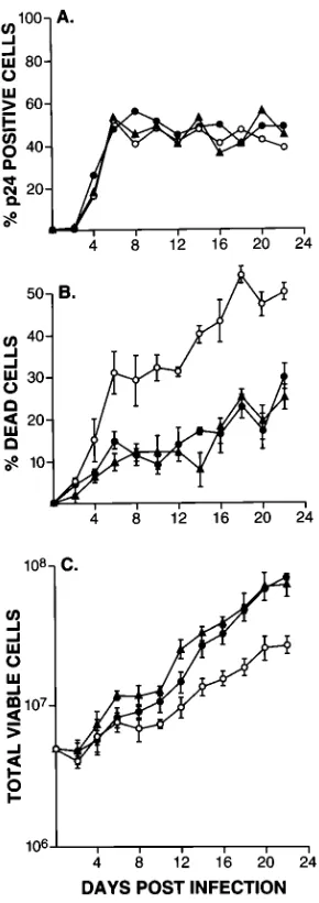
be expressed by greater single cell killing (4, 7, 47, 48). Figure
7 illustrates that this is the case. After infection with HXB2,
HXB2thr582, or HXB2thr582ser673, H9 cells reached stable
levels of productive infection within 6 days (Fig. 7A).
There-after, virus expression by the infected cell populations ranged
between 40 and 60% as measured by cells expressing the p24
core protein. Although the amounts of infecting virus were
similar and the same level of productive infection was achieved
by all three types of virus-infected cells, H9 cells infected with
HXB2thr582 exhibited much more extensive single cell killing
(Fig. 7B). As a result, HXB2- and HXB2thr582ser673-infected
H9 cells consistently showed a higher total viable cell count
beginning at approximately 1.5 weeks postinfection (Fig. 7C).
Although the experiment was terminated after 3 weeks of
culture, it illustrates how the HXB2thr582 variant would be
selected against in an in vitro system lacking any
immune-selective pressure once the back and compensatory mutations
were introduced into the viral population.
DISCUSSION
[image:5.612.362.507.68.478.2]The HXB2thr582 neutralization-resistant variant exhibits an
unstable phenotype in the absence of immune-selecting serum.
Here we showed that the molecular basis for reversion to
neutralization sensitivity is due, in part, to a back mutation at
position 582. This result reflects the highly conserved nature of
the 582 region and its functional importance. However, we also
showed that reversion can be due to a compensatory change
outside the 582 region. Compensatory changes can affect viral
functions such as infectivity and cell tropism (33, 60, 61). But
FIG. 7. Single cell killing by the parental, neutralization-resistant variant, and revertant viruses. (A) Kinetics of infection of H9 cells by the three viruses is shown. Virus infection was monitored by IFA for p24 expression. (B) Percentage of dead cells plus or minus the standard deviation. (C) Total number of viable cells plus or minus the standard deviation. Symbols:F, HXB2;E, HXB2thr582;
å, HXB2thr582ser673. The experiment was repeated three times with similar results. A representative experiment is shown.
a
Results (average of two to four experiments) are expressed relative to the value for the HXB2 parental virus.
b
The number of infected cells was first calculated based on the percentage of cells in the culture expressing p24 by IFA. Syncytium formation per 103
p24-positive infected cells was subsequently calculated and normalized to HXB2 values.
cND, not determined.
on November 9, 2019 by guest
http://jvi.asm.org/
[image:5.612.58.297.101.188.2]HIV escape mutants perhaps best exemplify the effects of point
mutations on conformational changes and their resultant
in-fluence on functions at distant sites. While some mutants
es-cape because of amino acid alterations within a neutralizing
epitope, either linear (31) or conformational (9), others,
in-cluding the HXB2thr582 variant, escape via conformational
changes affecting a site which is not part of the neutralizing
epitope (26, 34, 56, 62). Here we have shown that the impaired
recognition of the conformationally dependent CD4-binding
site by neutralizing human polyclonal and monoclonal
antibod-ies induced by the alanine to threonine substitution at amino
acid position 582 can be functionally restored on cell-free
viri-ons by a compensating alteration of phenylalanine to serine at
position 673.
Evidence of impaired recognition of the F105 epitope on
cells expressing HXB2thr582 compared with HXB2 and an
intermediate level of binding to cells expressing HXB2thr
582ser673 was obtained by FACScan analysis (Table 1).
How-ever, these results suggest that envelope conformation on the
surfaces of virions must differ from that of envelope expressed
on cell surfaces, because the decrease in F105 binding to
HXB2thr582 does not seem sufficient to account for the
re-markable ability of F105 to discriminate the parental from the
neutralization-resistant virus in a functional assay. More than
81-fold more F105 is necessary to neutralize cell-free
HXB2thr582 infection than that required to neutralize HXB2
infection (26). Moreover, the small increase in binding of F105
to cells expressing HXB2thr582ser673 compared with those
expressing HXB2thr582 does not correlate with the complete
restoration of functional F105 antibody activity observed with
the compensatory mutation (Fig. 6). A similar lack of
quanti-tative differences in binding to cell surface-expressed envelope
by other neutralizing monoclonal antibodies which
discrimi-nate between HXB2 and HXB2thr582 virions was previously
reported (26).
Virologic characterization of cells infected with the parental
HXB2 virus, the original HXB2thr582 virus, and the HXB2thr
582ser673 virus suggested that the in vitro pressure responsible
for reversion to the parental phenotype resulted from selection
for a less cytopathic virus. Two distinct mechanisms lead to
cytopathic effect in HIV-infected cells: syncytium formation
and single cell killing (7). We showed that both mechanisms
lead to increased cytopathicity of the HXB2thr582 variant
(Ta-ble 2 and Fig. 7). The observed increase in syncytium
forma-tion by HXB2thr582 differs from the result of Thali et al., who
reported that the Thr-582 mutation leads to a decreased ability
to form syncytia (52). The different result may be due to
dif-ferences in the systems studied. We used infectious
molecu-larly cloned viruses to study natural infection of target cells,
while Thali et al. use a complementation assay to study the
effects of either wild-type or mutant envelope expression. In
addition, we normalized the numbers of syncytia formed to the
number of cells in each infected population actually expressing
virus (Table 2). This was not done in our previous study (62),
cited by Thali et al. (52), and thus cannot be compared with the
results reported here.
In an in vitro tissue culture system in which HIV proviral
DNA is integrated into a proportion of immortalized T cells, a
highly cytopathic virus would be at a disadvantage. Cells
con-taining such a provirus would more likely die, while a more
benign viral type would be favored and tend to accumulate.
The overall dynamics of the system and length of time for a
revertant virus to be selected would depend on the proportion
of cells initially infected and expressing virus and on the cell
doubling time. A similar selection pressure probably does not
operate in vivo, however. In the absence of immortalization,
cells containing more benign proviruses would not tend to
accumulate. In fact, selection for a more virulent virus might
occur, as such a virus would tend to infect greater numbers of
susceptible T cells. This speculation is supported by numerous
reports illustrating the shift in viral characteristics from initially
‘‘slow-low,’’ non-syncytium-inducing, macrophage-tropic strains
early in infection to ‘‘rapid-high,’’ syncytium-inducing, T-tropic
strains later in disease progression (3, 15, 16, 51).
The nature of the molecular and biologic changes which
occurred in the immune-selected variant and the more benign
revertant illustrates important functional interactions between
gp120 and gp41 envelope components and highlights several
envelope domains believed to play a role in fusion events. The
precise mechanism by which HIV fuses with cells is not known.
The process is pH independent and thus involves direct fusion
of the virus envelope with the cell membrane (49). Whereas
gp41 by itself can induce fusion (37), the natural process
in-volves the initial binding of gp120 to the CD4 receptor,
fol-lowed by a conformational change which presumably uncovers
the gp41 fusion domain, allowing fusion to occur. This
mech-anism may ensure the maintenance of viral tropism by
prevent-ing promiscuous fusion with cells lackprevent-ing the CD4 receptor
(37). In addition to the CD4-binding region, other gp120
re-gions influence virus-cell fusion, including V1/V2 (50) and V3
(5, 19, 22, 36, 54). The actual fusion peptide, however, is
thought to be located at the amino terminus of gp41 (20).
Mutations in this region have led to decreased viral fusion (5,
7, 14, 18, 28, 29). Other gp41 domains influence fusion events
and viral function, however, including the leucine zipper,
en-compassing position 582 (6, 10); the transmembrane region
and residues proximal to it (24); and the 673 region (6). This
latter region has been shown to be inaccessible to specific
antipeptide antibodies upon oligomerization of the viral
enve-lope (35). However, oligomerization does not require the 673
region and can occur with only the first 129 amino-terminal
residues of gp41 present (11). In fact, gp41 amino acids 68 to
129, encompassing the 582 region, are critical for envelope
oligomerization (12). Further functional importance of the 582
and 673 regions has been demonstrated in recent studies in
which synthetic peptides representative of both regions have
displayed potent antiviral activities and inhibition of fusion (25,
57–59). These same two regions are postulated to interact and
play a critical role in the fusion process and viral entry (30).
The gp41 regions discussed are illustrated in Fig. 4.
We are intrigued that the mutations observed during the
progression of the parental HXB2 virus through the
neutral-ization-resistant variant to the spontaneous revertant involve
the CD4-binding region in gp120 and the 582 and 673 regions
in gp41, all believed to participate in fusion events.
Further-more, the changes observed in the fusion abilities of the three
viruses lend support to the proposed functional roles of the
three envelope regions. The initial variant selection resulted in
the neutralization-resistant HXB2thr582 which had a greater
propensity for forming syncytia (Table 2). The mutation in
gp41 altered the CD4-binding region of the viral envelope such
that recognition by conformationally dependent antibodies
specific for the region was impaired (26). Binding to CD4 is a
first step in the fusion process. Our results thus emphasize the
functional importance of gp120-gp41 interactions in the initial
events of viral infection.
The subsequent compensating change at position 673 which
restored neutralization sensitivity to CD4-binding site
antibod-ies and decreased syncytium formation to wild-type levels
il-lustrates the interplay between the 582 and 673 envelope
re-gions and indirectly supports previous findings implicating
both regions in the fusion process. Helseth et al. (24) have
on November 9, 2019 by guest
http://jvi.asm.org/
ing impaired. Alterations at positions 585 and 586 have
re-sulted in noninfectious viruses (62), and only conservative
amino acid substitutions are permitted at position 573 in order
for infectivity to be maintained (10). Position 582 can
accom-modate several amino acids and still retain infectivity (62). The
biochemical basis for threonine-induced neutralization
resis-tance by a conformational alteration involving the
CD4-bind-ing site is unknown, however. Because alanine, serine, and
glycine residues at position 582 lead to neutralization-sensitive
viruses (62), it has been suggested that a bulkier residue, such
as the threonine, might also cause neutralization resistance
(27). However, substitution of valine at position 582 of HXB2
produced a neutralization-sensitive virus (55a).
Cao et al. (6) have reported that, unlike our observations
here, substitution of phenylalanine with a proline residue at
position 673 led to increased syncytium-forming ability. In our
case, substitution of the phenylalanine with serine produced a
potential glycosylation site (NXT/S) (Fig. 4). Additional
exper-iments are necessary to determine whether glycosylation at this
site mediates the decrease in syncytium formation of the
re-vertant and the effect of the serine substitution by itself on
syncytium formation.
Overall, these studies highlight the conformational aspects
of immune escape and demonstrate the clear interaction
be-tween gp41 and gp120, which is important for viral function.
They illustrate that immune-selective pressure is not the only
force contributing to viral variability. Finally, they show the
interplay occurring between envelope domains which influence
virus-cell fusion and emphasize the plasticity and adaptability
of the viral envelope.
ACKNOWLEDGMENTS
We thank Anna Mazzuca for editorial assistance. The following reagent was obtained through the AIDS Research and Reference Reagent Program, Division of AIDS, NIAID, NIH: F105 monoclonal antibody from Marshall Posner.
REFERENCES
1. Albert, J., B. Abrahamsson, K. Nagy, E. Aurelius, H. Gaines, G. Nystrom,
and E. M. Fenyo.1990. Rapid development of isolate-specific neutralizing antibodies after primary HIV-1 infection and consequent emergence of virus variants which resist neutralization by autologous sera. AIDS 4:107–112. 2. Arendrup, M., C. Nielsen, J.-E. S. Hansen, C. Pedersen, L. Mathiesen, and
J. O. Nielsen.1992. Autologous HIV-1 neutralizing antibodies: emergence of neutralization-resistant escape virus and subsequent development of escape virus neutralizing antibodies. J. Acquired Immune Defic. Syndr. 5:303–307. 3. Åsjo¨, B., L. Morfeldt-Månson, L. J. Albert, G. Biberfeld, A. Karlson, K. Lidman, and E. M. Fenyo¨.1986. Replicative capacity of human immunode-ficiency virus from patients with varying severity of HIV infection. Lancet
ii:660–662.
4. Bergeron, L., and J. Sodroski. 1992. Dissociation of unintegrated viral DNA accumulation from single-cell lysis induced by human immunodeficiency virus type 1. J. Virol. 66:5777–5787.
5. Bergeron, L., N. Sullivan, and J. Sodroski. 1992. Target cell-specific determi-nants of membrane fusion within the human immunodeficiency virus type 1 gp120 third variable region and gp41 amino terminus. J. Virol. 66:2389–2397. 6. Cao, J., L. Bergeron, E. Helseth, M. Thali, H. Repke, and J. Sodroski. 1993.
leucine zipper of the human immunodeficiency virus type 1 transmembrane glycoprotein affect fusion and infectivity. J. Virol. 66:4748–4756. 11. Earl, P. L., R. W. Doms, and B. Moss. 1990. Oligomeric structure of the
human immunodeficiency virus type 1 envelope glycoprotein. Proc. Natl. Acad. Sci. USA 87:648–652.
12. Earl, P. L., and B. Moss. 1993. Mutational analysis of the assembly domain of the HIV-1 envelope glycoprotein. AIDS Res. Hum. Retroviruses 9:589– 594.
13. Feinberg, M. B., R. F. Jarrett, A. Aldovini, R. C. Gallo, and F. Wong-Staal. 1986. HTLV-III expression and production involve complex regulation at the levels of splicing and translation of viral RNA. Cell 46:807–817.
14. Felser, J. M., T. Klimkait, and J. Silver. 1989. A syncytia assay for human immunodeficiency virus type 1 (HIV-1) envelope protein and its use in studying HIV-1 mutations. Virology 170:566–570.
15. Fenyo¨, E. M., J. Albert, and B. Åsjo¨.1989. Replicative capacity, cytopathic effect and cell tropism. AIDS 3:S5–S12.
16. Fenyo¨, E. M., L. Morfeldt-Månson, F. Chiodi, B. Lind, A. von Gegerfelt, J. Albert, E. Olausson, and B. Åsjo¨.1988. Distinct replicative and cytopathic characteristics of human immunodeficiency virus isolates. J. Virol. 62:4414– 4419.
17. Fisher, A. G., E. Collalti, L. Ratner, R. C. Gallo, and F. Wong-Staal. 1985. A molecular clone of HTLV-III with biologic activity. Nature (London)
316:262–265.
18. Freed, E. O., D. J. Myers, and R. Risser. 1990. Characterization of the fusion domain of the human immunodeficiency virus type 1 envelope glycoprotein gp41. Proc. Natl. Acad. Sci. USA 87:4650–4654.
19. Freed, E. O., D. J. Myers, and R. Risser. 1991. Identification of the principal neutralizing determinant of human immunodeficiency virus type 1 as a fusion domain. J. Virol. 65:190–194.
20. Gallaher, W. R. 1987. Detection of a fusion peptide sequence in the trans-membrane protein of human immunodeficiency virus. Cell 50:327–328. 21. Gallaher, W. R., J. M. Ball, R. F. Garry, M. C. Griffin, and R. C. Montelaro.
1989. A general model for the transmembrane proteins of HIV and other retroviruses. AIDS Res. Hum. Retroviruses 5:431–440.
22. Grimaila, R. J., B. A. Fuller, P. D. Rennert, M. B. Nelson, M.-L.
Hammar-skjo¨ld, B. Potts, M. Murray, S. D. Putney, and G. Gray.1992. Mutations in the principal neutralization determinant of human immunodeficiency virus type 1 affect syncytium formation, virus infectivity, growth kinetics, and neutralization. J. Virol. 66:1875–1883.
23. Hahn, B. H., M. A. Gonda, G. M. Shaw, M. Popovic, J. A. Hoxie, R. C. Gallo,
and F. Wong-Staal.1985. Genomic diversity of the acquired immune defi-ciency syndrome virus HTLV-III: different viruses exhibit greatest diver-gence in their envelope genes. Proc. Natl. Acad. Sci. USA 82:4813–4817. 24. Helseth, E., U. Olshevsky, D. Gabuzda, B. Ardman, W. Haseltine, and J.
Sodroski.1990. Changes in the transmembrane region of the human immu-nodeficiency virus type 1 gp41 envelope glycoprotein affect membrane fu-sion. J. Virol. 64:6314–6318.
25. Jiang, S., K. Lin, N. Strick, and A. R. Neurath. 1993. HIV-1 inhibition by a peptide. Nature (London) 365:113.
26. Klasse, P. J., J. A. McKeating, M. Schutten, M. S. Reitz, Jr., and M.
Robert-Guroff.1993. An immune-selected point mutation in the transmem-brane protein of human immunodeficiency virus type 1 (HXB2-Env: Ala(3Thr)) decreases viral neutralization by monoclonal antibodies to the CD4-binding site. Virology 196:332–337.
27. Klasse, P. J., R. Pipkorn, J. Blomberg, K.-H. Han, B. Hilton, and J. A.
Ferretti.1993. Three-dimensional structure and antigenicity of transmem-brane-protein peptides of the human immunodeficiency virus type 1: effects of a neutralization-escape substitution. FEBS Lett. 323:68–72.
28. Kowalski, M., L. Bergeron, T. Dorfman, W. Haseltine, and J. Sodroski. 1991. Attenuation of human immunodeficiency virus type 1 cytopathic envelope glycoproteins. J. Virol. 65:281–291.
29. Kowalski, M., J. Potz, L. Basiripour, T. Dorfman, W. C. Goh, E. Terwilliger,
A. Dayton, C. Rosen, W. Haseltine, and J. Sodroski.1987. Functional regions of the envelope glycoprotein of human immunodeficiency virus type 1. Sci-ence 237:1351–1355.
30. Matthews, T. J., C. Wild, C.-H. Chen, D. P. Bolognesi, and M. L. Greenberg. 1994. Structural rearrangements in the transmembrane glycoprotein after receptor binding. Immunol. Rev. 140:93–104.
on November 9, 2019 by guest
http://jvi.asm.org/
31. McKeating, J. A., J. Gow, J. Goudsmit, L. H. Pearl, C. Mulder, and R. A.
Weiss. 1989. Characterization of HIV-1 neutralization escape mutants. AIDS 3:777–784.
32. Meltzer, S. J., S. M. Mane, P. K. Wood, L. Johnson, and S. W. Needleman. 1990. Sequencing products of the polymerase chain reaction directly, without purification. BioTechniques 8:142–148.
33. Morris, J. F., E. J. Sternberg, L. Gutshall, S. R. Petteway, Jr., and L. A.
Ivanoff.1994. Effect of a single amino acid substitution in the V3 domain of the human immunodeficiency virus type 1: generation of revertant viruses to overcome defects in infectivity in specific cell types. J. Virol. 68:8380–8385. 34. Nara, P. L., and J. Goudsmit. 1991. Neutralization resistant variants of HIV-1 escape via the hypervariable immunodominant V3 region (303-331 aa): evidence for a conformational neutralization epitope, p. 297–306. In R. A. Lerner, H. Ginsberg, R. M. Chanock, and F. Brown (ed.), Vaccines 90. Cold Spring Harbor Laboratory, Cold Spring Harbor, N.Y.
35. Neurath, A. R., N. Strick, and S. Jiang. 1992. Synthetic peptides and anti-peptide antibodies as probes to study interdomain interactions involved in virus assembly: the envelope of the human immunodeficiency virus (HIV-1). Virology 188:1–13.
36. Page, K. A., S. M. Stearns, and D. R. Littman. 1992. Analysis of mutations in the V3 domain of gp160 that affect fusion and infectivity. J. Virol. 66:524– 533.
37. Perez, L. G., M. A. O’Donnell, and E. B. Stephens. 1992. The transmembrane glycoprotein of human immunodeficiency virus type 1 induces syncytium formation in the absence of the receptor binding glycoprotein. J. Virol.
66:4134–4143.
38. Poiesz, B. J., F. W. Ruscetti, A. F. Gazdar, P. A. Bunn, J. D. Minna, and R. C.
Gallo.1980. Detection and isolation of type C retrovirus particles from fresh and cultured lymphocytes of a patient with cutaneous T-cell lymphoma. Proc. Natl. Acad. Sci. USA 77:7415–7419.
39. Popovic, M., M. G. Sarngadharan, E. Read, and R. C. Gallo. 1984. Detec-tion, isolation and continuous production of cytopathic retroviruses (HTLV-III) from patients with AIDS and pre-AIDS. Science 224:497–500. 40. Posner, M., T. Hideshima, T. Cannon, M. Mukherjee, K. H. Mayer, and
R. A. Byrn. 1991. An IgG human monoclonal antibody that reacts with HIV-1/gp120, inhibits virus binding to cells, and neutralizes infection. J. Immunol. 146:4325–4332.
41. Posner, M. R., H. Elboim, and D. Santos. 1987. The construction and use of a human-mouse myeloma analogue suitable for the routine production of hybridomas secreting human monoclonal antibodies. Hybridoma 6:611–625. 42. Reitz, M. S., Jr., C. Wilson, C. Naugle, R. C. Gallo, and M. Robert-Guroff. 1988. Generation of a neutralization-resistant variant of HIV-1 is due to selection for a point mutation in the envelope gene. Cell 54:57–63. 43. Robert-Guroff, M. 1990. Neutralizing antibodies, p. 179–185. In A. Aldovini
and B. D. Walker (ed.), Techniques in HIV research. Stockton Press, New York.
44. Robert-Guroff, M., M. Brown, and R. C. Gallo. 1985. HTLV-III neutralizing antibodies in patients with AIDS and AIDS-related complex. Nature (Lon-don) 316:72–74.
45. Robert-Guroff, M., A. Louie, M. Myagkikh, F. Michaels, M. P. Kieny, M. E.
White-Scharf, B. Potts, D. Grogg, and M. S. Reitz, Jr.1994. Alteration of V3 loop context within the envelope of human immunodeficiency virus type 1 enhances neutralization. J. Virol. 68:3459–3466.
46. Robert-Guroff, M., M. S. Reitz, W. G. Robey, and R. C. Gallo. 1986. In vitro generation of an HTLV-III variant by neutralizing antibody. J. Immunol.
137:3306–3309.
47. Somasundaran, M., and H. L. Robinson. 1987. A major mechanism of human immunodeficiency virus-induced cell killing does not involve cell fusion. J. Virol. 61:3114–3119.
48. Somasundaran, M., and H. L. Robinson. 1988. Unexpectedly high levels of
HIV-1 RNA and protein synthesis in a cytocidal infection. Science 242:1554– 1557.
49. Stein, B. S., S. D. Gowda, J. D. Lifson, R. C. Penhallow, K. G. Bensch, and
E. G. Engleman.1987. pH-Independent HIV entry into CD4-positive T cells via virus envelope fusion to the plasma membrane. Cell 49:659–668. 50. Sullivan, N., M. Thali, C. Furman, D. D. Ho, and J. Sodroski. 1993. Effect of
amino acid changes in the V1/V2 region of the human immunodeficiency virus type 1 gp120 glycoprotein on subunit association, syncytium formation, and recognition by a neutralizing antibody. J. Virol. 67:3674–3679. 51. Tersmette, M., R. E. Y. De Goede, B. J. M. Al, I. N. Winkel, R. A. Gruters,
H. T. Cuypers, H. G. Huisman, and F. Miedema.1988. Differential syncyti-um-inducing capacity of human immunodeficiency virus isolates: frequent detection of syncytium-inducing isolates in patients with acquired immuno-deficiency syndrome (AIDS) and AIDS-related complex. J. Virol. 62:2026– 2032.
52. Thali, M., M. Charles, C. Furman, L. Cavacini, M. Posner, J. Robinson, and
J. Sodroski.1994. Resistance to neutralization by broadly reactive antibodies to the human immunodeficiency virus type 1 gp120 glycoprotein conferred by a gp41 amino acid change. J. Virol. 68:674–680.
53. Thali, M., U. Olshevsky, C. Furman, D. Gabuzda, M. Posner, and J.
So-droski.1991. Characterization of a discontinuous human immunodeficiency virus type 1 gp120 epitope recognized by a broadly reactive neutralizing human monoclonal antibody. J. Virol. 65:6188–6193.
54. Travis, B. M., T. I. Dykers, D. Hewgill, J. Ledbetter, T. T. Tsu, S.-L. Hu, and
J. B. Lewis.1992. Functional roles of the V3 hypervariable region of HIV-1 gp160 in the processing of gp160 and in the formation of syncytia in CD41
cells. Virology 186:313–317.
55. Von Gegerfelt, A., J. Albert, L. Morfeldt-Månson, K. Broliden, and E. M.
Fenyo¨.1991. Isolate-specific neutralizing antibodies in patients with progres-sive HIV-1-related disease. Virology 185:162–168.
55a.Watkins, B., et al. Unpublished observations.
56. Watkins, B., M. S. Reitz, Jr., C. A. Wilson, K. Aldrich, and M. Robert-Guroff. 1993. Immune escape by human immunodeficiency virus type 1 from neu-tralizing antibodies: evidence for multiple pathways. J. Virol. 67:7493–7500. 57. Wild, C., T. Greenwell, and T. Matthews. 1993. A synthetic peptide from HIV-1 gp41 is a potent inhibitor of virus mediated cell-cell fusion. AIDS Res. Hum. Retroviruses 9:1051–1053.
58. Wild, C., T. Oas, C. McDanal, D. Bolognesi, and T. Matthews. 1992. A synthetic peptide inhibitor of human immunodeficiency virus replication: correlation between solution structure and viral inhibition. Proc. Natl. Acad. Sci. USA 89:10537–10541.
59. Wild, C. T., D. C. Shugars, T. K. Greenwell, C. B. McDanal, and T. J.
Matthews.1994. Peptides corresponding to a predictive alpha-helical do-main of human immunodeficiency virus type 1 gp41 are potent inhibitors of virus infection. Proc. Natl. Acad. Sci. USA 91:9770–9774.
60. Willey, R. L., E. K. Ross, A. J. Buckler-White, T. S. Theodore, and M. A.
Martin.1989. Functional interaction of constant and variable domains of human immunodeficiency virus type 1 gp120. J. Virol. 63:3595–3600. 61. Willey, R. L., D. H. Smith, L. A. Lasky, T. S. Theodore, P. L. Earl, B. Moss,
D. J. Capon, and M. A. Martin.1988. In vitro mutagenesis identifies a region within the envelope gene of the human immunodeficiency virus that is critical for infectivity. J. Virol. 62:139–147.
62. Wilson, C., M. S. Reitz, Jr., K. Aldrich, P. J. Klasse, J. Blomberg, R. C.
Gallo, and M. Robert-Guroff.1990. The site of an immune-selected point mutation in the transmembrane protein of human immunodeficiency virus type 1 does not constitute the neutralization epitope. J. Virol. 64:3240–3248. 63. Wong-Staal, F., G. M. Shaw, B. H. Hahn, S. Z. Salahuddin, M. Popovic, P.
Markham, R. Redfield, and R. C. Gallo.1985. Genomic diversity of human T-lymphotropic virus type III (HTLV-III). Science 229:759–762.