Latency-Associated Transcript Promoter
ANTHONY T. DOBSON,1TODD P. MARGOLIS,2WILLIAM A. GOMES,1
ANDLAWRENCE T. FELDMAN1*
Department of Microbiology and Immunology, School of Medicine, University of California at Los Angeles, Los Angeles, California 90024,1and F. I. Proctor Foundation and the Department of Ophthalmology,
University of California at San Francisco, San Francisco, California 941432
Received 7 October 1994/Accepted 27 December 1994
During herpes simplex virus latency, transcripts accumulate from a single transcription unit of the viral genome. The promoter for these latency-associated transcripts (LAT) has been located, and a number of studies have documented the specific regions of this promoter which are important in transient assays of neuronal cells in culture. To examine the regulation of this promoter from the viral genome, both in vitro and
in vivo, a series of seven promoter deletion viruses which drive the expression of the reporter geneb
-galac-tosidase was constructed. Rabbit skin cells were infected in cell culture with viruses bearing each promoter mutation, and the LAT promoter activity was compared with that obtained by infecting two neuronal cell lines, ND7 cells and C1300 neuroblastoma cells. Mouse dorsal root ganglia were also infected with these recombinant
viruses by footpad inoculations, andb-galactosidase activity was measured. Infected neuronal cells lines and
dorsal root ganglia exhibit much more LAT promoter activity than infected rabbit skin cells, suggesting that
the region upstream of2250 may contain one or several neuronal specific DNA-binding sites. However, a
comparison of LAT promoter activities within the deletion series revealed many differences between neurons of the dorsal root ganglia infected in vivo and the two neuronal cell lines infected in vitro. These results suggest that neurons may vary extensively in the quantity or kind of transcription factors they contain.
Herpes simplex virus (HSV) can infect and remain latent in a variety of peripheral and central nervous system neurons (5, 9, 27). During latency, lytic cycle genes are not expressed and RNA is detectable from a single region of the viral genome (6, 7, 26, 30, 31). We and others (10, 35) have further defined this region as a single transcription unit of 8.3 kb originating in the long repeat and crossing the joint region. The RNAs accumu-lating from this region appear as two to three bands on North-ern (RNA) blots ranging in size between 1.45 and 2.0 kb (32, 33) and are referred to as the latency-associated transcripts (LAT). These RNAs accumulate as stable introns in the nuclei of latently infected cells and are antisense to part of the third exon of the ICP0 mRNA (8, 11, 21, 23). A number of labora-tories have shown that recombinant viruses from which the LAT promoter has been deleted are inefficient at reactivating from the latent state compared with wild-type viruses (4, 13, 16). This suggests a biological function for some entity within the LAT transcription unit, and genetic studies are under way in several laboratories to try to define more precisely the func-tion required for reactivafunc-tion.
The TATA box for the LAT promoter or latency-associated
promoter (LAP promoter) is located 688 bp upstream of the 59
end of the stable LAT intron (10, 24, 34). Deletion of this TATA box and other upstream elements in recombinant vi-ruses causes the virus to be unable to express LAT region transcripts during a latent infection (10, 22). In addition, a
recombinant virus containing an insertion of ab-globin gene
just downstream of this TATA box synthesizesb-globin RNA
and not LAT RNA during latency (10). Transfection experi-ments with plasmid DNAs locating the LAT promoter to this
region are in agreement with the in vivo data (1, 2, 34). Finally, it is clear that the TATA box and other upstream elements control transcription of the entire transcriptional unit during latency, i.e., during latency there is no evidence for internal start sites. For example, removal of a 201-bp PstI fragment containing the TATA box and other upstream elements leaves approximately 600 bases of DNA upstream of the LAT intron; this virus was unable to make the LAT intron, a very stable
RNA, during latency (10). Second, insertion of the b-globin
gene immediately downstream of the TATA box leaves about
650 bp of DNA contiguous with and upstream of the 59end of
the LAT intron; this virus also does not make LAT during latency (10). Third, deletion of the TATA box and other up-stream promoter elements prevents the accumulation of RNAs outside the intron, the so-called minor LAT RNAs (22).
With these data in mind, we set out to investigate the fea-tures of the LAT promoter which cause it to be uniquely active during latency among all of the more than 70 HSV type 1 (HSV-1) promoters. Our approach has been to construct
re-combinant viruses containing the b-galactosidase gene as a
reporter. This approach has the advantage of examining tran-scription from the viral chromosome in neurons in vivo. During the course of these experiments, a number of reports have been published concerning the activity of the LAT promoter during transient transfections into various cell lines. In vitro studies of the LAT promoter performed by Batchelor and O’Hare have identified regions necessary for constitutive ex-pression as well as enhanced exex-pression in neuroblastomas relative to HeLa cells (1, 2). Leib and colleagues have shown that a functional Cyclic AMP (cAMP)-responsive element ex-ists immediately upstream of the LAT TATA box (16, 24). Zwaagstra et al. (36) have identified a protein, the LAT pro-moter-binding factor (LPBF), which can enhance transcription in both neuronal and nonneuronal cells. They have also shown that regions farther upstream appear to up-regulate promoter
* Corresponding author. Phone: (310) 1014. Fax: (310) 206-3865. Electronic mail address: [email protected]. edu.
2264
on November 9, 2019 by guest
http://jvi.asm.org/
activity in neuronally derived cells but down-regulate it in nonneuronal cells (35).
In order to examine the role of various regions of the
laten-cy-associated promoter, we constructed a series of 59
promot-er-deletion viruses which drive the expression of the reporter
gene b-galactosidase. These promoter-reporter constructs
were placed at the glycoprotein C locus to remove them from the possible influence of the repeat region or potential en-hancer elements. We reported previously that the constructs used here are active in neurons which are phenotypically latent during acute ganglionic infection, in that the LAT promoter is active, but they do not express viral antigen (20). The latent-specific activity of the LAT promoter allows us to examine a promoter deletion series in vivo and compare the results with those from infection of commonly used neuronal cells in vitro. LAT promoter activity in dorsal root ganglia was different from infection of commonly used neuronal cell lines. LAT promoter activity also differed significantly from what has been observed previously in plasmid transfection of neuronal cell lines (2). These results suggest that neurons of the dorsal root ganglia may have a unique and different set of transcription factors compared with those in cell lines routinely used to assay LAT promoter function in vitro.
MATERIALS AND METHODS
Construction of plasmids and viral recombinants.The recombinant virus KOS/1 has been described previously (20). It was derived from the plasmid JA1, which contains theb-galactosidase reporter gene construct (from pCH110; Phar-macia) located within the glycoprotein C locus. The promoter consists of a
SmaI-to-SacII DNA restriction fragment which extends to bp2863 relative to the LAT transcription start site. The deletion series was constructed as outlined in Fig. 1. Six constructs were selected according to the size of the promoter fragment by agarose-gel electrophoresis. Sequencing of alkali-denatured plas-mids using the Sequenase version 2.0 kit (U.S. Biochemical Corp.) was per-formed according to the manufacturer to determine the 59extent of the deletion. A primer (59-AGAACCCCGATTCCAATCC-39) which annealed to DNA im-mediately upstream of the deleted region, within the glycoprotein C sequence, was used. These constructs were then cotransfected with KOS(M) viral DNA, and recombinant viruses were isolated and plaque purified as previously de-scribed (9). Southern blot analysis (Fig. 2) was performed to confirm their structure (Fig. 3). All of the recombinant viruses are derivatives of the parental strain KOS(M); however, they have been named according to the size of the promoter fragment relative to that of the viral strain 171(16). KOS/1 has been renamed KOS863. The remainder of the deletion series were named as follows: KOS593, KOS506, KOS388, KOS252, KOS124, and KOS57 were derived from plasmids ATD120, ATD113, ATD114, ATD121, ATD116, and ATD117 respec-tively. The enzymes used in this study were obtained from Bethesda Research Laboratories, Boehringer Manheim Biochemicals, and New England Biolabs.
BglII and SpeI linkers were obtained from Pharmacia. Isotopes were obtained
from New England Nuclear and ICN Biomedicals, Inc.
Cells and viruses.Viral stocks were replicated in all cells maintained in minimal essential medium supplemented with fetal calf serum (0.5%), penicillin (250mg/ml), and fungizone (0.25 mg/ml). Viral stocks were stored at2708C prior to use. Recombinant viruses and KOS(M) replicated equally well in vitro and readily achieved titers ranging from 13109
to 53109
PFU/ml. The parental strain, KOS(M), typically replicated to somewhat higher titers within the ganglia of mice. This is presumably a result of its expression of the wild-type glycoprotein C status.
Animals and inoculations.Six-week-old female BALB/C or Swiss Webster
mice were inoculated with 4 3107
PFU of virus per footpad as described previously (15). Photographs were taken of ganglia from the Swiss Webster mice and are representative of all experiments. All other data was collected by using the BALB/C mice.
Histochemical analysis forb-galactosidase.Histochemical analysis was car-ried out as described previously (9). Briefly, the animals were sacrificed by CO2
inhalation and perfusion fixed, and their dorsal root ganglia (L3-S1) were re-moved. Histochemical staining of whole ganglia was performed with the reagent 5-bromo-4-chloro-3-indolyl-b-D-galactopyranoside (X-Gal; Sigma).
CPRG assay forb-galactosidase.Cellular extracts from either infected mice or transfected cell cultures were prepared as follows. (i) Following inoculation of mice, animals were sacrificed on day 4 by CO2inhalation and perfused with
phosphate-buffered saline (PBS). Dorsal root ganglia were removed, ground on ice in a 0.1-ml Dounce homogenizer, and stored in aliquots at2708C in a volume FIG. 1. Plasmid constructions. LH7, a parental plasmid containing a piece of
stuffer DNA, was opened at a unique SpeI site and digested with the bidirectional exonuclease BAL 31. The remaining stuffer DNA was removed by digestion with
BglII, the free ends of the DNA were blunted with T4 DNA polymerase, and a BglII linker was ligated into the plasmid deletion site. Dideoxy sequence analysis
was performed at the 59end of the promoter fragment from an oligonucleotide (open arrow) within the glycoprotein C gene.b-gal,b-galactosidase.
FIG. 2. Southern blot analysis of the viral recombinants. Viral DNA was digested with BglII and DraIII, electrophoresed on a 1% agarose gel, and blotted onto Magnagraph Nylon (MSI). A random-hexamer-primed probe fromb -ga-lactosidase (b-gal) was hybridized to the nylon membrane at 658C according to the method described by Maniatis et al. (18). Two bands hybridized as expected: a standard-size fragment exclusively from withinb-galactosidase and a second containing the deleted promoter region.
FIG. 3. Structures of the viral recombinants. As can be seen in the upper portion of the figure, a SmaI-to-SacII restriction fragment containing the LAT promoter was moved to the glycoprotein C locus cloned in front of ab -galacto-sidase (beta-gal) reporter gene construct (pCH110; Pharmacia). The deletion series is illustrated in the lower portion of the figure; the deletions are named according to their positions within strain 171. Several of the promoter elements are also shown. IRL, inverted repeat long; IRs, inverted repeat short; UL, unique
long region.
on November 9, 2019 by guest
http://jvi.asm.org/
times and microfuged to pellet cellular debris. For both the ganglionic and cell culture extracts, 10ml of supernatant was reacted with a solution containing 5 mM chlorophenol red–b–D-galactopyranoside (CPRG; Sigma), 50 mM potas-sium phosphate (pH 7.8), and 1 mM magnepotas-sium chloride in a total volume of 100 ml at 378C for 2 to 4 h. A573was measured. Transfections were normalized by
determining protein content with the Bio-Rad protein assay solution according to the manufacturer’s instructions.
RESULTS
Structure of the recombinant viruses.To facilitate in vivo
analysis of the LAT promoter, an 863-bp region consisting of the LAT TATA box and elements upstream of this was placed
in front of the b-galactosidase reporter gene construct and
inserted into HSV, disrupting the glycoprotein C gene. The
choice ofb-galactosidase as a reporter was based on the ease
of histochemical visualization of the signal, as shown by Ho and Mocarski (14). The decision to move an upstream DNA element to the glycoprotein C locus represented an attempt to isolate the promoter from the repeat region to determine if this piece of DNA could function to promote latent transcrip-tion.
A series of LAT promoter deletion viruses were subse-quently constructed. A stuffer DNA fragment was inserted
between the 59end of the LAT promoter and the 39end of the
glycoprotein C gene (Fig. 1). BAL 31 bidirectional deletions were performed on this DNA, which was subsequently digested
with BglII to remove the 59part of the stuffer DNA. Thus, each
of these deletions should resemble each other except at the 59
end of the LAT promoter. A series of plasmids was isolated. Following analysis by restriction digestion and sequencing, six plasmids were chosen for recombination into the viral strain KOS(M) at the glycoprotein C locus. Recombinant viruses
were screened by hybridization for the presence of theb
-ga-lactosidase gene, plaque purified at least three times, and an-alyzed by Southern blotting (Fig. 2). The structures of the resultant viral recombinants are illustrated in Fig. 3.
Histochemical analysis of dorsal root ganglia from mice
sacrificed 4 days postinoculation.Previous studies performed
by Margolis et al. (20) and Sawtell and Thompson (28) using the recombinant virus KOS/1 (KOS863) revealed distinct pop-ulations of neurons early in infection. Dual immunofluores-cence analysis of ganglionic sections of mice infected 4 days previously revealed that as many as 90% of the neurons
ex-pressing b-galactosidase were not expressing any detectable
viral antigen; conversely, most cells expressing viral antigen
were not expressingb-galactosidase. It was also noted that the
number of neuronal profiles expressing LAT did not signifi-cantly decrease between acute and latent time points (20, 28). This suggested that by day 4 postinoculation, the great majority of neurons had separated into two distinct populations: those in an acute phase of viral replication and those in the latent phase of viral infection. By combining in situ hybridization for the LAT with immunofluorescence for viral antigen, Speck and Simmons (29) were able to provide strong evidence for early divergence into acutely and latently infected neurons using wild-type HSV-1.
In order to assay LAT promoter activity, histochemical anal-ysis of dorsal root ganglia from mice sacrificed 4 days postin-oculation was performed. Promoter activity was measured by two methods. In the first method, whole mount ganglia were reacted with the chromogenic substrate X-Gal, and neurons
that expressedb-galactosidase turned blue (Fig. 4). With the
between bp 2388 and 2252 clearly specifies one or more
elements important for in vivo transcription. A more careful inspection of the ganglia photographed in Fig. 4 reveals more intensely stained neurons in KOS863-infected than in KOS593-infected ganglia. Although the numbers of blue sites
in each infection are similar, there appears to be moreb
-ga-lactosidase activity in the KOS863-infected neurons than in those infected with KOS593, which suggests that the additional region of DNA contained within KOS863 is also important for LAT transcription in vivo. This is seen more clearly by the
second method of measuringb-galactosidase activity.
In the second method, neuronal extracts at 4 days postin-oculation were reacted with CPRG, which forms a soluble product that can be measured by spectrophotometry. This method has the advantage of allowing quantitation of the
av-erage amount ofb-galactosidase activity per animal. As can be
seen in Fig. 5 (graph A), infection of mice with the largest promoter construct virus, KOS863, gives the greatest amount
of b-galactosidase activity. Extracts from mice infected with
KOS593, the next virus in the series, have an activity that is 40% of that of KOS863, a 2.5-fold decrease. KOS252, the fifth virus in the series, has only 2% of the activity of KOS863. The drop in X-Gal activity between KOS593 and KOS252 appears to be largely due to the 136-bp difference in promoter size between KOS388 and KOS252, consistent with the X-Gal data in Fig. 4. The drop in X-Gal activity between KOS388 and KOS252 represents a 15-fold decrease in activity. The activities of the extracts from the last two viral infections after KOS252 are too low to measure accurately. Therefore, we were unable to verify in vivo the apparent neuronal specificity which others (1, 35) have noted in this region by transient assays.
Specificity of expression within the ganglia.We have
previ-ously shown with KOS863 (KOS/1) that at 4 days postinfection, the infected neurons segregate into two distinct populations. Of interest here is that more than 90% of the neurons which
express the b-galactosidase gene from the LAT promoter do
not express any HSV-1 antigens and are thus phenotypically latent (20, 28). This is apparently due to the facts that the blue neurons are latently infected and that the productively infected neurons shut off the LAT promoter through autoregulation by the ICP4 protein (1, 3, 12). Although the LAT promoter in this
construct shuts off sometime during a latent infection, b
-ga-lactosidase activity can still be observed at 21 days postinfec-tion (our unpublished observapostinfec-tions). We have consistently ob-served by immunofluorescence staining that the productively infected neurons are cleared by day 8; thus, the neurons stain-ing at 21 days are clearly latently infected. Therefore, we at-tempted to verify that the expression that we observed for each of the viruses at 4 days postinfection was also similar at 21 days postinfection. Whole-mount ganglia were reacted with X-Gal, and the number of blue neurons was counted as described previously. There was an overall apparent decrease in the number and intensity of blue neurons at latent times
postin-oculation such that we were unable to quantitateb
-galactosi-dase activity at 21 days postinoculation by CPRG assays. How-ever, as measured by number of sites, the pattern of expression through the deletion series at 21 days postinoculation was similar to that at acute times on the basis of CPRG activity (Fig. 5, graph B). These data strongly suggest that the LAT promoter expression observed at day 4 for each promoter mutant was occuring in cells which had already established a latent infection.
on November 9, 2019 by guest
http://jvi.asm.org/
Thatb-galactosidase expression in vivo is driven by specific
promoter elements and is not due to spuriousb-galactosidase
expression is shown by several pieces of evidence. First, Ho and Mocarski, expressing the same gene from an early viral
pro-moter, reported nob-galactosidase expression at latent times
(14). Second, we have previously reported that the
neurofila-ment-L promoter, drivingb-galactosidase at gC, does not
ex-pressb-galactosidase during latency (20). Third, the deletion
series itself provides three internal control viruses, all from the
same original plasmid in gC, which express little or nob
-ga-lactosidase in vivo. Thus, the activity observed in these neurons is dependent on the presence of the upstream LAT promoter elements and is not dependent on the site of insertion into the viral DNA.
Efficiency of infection.Because decreases inb-galactosidase
activity could also be due to poor ganglionic infection, it was important to assess infection efficiency. This was performed by titrating the ganglionic extract for infectious virus by plaque assays. Table 1 shows that viral titers within the ganglia were equivalent among the recombinant viruses and thus cannot
account for any of the decreases inb-galactosidase activity.
In vitro infection of neuronal and nonneuronal cells in
cul-ture. A number of studies have been reported from other
laboratories on the activity of the LAT promoter as measured by transient transfection into various neuronal cell lines. Most of the commonly used neuronal cell lines are not of the same
lineage as neurons of the peripheral nervous system in which HSV establishes a latent infection, and thus these cell lines may vary considerably in the quantity and specificity of their transcription factors. In addition, cells in culture are rapidly dividing, as opposed to neurons of the dorsal root ganglia, which are postmitotic. It is possible that dividing cells also vary in the quantity of specific transcription factors compared with postmitotic cells. For these reasons, we decided to analyze the activity of each LAT promoter mutant in three cell lines and compare the results with those obtained from in vivo infections described above. For infection of a nonneuronal cell line, we have used rabbit skin cells, which is the cell line used to prop-agate the viruses described here. We also infected two different neuronal cell lines. ND7 cells are a fusion between mouse dorsal root ganglia and mouse neuroblastoma cells. This cell line has also been selected for its ability to inhibit the imme-diate-early gene expression upon infection, and it has been one of the cell lines used to study LAT promoter activity (36). The other neuronal cell line used in these studies is C1300 neuro-blastoma cells, a mouse neuroneuro-blastoma cell line of a different lineage than neurons of the peripheral nervous system such as dorsal root ganglia. It is important to note that the viral con-structs used in these infections retained the true LAT cap, which we and O’Hare have shown to be downregulated by the ICP4 gene product. The consequence of this is that the test promoters will be responsive mainly to the presence or absence
FIG. 4. Photomicrographs of whole mount dorsal root ganglia processed 4 days after footpad inoculation with the viral deletion series. Ganglia were stained with the chromogenic substrate X-Gal. Neurons expressingb-galactosidase appear as dark dots. (A) KOS863; (B) KOS593; (C) KOS506; (D) KOS388; (E) KOS252; (F) KOS124; (G) KOS57. Little to no expression could be seen in KOS252, KOS124, and KOS57, which appeared similar to the negative control [KOS(M)]-infected ganglia (data not shown).
on November 9, 2019 by guest
http://jvi.asm.org/
of transcription factors which can bind to the LAT promoter and will minimize the influence of the viral life cycle and viral DNA amplification on the pattern of expression. The results of in vitro infections with the panel LAT promoter mutants are shown in Fig. 6. As can be observed in Fig. 6A, the activity of the LAT promoter mutants in vivo, as measured by CPRG activity, decreases mainly between KOS863 and KOS593, with a more severe decline between KOS388 and KOS250. This is completely different from the pattern observed in infected rabbit skin cells, in which the promoter activity is very low throughout the deletion series and in which KOS124 exhibits the most promoter activity (Fig. 6A). Similar results were ob-tained by transfection of the promoter deletion plasmids into rabbit skin cells, that is, the total activity was very low, there was no significant decrease between KOS388 and KOS250, and the activity of the KOS124 promoter was relatively high com-pared with that observed in vivo (data not shown). For these reasons, we would suggest that the region of the LAT
pro-moter from 2863 through 2388 may contain transcription
factor-binding sites for neuron-specific or neuron-enhanced transcription factors.
However, a comparison of the apparent neuronal activity in vivo with that observed in two different cell lines in vitro sug-gests that there are many differences. The LAT promoter ac-tivity in infected ND7 cells differs from that observed in vitro
in two major areas. In the region between 2863 and 2593,
there is a significant decline in activity in vivo, whereas infected ND7 cells show an increase (Fig. 6B). Secondly, in the region
between2252 and2124, there is a small decline in vivo but a
very large increase in ND7 cells. However, the severe drop
between2388 and 2252 suggests the possibility at least that
there are common transcription factors which bind to this region. The LAT promoter activity in C1300 neuroblastoma cells is even more different, showing an increase between
re-gions2593 and2506 and a sharp decline between2506 and
2388, where none exists in vivo (Fig. 6B). Both neuronal cell
lines show a dramatic increase in transcription of KOS124, which is a far larger increase in transcription than that ob-served in vivo. This may reflect the binding of a factor or factors to this region which are common to dividing cells, since both neuronal cell types as well as rabbit skin cells showed this increase in promoter activity.
In addition to these differences between in vivo and in vitro
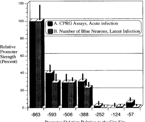
FIG. 5. (■), CPRG assay of ganglionic extracts from mice sacrificed 4 days postinoculation. Values are the means6standard errors of the means for three separate determinations performed on ganglionic extracts from a total of five mice per group, except for the KOS506 group, which had only three mice.3, average number of blue neurons 21 days postinoculation. Values are the means 6standard errors of the means and were determined from a total of five mice per group, except for the KOS124 group, which had only four mice. Values for both were plotted as percentages of the value of the largest promoter construct virus, KOS863.
[image:5.612.316.554.67.381.2]FIG. 6. (A) CPRG assay of ganglionic extracts prepared 4 days following footpad inoculation with the viral deletion series (white graphs). A 2.5-fold decrease in promoter activity is evident when the promoter size is decreased from bp2863 to2593. A 15-fold drop in activity occurs when the promoter is decreased in size from bp2388 to2252. Much less activity for KOS863 and other virus-infected cells occurs upon infection of rabbit skin cells in vitro (shaded graphs). KOS124 has the greatest activity of the promoter deletions. Data were calculated as described in the legend to Fig. 5. (B) The same promoter deletions are used to infect two different neuronal cell lines, ND7 (dark shaded squares) and C1300 (light shaded squares) neuroblastoma cells.
TABLE 1. Virus titers from ganglionic extracts processed 4 days after footpad inoculation with the viral recombinants
Virus Viral titer
(PFU/ml)
KOS863...9.83103
KOS593...4.53103
KOS506...6.73103
KOS388...5.23103
KOS252...6.83103
KOS124...1.13104
KOS57...7.53103
KOS(M)...2.33105
on November 9, 2019 by guest
http://jvi.asm.org/
[image:5.612.59.300.73.277.2] [image:5.612.58.297.628.725.2]expression, the LAT promoter in KOS252 represents a
dele-tion nearly identical to a bp2250 deletion plasmid described
by Batchelor and O’Hare (2). They reported this plasmid to have 98% of the activity of their largest promoter construct in IMR-32 neuroblastomas. By contrast, KOS252 expresses only approximately 2% of the activity of our largest promoter con-struct virus in vivo. Thus, there are many differences between the in vivo and the in vitro systems, indicating that the contri-bution of specific binding sites to LAT promoter activity should eventually be verified in vivo.
DISCUSSION
The LAT promoter is the sole promoter active during a latent infection. For this reason, a number of studies have focused on the LAT promoter and on potential sites on the promoter responsible for activity. All of these studies have utilized transfection assays of plasmid DNAs. Thus, Zwaagstra et. al. (36) have identified a protein, LPBF, which enhances transcription in both neuronal and nonneuronal cells. Leib and colleagues have identified a cAMP-responsive element imme-diately upstream of the LAT TATA box (16, 23). Batchelor and O’Hare (1, 2) have identified specific sites responsible for both constitutive and specific expression in neuroblastoma cells.
The recombinant viruses described here contain regulatory elements composed of no more than 863 bp of LAT promoter, and all of these sequences have been placed into the long unique region. It is apparent that these viruses do not contain
the full range of LAT promoter function, becauseb
-galacto-sidase activity is gradually lost over the course of several weeks. This suggests that the constructs are missing something re-quired for long-term expression, and recently we have obtained evidence suggesting that this region is downstream of the LAT transcription start site. First, other viral constructs not contain-ing the region downstream of the transcription start site turn off during latency (19, 20). Second, the region downstream of the transcription start site does not contribute apparent pro-moter activity, since in the LAT propro-moter deletion virus KOS29 this region is intact and no LAT intron is observed in latent infections of mouse dorsal root ganglia (10). Third, the addition of a similar upstream LAT promoter region to the long terminal repeat of Moloney murine leukemia virus was able to promote neuronal expression in vivo, initiating within the long terminal repeat when the LAT TATA box was re-moved (17). Since the long-terminal repeat itself did not con-tinue expression into latency, this demonstrates that the LAT promoter strength is derived from the upstream LAT pro-moter sequences, as shown also in the present study.
The present study has addressed transcription from the viral genome in vivo. The results from our studies suggest that two regions of the upstream 863 bases contribute to in vivo
func-tion. Of these, the more proximal region, between bp 2388
and 2252, clearly has neuron-specific elements. Important
transcription elements may also be present 39of bp2252, but
because of the low b-galactosidase levels generated by the
constructs in this region, we were unable to assay any differ-ences; in a second set of recombinant viruses will be necessary to examine these sequences in vivo. These results differ signif-icantly from those LAT for promoter activity as measured by infection of two neuronal cells in culture as well as by com-parison with transfections of these and other neuronal cell lines reported by others. The differences between our results in vivo and those obtained in vitro most likely can be attributed to the fact that the neuronal cells in culture do not accurately represent the kind or quantity of transcription factors that are
found in neurons of the peripheral nervous system. Our results also suggest the possibility that cells in culture contain a level of transcription factors which bind to the proximal part of the LAT promote higher than that found in neurons in vivo.
ACKNOWLEDGMENTS
We thank Mariwil G. Wong and Stephanie S. Liu for help with the animal studies, Sidne A. Omori for help with the in vitro transfections, Eric J. Black for technical assistance, James Adams for the construc-tion of plasmid JA1, and Lily Ho for the construcconstruc-tion of plasmid LH7. This work was supported by Public Health Service grants AI28338 and EY10008. L.T.F. is a recipient of Faculty Research award FRA-340 from the American Cancer Society, A.T.D. is supported by Public Health Service grant GM08042-06 from the National Institutes of Health to the UCLA Medical Scientist Training program, and T.P.M. is a recipient of a Research To Prevent Blindness career development award.
REFERENCES
1. Batchelor, A. H., and P. O’Hare. 1990. Regulation and cell-type-specific activity of a promoter located upstream of the latency-associated transcript of herpes simplex virus type 1. J. Virol. 64:3269–3279.
2. Batchelor, A. H., and P. O’Hare. 1992. Localization of cis-acting sequence requirements in the promoter of the latency-associated transcript of herpes simplex virus type 1 required for cell-type-specific activity. J. Virol. 66:3573– 3582.
3. Batchelor, A. H., K. W. Wilcox, and P. O’Hare. 1994. Binding and repression of the latency-associated promoter of herpes simplex virus by the immediate-early 175K protein. J. Gen. Virol. 75:753–767.
4. Bloom, D. C., G. B. Devi-Rao, J. M. Hill, J. G. Stevens, and E. K. Wagner. 1994. Molecular analysis of herpes simplex virus type 1 during epinephrine-induced reactivation of latently infected rabbits in vivo. J. Virol. 68:1283– 1292.
5. Cook, M. L., V. B. Bastone, and J. G. Stevens. 1974. Evidence neurons harbor latent herpes simplex virus. Infect. Immun. 9:946–951.
6. Croen, K. D., J. M. Ostrove, L. J. Dragovic, J. E. Smialek, and S. E. Straus. 1988. Latent herpes simplex virus in human trigeminal ganglia. Detection of an immediate-early gene ‘‘antisense’’ transcript by in situ hybridization. N. Engl. J. Med. 317:1427–1432.
7. Deatly, A. M., J. G. Spivack, E. Lavi, and N. W. Fraser. 1987. RNA from an immediate early region of the type 1 herpes simplex virus genome is present in the trigeminal ganglia of latently infected mice. Proc. Natl. Acad. Sci. USA
84:3204–3208.
8. Devi-Rao, G. B., S. A. Goodart, L. M. Hecht, R. Rochford, M. K. Rice, and
E. K. Wagner.1991. Relationship between polyadenylated and nonpolyade-nylated herpes simplex virus type 1 latency-associated transcripts. J. Virol.
65:2179–2190.
9. Dobson, A. T., T. P. Margolis, F. Sedarati, J. G. Stevens, and L. T. Feldman. 1990. A latent, nonpathogenic HSV-1-derived vector stably expresses beta-galactosidase in mouse neurons. Neuron 5:353–360.
10. Dobson, A. T., F. Sederati, G. Devi-Rao, W. M. Flanagan, M. J. Farrell, J. G.
Stevens, and L. T. Feldman.1989. Identification of the latency-associated transcript promoter by expression of rabbit beta-globin mRNA in mouse sensory nerve ganglia latently infected with a recombinant herpes simplex virus. J. Virol. 63:3844–3851.
11. Farrell, M. J., A. T. Dobson, and L. T. Feldman. 1991. Herpes simplex virus latency-associated transcript is a stable intron. Proc. Natl. Acad. Sci. USA
88:790–794.
12. Farrell, M. J., T. P. Margolis, W. A. Gomes, and L. T. Feldman. 1994. Effect of the cap region on acute expression of the herpes simplex virus type 1 latency-associated transcript promoter. J. Virol. 68:5337–5343.
13. Hill, J. M., F. Sedarati, R. T. Javier, E. K. Wagner, and J. G. Stevens. 1990. Herpes simplex virus latent phase transcription facilitates in vivo reactiva-tion. Virology 174:117–125.
14. Ho, D. Y., and E. S. Mocarski. 1988.b-Galactosidase as a marker in the peripheral and neural tissues of the herpes simplex virus-infected mouse. Virology 167:279–283.
15. Javier, R. T., J. G. Stevens, V. B. Dissette, and E. K. Wagner. 1988. A herpes simplex virus transcript abundant in latently infected neurons is dispensable for establishment of the latent state. Virology 166:254–257.
16. Leib, D. A., K. C. Nadeau, S. A. Rundle, and P. A. Schaffer. 1991. The promoter of the latency-associated transcripts of herpes simplex virus type 1 contains a functional cAMP-response element: role of the latency-associated transcripts and cAMP in reactivation of viral latency. Proc. Natl. Acad. Sci. USA 88:48–52.
17. Lokensgard, J. R., D. C. Bloom, A. T. Dobson, and L. T. Feldman. 1994. Long-term promoter activity during herpes simplex virus latency. J. Virol.
68:7148–7158.
on November 9, 2019 by guest
http://jvi.asm.org/
LAT promoter constructs. Virology 197:585–592.
20. Margolis, T. P., F. Sedarati, A. T. Dobson, L. T. Feldman, and J. G. Stevens. 1992. Pathways of viral gene expression during acute neuronal infection with HSV-1. Virology 189:150–60.
21. McGeoch, D. J., C. Cunningham, G. McIntyre, and A. Dolan. 1991. Com-parative sequence analysis of the long repeat regions and adjoining parts of the long unique regions in the genomes of herpes simplex viruses types 1 and 2. J. Gen. Virol. 72:3057–3075.
22. Mitchell, W. J., I. Steiner, S. M. Brown, A. R. MacLean, J. H. Subak-Sharpe,
and N. W. Fraser.1990. A herpes simplex virus type 1 variant, deleted in the promoter region of the latency-associated transcripts, does not produce any detectable minor RNA species during latency in the mouse trigeminal gan-glion. J. Gen. Virol. 71:953–957.
23. Perry, L. J., and D. J. McGeoch. 1988. The DNA sequences of the long repeat region and adjoining parts of the long unique region in the genome of herpes simplex virus type 1. J. Gen. Virol. 69:2831–2846.
24. Rader, K. A., C. E. Ackland-Berglund, J. K. Miller, J. S. Pepose, and D. A.
Leib.1994. In vivo characterization of site-directed mutations in the pro-moter of the herpes simplex virus type 1 latency-associated transcripts. J. Gen. Virol. 74:1859–1869.
25. Roberts, M. S., A. Boundy, P. O’Hare, M. C. Pizzorno, D. M. Ciufo, and G. S.
Hayward.1988. Direct correlation between a negative autoregulatory re-sponse element at the cap site of the herpes simplex virus type 1 IE175 (alpha 4) promoter and a specific binding site for the IE175 (ICP4) protein. J. Virol. 62:4307–4320.
26. Rock, D. L., A. B. Nesburn, H. Ghiasi, J. Ong, T. L. Lewis, J. R. Lokensgard,
and S. L. Wechsler.1987. Detection of latency-related viral RNAs in tri-geminal ganglia of rabbits latently infected with herpes simplex virus type 1. J. Virol. 61:3820–3826.
establishment and reactivation from latency. J. Virol. 66:2157–2169. 29. Speck, P. G., and A. Simmons. 1992. Synchronous appearance of
antigen-positive and latently infected neurons in spinal ganglia of mice infected with a virulent strain of herpes simplex virus. J. Gen. Virol. 73:1281–1285. 30. Spivack, J. G., and N. W. Fraser. 1987. Detection of herpes simplex virus
type 1 transcripts during latent infection in mice. J. Virol. 61:3841–3847. 31. Stevens, J. G., E. K. Wagner, G. B. Devi-Rao, and L. T. Feldman. 1987. RNA
complementary to a herpesvirus alpha mRNA is prominent in latently in-fected neurons. Science 235:1056–1059.
32. Wagner, E. K., G. Devi-Rao, L. T. Feldman, A. T. Dobson, Y. F. Zhang,
W. M. Flanagan, and J. G. Stevens.1988. Physical characterization of the herpes simplex virus latency-associated transcript in neurons. J. Virol. 62: 1194–1202.
33. Wechsler, S. L., A. B. Nesburn, R. Watson, S. M. Slanina, and J. Ghiasi. 1988. Fine mapping of the latency-related gene of herpes simplex virus type 1: alternative splicing produces distinct latency-related RNAs containing open reading frames. J. Virol. 62:4051–4058.
34. Zwaagstra, J., H. Ghiasi, A. B. Nesburn, and S. L. Wechsler. 1989. In vitro promoter activity associated with the latency-associated transcript gene of herpes simplex virus type 1. J. Gen. Virol. 70:2163–2169.
35. Zwaagstra, J. C., H. Ghiasi, A. B. Nesburn, and S. L. Wechsler. 1991. Identification of a major regulatory sequence in the latency associated tran-script (LAT) promoter of herpes simplex virus type 1 (HSV-1). Virology
182:287–297.
36. Zwaagstra, J. C., H. Ghiasi, S. M. Slanina, A. B. Nesburn, S. C. Wheatley, K.
Lillycrop, J. Wood, D. S. Latchman, K. Patel, and S. L. Wechsler.1990. Activity of herpes simplex virus type 1 latency-associated transcript (LAT) promoter in neuron-derived cells: evidence for neuron specificity and for a large LAT transcript. J. Virol. 64:5019–5028.