DISSERTATION ON
A STUDY ON CORRELATION BETWEEN SERUM CORTISOL AND STROKE SEVERITY
Submitted in partial fulfilment of
Requirements for
M.D. DEGREE BRANCH I GENERAL MEDICINE
Of
THE TAMILNADU DR.M.G.R. MEDICAL UNIVERSITY, CHENNAI.
MADRAS MEDICAL COLLEGE CHENNAI – 600 003.
CERTIFICATE
This is to certify that this dissertation entitled “A STUDY ON CORRELATION BETWEEN SERUM CORTISOL AND STROKE SEVERITY” submitted by Dr. RAMESH KUMAR A. C appearing for Part II M.D. Branch I General Medicine Degree examination in September 2006 is a
bonafide record of work done by him under my direct audience and supervision
in partial fulfillment of regulations of the Tamil Nadu Dr. M.G.R. Medical
University, Chennai. I forward this to the Tamil Nadu Dr.M.G.R. Medical
University, Chennai, Tamil Nadu, India
Dean,
Madras Medical College, Government General Hospital, Chennai – 600 003.
Director,
DECLARATION
I solemnly declare that the dissertation titled “A STUDY ON
CORRELATION BETWEEN SERUM CORTISOL AND STROKE
SEVERITY” is done by me at Madras Medical College & Govt. General
Hospital, Chennai during 2004-2005 under the guidance and supervision of
Prof. V. Sundaravadivelu, M.D.
The dissertation is submitted to The Tamilnadu Dr. M.G.R. Medical
University towards the partial fulfillment of requirements for the award of
M.D. Degree (Branch I) in General Medicine.
Place: Chennai
Date:
Dr. Ramesh kumar A.C., M.D. General Medicine Postgraduate Student
ACKNOWLEDGEMENT
I would like to express my sincere gratitude to my beloved Professor, Prof.
V. Sundaravadivelu, M.D., for his motivation, advice and valuable
criticism, which enabled me to complete this work.
I am extremely thankful to Assistant Professors of Medicine Dr.
Muthirulandi, M.D., DM., and Dr. Haridoss Sripriya Vasudevan., M.D., for
their co-operation and guidance.
I thank Dr. Balasubramaniam Ph.D., Professor of Neurochemistry for his
immense help in doing investigations.
I would always remember with extreme sense of thankfulness for the
co-operation and criticism shown by my Postgraduate colleagues.
I am immensely grateful to the generosity shown by the patients who
participated in this study. If at all, this study could contribute a little to
CONTENTS
Sl. No. Title Page No.
1. Introduction 1
2. Objectives of the study 3
3. Review of Literature 4
4. Materials and Methods 38
5. Statistical analysis 42
6. Observations 43
7. Charts & Graphs 48
8. Discussion 50
9. Conclusion 52
10. Scope for future studies 53
11. Proforma 54
12. Master chart 56
INTRODUCTION
A stress response consisting of increased levels of cortisol and catecholamines in
the first weeks after acute stroke has been known since the 1950s1-4, and a failure of
dexamethasone suppression of cortisol levels indicated a dysregulation of the hypothalamic
pituitary adrenal (HPA) system1.The cortisol response has been identified in both cerebral
infarction and intracerebralhaemorrhage1-4. High s-cortisol levels have been related to poor
outcome 2, 6. It is, however, not known whether this adrenal glucocorticoid stress response is
beneficial or harmful to the damaged brain.
The cortisol response is related positively to blood glucose 8 a parameter that
increases after severe stroke 6-9 possibly resulting from the stress response. It has been reported
that cortisol correlated positively to white blood cell count, fibrinogen, and other markers of
the inflammatory response after stroke10 as well as IL-611, and it was suggested that cytokines
modulate the cortisol response after acute stroke11 by stimulating the HPA axis leading to
increased levels of cortisol in the periphery12. Some researchers suggested that the association
between high stress hormone levels and less favourable outcome could be related to cardiac
abnormalities resulting from the increased levels of stress-hormones2, 3. In one study, the
degree of sympathetic activation was associated with the extent of the damage to the insula,
which is assumed to be involved in the regulation of the autonomic nervous system13. Insular
levels of catecholamines suggesting this as a mechanism for the cardiac complications
associated with stroke14.
It has also been observed that the normal circadian rhythm of cortisol is suspended during
acute stroke as equal levels of cortisol were found round the clock15.
Whether the stress response is just an epiphenomenon to stroke severity or
independently contributes to prognosis remains uncertain. Furthermore, the stress response
has not yet been put in perspective by evaluation in the context of parameters generally
assumed to be of importance in acute stroke.
AIM
1. The aim of the study was to investigate if a single serum cortisol
2. If cortisol is related to paraclinical parameters in acute stroke in order to
gain knowledge of the relations between cortisol and other paraclinical
REVIEW OF LITERATURE
A stroke (previously known as a cerebrovascular accident) is rapidly developing
clinical symptoms and/or signs of focal, and at times global (applied to patients in deep coma
and to those with subarachnoid haemorrhage) loss of brain function, with symptoms lasting
more than 24 hours or leading to death, with no apparent cause other than that of vascular
origin17. There is a wide range of severity, from recovery in a few days, through persistent
disability, to death.
A transient ischaemic attack (TIA) is an acute loss of focal brain or monocular
function with symptoms lasting less than I 24 hours and which is thought to be due to
inadequate cerebral or ocular blood supply as a result of arterial thrombosis, embolism, or
low flow, associated with arterial, cardiac, or haematological disease18. About 80 per cent
of all strokes are ischaemic, 10 per cent are due to primary intracerebral haemorrhage, about 5
per cent are due to subarachnoid haemorrhage.
After coronary heart disease and all cancers, stroke is the third most common cause
of death in the world, causing-about 4 million deaths in 1990, three-quarters of them in
developing countries19. A typical estimate of stroke prevalence is about 5/1000 population,
but clearly the exact figure depends on the population age and sex structure, and becomes
South Asian populations have a high prevalence of coronary heart disease,
central obesity (i.e. high waist-to-hip ratio), insulin resistance, non-insulin-dependent
diabetes, and hypertension21, and a high stroke mortality, but there is no good information
on incidence22. This seems to be due partly to genetic susceptibility (high serum
lipoprotein (a) levels) in these people, potentiated by dietary- and lifestyle-induced
changes in lipid levels 23.
Risk factors for ischaemic stroke
• Age
• Male sex
• Increasing blood pressure
• Cigarette smoking
• Blood lipids
• Diabetes mellitus
• Increasing plasma fibrinogen
• Raised factor VII coagulant activity
• Raised tissue plasminogen activator antigen
• Low blood fibrinolytic activity
• Raised haematocrlt
• Atrial fibrillation
• Sex hormones
• Excess alcohol consumption
• Obesity and diet
• Physical Inactivity
• Raised white blood cell count
• Recent Infection
• Hyperhomocysteinaemia
• Snoring
• Corneal arcus
• Psychological factors
• Vasectomy
• Low serum albumin
• Diagonal earlobe crease
• Impaired ventilatory function
• Family history of stroke
• Social deprivation
• Evidence of pre-existing vascular disease
• Cardiac failure
• Left ventricular hypertrophy
• Peripheral vascular disease
• Cervical arterial bruit and stenosis
• Transient Ischaemic attacks
Age
Age is the strongest risk factor for ischaemic stroke, primary intracerebral
hemorrhage, and subarachnoid hemorrhage24.
Sex
There is a small excess of males, which is most prominent in middle to old age, disappearing
in the very elderly and probably absent in the young.
Blood pressure
In healthy populations, in both sexes and allowing for the association with age, increasing
blood pressure is strongly associated with subsequent stroke risk, and probably with all the main
pathological types24. Although most of the information comes from consideration of the diastolic
blood pressure, the relationship with systolic blood pressure is similar and possibly stronger, and
The association between increasing blood pressure and stroke is less in the elderly than in
middle age. What is not quite so clear is whether hypertension is still a risk factor in the very elderly,
where stroke may be associated with low pressures, perhaps because low pressures are a reflection
of pre-existing cardiovascular and other disease 26. Hypertension probably increases stroke risk by
increasing the extent and severity of atheroma27 and the prevalence of small vessel disease in the
perforating arteries within the brain.
Cigarette smoking
Cigarette smoking has been better accepted as a risk factor for coronary heart disease than stroke.
However, there is no doubt of an association with stroke, there is a dose-response relationship,
males and females are equally affected, the association seems to become weaker in the elderly, and
there is perhaps an association with passive smoking28. Although smoking is a strong risk factor for
subarachnoid haemorrhage and for ischaemic stroke there appears to be less association with
primary intracerebral haemorrhage28, 29. Ex-cigarette smokers have a sustained excess risk of stroke for
some years
Blood lipids
Any relationship between blood lipids and stroke is much weaken, if it exists at all30, 31, although perhaps Increased serum lipoprotein (a) is predictive, Some attempts to
relate atheroma in the extra and intracranial circulation to blood lipid concentrations have
Diabetes mellitus
Diabetes mellitus has long been recognised as a risk factor for vascular disease and
about doubles the risk of stroke compared with non-diabetics, probably independently of
any association with other risk factors such as hypertension.32, 33
Pathophysiology of acute cerebral ischaemia
The brain normally derives its energy from the oxidative metabolism of glucose.
Because there are negligible stores of glucose in the brain, when CBF falls and the brain
becomes ischaemic, a series of neurophysiological and functional changes, which are
dependent on the oxidative metabolism of glucose to provide energy in the form of ATP,
occur at various thresholds of flow before cell death. Different mechanisms are responsible
for reversible loss of cellular function, and for irreversible cell death, and there are
differences between the mechanisms that cause death of neurons, glia, and endothelial cells,
and perhaps between white matter and grey matter34, 35
Cerebral ischaemia causes not only reversible and then irreversible loss of brain
function, but also cerebral oedema36 Ischaemic oedema it partly 'cytotoxic’ and partly
'vasogenic'. Cytotoxic oedema starts within minutes of stroke onset, and affects the grey
more than the white matter, with damaged cell membranes allowing intracellular water
Vasogenic oedema, which starts rather later, within hours of stroke onset affects
the white matter more than the grey the damaged blood-brain barrier allowing plasma
constituents to enter the extracellular space, Ischaemic cerebral oedema when its
maximum in 2-4 daysand then subsides over a week or two.
Hyperglycemia is associated with a poor outcome after stroke, either because
the consequences of ischaemia are exacerbated in the presence of high blood glucose
concentrations, perhaps mediated by excess lactate production37, or because
hyperglycemia reflects the stress response, and so the severity of the initial stroke38, 39. Clearly, trials of glucose lowering need to be done to sort this out.
Fever is associated with a worse outcome and hypothermia with a better outcome
in stroke but, like blood sugar, it is not clear whether this association represents a causal
relationship, and therefore whether any intervention would be worthwhile. Dehydration,
increasing haematocrit, and raised whole blood viscosity are further potential
exacerbating factors.
The causes of stroke
Cerebral ischaemia and infarction are usually caused by sudden occlusion of an
artery supplying the brain or, less often, by low flow distal to an already occluded or
wall; embolism from the heart; haematological disorders; and various rare, but
sometimes treatable, conditions which are proportionately more common in young
stroke patients (where degenerative arterial disease is unusual) but which can still be a
cause of stroke in the elderly.
Causes of ischaemia and infarction
• Arterial wall disorders
o Atheroembolism
o Intracranial small vessel disease
o Trauma
o Dissection
o Fibromuscular dysplasia
o Congenital arterial anomalies
o Moyamoya syndrome
o Embolism from arterial aneurysms
o Leukoaraiosis
o Irradiation
o Infection
• Haematological disorders
• Miscellaneous conditions
• Pregnancy/puerperium
• Oral contraceptives and other female sex hormones
• Drug abuse
• Cancer
• Perioperative
• Migraine
• Inflammatory bowel disease
• Homocystinaemia
• Fabry's disease
• Mitochondrial cytopathy
• Hypoglycaemia
• Fibrocartilaginous embolism
• Snake bite
• Fat embolism
• Epidermal naevus syndrome
• Susac's syndrome
Atheroma seems to be an almost inevitable accompaniment of ageing, at least in
developed countries. It is by far the most common arterial disorder and, when
complicated by thrombotic or embolism, Is the most frequent, but by no means only,
cause ofcerebral ischaemia and infarction.
The approximate relative frequency of the main causes of ischaemic stroke and TIA
Atherothrombosis affecting large and
medium-sized arteries between the
heart and the brain
50%
Intracranial small vessel disease 25%
Embolism from the heart 20%
Rare disorders 5%
Distribution of atheroma
Atheroma mainly affects large and medium-sized arteries at places of arterial
branching, tortuosity , and confluence40. These are sites of haemodynamic sheer stress
and thus endothelial trauma; boundary layer separation, blood stagnation, and the
accumulation of platelets; and of turbulence, all of which are likely to promote
thrombosis41 .Atheroma starts in childhood, it is thought in response to endothelial
which inmates blood coagulation and subsequent thrombosis.
Causes of dissection of the extra- and Intracranial arteries
Traumatic
Penetrating injury
Non-penetrating Injury
Spontaneous
Fibromuscular dysplasia
Cystic medial necrosis
Marfan's syndrome
Ehlers-Danlos syndrome
Pseudoxanthoma elasticum
Inflammatory arterial disease
Infective arterial disease (e.g. syphilis)
Spontaneous intracranial haemorrhage
Spontaneous intracranial haemorrhage occurs within the brain (primary
intracerebral haemorrhage), into the subarachnoid space (subarachnoid haemorrhage),
sometimes into the ventricles (intraventricular haemorrhage), and rarely into the subdural
space (subdural haemorrhage). The exact site of origin may not necessarily be
immediately obvious because, for example, a saccular aneurysm can rupture into the
can cause intraventricular haemorrhage as well as a basal ganglia haematoma. Even at
post-mortem, there may be uncertainty because the source of the haemorrhage may well
have been destroyed, particularly if small (e.g. a tiny intracranial vascular malformation).
The causes of intracranial haemorrhage are much the same, whatever the primary site of
the bleeding, although their relative frequency varies somewhat with the site.
Causes of spontaneous Intracranial haemorrhage
.o Hypertension
o Aneurysms
Saccular
Atheromatous
Mycotic
Myxomatous
Dissecting
o Cerebral amyloid angiopathy
o Intracranial vascular malformations
Arteriovenous (cerebral, dural)
Venous
Cavernous
Telangiectasis
o Haemostatic failure
Thrombocytopenia
Thrombotic thrombocytopenic purpura
Anticoagulation
Therapeutic thrombolysis
Antiplatelet drugs
Polycythaemia rubra vera
Essential thrombocythaemia
Paraproteinaemias
Disseminated intravascular coagulation
Renal failure
Liver failure
Snake bite
o Inflammatory vascular disease
o Haemorrhagic transformation of cerebral infarction
o Intracranial venous thrombosis
o Sickle-cell disease/trait
o Moyamoya syndrome
o Carotid endarterectomy
o Posterior fossa and other intracranial surgery
o Alcoholic binge
o Wernicke's encephalopathy
o Vascular tumours
o Melanoma
o Choriocarcinoma
o Malignant astrocytoma
o Oligodendroglioma
o Medullobiastoma
o Haemangioblastoma
o Choroid plexus papilloma
o Hypernephroma
o Endometrial carcinoma
o Bronchogenic carcinoma
o Drug abuse
o Infections
Herpes simplex
Leptospirosis
Anthrax
Chronic meningitis
o Silastic dural substitute
Primary Intracerebral haemorrhage
Primary intracerebral haemorrhage (PICH) is somewhat more
frequent than subarachnoid haemorrhage, the incidence
increases with age. It is most commonly due to intracranial small vessel disease
associated with hypertension, cerebral amyloid angiopathy, and intracranial vascular
malformations, but there is usually a combination of different factors operating in any
one individual, e.g. hypertension and cerebral amyloid angiopathy, therapeutic
throm-bolysis and a vascular malformation, etc.43Less common causes include saccular
aneurysms; haemostatic defects, particularly induced by anticoagulation44, therapeutic
thrombolysis, perhaps with cerebral amyloid angiopathy, and possibly antiplatelet drugs;
cerebral vasculitis and drug abuse.
The site of PICH, shown on CT, provides some clue to the cause; 'hypertensive'
haemorrhages tend to occur slightly more in the basal ganglia, thalamus, and pons, while
lobar haemorrhages (i.e. superficial in cerebrum) tend to be somewhat more often due to
cerebral amyloid angiopathy, vascular malformations, and haemostatic failure.
Occasionally PICHs occur in different parts of the brain simultaneously, or over a short
period of time. Rarely, PICH is familial.
The incidence of spontaneous subarachnoid haemorrhage (SAH) increases with
age and is about 5-10/100 000 population/annum, being somewhat more frequent in
women than men. A ruptured saccular aneurysm is by far the most common cause45.
Some SAHs are due to bleeding from an intracranial vascular malformation, a few are
due to rarities, and depending on the intensity of investigation in about 15 per cent no
cause can be identified in life 46.Primary intraventricular haemorrhage is very unusual,
except in premature babies. In adults, a cause is not always found. Some may be due to a
vascular malformation in the ventricular wall47. The clinical features are so similar to
SAH that it can only be differentiated on CT, or at post-mortem.
Subdural haemorrhage is more often traumatic, or due to ventricular
decompression for hydrocephalus, than spontaneous (but remembering that trauma can
so easily be ignored or forgotten). Rupture of a vascular malformation in the dura or of a
very peripheral aneurysm (mycotic much more likely than saccular), a haemostatic defect
(particularly therapeutic anticoagulation), or a peripheral cerebral tumour can be
responsible. It is also a very rare complication of lumbar puncture.
Transient ischaemic attacks
About 15 per cent of first stroke patients have had earlier TIAs, but only about half
of them consult a doctor. Therefore, the incidence of TIAs presenting to medical
attention (about 0.5 per 1000 population per annum) must be an underestimate of the real
residual neurological signs of no functional significance, e.g. reflex asymmetry. About 25
per cent have focal hypodensity on CT, relevant to the symptoms in about half and
therefore perhaps representing recent infarction49.
An even higher proportion has focal lesions on MRI50. However, the diagnosis
depends not on either neurological signs or imaging but essentially on the nature and
duration of the symptoms in the right 'vascular' milieu (elderly, vascular risk factors,
absent pulses, and bruits, etc.). Fortunately, in general, there is less inter observer
disagreement about symptoms, which are remembered, than signs which are likely to
attenuate and disappear51. The main use of brain imaging is to rule out the very
occasional structural lesion causing 'transient focal neurological attacks'.
Causes of transient focal neurological attacks
o Focal cerebral ischaemia (i.e. TIA)
o Migraine with aura
o Partial epileptic seizures
o Structural intracranial lesions
o Tumour
o Chronic subdural haematoma
o Giant aneurysm
o Multiple sclerosis
o Labyrinthine disorders
o Peripheral nerve or root lesion
o Metabolic
Hypoglycaemia
Hyperglycaemia
Hypercalcaemia
Hyponatremia
o Psychological
Symptoms of T I A
• Unilateral weakness, heaviness, or clumsiness
• Unilateral sensory symptoms
• Dysarthria
• Transient monocular blindness
• Dysphasia
• Bilateral simultaneous blindness
• Vertigo
• Homonymous hemianopia
• Diplopia
• Bilateral motor loss
• Dysphagia
• Crossed sensory and motor loss
Clinical features.
The diagnosis of stroke is straightforward if there is a clear history of focal brain
dysfunction which started suddenly, or was first noticed on waking, particularly if the
patient has not had a previous stroke, is over the age of 50, and has vascular risk factors
or disorders. There may be some progression over the first few minutes or hours, but
usually the deficit stabilizes by 12-24 hours and, if the patient survives, recovery starts
within a few days in most cases. The severity ranges from a trivial deficit, which is gone
in a day, through a persistent deficit with or without disability, to death within hours of
onset.
If the history is clear-cut, the chance of a CT or MRI brain scan showing anything
infarction or the lesion is very small) is under five per cent52. If there is doubt about the
speed of onset of a focal deficit, then the diagnosis is rather more likely to be an
intracranial mass lesion, such as a tumour or chronic subdural haematoma. If the onset
was clearly sudden, but there was no obvious focal deficit, then brain imaging may show
a thalamic or cerebellar infarct or haemorrhage. Clinical clues to an intracranial tumour
are recent headaches, seizures, papilledema, a worsening deficit over days or weeks, and
any suggestion of a primary tumour out with the brain. Clues to a chronic subdural
haematoma are head injury in the previous few weeks; more drowsiness, confusion and
headache than anticipated from the severity of the neurological deficit; a fluctuating
course; and a patient on anticoagulants.
Other diagnoses are usually but not necessarily obvious: multiple sclerosis (young
age); peripheral nerve or root lesion (clinical signs); post-seizure hemiparesis (history);
metabolic encephalopathy (global rather than focal neurological features); somatization
and hysteria (young age, signs); encephalitis (fever, clinical symptoms and signs,
diffusely abnormal EEG); and intracranial abscess (fever and predisposing cause such as
sinusitis, congenital heart lesion, etc.)53. Occasionally, head injury causing intracerebral
haemorrhage can be missed if the patient is amnesic for the injury itself and has an
unmarked scalp; while ischaemic stroke shortly after an obvious head injury may be due
to neck artery dissection.
the CT scan shows ‘primary intracerebral haemorrhage' or 'subarachnoid haemorrhage'
and the circumstances at the onset are unclear54. If there are persisting signs from a
previous stroke, and the patient then falls ill for some other reason such as an infection,
or has an epileptic seizure, the old signs may appear to worsen and so mimic stroke
recurrence.
Determining the site of the lesion depends on classical
clinicoanatomical correlation55. A simple system, which does not require great
neurological skill, divides stroke patients first into four main clinical syndromes: total
anterior circulation syndrome (TACS), partial anterior circulation syndrome (PACS),
lacunar syndrome (LACS), and posterior circulation syndrome (POCS). Occasionally one
has to put the patient in an uncertain category. This division depends entirely and only on
the symptoms and signs, which are accessible to everyone, irrespective of the availability
or standard of any technology.
Next, based on brain CT or perhaps MRI, patients with primary intracerebral
haemorrhage are separated off. The rest, where the scan is either normal or shows
infarction in a relevant area, can then be divided into: total anterior circulation infarct,
about 15 per cent of the total in community-based studies (TACI); partial anterior
circulation infarct, 35 per cent (PACI); lacunar infarct, 25 per cent (LACI); and posterior
information for survival, residual disability, and recurrence, and an indication of the
cause of the stroke57
Total anterior circulation syndrome
(TACS
)A large haematoma in one cerebral hemisphere, or an infarct large enough to affect
the cortex, basal ganglia, and internal capsule, causes a characteristic clinical syndrome
of contralateral hemiparesis, with or without a sensory deficit, involving the whole of at
least two of the three body areas (face, upper limb, lower limb), a homonymous visual
field defect, and new higher cerebral or 'cortical' dysfunction (dysphasia, neglect,
visuospatial problems, etc., depending on cerebral dominance). Total anterior circulation
infarcts. (TACIs) ate usually due to acute occlusion of the internal carotid artery
(normally atherothrombotic), or embolic occlusion of the proximal middle cerebral artery
from a cardiac or proximal arterial source58.
Partial anterior circulation syndrome
(PACS)
A lobar haemorrhage, or a cortical infarct, causes a more restricted clinical
syndrome consisting of only two of the three components of the total anterior circulation
syndrome; or just isolated higher cortical dysfunction such as dysphasia; or a
pre-dominantly proprioceptive deficit in one limb; or a motor/ sensory deficit restricted to
one body area or part of one body area59. If the 'cortical' signs are rather subtle (dressing
apraxia, neglect, dysphasia mistaken for dysarthria, etc.),the patient may be misclassified
as 'lacunar'.
branch of the middle cerebral artery, or rarely the trunk of the anterior cerebral artery,
usually as a consequence of embolism from the heart or proximal atherothrombosis, in
the same ways as TACIs. Anterior cerebral artery infarcts cause contralateral weakness,
predominantly of the lower limb, perhaps with some cortical sensory loss, and aphasia if
in the dominant hemisphere.
Lacunar syndrome
(LACS
)Lacunar syndromes are defined clinically and are highly predictive of small, deep
lesions affecting the motor and/or sensory pathways, i.e. in the corona radiata, internal
capsule, thalamus, cerebral peduncle, and pons59, 60.
The four main lacunar syndromes are most reliably defined if there has been no
previous stroke and if the patients are examined at the time of their maximal deficit
1pure motor stroke
2 pure sensory stroke
3 sensorimotar stroke
4 ataxic hemiparesis
Posterior circulation syndrome (POCS)
Brainstem, cerebellar, thalamic, or occipital lobe signs normally indicate infarction
in the distribution of the vertebrobasilar (i.e. posterior) circulation61, or a localized
haemorrhage. A combination of brainstem and occipital lobe signs is highly suggestive of
infarction due to thromboembolism within the basilar and posterior cerebral artery (PCA)
perhaps midbrain infarction to cause some contralateral hemiparesis and sensory loss. A
marked cognitive deficit such as aphasia, as well as the expected homonymous
hemianopia, and so be confused with occlusion of the middle cerebral artery or one of its
branches62; this is the so-called 'walking total anterior circulation syndrome (TACS)'
because although it fulfils the definition of a TACS, the motor loss is mild. The causes of
infarction in the vertebrobasilar territory are rather heterogeneous and, in individual.
Investigations of stroke
Apart from routine investigations
• Full blood count
• ESR
• Electrolytes
• Urea
• Plasma glucose
• Plasma cholesterol
• Urine analysis
• Electrocardiogram
C T Brain.
When it is essential to exclude PICH, there is no alternative to a CT scan,
Ischaemic stroke, the CT scan is normal immediately after onset, and if the lesion
is small (less than about 0.5 cm in diameter), or in the posterior fossa, the scan may
remain normal. With larger infarcts, a diffuse low-density area begins to appear, due to
increasing brain water content, within a few hours. This may be accompanied by subtle
effacement of sulci and loss of the normal grey-white matter differentiation, loss of the
insular ribbon, loss of outline of the lentiform nucleus, and compression of the adjacent
ventricle63.
When the lesion is large, more obvious infarct swelling, brain shift, and herniation
may be seen a few days after onset. In addition, CT can show haemorrhagic
transformation, either asymptomatic or symptomatic, and although this tends to occur a
few days after stroke onset in large infarcts, it can happen within hours and the
haemorrhagic area can look very like a primary haemorrhage 64. MRI is more sensitive
but less specific than C T.
Primary intracerebral haemorrhage (PICH) appears at once on CT as a
well-demarcated high-density round or oval area, with or without rupture into the ventricles or
on to the surface of the brain. Lesions as small as 0.5cm in diameter can be picked up.
Mixed-density haemorrhages, suggesting blood of different ages, is rather characteristic
sort65.
MRI is less; available than CT and patients have to lie still for longer, which for
acute stroke makes CT the preferred immediate imaging technique, particularly since it
displays intracerebral haemorrhage more reliably66. MRI is not necessarily superior to CT
in detecting the very earliest signs of cerebral infarction 67.
Cortisol
Cortisol is the predominant corticosteroid secreted from the adrenal cortex in
humans. In a healthy, unstressed person, cortisol is secreted according to a diurnal pattern
under the influence of corticotropin released from the pituitary gland. Corticotropin
secretion, in turn, is under the influence of hypothalamic corticotropin - releasing
hormone and both hormones are subject to negative feedback control by cortisol itself.
Circulating cortisol is bound to corticosteroid binding globulin, with less than 10
percent in the free, bioavailable form.
With severe infection, trauma, burns, illness, or surgery, there is an increase in
cortisol production by as much as a factor, of six that is roughly proportional to the
severity of the illness68. Diurnal variation in cortisol secretion is also lost. These effects
are due to increased production of cortisol secretion is also lost. These effects are due to
reduction in negative feedback from cortisol69. Stimulation of the hypothalamic -
pituitary - adrenal axis in this context is caused by elevated levels of circulating
cytokines, among other factors70.
Adrenal responsiveness to exogenous corticotropin is normally maintained during
acute illness71. In addition, during critical illness, levels of corticosteroid binding globulin
decrease rapidly72, leading to increase at sites of inflammation owing to the cleavage of
corticosteroid - binding globulin by neutrophil elastate, an effect that liberates cortisol75.
In addition to having systemic actions, inflammatory metabolism10 and can increase the
affinity of glucocorticoid receptors for cortisol11. These changes in cortisol action appear
to be important adaptive mechanisms regulating the inflammatory response70.
During severe illness, many factors can impair the normal corticosteroid response.
These factors include preexisting conditions affecting the hypothalamic - pituitary -
adrenal axis76, but corticosteroid insufficiency can also occur during the course of acute
illness. Responses involving corticotropin releasing hormone and corticotropin can be
impaired by head injury, central nervous system depressants, or pituitary infarction77.
Adrenal cortisol synthesis can be impaired by multiple meachanisms71,76. The
anesthetic agent etomidate and the antifungal agent ketoconazole inhibit the activity of
especially those with septicemia and underlying coagulopathy, and adrenal insufficiency
can occur when there is extensive destruction of adrenal tissue caused by tumors or
infection. The high levels of inflammatory cytokines in patients with sepsis can also
directly inhibit adrenal cortisol synthesis79.
Ischaemic stroke is a stress factor triggering a complex defensive reaction called
"alarm reaction" by Selye. Stress gives rise to liberation of catecholamines, dopamine
beta-hydroxylase in the blood, cerebro-spinal fluid and urine. Patients with ischaemic
stroke were found to have increased adrenaline, noradrenaline, and
3-metoxy-4-hydroxymandelic acid level in urine and increased cortisol level in blood serum. Patients,
especially those with severe ischaemic stroke have increased concentrations of glucose
metabolites in blood and cerebro-spinal fluid: pyruvate acid, lactic acid, acetylacetic acid
and hydroxybutyric acid.
In acute illness, cortisol secretion increases whereas that of the adrenal androgens,
and dehydroepiandrosterone sulfate declines. The period following ischemic stroke can
be considered as a reaction to a stressful event. Changes in cortisol secretion are one of
the indicators of stress reaction
Increased levels of glucocorticoids (GCs) released from the adrenal cortex during
stress have negative effects on many organ systems and may impair the immune system,
myocardial function and affect carbohydrate, protein and fat metabolism. In vitro and in
hypoxic injury to neurones80. Experimental ischaemic injury to neurones is thus
potentiated by high physiological levels of GCs and attenuated after adrenalectomy81-83.
Prolonged sustained exposure to GCs may also influence cognition and mood84.
Central for these effects is the hippocampal formation, important for mood and cognition
and with a high density of corticosteroid receptors85, 86. However, adrenalectomy per se
can also induce neuronal injury and adequate circulating GC levels are essential for
maintaining body homeostasis87.
Acute stroke is associated with increased activity in the
hypothalamic-pituitary-adrenal axis resulting in increased cortisol production and elevated circulating cortisol
level88-90. Previous research have shown that excessive cortisol levels after stroke is
associated with cognitive dysfunction89-91, severity of hemiparesis89,92 and may predict
stroke outcome and the later development of depression93 . In contrast, low circulating
cortisol levels have been associated with poor outcome after burns and septicemia94-97,
stressing the importance of cortisol for the maintenance of vital functions.
During the first few days after stroke, there is an initial rise of ACTH and cortisol
but a subsequent, rapid decline of ACTH with a persisting increase of cortisol. This
dissociation between ACTH and cortisol is most likely due to a cortisol-induced
depression of ACTH levels combined with an increased responsiveness at the adrenal
levels88, 90.
increased cytokine drive in stroke patients98, i.e. release of interleukin (IL)-1, IL-6 and
tumour necrosis factor alpha, which act on several levels of the HPA axis99-100. Thus, an
increased cytokine drive may be partially responsible for the observed cortisol/adrenal
dissociation.
Increased cortisol levels may induce cognitive dysfunction101 and in experimental
studies GCs in conjunction with ischaemia exert a toxic effect on neurones, especially
those in the hippocampus81, 102, where it can induce hippocampal pyramidal neurone
degeneration. Hippocampal stimulation during surgery in man is known to inhibit the
cortisol axis, and the hippocampus is suggested to be important for the feedback
regulation for the cortisol axis84, 103. Hippocampal dysfunction may thus lead to
hypercortisolism that, in turn, may aggravate existing damage, resulting in a vicious
circle.
Even minor doses of exogenously administered cortisol cause a decreased
cognitive performance101, suggesting that the increases seen in the present report may
contribute to the observed disorientation. Furthermore, disorientation may be an
MATERIALS AND METHODS
Patients
The study included 60 patients with acute stroke in the study within 24 h of stroke
onset after informed consent. The patients were recruited from medical wards in
government general hospital. Of them, 13 patients we excluded as per exclusion criteria.
The remaining 47 patients were selected for the study.
Exclusion criteria
1. Pregnancy
2. Liver disease
3. Age <18
4. Patients who are taking following drugs
a.phenytoin
b. rifampicin
c. ketaconazole
d. steroids
Methodology
Blood pressure, pulse rate, body temperature, and Scandinavian Stroke Scale (SSS) were
recorded in all patients at their arrival in hospital. Diagnosis of cerebral infarction or
intracerebral haemorrhage was based on clinical findings and CT-scan in all patients.
Single-measurement serum cortisol was chosen as a previous study has shown that a
circadian rhythm cannot be demonstrated in acute stroke(15).
Scandinavian stroke Scale
(Scandinavian Stroke Study Group, 1985)
Consciousness: fully conscious - 6, somnolent, can be awaked to full consciousness - 4, reacts to verbal command, but is not fully conscious - 2, coma - 0;
Orientation: correct for time, place and person - 6, two of these - 4, one of these -
2, completely disorientated - 0;
Speech: no aphasia - 10, impairment of comprehension or expression disability -
6, more than yes/no, but not longer sentences - 3, only yes/no or less - 0;
Eye movement: no gaze palsy - 4, gaze palsy present - 2, forced lateral gaze - 0;
Facial palsy: none/dubious - 2, present - 0;
Arm, motor power (assessed only on affected side): raises arm with normal strength - 6, raises arm with reduced strength - 5, raises arm with flexion in elbow - 4, can move, but not against gravity - 2, paralysis - 0;
Hand, motor power (assessed only on affected side): normal strength - 6, reduced strength in full range - 4, some movement, fingertips do not reach palm - 2, paralysis - 0;
Leg, motor power (assessed only on affected side): normal strength - 6, raises straight leg with reduced strength - 5, raises leg with flexion of knee - 4, can move, but not against gravity - 2, paralysis - 0;
Foot paresis: none - 2, present - 0.
Serum cortisol
Serum cortisol is estimated by competitive immunoenzymatic calorimetric method
(DiaMetra kit). The analyzing laboratories were blinded to all clinical information about
patients.
Cortisol (antigen) in the sample competes with horseradish peroxidase-cortisol
(enzyme labeled antigen) for binding on to the limited number of anti-cortisol (antibody)
performed by a simple solid phase washing. The enzyme substrate (H2O2) and the
TMB-substrate are added. After an appropriate time is elapsed for maximal color development,
the enzyme reaction is stopped and absorbances are determined. Cortisol concentration in
the sample is based on a series by a set of standard. The color intensity is inversely
proportional to the cortisol concentration in the sample.
Statistical analysis
Statistical analysis was performed by SPSS 11.5 for Windows. Normal distribution
was assessed by Kolmogorov Smirnov’s one-sample test. Spearman’s - correlation was
applied in testing correlations in not continuous variables. Student’s t test was used in
comparing means of independent, normally distributed, continuous variables.
Stratifications were based on the median of SSS score. Univariate logistic regression
analysis and variables reaching a significance level of 0.1 in univariate analysis were
done.
A significance level of 0.05 was selected for the final model. Stroke severity as
assessed by SSS and blood glucose, body temperature, pulse rate, total WBC count,
systolic and diastolic blood pressure were correlated with serum cortisol. Statistical
OBSERVATIONS
Table No. 1
Patient characteristics
Patient characteristics
N=47 Percentage or Median with
quartiles
Age Male sex H/o Hypertension
H/o Diabetes H/o CAD H/o Alcoholism
60 yrs (50-65) 55.3 %
42.6% 27.7% 14.9% 25.0%
Table No.2
Patient profile on admission
Patient indicators Mean Standard deviation
SBP 158.21 15.59
DBP 93.45 11.00
Pulse rate 85.45 8.35
Temperature 99.6 1.21
Total count 8348 1873.4
Blood sugar 103.36 21.29
Serum cortisol 33.52 11.20
[image:43.612.161.457.498.649.2]Table No.3
Levels of correlation with SSS
Factor Correlation coefficient Significance P value
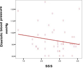
SBP -0.166 0.266
DBP -0.108 0.469
Pulse -0.057 0.703
Temperature -0.292 0.046
TC -0.161 0.279
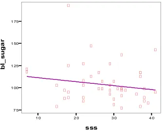
Blood sugar -0.171 0.251
Serum cortisol -0.437 0.002
Table No.4
Levels of correlation with Serum cortisol
Factor Correlation coefficient Significance P value
SBP 0.282 0.055
DBP 0.167 0.262
Pulse 0.423 0.003
Temperature 0.312 0.001
TC 0.248 0.032
Blood sugar 0.248 0.093
SSS -0.437 0.002
Data from 47 patients participated in this study is finally entertained for analysis of
which 26 patients are males (55.3%). The mean age of presentation is 58.21 yrs as shown
[image:44.612.97.519.366.517.2]The mean duration of presentation i.e. from the time of onset of stroke and
presentation to the hospital is 13.62 hours. Of the 47 patients enrolled for the study, 13
persons were known diabetics (27.7%) and 20 persons were known hypertensives
(42.6%).
Of the 47 patients enrolled for the study, 8 (17%) patients were having
hemorrhagic stroke and the remaining were suffering from ischemic strokes. The most
common site for ischemic stroke was MCA territory including its lenticulostriate
branches.
Of the 47 patients, 12 persons (25.5%) were alcoholics and 17 persons were
smokers (36.2%). Alcoholism and smoking were exclusively seen in males.
On admission, almost all the patients were having systolic blood pressure more
than 140 mmHg. 29 patients (61.7%) were having diastolic blood pressure more than 90
mmHg. The average temperature is about 99.6 degree F and the mean pulse rate is about
85.45.
The investigations showed total WBC count of about 8400 cells/cu.mm as an
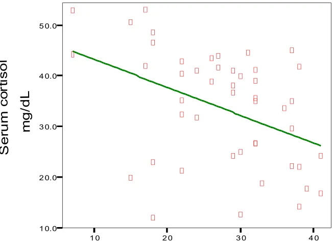
Serum cortisol measured as the time of admission showed a mean of about 33.52
mg/dL and a standard deviation of about 11.20 mg/dL. The value varied between 11.6 to
52.8 mg/dL.
The Scandinavian stroke scale assessment at the time of admission ranged from 7
to 41 in our patients for a total of 60. The mean SSS score is about 27.55 with standard
deviation is about 8.56.
The correlation between SSS and serum cortisol were analyzed. The correlation
coefficient (rho ) is about -0.437 with a P value of 0.002.
The relationship between SSS score and other parameters were also analyzed. The
correlation coefficient was more for body temperature (-0.292) with a P value of 0.045.
The correlation coefficients for other parameters were depicted in Table 3 with
corresponding P values
To analyse the relationship of serum cortisol and the important parameters
correlation coefficients were computed. The highest magnitude of correlation was
obtained for SSS score as depicted above. There is also good correlation between serum
CHARTS AND GRAPHS
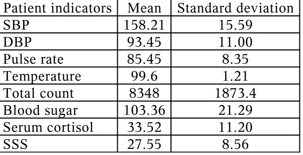
Fig.1 Age distribution
11 11
20
5
0 2 4 6 8 10 12 14 16 18 20
40-49 yrs 50-59 yrs 60-69 yrs >70 yrs
[image:47.612.145.485.454.670.2]Age
Fig.2 Sex distribution
26
21
0 5 10 15 20 25 30
Male Female
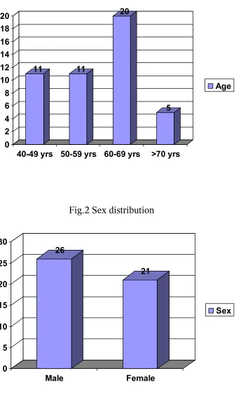
Fig.3 Duration of presentation
8
21
18
0 5 10 15 20 25
<8 hrs 9-15 hrs 16-24 hrs
Duration
Fig.4 Stroke severity - SSS
4
24
19
0 5 10 15 20 25
upto 15 16-30 >30
[image:48.612.145.491.123.683.2]SSS
8 39 0 5 10 15 20 25 30 35 40 Hemorrhage Infarct Type
Fig. 6 Correlation between SSS and Serum cortisol levels
Linear Regres sion
1 0 2 0 3 0 4 0
Scandinavian stroke scale
1 0.0 2 0.0 3 0.0 4 0.0 5 0.0 S e ru m c o rt is o l m g /d L
[image:49.612.129.455.398.641.2]Linear Regres sion
1 0 2 0 3 0 4 0
[image:50.612.116.469.63.660.2]sss 7 5 1 00 1 25 1 50 1 75 b l_ s u g a r
Fig.8 Correlation between SSS and Total count
Linear Regres sion
1 0 2 0 3 0 4 0
SSS 4 00 0
6 00 0 8 00 0 1 00 0 0 1 20 0 0
T o ta l C o u n t C e ll s /c u .m m
tc = 9165.25 + -29.63 * sss R-Square = 0.02
[image:50.612.130.463.66.337.2]Linear Regres sion
1 0 2 0 3 0 4 0
[image:51.612.139.455.55.317.2]SSS 1 25 1 50 1 75 2 00 S y s to li c B lo o d p re s s u re m m H g
Fig.10 Correlation between Diastolic blood pressure and SSS
Linear Regres sion
1 0 2 0 3 0 4 0
SSS 8 0 9 0 1 00 1 10 1 20 D ia s to li c b lo o d p re s s u re m m H g
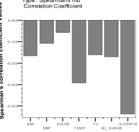
[image:51.612.141.456.363.620.2]S BP DBP
P UL S E T EM P
T C
B L_ S UG AR S .CO RT IS -0 .4 0 0
-0 .3 0 0 -0 .2 0 0 -0 .1 0 0 0 .00 0
S p e a rm a n 's c o rr e la ti o n c o e ff ic ie n t v a lu e s Correlations
[image:52.612.160.437.66.329.2]Type : Spearman's rho Correlation Coefficient
Fig.12 Correlation coefficients for Serum cortisol
S SS S BP DBP P UL S E T EM P T C B L_ S UG AR -0 .2 5 0
0 .00 0 0 .25 0 0 .50 0
DISCUSSION
In this study on Indian population involving 47 patients with stroke, we
evaluated the relationship between serum cortisol level and stroke severity as assessed by
Scandinavian stroke scale score. This study shows a statistically significant correlation
between serum cortisol and stroke severity (P<0.05).
Earlier studies have shown that elevated serum cortisol is an indirect
indicator of stroke severity. This is evident from our study that showed an inverse
correlation between SSS scores which is inversely related to the stroke severity and
increasing serum cortisol.
We also tried to show correlation between serum cortisol and other clinical
and paraclinical parameters of relevance in stroke. The most significant correlation is
between serum cortisol and temperature (P=0.001). There is also statistically significant
correlation between serum cortisol with total WBC count (P=0.032) and pulse rate
(P=0.003)
Eventhough earlier studies showed that blood glucose is related significantly
to stoke severity and serum cortisol, in our study there is no significant correlation.
systolic and diastolic blood pressure, statistical significance was not obtained (P= 0.266
and 0.469 respectively).
The limitation in our study is that ACTH, nor-adrenaline and adrenaline and
CONCLUSION
The following conclusions were derived from our study
1) High serum cortisol correlated with severity of stroke
2) High serum cortisol also correlated with pulse rate, body temperature and WBC
count.
SCOPE OF FUTURE STUDIES
This study conducted in Indian population has significant observations and
potential therapeutic implications. Still few questions remain unanswered. Although
serum cortisol has been convincingly proved an independent indicator of severity of
stroke, the exact pathophysiological mechanism of relationship has to be elucidated by
further studies.
Estimation of other harmones involved in stress response such as ACTH,
epinephrine, cytokines can be undertaken to correlate the severity of stroke.
Proforma
Name: Age&Sex: IP No.
Time duration: Presenting complaints:
LOC: Seizures: Fever: Others:
History
HTN DM PT BA IHD
CVA Seizures Smoking Alcoholism
General Examination
GCS: BP: PR: JVP: Temperature:
Cranial nerves: VII CNS examination SSS:
1. Consciousness 2. Orientation 3. Speech
4. Eye movements 5. Facial palsy 6. Gait
9. Leg power 10. Foot paresis
Investigations:
Blood Hb: TC: DC: ESR:
Blood Sugar: Urea: Creatinine: Electrolytes:
Lipid profile: Serum cortisol: USG abdomen: ECG:
Bibliography
[1] Oka M. Effect of cerebral vascular accident on the level of 17-hydroxy- corticosteroids in plasma. Acta Scand Med 1956; 156:221- 6.
[2] Feibel JH, Hardy PM, Campbell GC, Goldstein MN, Joynt RJ. Prognostic value of the stress response following stroke. JAMA 1977;238: 1374 -6.
[3] Myers MG, Norris JW, Hachinski VC, Sole MJ. Plasma norepinephrine in stroke. Stroke 1981; 12:200-4.
[4] Meyer JS, Stoica F, Pascu I, Shimazu K, Hartmann A. Chatecholamine concentrations in CSF and plasma of patients with cerebral infarction and haemorrhage. Brain 1973; 96:277-88.
[5] Olsson T. Urinary free cortisol excretion shortly after ischaemic stroke. J mt Med 1990; 228:177-81.
[6] Murrus K, Fogelhulm R, Kettunen 5, Vuorela AL. Serum cortisol and outcome of ischemic brain infarction. J Neurol Sci 1993; 116:12-7.
[7] Fassbender K, Schmidt R, Mossner R, Daffertshofer M, Hennerici M. Pattern of activation of the hypothalamic pituary adrenal axis in acute stroke. Relation to acute confusional state, extend of brain damage, and clinical outcome. Stroke 1994; 25:1105-8.
[8] O’Neill PA, Davies I, Fullerton KJ, Bennett D. Stress hormone and blood glucose response
following acute stroke in the elderly. Stroke
1991; 22:842-7.
[9] Christensen H, Boysen G. Blood glucose increased early after stroke onset. A study on serial measurements. Eur J Neurol 2002; 9:297-301.
[11] Johansson A, Ahrén B, Nãsman B, Carlstrom K, Olsson T. Cortisol axis abnormalities early after stroke relationships to cytokines and leptin. J int Med 2000; 247:97-187.
[12] Chrousos GR The hypothalamic pituitary adrenal axis and immune mediated inflammation. NEJM 1995;332:1354-61.
[13] Sander D, Klingelhofer J. Stroke-associated pathological sympathetic activation related to size of infarction and extent of insular damage. Cerebrovasc Dis 1995; 5:381-5.
[14] Smith KE, Hachinski VC, Gibson CJ, Ciriello J. Changes in plasma catecholamine levels after insula damage in experimental stroke. Brain Res 1986;375:182-5.
[15] Franceschini R, Gandolfo C, Cataldi A, Del Sette M, Cianciosi P, Finocchi C, et al. Twenty-four-hour j3-endorphin secretory pattern in stroke patients. Stroke 199425:2142-5
16) Brain’s Diseases 0f the Nervous System. Eleventh edition, chapter 27, page 775-896.
17) Hatano (1976).experience from a multicentre stroke register: a preliminary report. WHO Bull.,54,541-53.
18) Hankey, G.J., and Warlow,C.P.(1994). Transient ischaemic attack of brain and eye. Saunders, London.
19) Murray.C.J.L. and Lopez,A.D.(1997). Mortality by cause for eight regions of the world: global burden of disease study. Lancet, 349,1269-76.
20) Wyller, T.B., Bautz-Holter, E., and Homen, J.(1994). Prevalence of stroke and stroke-related disability in north Trondelag County, Norway. Cerebrovasc. Dis., 4, 421-7.
21) McKeigue, P. M.,Shah,B., and Marmot,M. G.(1991). Relation of central obesity and insulin resistance with high prevalence and cardiovascular risk in south Asians. Lancet, 337, 971-3.
22) Balarajan, R. (1991). Ethinic differences in mortality from ischaemic heart disease and cerebrovascular disease in England and Wales
24) Bamford. J. M., Dennis M. et al. (1990). A prospective study of acute cerebrovascular disease in community ; the oxford community stroke project,1981-86. 2. Incidence, case fatality rates and overall outcome at one year of cerebral infarction, primary intra cerebral hemorrhage and subarachnoind hemorrhage. J. Neurol. Neurosurg. Psychiatry, 53, 16-22.
25) Shaper, A. G et al (1991). Risk factors for stroke in middle aged british men. BMJ, 302, 1111-15.
26) Evans, J. G. (1987). Blood pressure and stroke in elderly English population. J. Epidemiol. Community Hlth, 41, 175-82.
27) Chobanian, A. R. (1983). The influence of hypertension and other hemodynamic forces in atherogenesis. Progr. Cardiovasc. Disc., 26, 177-96.
28) Shinton, R and Beevers, G (1989). Meta-analysis of cigerrate smoking and stroke. BMJ 298, 789-94.
29) Kawachi et al. (1993). Smoking cessation and decreased risk of stroke in women. JAMA, 269, 232-6
30) Lindenstrom et al. (1994). Influence of total cholesterol, HDL cholesterol, and TGL on risk of cerebrovascular disease, and Copenhagen city heart study. BMJ, 309, 11-15.
31) Prospective Studies Collaboration (1995). Cholesterol, diastolic blood pressure, and stroke: 13,000 strokes in 450,000 people in 45 prospective cohorts. Lancet, 346, 1647-53.
32) Rosengren et al. (1989). Impact of coardiovascular risk factors on coronary heart disease and mortality among middle aged diabetic men: a general population study. BMJ, 229, 1127-31.
33) Burchfiel, C. M., et al. (1994). Glucose tolerance and 22 year old stroke incidence. The Honolulu Heart program. Stroke, 25, 951-7.
34) Dearden, N. M. (1985). Ischaemic brain. Lancet 2, 225-9.
35) Pulsinelli, W. (1992)Pathophysiology of acute ischaemic stroke. Lancet, 339, 605-9.
Stroke, 7, 547-54.
37) Weir, C.J. et al. (1997). Is hyperglycemia an independent predictor of poor out come after stroke? Results of long term follow up study.BMJ, 314, 1303-6.
38) Toni, D., et al. (1992). Does hyperglycemia play a role on the out come of acute ischaemic stroke patients? J. Neurol., 239, 382-6.
39) Tracey et al. (1993). Hyperglycemia and mortality from acute stroke. QJM, 86, 439-46. 40) Fisher, C. M. (1954). Occlusion of the carotid arteries. Arch. Neurol. Psychiatry, 72, 187-204.
41) Grady, P.A. (1984). Pathophysiology of extracranial cerebral artery stenosis – A critical review. Stroke, 15, 224-36.
42) Ross, R. (1999). Atherosclerosis – an inflammatory disease. NEJM., 340, 115 - 26
43) Warlow, C. P. et al (2000). Stroke ;a practical guide to management, 2nd edn., chapter 18. Blackwell Scientific, oxford.
44) Hart, R. G., et al. (1995). Oral anticoagulants and intra cranial hemorrhage. Facts and hypothesis. Stroke, 26, 1471-7.
45) Vermeulen, M. et al.(1992). Subarachnoid hemorrhage. Saunders, London.
46) Rinkel, G. J. E., e t al. (1993). Subarachnoid hemorrhage without detectable aneurysm. A review of causes. Stroke, 24, 1403-9.
47) Gates, G. C., et al (1986). Primary intra cerebral hemorrhage in adults. Stroke, 17, 872-7. 48) Brown, R. D., et al. (1998). Incidence of transient ischaemic attack in Rochester, Minnesota, 1985-1989. Stroke, 29, 2109-13.
49) Dennis, M., et al. (1990). CT in patients with T I A: When is a T I A not a T I A but a stroke? J. neurol., 237, 257-61.
51) Tomasello, F., et al. (1982). Assessment of inter-observer differences in italian multicenter study on reversible cerebral ischaemia. Stroke,13, 32-5.
52)Sandercock, P. A. G., et al.(1985). Value of C T in patients with stroke: oxfordshire community stroke project. B M J, 290, 193-7.
53) Norris, J. W and Hachinski, V. C., (1982). Misdiagnosis of stroke. Lancet , 1, 328-31
54) Berlit et al. (1991). Differential diagnosis of spontaneous and traumatic intracranial hemorrhage. J. neurol. Neurosurg. Psychiatry, 54, 1118.
55) Caplan, L. R. and Stein, R.W. (1986). Stroke: a clinical approach. Butterworths, Boston. 56) Bamford, J., et al. (1991). Classification and natural history of clinically identifiable subtypes of cerebral infarction. Lancet, 337, 1521-6.
57) Olsen, T. S., et al.(1985). Causes of cerebral infarction in the carotid territory. Its relation to the size and location of the infarct and to the underlying vascular lesion. Stroke, 16, 459-66.
58) Caplan, L. R., 1993. Brain embolism, revisited. Neurology, 43, 1281-7.
59) Boiten, J. and Lodder, J. 1991. Lacunar infarcts. Pathigenesis and validity of clinical syndromes. Stroke, 22, 1374-8.
60) Bamford,J. M and Warlow, C. P. (1988). Evolution and testing of lacunar hypothesis. Stroke, 19, 1074-82.
61) Castaigne, P., et al. (1973). Areterial occlusion in the vertebrobasilar system. A study of 44 patientswith postmortem data. Brain, 96, 133-54.
62) Argentino, C. et al 1996. posterior circulation infarcts simulating anterior circulation stroke: perspective of the acute phase. Stroke, 27, 1306-9.
63) Horowitz, S. H et al 1991. C T – angiographic findings within five hours of cerebral infarction. Stroke, 22, 1245-53.
65) Pfleger, M. J. et al 1994. sensitivity and specificity of fluid blood levels for coagulopathy in acute intra cerebral hematomas. Am. J. neuroradiol., 15, 217-23.
66) Patel, M. R. et al. 1996. Detection of hyperacute primary intraparenchymal hemorrhage by M R I. stroke, 27, 2321-4.
67) Mohr, J. P. et al 1995. M R I versus C T imaging in acute stroke. Stroke, 26, 807-12.
68) Chernow B, Alexander HR, Smallridge RC, et al. Hormonal responses to graded surgical stress. Arch Intern Med 1987;147:1273-1278.
69) Perrot D, Bonneton A, Dechaud H, Motin J, Pugeat M. Hypercortisolism in septic shock is not suppressible by dexamethasone infusion. Crit Care Med 1993;21:396-401.
70) Chrousos GP. The hypothalamic-pituitary-adrenal axis and immune-mediated inflammation. N Engl J Med 1995;332:1351-1362.
71) Lamberts SWJ, Bruining HA, de Jong FH. Corticosteroid therapy in severe illness. N Engl J Med 1997;337:1285-1292.
72) Beishuizen A, Thijs LG, Vermes I. Patterns of corticosteroid-binding globulin and the free cortisol index during septic shock and multitrauma. Intensive Care Med 2001;27:1584-1591.
73) Hammond GL, Smith CL, Paterson NA, Sibbald WJ. A role for corticosteroid-binding globulin in delivery of cortisol to activated neutrophils. J Clin Endocrinol Metab 1990; 71:34-39.
74) Cooper MS, Bujalska I, Rabbitt E, et al. Modulation of 11 -hydroxysteroid dehydrogenase isozymes by proinflammatory cytokines in osteoblasts: an autocrine switch from glucocorticoid inactivation to activation. J Bone Miner Res 2001;16:1037-1044.
75) Franchimont D, Martens H, Hagelstein MT, et al. Tumor necrosis factor alpha decreases, and interleukin-10 increases, the sensitivity of human monocytes to dexamethasone: potential regulation of the glucocorticoid receptor. J Clin Endocrinol Metab 1999;84:2834-2839.