ANATOMY OF THE RADIAL ARTERY, ITS
BRANCHING PATTERN AND VARIATIONS
WITH ITS CLINICAL APPLICATIONS.
Dissertation submitted for
M.S. ANATOMY BRANCH – V
DEGREE EXAMINATION
THE TAMILNADU DR. M.G.R. MEDICAL UNIVERSITY CHENNAI, TAMILNADU
CERTIFICATE
This is to certify that the dissertation on “RADIAL ARTERY, ITS BRANCHING PATTERN AND VARIATIONS WITH ITS CLINICAL APPLICATIONS” is the bonafide work done by Dr. M.JEYANTHI, in the Institute of Anatomy, Madras Medical College, Chennai – 600 003, during
2005 – 2008 under my supervision and guidance in partial fulfillment of the
regulation laid down by Tamilnadu Dr. M.G.R. Medical University, for the
M.S. Anatomy Branch V examination to be held in March 2008.
Dr. T.P.KALANIDHI, M.D.
Dean ,
Madras Medical College, Chennai – 600 003.
Date :
Station :
Dr. CHRISTILDA FELICIA JEBAKANI, M.S.
Director,
Institute of Anatomy, Madras Medical College, Chennai – 600 003.
Date :
ACKNOWLEDGEMENT
I would like to record my acknowledgement to my teacher and guide
Dr. CHRISTILDA FELICIA, M.S, Director and Professor, Institute of Anatomy, Madras Medical College, who was with me all through this
dissertation work as a friend, philosopher and guide. This dissertation work
would not have seen its day without her expertise guidance, kind concern and
support. It has been a unique experience to work with her and she has been a
source of inspiration in everyway and a great source of support whenever there
was a slackness on my part.
I wish to acknowledge my gratefulness to
Dr. T.P.KALANIDHI, M.D, Dean, Madras Medical College, Chennai for having given permission to freely use all resources in the college
during the course of my study.
I sincerely thank Dr. I.JAYARAJ, Dr. B.CHEZHIAN and
Dr. P.MURUGESAN for their appreciation and support throughout my study.
For their constant encouragement and sincere help
throughout my dissections, I would like to extend thanks to Mrs. M.S.THENMOZHI, Dr. V.LOGANAYAKI, Dr. V.SATHYALAKSHMI
I would like to particularly thank Dr. K.HARSAVARDHANAN, M.Ch, Professor and Head of the Department and Dr. N.NAGARAJAN, M.Ch, Assistant Professor of Cardiothoracic Surgery, Madras Medical College, Chennai, who helped me throughout my dissertation work and to
collect materials for the study.
I wish to acknowledge my sincere thanks to
Dr. T.S.SWAMINATHAN, M.D, DMRD, Director and Professor of Barnard Institute of Radiology, Madras Medical College, Chennai for having permitted
me to observe the Angiogram procedures and having allowed to do adult
cadaveric angiogram study and to collect pictures.
I wish to acknowledge my indebtedness to Dr.T.VIDYASAGARAN, M.S., M.Ch, Professor and Head of the Department of Vascular Surgery, Madras Medical College, Madras Medical College, Chennai who guided me
through the clinical study.
I would also like to extend my sincere thanks to
Dr. M.P.NAMASIVAYAM, M.S., M.Ch, Professor and Head of the Department, Plastic Surgery, Madras Medical College, Chennai to collect
pictures regarding my study.
My sincere thanks to my friends, colleagues, juniors who supported and
I would like to express my thanks to all technicians and staff members
who helped with making arrangements for dissections and in the histological
sectioning and slide preparation related to my study.
It is the constant encouragement and support given by my relatives
particularly my husband, my daughter K. SWATHI and my son
K. SANDEEP.
CONTENTS
CHAPTER TITLE PAGE NO.
I INTRODUCTION 01
II AIM OF THE STUDY 04
III REVIEW OF LITERATURE 08
IV DEVELOPMENTAL ANATOMY 36
V MATERIALS AND METHODS 39
VI OBSERVATION 46
VII DISCUSSION 55
VIII CONCLUSION 80
INTRODUCTION
ANATOMY – ITS IMPORTANCE
“The body is an instrument, the mind its function, the witness and reward of its operation”
-George Santayana.
Andreus Vesalius, father of modern anatomy, wrote in 1543 in the preface to his De Humane Corporis Fabrica, “Anatomy should rightly be
regarded as the firm foundation of the whole art of Medicine and its essential
preliminary”.
Hence anatomy forms the first foundation stone of the medical career.
Knowing anatomy is one thing and knowing it in detail is quite another. With
anatomical knowledge the sails have already been put to hoist but with the
details in it, the winds have started blowing. The resultant effect is that the ship
is on the move. Also with detailed knowledge in anatomy precision teaches its
zenith with a happy out come for the concerned and the considerate.
By the way, even in appreciating the deep realms of pathology, anatomy
stands as a fathom stick. The systemic arteries all stem from the aorta and by
branching and re-branching from what is some times spoken of as the ‘arterial
tree’. Its ultimate branches or arterioles lead into the capillaries by which the
The situation of the larger arteries in the trunk and limbs affords them
natural protection from injury. Thus, the main artery of each limb passes from
the trunk into the medial side of the limb and runs directly on the flexor aspect;
accordingly as the limb is bent, the vessel is increasingly safeguarded. Further,
its position on the flexor aspect lessens the tension exerted upon it during
movements of the limb joints.
Around the limb joints there are notable anastomoses between medium
sized arteries; those anastomoses are of clinical importance as they are the
means by which the blood supply to the distal part of the limb is maintained by
the opening up of a collateral circulation should the main artery be blocked by
ligature or disease. Quiring (1949) provides as convenient, illustrated survey of
the chief anastomoses both arterial and venous, in the body.
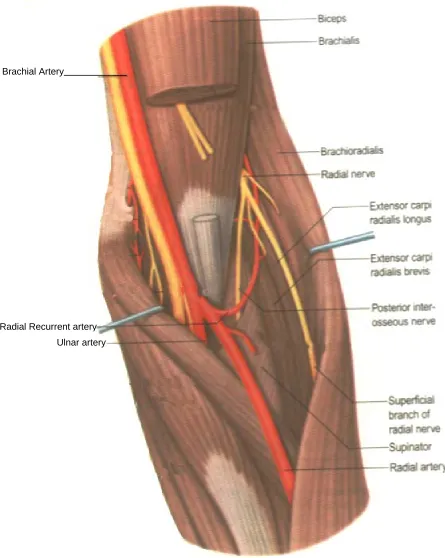
The radial artery is smaller than the ulnar artery, yet appears a more
direct continuation of the brachial artery. It normally starts 1cm distal to the
flexion crease of the elbow (fig 1). It descends along the lateral side of the
forearm, accompanied by paired venae comitantes, from the medial side of the
neck of the radius to the wrist, where it is palpable between flexor carpi radialis
medially and the salient anterior border of the radius (fig – 2).
Proximally it is overlapped anteriorly by the belly of brachioradialis, but
elsewhere in its course it is covered only by the skin, and superficial and deep
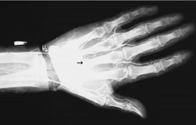
fasciae. At the wrist the radial artery passes on to the dorsal aspect of the
carpus between the lateral carpal ligament and the tendons of abductor pollicis
It crosses the scaphoid bone and trapezium (in the ‘anatomical snuff
box’) and as it passes between the heads of the first dorsal interosseous muscle
it is crossed by the tendons of extensor pollicis longus(fig – 5). Between the
thumb extensors, it is crossed by the start of the cephalic vein and the digital
branches of the radial nerve which supply the thumb and index finger.
In the hand the radial artery passes through the first interosseous space
between the heads of the first dorsal interosseous and crosses the palm. At first
it lies deep to the oblique head of adductor pollicis and then passes between its
oblique and transverse heads or through transverse head. At the fifth
metacarpal bone it anastomoses with the deep branch of the ulnar artery,
completing the deep palmar arch (fig – 4a).
Branches in the forearm are, radial recurrent artery, cutaneous branches,
muscular branches.
Branches in the wrist and palm are palmar carpal branch, superficial
palmar branch, dorsal carpal branch, first dorsal metacarpal artery, arteria
princeps pollicis, arteria radialis indicis and the terminal part which forms the
AIM OF THE STUDY
Variations and abnormalities of the vascular system are the special
interest to the anatomist because of their morphological significance. The
vascular system is rich in such variation, many of which are of great practical
importance.
With the exception of those directly due to the effect of morbid
conditions and external influence, all variations are the result of modifications
of normal developmental processes. The exception referred to are, however,
very numerous; thus disease or injury may lead to the obliteration of vessels – a
condition which is invariably associated with the enlargement of collateral
vessels. Variations which are determined by or are dependant upon
modifications of the usual developmental processes are of greater interest.
Anomalies of the forelimb arterial tree are fairly common, probably
because they have multiple and plexiform sources. In general anomalous
pattern may present as differences in mode and proximodistal level of
branching, the presence of unusual compound arterial segments, aberrant
vessels, arcades, plexuses.
The radial artery is superficial in the distal forearm and lies on the lateral
side of flexor carpi radialis tendon. It’s pulsation can readily be felt, supplying
information of clinical importance, such as rate, rhythm, compressibility and
condition of arterial wall. The arterial variations of the upper limb have been
brachio ulnar arteries have been encountered during elevation of the radial
forearm flap.
Possible intra arterial injection of drugs due to the proximity of normal
vein puncture sites has also been reported as well as possible arteriographic
misinterpretations when the contrast dye is injected distal to the origin of these
variant arteries. The existence of a superficial radial artery implies the absence
of the normal radial pulse at wrist level and also it produces problems in
cannulation for operation monitoring.
The absence of the ulnar artery was responsible for hand ischaemia after
radial artery conduit for coronary bypass. So preoperative assessment of the
radial artery and collateral hand circulation is mandatory to avoid hand
ischaemia in patients with inadequate collateral circulation.
Radial artery is the commonest one affected in some vascular diseases,
crush injuries which will lead to ischaemia of the hand where the role of
vascular surgeons is very essential to manage. Radial artery is the foremost one
used for arterio-venous fistula by nephrologists in chronic renal failure and by
diabetologists for dialysis purpose.
Because of its histological similarity with coronary artery, it is being
used by cardiothoracic surgeons for coronary revascularization which results in
long term patency, improves long term outcome, leads to low morbidity, and
good functional outcome of the hand after harvesting the radial artery
The feasibility of performing coronary angiography or percutaneous
coronary intervention through radial artery has been broadly evaluated and
major advantages of this approach compared to classic femoral one, mainly
include reduction of local vascular complications and patient’s short
ambulation.
Extensive studies have been done by many scientific experts regarding
variation pattern of radial artery and also by many cardiothoracic surgeons
regarding its merits and demerits in coronary artery bypass grafting. (CABG)
Even though the branches of radial artery are very few, the variations are
not uncommon. So the knowledge about the radial artery, its branches and its
variations are very much important for the vascular surgeons, nephrologists,
plastic surgeons and cardio – thoracic surgeons; according to which they can
modify the surgical procedures in a most satisfactory way.
So the present study of radial artery was undertaken by me to study the
course, branching pattern, its variation and also the histological similarity with
coronary artery. The radial artery is studied under the following parameters.
1. Origin of the radial artery
2. Course of the radial artery
3. Branches
Brachial
a) Radial recurrent branch – Origin
b) Superficial palmar branch – its origin and its contribution to the
completion of the Superficial Palmar
Arch.
dorsal side
c) Arteria princeps pollicis origin
palmar side
dorsal side
d) Arteria radialis indicis origin
palmar side
e) Palmar carpal branch
f) First dorsal metacarpal artery
g) Dorsal carpal branch
h) Origin of interosseous artery from the radial artery
4) Completion of superficial palmar arch
REVIEW OF LITERATURE
Quains (1844) was believed to be the first person to provide sufficient data for better statistical evaluation regarding radial artery. In his series of dissection
with 481 extremities he encountered 19.5% of anomalies of radial artery
affecting its origin or course.
Henry Gray (1858) described, though smaller than the ulnar, the radial artery appears of more direct continuation of the brachial artery and it begins about
1cm distal to the bend of elbow (at the level of neck of radius) and traverses up
to its styloid process in the distal part. Then the radial artery descends along the
lateral side of the forearm to wrist and then curls posterolaterally round the
carpus, beneath tendons of abductor pollicis longus, extensor pollicis brevis
and extensor pollicis longus to the proximal end of the first inter metacarpal
space, swerving medially between heads of the first dorsal interosseous into the
palm and then crossing medially to form the deep palmar arch with the deep
branch of the ulnar artery. Proximally it is overlapped by the belly of
brachioradialis, rest is covered by skin, superficial and deep fasciae. Sometimes
the radial artery arises proximally, usually from the axillary artery. He
mentioned that the arteria princeps pollicis is the usual nutrient artery supply to
the first metacarpal bone. The Superficial palmar branch arises from the radial
artery just before it curves round the carpus, passes through and occasionally
over the thenar muscles, which it supplies, sometimes anastomoses with the
Anterior carpal branch, posterior carpal branch and the first dorsal
metacarpal branch are the slender branches arising from the radial artery
anterior and dorsal part of the wrist. The arteria princeps pollicis et radialis
indicis may be continued as the first palmar metacarpal artery. They may arise
from the superficial palmar arch. The deep palmar arch is formed by
anastomosis of the end of the radial artery with the deep branch of the ulnar
artery. The radial artery can give rise to the common interosseous artery.
Poirier (1886), In his text book of anatomy, “superficial brachial artery” is discussed. In 100 dissections the brachial artery crossed superficial to the
median nerve 6 times “High origin” of the radial artery according to Poirier, is
rarer.
Charles (1894) recorded a case of absence of the radial artery, its place at the wrist was taken by the anterior interosseous artery which wound around the
radial border of the wrist deep to the long tendons of the thumb and entered the
palm of the hand by passing between the first and second metacarpal bones in
the fashion of a normal radial artery.
Prof. Johnston (1921) in his synopsis of “Regional Anatomy” described that , the radial artery is comparatively superficial throughout its whole course, but
particularly in its lower half. It arises from the brachial artery in cubital fossa
and runs forwards, curving laterally to the wrist. The radial artery is overlapped
by the brachioradialis, but in the lower half of its extend it is covered only by
the skin, the superficial and deep fasciae. The superficial palmar branch of
arch represents the direct continuation of the radial artery into the palm, the
arch being completed on the medial side by union with the deep branch of the
ulnar artery.
Dubreuil Chambardel (1926) mentioned that the median artery contributed to the formation of superficial palmar arch seen in 4% of 1200 cases.
Beuntaro Adachi (1928) a Jaspanese anatomist described 3 types of superficial palmar arch.
a) The “ulnar type” in which the contribution by the radial artery is absent
or minimal.
b) The “radio-ulnar” type
c) The “medio-ulnar” type in which a median artery is sufficiently strong
to reach the palm of the hand and to take part in the formation of arch.
According to him the ulnar type is the more frequent (59%), next the
radio-ulnar (32%) and the medio-ulnar type the least common (9%).
Adachi also describes a rare abnormality which was seen 8 times in 698
dissection (1.1%). It may be called superficial radial artery, which is given off
at varying level in the forearm. It is thicker than the continuing trunk, travels
dorsally across the surfaces of the brachioradialis muscle and runs downwards
in the superficial fascia. Over the thumb tendons and ends like the normal
radial artery by piercing the first interosseous space.
The continuing smaller trunk of the radial artery takes a normal course
to the wrist then deep in the thumb tendons and usually ends in the posterior
Degaris and Swartley (1928) recognized 23 different patterns of axillary artery and its branches in their study based on 512 dissection and found 7.7%
of high origin of the radial artery. Among these 2.7% are of white 5% of negro
arms.
George A.Piersol (1930) described that the radial artery is the smaller of two terminal branches of brachial artery and arises at the bend of the elbow and
passes down the lateral border of the forearm to the level of the styloid process
of radius where it bends laterally and extends downwards over the dorsum until
it reaches the interval between the first and second metacarpal bones and again
changes its direction and passes forwards into the palmar surface of the hand,
across which it is continued medially over the anterior surface of the second,
third and fourth metacarpals forming deep volar arch. It terminates opposite the
proximal part of the fourth metacarpal interspace by anastomosing with deep
volar branch of the ulnar artery.
The volar radial carpal, the dorsal radial carpal and the first dorsal
metacarpal arteries are very slender branches and make anastomosis with
branches from volar ulnar carpal, dorsal ulnar carpal and the first dorsal
metacarpal artery passes distally along the radial border of the second
metacarpal and terminates upon the first phalanx of the index finger.
The superficial volar branch occasionally arises high upon the radial and
frequently it takes no part in the formation of the superficial volar arch and may
terminate in the muscles of the thenar eminence, the digital branches being
princeps pollicis arises from the radial just as it emerges from between the two
heads of dorsal first interosseous muscle. He also stated that the arteria
princeps pollicis and the arteria dorsalis indicis may be well developed and
arise directly from the deep volar arch.
Thomas Walmsley (1934) has described the radial artery as the smaller of the division of brachial artery though it appears to be the more direct continuation
of brachial artery. Upper part of radial artery is undercover of medial part of
fleshy belly of brachioradialis and lower part is deep to deep and superficial
fasciae and skin. He also says that sometimes the radial artery lies superficial to
the deep fascia, instead of beneath it, in its whole course and sometimes it
arises from the axillary artery or the upper part of the brachial artery.
The superficial palmar artery, arise near the root of the thumb and runs
distally first superficial to and then among its short muscles. It often completes
the superficial palmar arch.
Radial artery enters palm from dorsum of hand by passing through first
interosseous space and in the palm, it lies undercover or amongst the fibres of
the obliquous portion of the adductor pollicis and emerges between the border
of this and the transverse part of muscle and then passes across the palm as
deep palmar arch which is completed on the medial side by anastomosis with
the deep branch of the ulnar artery. According to him, from the radial artery in
the palm, there usually arises two branches. The arteria princeps pollicis, which
The second branch, arteria radialis indicis passes forwards to the radial
side of index finger. It may give off a branch which completes the superficial
palman arch. Anterior carpal branch, posterior carpal branch are slender
branches arising from the radial artery anteriorly and dorsally. Slender dorsal
metacarpal arteries arising from the posterior carpal arch.
J.E. Frazer (1937) has described that the radial artery is one of the two terminal branches of the brachial artery which in the cubital fossa opposit the
upper part of the neck of the radius divides into radial and ulnar arteries. Radial
artery is smaller than the ulnar, its direction is at first continuous with parent
trunk. He subdivided the radial artery into 3 parts.
I part- Occupying forearm – from origin to the styloid process of the radius.
II part- the back of the wrist
III part- the palm.
The artery may arise from the upper part of the brachial or from the
axillary – then it may descend superficially to the bicipital aponeurosis of the
biceps and deep fascia of the forearm. It may end at the lower part of the
forearm, its distribution being replaced by branches of the ulnar, median or
anterior interosseous arteries. It gives radial recurrent branch close to its
commencement.
The third part forms greater portion of the deep palmar arch, extends
inwards from the proximal end of the first inter metacarpal space to join the
The princeps pollicis artery arises from the radial extremity of the deep
arch. The radialis indicis artery passes downwards on the outside of the second
metacarpal bone. The princeps pollicis and radialis indicis arteries not
infrequently arises by a common trunk or may be derived from the superficial
arch, from an enlarged median artery or from the superficial palmar branch.
Superficial palmar arch is completed by superficial palmar branch of the radial,
or failing this by a branch from the radialis indicis artery or from the princeps
pollicis artery. Anterior, posterior carpal branches are slender branches from
the radial artery. The first dorsal meta carpal artery arises directly from the
radial or from the posterior carpal branch.
Miller R.A (1939) found bilateral high origin of the radial artery in 15 of 480 bodies about 3 percent.
C. Latimer Callander, Dean Lewis (1939) have mentioned in their book, “Surgical Anatomy” that the radial artery arises at the level of the coronoid
process of ulna and the neck of radius, continuous in direction of the brachial
trunk. It runs a fairly straight course through the forearm. In the distal third, it
is subcutaneous and lies on the radius and on the flexor pollicis longus muscle.
It gives off two large branches in the forearm, the radial recurrent artery near its
origin and the superficial volar artery in the distal part of the forearm. The
superficial volar artery, runs through the short muscles of the thumb to meet the
ulnar artery and complete the superficial palmar arch.
radial side of forearm as far as the styloid process and curving over the lateral
side and back of the wrist, enters the palm between the bases of the first and
second metacarpal bones and ends by anastomosing with deep branch of ulnar
to form deep volar arch. The course is divisible into 3 parts – that in forearm,
that at the wrist and that in the palm of the hand.
He described about radial recurrent artery ; which usually arises from
the lateral side of the radial just beyond its origin from brachial.
The superficial volar branch courses forward over the short muscles of
the ball of the thumb, and anastomoses with the superficial branch of the ulnar
artery to complete the superficial volar arch. The volar radial carpal, dorsal
radial carpal and the first dorsal metacarpal arteries are the other slender
branches of the radial artery.
Cohen S.M. (1948) reviewed 12 cases of intra arterial injection of thiopental sodium of which six required forearm amputation. He concluded that spasm of
the distal arterioles and late thrombosis of the arteries were the significant
lesions.
Barry J. Anson. (1950) pointed out in his book – ‘An Atlas of Human Anatomy’ about the high division of brachial artery giving rise to smaller radial
artery or a large radial artery.
Emanuel B.Kaplan (1953) has mentioned in his book “Functional and surgical anatomy of the hand” that the superficial arterial arch is formed by anastomosis
with superficial branch of radial artery. He describes the variations of
1. The superficial palmar arch may have three variants.
The first variant, in which the ulnar artery is responsible for the
formation of the superficial palmar arch, is encountered in about 66% of the
cases.
2. The second variant of the superficial palmar arch in which the
superficial radial palmar artery participate in the formation of the
arch in encountered in about 30% of the cases.
3. The third variant of the superficial arch in which the median artery
contributes to the formation of the arch. Very infrequently an
additional subcutaneous branch of the superficial branch of the radial
and a subcutaneous branch of the ulnar artery is found.
Variations of the deep palmar arch
The most frequent variation is the regular anastomosis between the
radial artery and the deep branch of the ulnar artery. The anterior interosseous
artery may participate in the formation of the deep palmar arch with the radial
artery.
Lawrence J. Mc Cormack, Cauldwell E.W. and Anson B.J. (1953) observed the variation occurred in 139 of 750 extremities (18.53%) considered on the
basis of the 364 (of 386) cadavers with both upper extremities represented,
major arterial variations occurred in 112 (30.77%) Bilateral occurrence was
recorded in 23 cadavers (6.32%) while the remaining 89 (24.45%) revealed
Mr.Cormack and his co-workers found a high origin of the radial artery to be
far the most common variation of the arteries of the arm for it represented 77%
of all the variations they observed, and was found in 14.27% of all specimens.
In 2.13% of limbs the radial artery arose from the axillary artery, and in
12.41% from some part of the brachial artery.
In 6 specimens there occurred anomalous terminal distribution of the
radial artery. In 5 of the 6 specimens, the radial artery divided in the distal
fourth of the forearm into two large branches one of which continued along the
medical border of brachio radialis muscle tendon and to enter the deep aspects
of the palm. The second branch provided a strong contribution to the thumb
and index finger and within the palm contributed the radial portion of the deep
volar arch. In 80 of 750 specimen, they studied about superficial volar arch. In
only 15 specimens was a volar arch is formed in the usual manner by a branch
of the radial artery. In 41 cases a superficial volar arch was formed by a
delicate arterial twig contributed to the ulnar artery.
Weathersby (1954) said that the superficial palmar arch is completed by the superficial palmar branch of the radial in about 35 percent of hands. In 12%,
there was no connection of the arch to branches of the radial artery. On the
radial side, sometimes it was with a branch of the princeps pollicis alone.
In 10% the first dorsal metacarpal artery passed between the thumb and
forefinger to join the superficial arch or its branches. He also found the origin
of the radialis indicis artery in 13% of hands from the radial side of the
J.D.Boyd et al (1956) stated that the brachial artery ends in the middle of the cubital fossa by dividing, at about the level of the neck of the radius into the
radial and ulnar arteries. It descends on the lateral side of the forearm. Then it
turns downwards and backwards below the styloid process of the radius deep to
the extensor tendons of the thumb. It passes downwards and inwards between
the proximal ends of the first and second metacarpal bones and two heads of
first dorsal interosseus muscle and it traverses the adductor pollicis muscle to
enter the palm. In the palm it ends by anastomosing with the deep division of
the ulnar artery to form deep palmar arch. On the anterior aspect, the radial
artery gives superficial palmar branch which anastomoses with superficial
division of the ulnar artery to form superficial palmar arch.
J.C.B. Grant, James Couper Brash (1957) have described that the radial artery is the smaller of the two terminal branches of brachial artery but it is
more direct line with the parent trunk . Begins in the cubital fossa opposite the
neck of radius and ends in the palm of the hand by anastomosing with the deep
branch of the ulnar artery and thus completing the deep palmar arch. He also
pointed out that the radialis indicis artery not uncommonly anastomoses with
the superficial palmar arch. He also indicated that, the radial artery may be
absent, its place being taken by branches of the ulnar or interosseous arteries. It
may end in muscular branches in forearm or as the superficial palmar or in
carpal branches.
The radial artery may run a superficial course, or and especially when
wrist across the brachioradialis and it may lie superficial to the extensor
tendons of the thumb, instead of deep to them. Its branches may be diminished
or increased in number. The radial recurrent may spring from the brachial or
ulnar arteries. The princeps pollicis and radialis indicis arteries may be absent,
their places being taken by branches of superficial arch. The superficial palmar
arch is sometimes absent, its branches are then given off from the deep arch.
The deep palmar branch of the radial artery is much more rarely absent than the
superficial arch. When absent its branches are given by superficial palmar arch.
Arnold K. Henry (1957) has told that the radial artery begins at the medial side of the biceps tendon and the artery is bound by fascia to pronator teres.
Russell T. Woodburne (1957) has described in his “Essentials of Human Anatomy” that a high origin of the radial artery from the brachial or even from
the axillary artery, occurs in 15% of cases. The radial recurrent artery arises
just below the origin of radial artery. The superficial palmar branch passes over
or through the muscles forming the eminence of thumb. The superficial palmar
artery is usually slender.
The deep palmar arterial arch is formed by the terminal portion of the
radial artery in conjunction with the deep branch of ulnar artery and the
branches of deep palmar arch are, the arteria princeps pollicis, the arteria
radialis indicis, 3 palmar metacarpal , recurrent carpal and dorsal perforating
R.D.Lockhart et al (1959) said that the brachial artery lying on brachialis in the cubital fossa, divides opposit the neck of the radius into the radial artery, its
more direct continuation and the ulnar artery.
The radial artery, crossing the tendon of biceps , descends medial to the
radial nerve, undercover of the belly of brachioradialis and having crossed
supinator, pronator teres and flexor digitorum sublimis continues over the
flexor pollicis longus and it crosses the floor of the anatomical snuff box on the
scaphoid and trapezium passing deep to extensor pollicis longus, it reaches the
proximal end of the space between the first and second metacarpal bones and
turning forwards between the two heads of the first dorsal interosseous muscle,
enters the palm between the two heads of adductor pollicis. There it gives
origin to the princeps pollicis and radialis indicis and continues as the deep
palmar arch. Just above the wrist it gives superficial palmar branch which
crosses or penetrates the thenar muscles and may complete the superficial
palmar arch.
Sir Solly Zuckerman (1961) has mentioned that the superficial palmar arch is completed laterally by a branch from one of the terminal branches of the radial
artery either the superficial palmar branch of the radial artery, a small vessel
which passes over the thenar muscles from the point where the radial artery
turns backwards behind the tendons of the muscles of the thumb to the back of
the wrist, or a branch from either the princeps pollicis artery or the radialis
when it returns to the deep aspect of the palm. Sometimes the princeps pollicis
and radialis indicis arteries arise by a common stem.
Coleman S.S. and Anson B.J. (1961) said that the superficial palmar arch is completed by a superficial branch of radial in about 35% of hands. In 37% the
superficial branch of radial artery ending in thumb muscles. In 18% of hands
the first dorsal metacarpal artery passed between the thumb and forefinger to
join the superficial palmar arch or its branches. They listed the arch as
incomplete when it did not send a branch to the thumb and index finger, more
than half of their 21.5% of incomplete arches were of this type.
Regarding deep palmar arch they reported a few instances in which the
arch was incomplete with the radial artery supplying the thumb and radial side
of the index finger, or doing this and ending by anastomosing with a
perforating artery from a dorsal metacarpal, while the ulnar end of the arch was
formed by the deep branch of the ulnar and another perforating artery. They
found one case also in which the deep arch was formed entirely by the deep
ulnar and a perforating artery, there being no deep branch of the radial artery.
J.A.Keen (1961) has pointed out that among 284 dissections the superficial brachial artery including those which continued as superficially placed radial
and ulnar arteries was found 35 times (12.3%). He also pointed out that the
level of bifurcation of the brachial artery was related to the level of the radio-
humeral joint. Among 284 dissections a “superficial radial artery” was seen
about the middle the forearm as far as the first interosseous space. The third
instance was on right side only.
Sir John Bruce, Robert Walmsley, James A. Ross (1964) described about the course of the radial artery as that, in the proximal two thirds, it lies
undercover of brachioradialis and in the proximal third lies on the supinator. In
the middle third it lies on the pronator teres and the radial head of the flexor
digitorum superficialis. The distal third of the artery is subcutaneous and lies
upon the flexor pollicis longus, the pronator quadratus and the radius. It gives
radial recurrent artery near its origin. The superficial palmar branch runs
through or superficial to the short muscles of thumb and sometimes joins the
terminal part of the ulnar artery to complete the superficial palmar arch.
He also stated that when the radial artery is wounded at the wrist, both
ends must be ligatured owing to the free anastomosis in the hand.
G.J.Romanes (1964) stated that the radial artery which is a branch of brachial artery given off in the cubital fossa opposit the neck of radius, smaller than the
ulnar, is in more direct line with brachial artery. It ends by anastomosing with
the deep palmar branch of the ulnar artery, thus completing the deep palmar
arch. The superficial palmar branch usually pierce the muscles of the thenar
eminence, and ends either in their substance or by joining the ulnar artery to
complete the superficial palmar arch.
He also noted that “high division” of the brachial artery occurs most
commonly in the proximal third of the upper arm. When high division occurs,
descend in the forearm in the superficial fascia or it may pass behind the tendon
of biceps. He also mentioned that the radial artery may arise from the axillary
artery and it may pass to the back of the wrist superficial to the extensor
tendons or it may lie on the superficial fascia of the forearm. The superficial
palmar arch is completed by superficial branch of the radial or by the radialis
indicis or the princeps pollicis artery.
Ben Pansky, Earl Lawrence House (1964) in their review of ‘Gross Anatomy’ mentioned, that the radial artery extends from the neck of radius to
medial side of radial styloid process.
Ernest Gardner, Donald J Gray, Ronan O’ Rahilly (1967) told that the radial artery is the smaller terminal division of the brachial artery and begins in
the cubital fossa, opposite the neck of radius. The radial artery varies and it
may be absent, it may arise in the arm, or even from the axillary artery.
Occasionally, it runs a very superficial course throughout the forearm. The
completion of the superficial palmar arch on the radial side is extremely
variable. Commonly the superficial palmar arch was completed by the radialis
indicis, by the superficial palmar branch of the radial, or by the princeps
pollicis. A large median artery may contribute to it.
W. Henry Hollinshead (1969) described that the radial artery is in its course the more direct continuation of the brachial artery and it has the usual course.
The deep palmar arch is the chief continuation of the radial artery into
the hand and is described as being, with the smaller princeps pollicis, one of the
Barry J. Anson, Chester B.Mcvay (1971) described that the radial artery runs a fairly straight course through the formation and terminates by takes part in
the forearm of the deep palmar arch and because the artery is superficial,
ligations of the radial artery is performed readily at any part of its course in the
forearm. It gives radial recurrent artery near its origin. In ligation in the upper
third of the forearm the artery is found deep to the interval between the brachio
radialis and pronator teres muscle.
For ligation of the artery at the wrist an incision is made over the artery
midway between the outer border of the radius and the tendon of the flexor
carpi radialis. The vessel lies upon the pronator quadratus muscle.
Richard S. Snell (1973) said that the radial artery is the smaller of the terminal branches of the brachial artery. It begins in the cubital fossa at the level of the
neck of radius. He also described that the deep palmar arch is a direct
continuation of the radial artery and on entering the palm, the radial artery
gives off the arteria radialis indicis and the arteria princeps pollicis.
W.J.Hamilton (1976) stated that the radial artery is accompanied by the superficial division of the radial nerve on its lateral side and traverses the
lateral part of the wrist by passing backwards below the styloid process of
radius and enters palm in between the proximal ends of the 1st and 2nd
metacarpal bones and in the palm it ends by anastomosing with deep division
J.P.Mall et al (1976) in their article “Arterial pattern of brachial artery in relation to brachial plexus” mentioned the anomalous patterns of brachial
artery which they found in 33.4% cases in 96 limbs dissected. The patterns are
1. Those “superficial brachial arteries” which continue into the cubital
fossa and then bifurcate as usual (2.1%)
2. Those “superficial brachial arteries” which join to radial artery (2.1%)
3. Those “superficial brachial arteries” which continue as the radial
artery, called high origin of the radial artery (2.1%)
Keith L. Moore (1980) has described that the radial artery begins in the cubital fossa just medial to the biceps brachii tendon at the level of the neck of radius.
The radial recurrent branch arises from lateral side of radial artery just distal to
its origin. As it crosses the floor of the anatomical snuff box it lies on the
scaphoid and the trapezium and ends by completing the deep palmar arch in
conjunction with the ulnar artry. The common place for taking the pulse is
where the radial artery lies on the anterior surface of the distal end of the radius
lateral to the tendon of the flexor carpi radialis muscle. Here it is covered only
by deep and superficial fasciae, skin.
Erlandson et al (1981) has given the frequency of normal anatomic variations of upper extremity arteries as follows:
Sub clavian artery:-
Anomalous origin from the aortic arch 2%
High origin of radial artery 14%
1. From axillary artery 12%
2. From brachial artery 2%
High origin of ulnar artery 3%
Palmar Arch: Incomplete palmar arch 20%
Complete palmar arch 80%
Ulnar and radial artery origin 36%
Ulnar artery origin only 37%
Others 7%
Radial and ulnar arch 3%
Ulnar artery origin only 13%
Other 4%
Wilson SE. Stabie BE, Williams R.A. Olwen MC (1982) mentioned that in the upper extremity, the radial artery has been chosen by most surgeons to be
the primary choice for construction of an arteriovenous graft.
Priver, Lysenbov, Bushkovich (1985) have described that the princeps pollicis branches off the radial artery as soon as the latter penetrates the first
interosseous space on the palm of the hand. It runs over the palmar surface of
the first metacarpal bone and divides into branches, digitales palmares, to both
sides of the thumb and to the radial side of the index finger.
Ugawa, Ikeda (1985) studied about the arterial patterns of the hands in primates (13 macaca fuscates) and they observed that in the dorsum of the
hand, there are four dorsal metacarpal arteries. The 1st, 2nd and 3rd dorsal
metacarpal arteries arise from the radial artery, while the 4th one originates
from the ulnar marginal artery. The radial artery lies in the inter muscular cleft
diagonally into the dorsum of the hand from a point a little beyond the styloid
process of radius.
The deep palmar arterial arches are formed by the perforating branches
of the 2nd dorsal metacarpal artery and are usually composed of two proximal
arches (C. volaris proximalis and a. volaris profundus) and one distal arch (C.
volaris distalis). Volaris profundus (Deep palmar arch in man) follows the
course of deep branch of ulnar artery and then divides terminally into radial
and ulnar branches. The superficial palmar arterial arch is formed by the distal
curved portion of the ulnar artery and completed by the superficial palmar
branch of the radial artery.
Poteat WL (1986) reported a case of unilateral absence of the radial artery. The arterial system of the specimen was developmentally primitive with the
anterior interosseous artery the chief blood supply to the forearm and hand.
Kanaga suntheram, Sivananda Singham, Krishnamurti (1987) said that the radial artery is the other terminal branch of the brachial artery. It extends from
the neck of the radius to the medial side of the styloid process of the radius
which is the first part and from distal part of styloid process it proceed towards
the first interosseous space. This forms the second part of the course of the
artery.
The third part of artery enters the palm by passing between two heads of
the first dorsal interosseous muscle. In the palm, it gives off the princeps
pollicis artery which divides into two to supply the medial and lateral sides of
lateral side of the index finger. After this, the radial artery curves medially and
continues as deep palmar arch which is completed on the medial side by the
deep branch of the ulnar artery.
Michael Sachs (1987) during the clinical investigations of 570 soldiers, found that the radial artery pulse was not felt in 5 cases at the typical place. In these 5
cases, he was able to feel a subcutaneous artery which coursed superficial to
the anatomical snuff box and crossed superficial to the tendon of extensor
pollicis longus muscle.
The radial artery divides in the distal fourth of the forearm (5-7cm
proximal to the wrist joint) into two branches. The dorsal branch courses
subcutaneously over the tendon of the brachioradialis muscle, and runs over the
tendon of the extensor pollicis longus muscle to enter the deep aspect of the
palm in the first metacarpal space. This dorsal branch courses parallel to the
superficial branch of the radial nerve. The palmar branch can be regarded as the
‘normal’ radial artery which continues along the medial border of the
brachioradialis muscle and courses deep under the tendons of the dorsal
muscles of the thumb.
John V. Basmasjian, Charles E.Slonecker (1989) described that the trunk of radial artery is not crossed by any muscle, but the brachio radialis is lateral and
overlaps it. The flexor carpi radialis is medial in distal two-thirds, immediately
behind it are 6 muscles that clothe the anterior aspect of the radius and beyond
these are the distal end of the radius and the capsule of the wrist joint. The
deep to the abductor pollicis longus. Here the radial artery gives an important
nutrient artery to the scaphoid bone. The radial artery continuation in the palm
is completed by the deep branch of the ulnar artery and forms deep palmar
arch.
Yao. JST (1989) stated that occlusion of the radial artery, in particular is common following direct penetrating or blunt trauma or from transarterial
catheters for arterial pressure monitoring or blood gas analysis. Patients with an
incomplete palmar arch are at an increased risk of developing chronic hand
ischaemia due to injury of the radial or ulnar artery.
Mezzogiorno et al (1994) from their study of 60 vascular casts of upper extremity found that the deep palmar circulation is constituted by the deep
palmar arch. In most cases this is a complete arch formed by the radial artery
and its continuation to a deep branch of the ulnar artery. In a few cases, the
deep palmar ciculation is formed only by the radial or the ulnar artery. Only
rarely is there a complete absence of the deep palmar arch. Four anatomic
patterns were identified. (Table-1)
1. Radio ulnar (66.67%)
2. Radial anastomotic (21.67%)
3. Radial (8.33%) and
4. Ulnar (3.33%)
Two distinct types of the radio ulnar variant were observed, a proximal
William J. Zwiebel (1995) described the more commonly encountered variants of upper limb as follows.
1. Radial artery origin from the axillary artery 1-3%
2. Each division of the brachial artery
a. High origin of the radial artery 19%
b. Accessory (duplicated) brachial artery.
3. Ulnar artery origin from brachial or axillary artery 2-3%
4. Low origin of ulnar artery < 1%
5. Persistent median artery 2-4%
Omori o et al (1996) during the dissection course of Kobe university school of medicine, they found bilateral superficial brachial arteries that continued to the
radial arteries.
Celik H.H., Sargon MF, Konan A, Kural. E (1996) described that high division of the brachial artery was observed in 2 cadavers during routine
dissection of upper extremities. In the first case, the brachial artery of the right
upper extremity divided into two terminal branches immediately after passing
between the lateral and medial roots of the median nerve and just below the
origin of profunda brachial artery. The lateral branch was the radial artery.
In the second case, the brachial artery was divided into its two terminal
branches just below the origin of the profunda brachial artery.
Renan Uflacker (1997) stated that the radial artery is the more direct continuation of the brachial artery, arising about 1cm below the bend of elbow
of the radial artery one in the forearm, one at wrist, and one in the hand. The
radial artery may originate at the axillary or upper part of the brachial artery
which he mentions as variation.
Nunoo – Mensah (1998) mentioned that the absence of the ulnar artery was responsible for hand ischaemia after radial artery grafting for coronary bypass.
P.H. Grossman et al (1998) stated that the radial forearm flap is a workhorse for coverage of soft tissue defects of the hand, when distally based; it can be
based proximally to cover defects in the elbow region (fig – 8). The radial
forearm fascial flap without inclusion of overlying skin, avoids donor-site
morbidity and provides a pliable and thin coverage for the recipient defect.
The radial forearm fascial flap is ideally suited for reconstruction of soft
tissue defects of the hand, when thin, pliable coverage is needed and long
periods of immobilization are contra indicated. The flap is thin enough to have
proven very satisfactory for resurfacing the thumb & complete palmar arch
with good retrograde flow into the radial artery, is a pre requisite for the
distally based flap (fig – 9, 10, 11,).
Brian F. Buxton (1998) mentioned that the radial artery has a number of anatomical variations. The most common and important variation is the high
origin of the radial artery which occurs in 14% of upper limbs.
In these, the radial artery is found to originates in the proximal half of
the brachial artery in 11% with 2% arising from the axillary artery and 1%
The ‘classic’ type of superficial palmar arch, in which the superficial
branch of the radial artery joins the superficial palmar arch of the ulnar artery,
was found to be relatively uncommon 12.5%. The complete deep palmar arch,
in which continuity exists between the deep palmar branches of the radial and
ulnar arteries, was found in 87.5% of hands. The radial artery has a relatively
higher prevalence of intimal disease compared with the interal thoracic artery.
Ultrasound examination of the radial artery is therefore, desirable to identify
arteries that have intimal plaques and calcification.
Ultrasound examination of the forearm arteries identifies diseases in the
ulnar artery and any anatomical variations of the radial artery, such as a high
origin from the brachial artery.
Kulshrestha P et al (1999) stated that the use of the radial artery as a conduit for coronary artery bypass grafting has several technical advantages; the artery
is approximately 20 cm long; it has a diameter greater than that of the internal
mammary artery; it is normally subjected to systemic blood pressure; it has
thick and resistant walls; and it is rarely affected by atherosclerosis
(fig – 12, 13).
Gianfederic Ponsati, Mario Gaudino (2000) stated that the radial artery was re-introduced in coronary artery bypass surgery in the early 1990s and, due to
its favourable anatomical position, caliber and length, soon gained good
popularity. They have also mentioned that from a histological point of view the
radial artery is a thick walled muscular artery, whose vascular wall is irrorated
discontinuities of the internal elastic lamina. The abundant muscular
component of the radial artery is the anatomical background of the hyper
spastic attitude of the artery.
D.Eugene Strandness (2001) stated that occlusion of the radial artery, in particular is common following direct penetrating or blunt traumas or from
transarterial catheters for arterial pressure monitoring or blood gas analysis.
Patients with an incomplete palmar arch are at an increased risk of developing
chronic hand ischaemia due to injury to the radial or ulnar artery.
Microembolism to the radial, ulnar or digital arteries can also produce chronic
hand and or digit ischaemia.
Malcic – Gurbuz-J, Gurunlooglu. R. Ozdogmus Yalin – A (2002) reported a trifurcation of the brachial artery that divided into radial, ulnar and superior
ulnar collateral arteries high in the arm.
Durgun B, Yucel AH, Kzil Kenat, Dere F (2002) Presented a case report of multiple arterial variations involving the arteries of the upper limb in a single
cadaver. On the right side, the sub scapular artery gave rise to a large posterior
circumflex scapular arteries. On the left side, the radial and ulnar arteries arose
from the brachial artery at the level of arm, with their origin being opposite to
the usual arrangements. There was an arciform anastomosis between the radial
and ulnar arteries with the radial recurrent arteries arising from the concavity of
the arch. The course of both the radial and ulnar arteries was normal at the
wrist and hand, except for the absence of the first palmar metacarpal artery and
M.Rodriguez – Nied enfuhr et al (2003) reported that the superficial radial artery coursing over the tendons defining the snuff box. This is a rare finding
with an incidence around 0.4% of adult upper limbs (2/480 dissections)
The absence of the radial artery has been rarely reported with an
estimated incidence lower than 0.2% (<1/480 dissections). In those cases, the
radial blood supply territory was provided by the anterior intereosseous or the
median artery.
John Gourassas et al (2003) presented a case report of a patient with a failed radial coronary angiography approach, due to the anomalous origin of the
radial artery from the brachial artery. The radial artery was hypoplastic and
exhibited a severe spasm resistant to vasodilators
Bilodi AK, Sanikop MB (2004) studied 20 human upper limbs during routine dissection and found trifurcation of brachial artery in left limb of one specimen.
Here the brachial artery terminated into ulnar and radial arteries and the
common interosseous artery in the left upper limb. In the right upper limb of
the same body, the brachial artery terminated into ulnar and radial arteries and
the common interosseous were arising from the radial artery but not from ulnar
artery.
Mary L. Brandt M.D., Stuart Goldstein MD (2004) says that chronic haemodialysis access can be obtained in children by creation of a primary
arteriovenous fistula. Primary fistulae are most commonly created between the
Robert B. Rutherford (2005) classified the superficial volar arch as complete arch 80% or an incomplete arch 20% The deep volar arch is formed primarily
by the radial artery which goes on to anastomose with the deep volar ramus of
the ulnar artery. It has less variability than the superficial volar arch. The deep
volar arch can either be complete (97%) or incomplete (3%)
Vollala VR, Rao M, Prasad Deepthinath (2005) reported multiple variations in external pattern of upper extremity of a female cadaver.
1. Presence of an anastomotic artery which connected brachial artery to
the radial artery.
2. Radial artery passed deep to the tendon of biceps brachii in the cubital
fossa.
3. Median artery arose directly from ulnar artery and pierced the median
nerve.
4. Formation of superficial palmar arch by superficial branch of ulnar
DEVELOPMENTAL ANATOMY
DEVELOPMENTAL ANATOMY OF UPPER LIMB VASCULATURE
Normal Development
When the upper limb buds are formed in the 4th week of intrauterine life,
a number of small arteries arise from the dorsal aorta and pass into the limb
bud to form capillary network which drain into the anterior cardinal veins. Out
of these small arteries, only one persists as the axis artery of the upper limb and
it shifts its origin to the lateral branch of the 7th cervical inter segmental artery.
This trunk is the axis artery which is divisible into sub clavian, axillary,
brachial and anterior interosseous segment which extends upto the level of
carpus where it terminates as a deep plexus in the developing hand. A branch
from the main trunk passes dorsally between the early radius and ulna as the
posterior interosseous artery (fig – 14).
A second branch accompanies the median nerve into the hand, where it
ends in a superficial capillary plexus. The radial and ulnar arteries are the latest
arteries to appear in the forearm. At first the radial artery arises more
proximally than the ulnar, crosses in front of the median nerve, and supplies
biceps. Later, the radial artery establishes a new connection with the main trunk
at or near the level of origin of the ulnar artery and the upper portion of the
original stem usually largely disappears. On reaching the hand, the ulnar artery
links up with the superficial palmar plexus, from which the superficial palmar
and is reduced to a small vessel. The radial artery passes to the dorsal surface
of the hand, but after giving off dorsal digital branches, it traverses the first
inter metacarpal space and joins the deep palmar arch.
M. Rodriguez – Niedenfuhr, Varquez Parkin described in their new
theory, that the formation of the arterial system in the upper limb takes place as
a dual process. An initial capillary plexus originating from the dorsal aorta
enters the limb bud during stage 12 when the limb bud begins its out growth.
Thin plexus develops at the same rate as the limb. At stage 13, the capillary
plexus begins a maturation process involving the enlargement and
differentiation of selected canals. This remodeling process starts in the aorta
and continues in a proximal to distal sequence. By stage 15, the differentiation
has reached the subclavian and axillary arteries , by stage 17 the brachial
artery as far as the elbow, by stage 18 the forearm arteries except for the distal
part of the radial and finally by stage 21 the whole arterial pattern is present in
its definitive morphology.
Embryological justification of the existence of the arterial variations in the adult upper limb
Based on the results of Rodriguez Nilenfuhr et al 2001, injection studies
on animal embryos and experimental data which showed that when an
endothelial tube gets a muscular coating it looses its remodeling ability. The
sprouting theory described in many of the embryological and anatomical text
books and reproduced in (fig – 15) was absolute. The new findings suggest that
by a proximal to distal differentiation of certain capillary vessels, and the
regression of others (fig – 16). It is suggested that the persistence, enlargement
and differentiation of capillaries forming the initial capillary plexus, which
would normally remain in a capillary state or even regress, give rise to arterial
variations of the definitive arterial pattern, rather than the sprouting of aberrant
vessels.
The establishment of the superficial brachial, accessory brachial and the
brachial part of those variation affecting the arm and forearm must be
determined before stage 17 as then, the arteries until the elbow would have
already got a definitive morphology of its wall and no further remodeling
would be possible. The variations affecting the forearm arteries as well as the
ante-brachial part of these variations affecting the arm and forearm, have to be
established before stage 18 as then the forearm arteries have got their definitive
wall morphology except the distal part of the radial. The superficial radial and
the distal part of the superficial brachial radial arteries would be determined
MATERIALS AND METHODS
STUDY MATERIALS
The study materials consist of
a) 50 upper limb specimens from 25 adult cadavers (19 males, 6 females)
b) 2 clinical cases.
METHODS OF STUDY
I. Conventional dissection method in adult cadavers after injecting
red oxide.
II. Adult cadaveric Angiographic study.
III. Clinical study
a) by angiography
b) by doppler study
IV. Histological study.
I. Conventional dissection Method
25 Adult human cadavers were selected from the cadavers allotted to the
I MBBS students at the Institute of Anatomy, Madras Medical College.
Red Oxide dye preparation:-
The red oxide with melted bull’s fat was injected as a method for better
identification of the branches of the radial artery. This injecting medium was
prepared by mixing the bull’s fat, turpentine oil and vegetable oil in the
proportion of 2:1:1. Red oxide powder was added for colouring. First the bull’s
and mixed thoroughly. The turpentine oil and vegetable oil were added to the
above mixture to prevent the bull’s fat from solidifying quickly.
Now the axillary artery was exposed by making a small skin incision
just below the clavicle in the lateral aspect. A small caliber metal cannula was
introduced through a linear incision on the arterial wall about 3cm. into the
artery. Then the axillary artery was ligated proximal to this.
About 20-30ml of the mixture was then loaded in as metal syringe when
it is still hot and in liquid form and injected into the cannula and the specimen
was allowed to settle down for at least 6 hours before carrying out the
dissection on it.
The skin and superficial fascia over the flexor aspect of arm were
reflected by making a transverse incision at the middle part of arm and the
incision was extended to the forearm, palm over the flexor aspects as shown in
the (fig – 17).
The skin over the dorsum of hand was also carefully reflected along the
conventional anatomical lines. Then the deep fascia was removed, flaps
reflected to uncover biceps brachii muscles and its tendon, aponeurosis and
brachio radialis muscles. The brachio radialis muscle was slightly displaced
laterally.
The principal neurovascular bundle of the forearm in the cubital fossa
was identified. Then the structure in this fossa were traced proximally up to the
lower 1/3 of the arm. The neurovascular bundle consists of the brachial artery
comitantes. Then the radial artery was traced from its origin in the cubital
fossa and then along the medial border of brachioradialis muscle in the upper
2/3 of forearm and traced to lower 1/3 of forearm upto styloid process of
radius. In the lower part of its course, it lies just below the skin superficial and
deep fasciae.
During the above procedure, the radial recurrent branch was identified
along the posterolateral aspect of the radial artery just distal to its origin. It
ascends between the brachioradialis and brachialis muscles and on the
supinator. It was traced above up to its anastomosis with radial collateral artery
which is a branch of profunda brachii artery. Few muscular branches were
traced in the forearm. Palmar carpal branch traced from its origin from the
radial artery near the distal border of pronator quadratus muscle.
The superficial palmar branch was traced just proximal to the wrist,
which passes through the muscles of thenar eminence and then it was traced up
to its anastomosis with the terminal part of ulnar artery to form the a superficial
palmar arch. A slender dorsal carpal branch arose from the radial artery deep to
extensor pollicis tendons was noted in all the 50 specimens.
Just before the radial artery passes between the heads of 1st dorsal
interosseous muscle, the first dorsal metacarpal artery was given off from the
radial artery which was very thin.
From the lateral part of wrist, the radial artery was traced dorsally which
goes deep to the tendons of abductor pollicis longus and extensor pollicis
entry into the palm by passing between the heads of the first dorsal
interrosseous muscle. Then the princeps pollicis artery was dissected in the
first inter digital cleft in the palmar side and its 2 divisions were also identified
along both sides of thumb in the palmar aspect.
Another branch, the arteria radialis indicis was also traced just nearer to
the arteria princeps pollicis and was cleared up to the radial side of the index
finger. Then the terminal part of the radial artery was dissected inbetween the
two heads of the adductor pollicis muscle upto the base of the 4th metacarpal
bone where it joins with the deep branch of ulnar artery to complete the deep
palmar arch. The convexity of this arch is towards the digits and this arch is
situated proximal to the superficial palmar arch. The deep branch of ulnar
nerve lies in the concavity of this arch. During the above cadaveric dissection
in the adult upper limbs, the variations of the radial artery and its branches
were photographed for documentation. The findings of the observation were
noted down as per the parameters taken for this study.
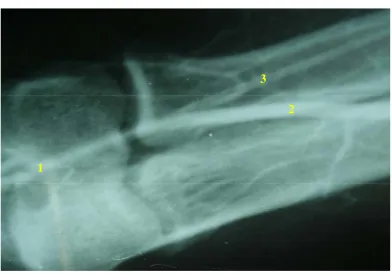
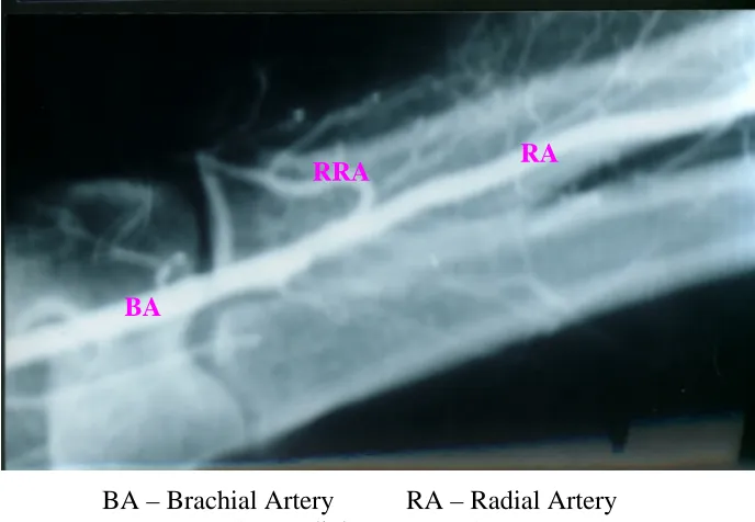
II. Adult Cadaveric Angiographic Study
2 disarticulated upper limbs were taken to the Barnard Institute of
Radiology, Government General Hospital, Chennai – 3 and under the guidance
of radiologist, the urograffin solution (contrast) was injected into the brachial
artery which was exposed in the cubital fossa by making a small incision over
that part. Then the branching pattern, course of the radial artery were studied
from 5 minutes after injecting the contrast. Then the conventional dissection
was carried out in these limbs.
III. CLINICAL STUDY
2 Clinical cases from the Vascular Surgery Department, Government
General Hospital, Chennai –3 have been selected with the age of 30 and 35
years with the following diagnosis
Sl.No. Sex / Age Diagnosis
1 male / 30 Ulnar artery occlusion
2 male / 35 Pseudoaneurysm of radial
recurrent artery.
In the above two cases the branching pattern of the radial artery was
observed by the angiographic study.
Angiographic Procedure
Retrograde axillary artery Catheterization
First the patient was put in supine position. Then the left arm was
abducted to the extreme and the hand was placed under the patient’s head. The
puncture site of the axillary artery was located along the lateral axillary fold
over the proximal part of the humerus so that the underlying bone provides
support during compression.
The axillary artery was palpated and fixed by the left index and middle
finger. Under local anesthesia, a small superficial skin nick was made with a
no. 11 blade directly over the arterial pulse. The course of the artery was