0022-538X/97/$04.00
1
0
Copyright
q
1997, American Society for Microbiology
Role of CD4 Epitopes outside the gp120-Binding Site during
Entry of Human Immunodeficiency Virus Type 1
JAMES H. M. SIMON,
1* PHIL STUMBLES,
2† NATHALIE SIGNORET,
3CHAMORRO SOMOZA,
2‡
MIKE PUKLAVEC,
2QUENTIN J. SATTENTAU,
3A. NEIL BARCLAY,
2ANDWILLIAM JAMES
1Sir William Dunn School of Pathology
1and MRC Cellular Immunology Unit, Sir William Dunn School of Pathology,
2University of Oxford, Oxford OX1 3RE, United Kingdom, and Centre d’Immunologie de Marseille-Luminy,
13288 Marseille Cedex 9, France
3Received 13 June 1996/Accepted 17 October 1996
CD4 is the primary receptor for human immunodeficiency virus (HIV). The binding site for the surface
glycoprotein of HIV type 1 (HIV-1), gp120, has been mapped to the C
*
-C
(
region of domain 1 of CD4.
Previously, we have shown that a mutant of rat CD4, in which this region was exchanged for that of human
CD4, is able to mediate infection of human cells by HIV-1, suggesting that essential interactions between HIV
and CD4 are confined to this region. Our observations appeared to conflict with mutagenesis and antibody
studies which implicate regions of CD4 outside the gp120-binding site in postbinding events during viral entry.
In order to resolve this issue, we have utilized a panel of anti-rat CD4 monoclonal antibodies in conjunction
with the rat-human chimeric CD4 to distinguish sequence-specific from steric effects. We find that several
antibodies to rat CD4 inhibit HIV infection in cells expressing the chimeric CD4 and that this is probably due
to steric hinderance. In addition, we demonstrate that replacement of the rat CDR3-like region with its human
homolog does not increase the affinity of the rat-human chimeric CD4 for gp120 or affect the exposure of gp41
following binding to CD4, providing further evidence that this region does not play a crucial role during entry
of virus.
The primary receptor for human immunodeficiency virus
type 1 (HIV-1) is CD4 (13, 27), a differentiation marker
ex-pressed on the surface of T lymphocytes, on a certain
popula-tion of dendritic cells, and, to a lesser extent, on macrophages
(44). CD4 is a member of the immunoglobulin (Ig)-like
super-family; its extracellular portion is comprised of four Ig domains
(4, 45, 53). The binding site for HIV gp120 has been mapped
to the second complementarity-determining region
(CDR2)-equivalent region of the amino-terminal domain of CD4 by use
of monoclonal antibodies (MAbs) (32, 47) and mutagenesis (1,
2, 6, 10, 33, 39, 49).
Binding of HIV to CD4 induces conformational changes in
the envelope glycoproteins that lead to fusion of the virus and
cellular membranes (35, 36, 48). Regions of CD4 outside the
gp120-binding site have been implicated in these postbinding
events. In particular, the F-G turn, or CDR3-equivalent
re-gion, of domain 1 (D1) has been implicated by numerous
authors (3, 8, 26, 29, 30, 38, 52). However, the weight of
evidence now suggests that this region is not involved in
post-binding events (5, 34, 43, 51).
MAbs to regions of human CD4 distant from the
gp120-binding site have been used in an effort to distinguish epitopes
of CD4 that are involved in binding gp120 from epitopes that
may be involved in postbinding events. Notably, MAbs against
CD4 D1 (12, 52), D2 (7, 37), D3 (24), and D3D4 (23) have all
been shown to inhibit virus infection, apparently with little or
no effect on virus binding.
Previously we generated a mutant of rat CD4 which binds
gp120 (49) and acts as an efficient receptor for HIV-1 (51),
implying that regions of CD4 outside the gp120-binding site
are unlikely to be essential for HIV interaction. In this study,
we investigated the ability of a panel of anti-rat CD4 MAbs,
none of which bind human CD4, to influence HIV infection of
cells expressing this chimeric receptor. We show that MAbs
which inhibit virus entry in this system bind epitopes of rat
CD4 analogous to those of human CD4 bound by inhibitory
anti-human CD4 MAbs. Furthermore, we show that although
these MAbs have little effect on the binding of monomeric
recombinant gp120 to cell surface CD4, the MAbs directed to
D1 and D2 are unable to form a ternary complex with a soluble
form of the receptor bound to oligomeric envelope protein
(Env) on the surface of HIV-infected cells. We conclude that
the principal mechanism by which D1 CDR3- and D2-directed
MAbs inhibit HIV infection is by the inhibition of appropriate
binding of CD4 to Env oligomers in the crowded environment
of the virion and virus-infected cells.
MATERIALS AND METHODS
Cell lines.BHK-21 cells (31), HeLa cells (21), HeLa m6 cells (expressing the rat-human chimeric CD4 molecule mutant [m6] [51]), and the HIV-1 Env-expressing HeLa cell line H32 (obtained from Lee Bacheler, DuPont Co., Wil-mington, Del.) were cultured in Dulbecco modified Eagle medium (Gibco BRL) containing 10% (vol/vol) fetal bovine serum (FBS) (Gibco BRL), 2 mML
-glutamine (Sigma), and antibiotics (50 U of penicillin per ml, 50mg of strepto-mycin per ml, and 100mg of neomycin per ml). In addition, HeLa m6 and H32 cells were maintained in 0.6 mg of Geneticin G418 (Sigma) per ml. C8166 cells (46) and H9 cells (40) were obtained from the National Institutes of Health AIDS Research and Reference Reagent Program and were maintained in RPMI 1640 medium (Gibco BRL) with 10% FBS,L-glutamine, and antibiotics.
Preparation of soluble and truncated forms of CD4.Soluble forms of CD4 consisting of either D1 and D2 [sCD4(d112)], D3 and D4 [sCD4(d314)], or the four extracellular domains of rat CD4 (sCD4) were produced from Chinese
* Corresponding author. Present address: Howard Hughes Medical
Institute, University of Pennsylvania School of Medicine, Clinical
Re-search Building, Room 350, Philadelphia, PA 19104. Phone: (215)
573-3494. Fax: (215) 573-2172. E-mail: [email protected]
.edu.
† Present address: Division of Cell Biology, TVW Telethon Institute
for Child Health Research, Subiaco, W.A. 6008, Australia.
‡ Present address: DNAX Research Institute of Molecular and
Cel-lular Biology Inc., Palo Alto, CA 94304-1104.
1476
on November 9, 2019 by guest
http://jvi.asm.org/
hamster ovary (CHO) cell lines transfected with the modified cDNA for rat CD4 by using the glutamine synthetase selection system (4, 15).
Production of anti-rat CD4 MAbs.BALB/c mice were immunized with 30 mg of sCD4 in complete Freund’s adjuvant subcutaneously at an interval of 2 weeks. After 8 weeks, mice were boosted with 60 mg of sCD4 in phosphate-buffered saline (PBS). For the final boost prior to taking the splenocytes for fusion with NSO myeloma cells, a mixture of free sCD4 and sCD4 bound to cells was given intravenously. Anti-CD4 MAb W3/25 (18 mg) was coupled to 0.1 ml of a 5% suspension of sheep erythrocytes by the chromic chloride method. After being washed, the cells were incubated with excess sCD4 (100 mg) and washed once, and a further 10 mg of sCD4 was added prior to intravenous injection. The spleen was taken for fusion with the myeloma NSO cells as described previously (54). The fusion was plated into four 96-well microtiter plates in the presence of irradiated thymocyte feeders, and hypoxanthine-aminopterin-thymidine selec-tion was applied. Supernatants were assayed after 10 days for binding to cells or sCD4 immobilized on polystyrene plates by enzyme-linked immunosorbent as-say.
Analysis of anti-CD4 MAb binding to sCD4 and fragments.The anti-CD4 MAbs were analyzed by binding to sCD4 or fragments immobilized by passive absorption to polystyrene plates and then reacting with125I-labelled rabbit
anti-mouse (RAM) immunoglobulin G (IgG) polyclonal antiserum as described pre-viously (16, 54) or with alkaline phosphatase-conjugated RAM IgG followed by color development with the alkaline phosphatase substrate 4-nitrophenylphos-phate. In addition to use of direct binding, various preparations of sCD4 were tested for the ability to inhibit these binding assays.
Viruses.HIV-1IIIB(42) was obtained from the National Institutes of Health
AIDS Research and Reference Reagent Program and propagated by acute infection of C8166 cells. The culture supernatants were clarified, filtered through 0.45-mm-pore-size filters, treated with 40 U of DNase I (Sigma) per ml for 30 min at room temperature to remove contaminating proviral DNA, and stored in 1-ml aliquots at2808C.
Cocal virus (COC) (25) was obtained from Robert Shope (Yale Arbovirus Unit) and propagated in BHK-21 cells. COC(HIV) pseudotype virus was made by superinfecting acutely HIV-1IIIB-infected H9 cells with COC, and pseudotype
plaque assays were performed essentially as described previously (51). These pseudotypes have the genome of COC and the spike glycoproteins of HIV, permitting HIV Env-mediated penetration of cells by COC, which leads to formation of COC plaques. The amount of gp120-mediated entry is calculated as the difference between the number of plaques produced when cells are chal-lenged with virus neutralized with anti-COC antiserum and the number of back-ground plaques when cells are challenged with COC(HIV) neutralized with both anti-COC antiserum and anti-gp120 polyclonal antibody.
PCR-based HIV-1 entry assay.Entry of HIV-1 was detected by PCR and was quantified essentially as previously described (11). HeLa m6 cells were chal-lenged with HIV-1IIIBand incubated at 378C for 2 h. The cells were then washed
and incubated in fresh medium. Twenty hours after challenge, the cells were washed and lysed. Proviral DNA was amplified by PCR with the U31-U52 primer pair (with U31radiolabelled with32
P), and amplification products were electrophoresed on agarose gels and blotted onto Hybond N1positively charged nylon (Amersham), which was then analyzed with a PhosphorImager. The bands were quantitated by using the ImageQuant program.
Flow cytometric analysis of MAb and gp120 binding to HeLa m6 cells.Binding of anti-rat CD4 MAbs and gp120 to HeLa m6 cells was analyzed by indirect flow cytometry with a FACScan (Becton Dickinson and Co., San Jose, Calif.). Bac-ulovirus-derived gp120 (HIV-1IIIBstrain) was obtained from the Medical
Re-search Council AIDS-Directed Programme. Cells were detached by treatment with PBS–0.5 mM EDTA, and 53105cells were incubated with 50ml of gp120
(at the appropriate concentration) or anti-rat CD4 MAb for 1.5 or 1 h, respec-tively, on ice. After being washed in ice-cold PBS–0.5% bovine serum albumin (BSA), the cells were incubated for a further 45 min on ice with 50ml of biotinylated anti-gp120 MAb (D47, which binds the V3 loop; a gift from Bob Doms), to detect gp120 or with fluorescein isothiocyanate-conjugated RAM Ig (RAM-FITC) (Serotec) to detect anti-rat CD4 MAbs. To detect bound gp120, cells were then washed and stained with streptavidin-FITC (Amersham).
Formation of ternary complexes between sm6, anti-rat CD4 MAbs, and oli-gomeric Env on the surface of chronically HIV-1-infected H9 cells.Indirect flow cytometry was used to analyze ternary complex formation between anti-rat CD4 MAbs and a soluble form of m6 (sm6) (49) bound to H9 cells chronically infected with the HXB-2D strain of HIV-1. Half a million cells were washed twice in PBS–0.5% BSA by pelleting and resuspended in 50ml of PBS–0.5% BSA con-taining sm6 at the appropriate concentration. The cells were incubated on ice for 2 h, washed, and resuspended with 50ml of anti-rat CD4 MAb. After a further 1 h on ice, the cells were washed and stained with RAM-FITC for 30 min at 48C, fixed with PBS–2% formaldehyde overnight, and analyzed by flow cytometry.
Measurement of sCD4 binding and gp41 exposure on the surface of HIV-1-infected H9 cells.Chronically infected H9 cells were washed in RPMI containing 2% FBS and resuspended at 23107cells/ml. Fifty microliters of sCD4 (human,
rat, m6, or m11), at the appropriate concentration, was added to an equal volume of cells. Half of the cells were then incubated at 48C to measure binding of sCD4, and the remaining half were incubated at 378C to measure exposure of gp41. After a 2-h incubation, cells were pelleted and washed twice in PBS containing 1% FBS and 0.02% sodium azide. Bound sCD4 was detected by staining first
with MAb L120 (D. Buck, Becton Dickinson) for human CD4 or with MRC-OX-71 for rat CD4 and m6 and m11 and then with a phycoerythrin-conjugated anti-mouse Ig (Immunotech SA, Marseille, France). Exposure of gp41 was de-tected by staining with MAb 50-96 (a gift from S. Zolla-Pazner) followed by phycoerythrin-conjugated anti-human Ig (Immunotech SA). Cells were washed twice, fixed overnight in PBS–2% formaldehyde, and then analyzed by flow cytometry.
RESULTS
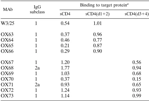
Anti-CD4 MAb binding to fragments of sCD4.
The MAbs
obtained were tested for their reactivities with sCD4(d1
1
2)
and sCD4(d3
1
4), and a total of 11 were cloned for further use.
The reactivities of these MAbs are shown in Table 1. As
ex-pected, those antibodies reactive with sCD4(d1
1
2) gave no
reaction with sCD4(d3
1
4) and vice versa (Table 1). All of the
new MAbs were shown to bind to cell surface CD4 by
fluores-cence-activated cell sorter analysis (data not shown).
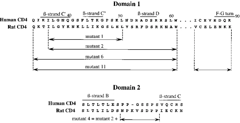
Epitope mapping of anti-CD4 MAbs recognizing the
amino-terminal domains of CD4.
The determinants recognized by
MAbs binding to D1 and D2 of rat CD4 generated in this and
previous studies were characterized by using a series of
muta-tions that had been produced in D1 and D2 in another project
(49). Rat CD4 does not bind gp120 of HIV-1, but residues
identified as important in the HIV-1-binding site of human
CD4 were substituted in the rat sequence in an attempt to
make rat CD4 bind HIV-1, as illustrated in Fig. 1.
Mutants were prepared in the CDM8 vector and transiently
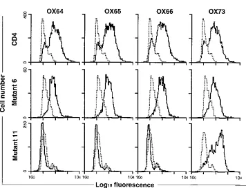
expressed in COS cells. MRC-OX-35 and MRC-OX-36 bound
to m1 (data not shown) but not to m4 (Fig. 2A) indicating that
epitopes in the B-C loop of D2 are required for binding of
these MAbs. m11, which contained changes in the F-G loop in
addition to changes in the C
9
-D region, caused the loss of
MRC-OX-64, -65, and -66 reactivity (Fig. 2B). As m6
con-tained only the C
9
-D region changes, the epitope for these
MAbs must involve the F-G turn. In summary, three groups of
MAbs were mapped: W3/25 (with MRC-OX-37, -38, and -63)
to the C
9
-C
0
region of D1 (data not shown) (14); MRC-OX-64,
-65, and -66 to the F-G turn in D1; and MRC-OX-35 and -36
to the B-C region of D2.
[image:2.612.315.556.81.244.2]Inhibition of viral entry by anti-rat CD4 MAbs.
m6 binds
gp120 with an affinity of two- to threefold less than that of
human CD4 (49) and is an efficient receptor for HIV-1 when
expressed on the surface of HeLa cells (51). HeLa m6 cells
were preincubated with each of the m6-binding MAbs for 1 h
TABLE 1. Binding of anti-CD4 MAbs to domains of rat sCD4
MAb IgG
subclass
Binding to target proteina
sCD4 sCD4(d112) sCD4(d314)
W3/25
1
0.54
1.01
OX63
1
0.37
0.96
OX64
1
0.46
0.77
OX65
1
0.21
0.87
OX66
1
0.29
0.90
OX67
1
1.20
0.56
OX68
2a
1.77
0.94
OX69
1
1.03
0.68
OX70
1
0.37
0.15
OX71
2a
0.93
0.65
OX72
1
1.24
0.93
OX73
1
1.14
0.99
aMean optical density at 405 nm for MAb binding to duplicate microtiter plate wells. No binding to any sCD4 preparation by an irrelevant IgG1 control MAb (OX21) was observed.
V
OL. 71, 1997
EPITOPES OF CD4 INVOLVED IN HIV ENTRY
1477
on November 9, 2019 by guest
http://jvi.asm.org/
at 37
8
C. MRC-OX-63 was used as a positive control, as it binds
the CDR2 region of rat CD4 and, consequently, does not bind
m6, in which this region has been replaced with its human
homolog. The cells were then challenged with HIV-1
IIIB, and
the level of virus entry was quantitated by using a PCR-based
technique as described in Materials and Methods. (CD4 MAbs
were present at all times during the course of the experiment,
including during challenge with virus and after washing of
cells.) The MAbs that bind the CDR3-equivalent region of
CD4 D1 (MRC-OX-64, MRC-OX-65, and MRC-OX-66), the
D2-binding MAbs (MRC-OX-35 and MRC-OX-36), and two
of the D3D4-binding MAbs (MRC-OX-67 and MRC-OX-68)
all significantly inhibited viral entry, while the control MAb
and those binding other epitopes did not (Fig. 3A).
As a second measure of viral entry, HeLa m6 cells which had
been preincubated with the MAbs at 37
8
C for 1 h were
chal-lenged with COC(HIV) pseudotype virus as described in
Ma-terials and Methods. The results confirm the PCR data, with
the same set of MAbs inhibiting entry of pseudotype virus (Fig.
3B).
Several anti-rat CD4 MAbs inhibit syncytium formation.
HeLa m6 cells were preincubated with each of the anti-rat
CD4 MAbs and then mixed with an equal number of HIV
Env-expressing H32 cells. After overnight incubation in the
continual presence of antibody, the cells were fixed and
stained, and the number of syncytia was quantitated (Fig. 4).
As expected, all of the MAbs that inhibited viral entry also
inhibited syncytium formation. However, the D3D4-specific
MAb, MRC-OX-71, also interfered with syncytium formation,
although it had no effect on virus entry (Fig. 3). Very slight
enhancement of syncytium formation was observed in the
pres-ence of MRC-OX-69.
Effect of the anti-rat CD4 antibodies on binding of
mono-meric gp120 to HeLa m6 cells.
In order to determine whether
binding of any of the anti-rat CD4 MAbs to HeLa m6 cells
affected binding of recombinant monomeric gp120, a flow
cy-tometry assay was developed. After a 1-h incubation of HeLa
m6 cells with each of the anti-rat CD4 MAbs, gp120 was added
to a final concentration of 10
m
g/ml. This concentration is
sufficient to saturate the gp120-binding sites on the surface of
HeLa m6 cells (results not shown). The cells were incubated
for a further 1.5 h on ice, and the amount of bound gp120 was
determined by flow cytometry as described in Materials and
Methods. The results presented in Fig. 5 demonstrate that
binding of soluble, monomeric gp120 was not inhibited by any
of the anti-rat CD4 MAbs.
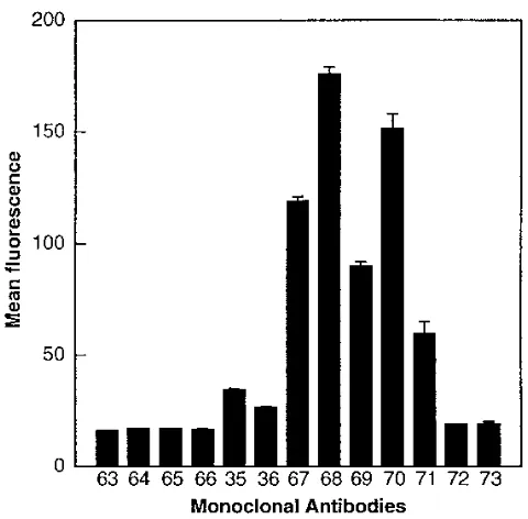
Effect of gp120 on binding of anti-rat CD4 MAbs to HeLa
m6 cells.
In a reciprocal experiment, HeLa m6 cells were
incubated with 50
m
l of either PBS–0.5% BSA or gp120 (10
m
g/ml) for 1.5 h at 4
8
C. These cells were then washed and
stained with each of the m6-binding anti-rat CD4 MAbs and
with MRC-OX-63 (as a negative control), as described in
Ma-terials and Methods. Prebinding of gp120 resulted in a
reduc-tion of at least 50% in binding of the D1 CDR3-directed MAbs
(MRC-OX-64, MRC-OX-65, and MRC-OX-66) and of the
D3-binding MAbs (MRC-OX-67 and MRC-OX-68) (Fig. 6).
There was no significant reduction in binding of any of the
other anti-rat CD4 MAbs.
The loss of binding of certain MAbs induced by monomeric
gp120 was unexpected, as these MAbs did not affect binding of
monomeric gp120 to CD4. One possible explanation is that
gp120 induces a conformational change in CD4 that leads to a
loss of the epitopes bound by these MAbs. In the case of the
D1 CDR3-directed MAbs, this seems unlikely, as there would
have to be a conformational change within an Ig domain, a
structural motif known to be rigid. A more plausible
explana-tion is that there is a conformaexplana-tional change in gp120 that leads
to a masking of the D1 CDR3 loop. A gp120-induced
confor-mational change in CD4 leading to the loss of the
MRC-OX-67/68 epitope is conceivable, as this would require interdomain
changes. The possibility of such conformational changes
be-tween the D1D2 and D3D4 halves of CD4, around the hinge
region, has previously been proposed (24).
Numerous epitopes of m6 are masked after binding to cell
surface, oligomeric Env.
On the surface of virions, Env is
oligomeric, and consequently, binding of CD4 to virions may
differ from binding of CD4 to recombinant, monomeric gp120.
Binding of a soluble form of m6 (sm6) to oligomeric Env was
assessed by flow cytometry of chronically HIV-1
IIIB-infected
H9 cells (H9/HIV-1
IIIB), as described in Materials and
Meth-ods.
The concentration of sm6 necessary to saturate
H9/HIV-1
IIIBcells (10
m
g/ml) was determined as described in Materials
[image:3.612.111.502.71.272.2]and Methods (results not shown). This concentration is greater
FIG. 1. Sequences of rat CD4 mutants constructed by substitution of human CD4 residues.
on November 9, 2019 by guest
http://jvi.asm.org/
FIG. 2. Epitope mapping of D1- and D2-directed MAbs. Fluorescence-activated cell sorter histograms of MAb tissue culture supernatants binding to COS cells transfected with the mutants of CD4 are shown, with dotted lines showing staining with the negative control MAb MRC-OX-21. (A) Localization of the epitopes for MRC-OX-35 and -36 to D2 of CD4 (m4). (B) Localization of the epitopes for MRC-OX-64, -65, and -66 to the F-G turn of D1 of CD4 (m11).
V
OL. 71, 1997
EPITOPES OF CD4 INVOLVED IN HIV ENTRY
1479
on November 9, 2019 by guest
http://jvi.asm.org/
than that previously determined for human CD4 (48), and this
is likely to reflect the lower affinity for gp120 of m6 compared
to human CD4 (49).
Next, H9/HIV-1
IIIBcells were saturated with sm6 and
stained with each of the anti-rat CD4 MAbs as described in
Materials and Methods. The epitopes for the D1 MAbs
(MRC-OX-64, MRC-OX-65, and MRC-OX-66), the D2 MAbs
(MRC-OX-35 and MRC-OX-36), and some of the D3D4
MAbs (MRC-OX-72 and MRC-OX-73) were masked (Fig. 7).
There were various fluorescence intensities for all of the other
MAbs (67, 68, 69,
MRC-OX-70, and MRC-OX-71), which may reflect various degrees of
exposure of the epitopes bound by these MAbs or a variation
in the valency of binding to sm6.
The CDR3-equivalent region of CD4 does not have an effect
on affinity of CD4 for oligomeric Env or exposure of gp41
epitopes.
Previously, we demonstrated that the
CDR3-equiva-lent region of D1 of CD4 was unlikely to be involved in
post-binding events that lead to fusion between viral and cellular
membranes, since m6 acts as an efficient receptor for HIV-1
despite having a markedly different sequence in this region
(51). However, m6 is not as efficient a receptor as wild-type
CD4. We believe that this is because it has a slightly lower
affinity for gp120 than does human CD4 (49), but we cannot
formally rule out an involvement of the CDR3 region. In
ad-dition, the partial occlusion of MAb binding to the CDR3 loop
following soluble gp120 binding may indicate the presence of
an interaction, albeit of low affinity, between the CDR3 loop
and gp120.
[image:5.612.61.301.67.513.2]In order to address this point more specifically, we
con-structed a mutant (m11) in which the F-G turn of m6 was
replaced with the equivalent residues of human CD4 (Fig. 1).
The affinities of sm6 and sm11 for HIV-infected H9 cells and
their abilities to induce exposure of gp41 were measured as
described in Materials and Methods. The results presented in
Fig. 8. demonstrate that the replacement of rat with human
sequence in the CDR3 region of m6 has no effect either on its
affinity for oligomeric Env or on exposure of gp41 epitopes.
Instead, we found that the degree of gp41 exposure could be
directly correlated to the affinity of CD4 for gp120 by using a
FIG. 3. Inhibition of viral entry into HeLa m6 cells by anti-rat CD4 MAbs. (A) HeLa m6 cells were preincubated with each of the m6-binding MAbs for 1 h at 378C and then challenged with HIV. As a negative control, untransfected HeLa cells (H) were used, and as positive controls, HeLa m6 cells preincubated with MRC-OX-63 (63) or growth medium (C) were used. Twenty hours after challenge, the cells were lysed and proviral DNA was amplified by PCR with radiolabelled primers (U31and U52). The relative levels of proviral DNA were quantitated with a PhosphorImager and are shown as a means with standard errors for triplicate determinations. Arbitrary PhosphorImager units (APUs) are used. Each of the MAbs is indicated by its MRC-OX number. (B) As a second measure of viral entry, HeLa m6 cells were challenged with COC(HIV) pseudotypes after preincubation of cells with the anti-rat CD4 MAbs. The rel-ative levels of pseudotype entry are shown as the mean number of PFU 6 standard error for duplicate determinations.
FIG. 4. Inhibition of syncytium formation by anti-rat CD4 MAbs. HeLa m6 cells were preincubated with each of the MAbs for 1 h at 378C and then mixed with an equal number of gp160-expressing HeLa H32 cells. After an overnight incubation, the cells were fixed, stained, and counted. The graph shows the mean numbers of syncytia, with standard errors, per field for 10 fields. Each of the MAbs is indicated by its MRC-OX number. As a negative control, untransfected HeLa cells (H) were mixed with HeLa H32 cells, and as positive controls, HeLa m6 cells preincubated with MRC-OX-63 (63) or growth medium (C) were mixed with HeLa H32 cells.
on November 9, 2019 by guest
http://jvi.asm.org/
[image:5.612.317.553.68.300.2]panel of rat-human chimeric CD4 molecules with various
gp120 affinities (results not shown).
DISCUSSION
In the work presented here, we have used a chimera of rat
and human CD4, which acts as a functional receptor for HIV-1
(49, 51), to examine the role played by regions of CD4 distant
from the gp120-binding site during the postbinding events that
lead to membrane fusion. A panel of anti-rat CD4 MAbs,
which bind this chimera but not human CD4, was used in an
effort to determine the mechanisms by which anti-CD4 MAbs
interfere with virus entry and syncytium formation without
apparently blocking binding of virus (summarized in Fig. 6).
D1.
The binding site for gp120 is contained within the
CDR2-equivalent region of CD4 D1 (1, 2, 6, 10, 33, 39, 47, 49).
Following binding to CD4, the spike glycoproteins of HIV
undergo a series of conformational changes that lead to
virus-cell fusion (35, 36, 48). It has become evident that these
con-formational changes cannot result from specific interactions of
gp120 with the D1 CDR3 region of CD4 (5, 34, 43, 51),
al-though subtle strain-specific effects have been reported (50).
However, MAbs that bind this region of human CD4 inhibit
virus replication and syncytium formation (12, 52). One
expla-nation for this is that antibodies to CDR3 do not interfere with
the binding of CD4 to a gp120 monomer or to some of the
available sites within an Env oligomer but that they sterically
hinder the binding of CD4 molecules to all of the gp120 sites
within an oligomer, as previously suggested (28, 34, 35). This
model would predict, and our results confirm, that the D1
CDR3 epitope is masked when sm6 (a soluble form of the
chimeric CD4) occupies all of the available sites in an Env
oligomer. Consequently, the inhibition of virus entry and
syn-cytium formation by these antibodies may be by inhibiting the
binding of CD4 to multiple sites in an Env oligomer, which has
been shown to be necessary to trigger membrane fusion (35).
The demonstration that exposure of gp41 is unaffected by the
sequence of the CDR3 loop provides further evidence that this
region is not involved in postbinding events.
A previous study (12) reported that an anti-CDR3 loop
MAb (13B.8.2) inhibited replication of HIV-1 and that this
inhibition occurred at a step after virus entry. In our hands,
however, MAbs specific for rat CDR3 were efficient inhibitors
of HIV-1 entry. The reason for this difference remains unclear.
D2.
The anti-rat CD4 MAbs MRC-OX-35 and MRC-OX-36
bind a region of rat CD4 equivalent to that bound by the
anti-human CD4 MAb 5A8, which was found to inhibit virus
replication and syncytium formation (7). The mechanism by
which the MAbs to this D2 epitope inhibit virus entry and
syncytium formation appears to be similar, although not
iden-tical, to that of the anti-D1 CDR3 MAbs (34, 37, 52). MAbs
that bind either of these epitopes reduce binding of sCD4 to
membrane-bound oligomeric Env but do not affect binding of
monomeric gp120 to CD4 (34, 37). In addition, we have shown
that both of these epitopes are masked when sm6 has saturated
the oligomeric Env sites on the surface of HIV-1
IIIB-infected
cells. However, the D2 epitopes differ from the D1 CDR3
epitope, as they are not masked following binding to
mono-meric gp120. A possible explanation for this is that the binding
of multiple molecules of CD4 to oligomeric Env orients the
epitope toward the interior of the complex, thereby concealing
it from antibody.
D3 and D4.
A role for the D3D4 moiety of CD4 in
post-binding events has previously been implicated from work with
anti-human CD4 MAbs (23, 24) and mutagenesis studies (22,
FIG. 5. Effect of anti-rat CD4 MAbs on the binding of monomeric gp120 to HeLa m6 cells. HeLa m6 cells were incubated for 1 h at 48C with each of the MAbs, washed, and then incubated for a further 1.5 h at 48C with 10mg of gp120 per ml. The cells were then washed and incubated with biotinylated anti-gp120 MAb for 45 min on ice. After further washing, the cells were stained with streptavidin-FITC and analyzed by flow cytometry. The graph shows the mean fluorescence channels with standard errors for duplicate samples. As a negative control (C), untransfected HeLa cells were used, and as a positive control, cells preincubated with MRC-OX63 were used. Each of the MAbs is indicated by its MRC-OX number.
FIG. 6. Effect of monomeric gp120 on the binding of anti-rat CD4 MAbs to HeLa m6 cells. HeLa m6 cells were incubated for 1.5 h at 48C with either PBS–0.1% BSA (filled bars) or 10mg of monomeric gp120 per ml (hatched bars). The cells were then washed and incubated with each of the anti-rat CD4 MAbs (shown by their MRC-OX numbers) for a further 1 h. After further washing, the cells were stained with RAM-FITC and analyzed by flow cytometry. The graph shows the mean fluorescence channels for duplicate determinations with stan-dard errors.
V
OL. 71, 1997
EPITOPES OF CD4 INVOLVED IN HIV ENTRY
1481
on November 9, 2019 by guest
http://jvi.asm.org/
41). The MAbs against D3D4 of rat CD4 can be placed in
distinct functional groups as a result of our experiments.
MRC-OX-67 and MRC-OX-68 form the first group. These MAbs
partially inhibit cell-free virus entry and syncytium formation,
and their epitopes are partially masked by prebinding of
mono-meric gp120. However, in contrast to the D1 CDR3 and D2
antibodies, they bind normally to sm6 complexed with
oligo-meric Env. In these respects, they appear to be analogous to
the anti-human CD4 MAb Q425, which binds close to the top
of D3, near the hinge region (24). Studies with the Q425 MAb
have led to the suggestion that flexion about the hinge of CD4
may be involved in the postbinding events that lead to fusion
(24).
The second group of D3D4 MAbs comprises MRC-OX-69
and MRC-OX-71, both of which modulate syncytium
forma-tion, albeit modestly, but have no effect on entry of cell-free
virus. This suggests that there may be differences in the mode
of interaction between CD4 and gp120 during syncytium
for-mation and entry of cell-free virus. However, a modest
alter-ation in the kinetics of fusion caused by binding of
MRC-OX-69 or MRC-OX-71 could explain this observation, given
that syncytium formation requires more interactions between
Env and CD4 than does entry of cell-free virus.
A third group of D3D4 MAbs includes MRC-OX-72 and
MRC-OX-73, which do not interfere with HIV infection or
syncytium formation and yet have epitopes that are masked by
interaction with oligomeric but not monomeric gp120. This
shows that the results obtained from this system to examine
binding of CD4 to oligomeric Env, although more reliable than
those obtained from investigating monomeric interactions,
must still be treated with caution.
The system utilized for analyzing CD4-gp120 interactions is
critical, particularly when assessing the effects of MAbs on this
interaction. Although analysis of the binding of recombinant,
[image:7.612.58.298.67.303.2]monomeric gp120 to sCD4 or cell surface CD4 reveals
impor-tant clues, the true physiological picture represents an even
greater level of complexity, since both the Env glycoproteins
and CD4 will be in an oligomeric, membrane-bound form. The
packing of gp120-CD4 complexes is therefore more
con-strained in a virus-cell interaction than in monomeric
gp120-CD4 interactions, and consequently, a MAb that has little or
[image:7.612.318.552.67.541.2]FIG. 7. Formation of ternary complexes between sm6 anti-rat CD4 MAbs and oligomeric Env on the surface of chronically HIV-infected H9 cells. Chron-ically HIV-infected H9 cells were incubated for 2 h on ice with 10mg of sm6 per ml. These cells were then washed, incubated with each of the anti-rat CD4 MAbs (shown by their MRC-OX numbers) for 1 h, stained with RAM-FITC, and analyzed by flow cytometry. The graph shows the mean fluorescence channels for duplicate samples with standard errors.
FIG. 8. The sequence of the CDR3 loop of CD4 does not affect affinity for gp120 or exposure of gp41. Chronically HIV-infected H9 cells were incubated for 2 h on ice (A) or at 378C (B) with the appropriate concentration of soluble m6 (closed circles), m11 (open triangles), or human (open circles) or rat (closed triangles) CD4. The cells were then stained with L120 (human CD4) or MRC-OX-71 (rat CD4, m6, and m11) followed by phycoerythrin-conjugated anti-mouse Ig to detect the levels of bound CD4 (A) or with MAb 50-96 followed by phycoerythrin-conjugated anti-human Ig to determine the level of gp41 exposure (B) and analyzed by flow cytometry. The mean fluorescence intensity as a func-tion of the concentrafunc-tion of sCD4 is shown.
on November 9, 2019 by guest
http://jvi.asm.org/
no effect on formation of this complex when the constituents
are monomeric may well interfere with appropriate binding in
the oligomeric system.
In summary, anti-rat CD4 MAbs affect virus entry and
syn-cytium formation of m6-expressing cells in a way analogous to
that of MAbs against human CD4. For example, the
CDR3-directed mAbs, MRC-OX-64, -65, and -66, may be analogous
to the anti-human CD4 MAb L71 (52); the D2-directed MAbs,
MRC-OX-35 and -36, may be analogous to 5A8 (7); and the
D3 MAbs, MRC-OX-68 and -69, may be analogous to Q425
(24). This conservation of function shows that while the
pri-mary sequence of CD4 epitopes outside the gp120-binding site
does not appear to influence cell-virus fusion, binding of MAbs
to some of these regions does. One mechanism by which these
inhibitory MAbs may act is by sterically interfering with
pack-ing of CD4 molecules to oligomers of Env in the crowded
environment on the virion surface. In addition, some MAbs
may sterically hinder molecular rearrangements induced by the
CD4-gp120 interaction either in gp120-gp41 oligomers or
around the hinge of CD4.
Recently, several HIV-1 fusion cofactors have been
identi-fied (9, 17–20). The relationship of these cofactors to the
Env-CD4 interaction remains to be elucidated at the molecular
level. As there is little sequence identity between m6, a
func-tional receptor for HIV-1 (51), and human CD4 outside the
gp120-binding site, it is unlikely that regions of CD4 outside
this domain could interact directly with any of the fusion
co-factors. However, it is possible that some of the MAbs
de-scribed in this study may act by sterically inhibiting interactions
between Env and a fusion cofactor after docking of the virus to
CD4.
ACKNOWLEDGMENTS
This work was funded by the MRC AIDS-Directed Programme; by
the ANRS, CNRS, and INSERM of France; and by the EC Concerted
Action “Virus-cell membrane interactions.”
We thank Simon Davis and John Moore for useful discussions and
Vinod Aachen and Harsh Sharma for help in preliminary
character-ization of the CD4 MAbs.
REFERENCES
1. Arthos, J., K. C. Deen, M. A. Chaikin, J. A. Fornwald, G. Sathe, Q. J. Sattentau, P. R. Clapham, R. A. Weiss, J. S. McDougal, C. Pietropaolo, R. Axel, A. Truneh, P. J. Maddon, and R. W. Sweet.1989. Identification of the residues in human CD4 critical for the binding of HIV. Cell 57:469–481. 2. Ashkenazi, A., L. G. Presta, S. A. Marsters, T. R. Camerato, K. A. Rosenthal,
B. M. Fendly, and D. J. Capon.1990. Mapping the CD4 binding site for human immunodeficiency virus by alanine-scanning mutagenesis. Proc. Natl. Acad. Sci. USA 87:7150–7154.
3. Berger, E. A., J. D. Lifson, and L. E. Eiden. 1991. Stimulation of glycoprotein gp120 dissociation from the envelope glycoprotein complex of human im-munodeficiency virus type 1 by soluble CD4 and CD4 peptide derivatives: implications for the role of the complementarity-determining region 3-like region in membrane fusion. Proc. Natl. Acad. Sci. USA 88:8082–8086. 4. Brady, R., E. Dodson, G. Dodson, G. Lange, S. Davis, A. F. Williams, and
A. N. Barclay.1993. Crystal structure of domains 3 and 4 of rat CD4: relation to the NH2-terminal domains. Science 260:979–983.
5. Broder, C. C., and E. A. Berger. 1993. CD4 molecules with a diversity of mutations encompassing the CDR3 region efficiently support human immu-nodeficiency virus type 1 envelope glycoprotein-mediated cell fusion. J. Vi-rol. 67:913–926.
6. Brodsky, M. H., M. Warton, R. M. Myers, and D. R. Littman. 1990. Analysis of the site in CD4 that binds to the HIV envelope glycoprotein. J. Immunol. 144:3078–30786.
7. Burkly, L., D. Olson, R. Shapiro, G. Winkler, J. Rosa, D. Thomas, C. Williams, and P. Chisholm.1992. Inhibition of infection by novel CD4 domain 2-specific monoclonal antibodies. J. Immunol. 149:1779–1787. 8. Camerini, D., and B. Seed. 1990. A CD4 domain important for
HIV-medi-ated syncytium formation lies outside the virus binding site. Cell 60:747–754. 9. Choe, H., M. Farzan, Y. Sun, N. Sullivan, B. Rollins, P. D. Ponath, L. Wu, C. R. Mackay, G. LaRosa, W. Newman, N. Gerard, C. Gerard, and J. Sodroski.1996. The b-chemokine receptors CCR3 and CCR5 facilitate
infection by primary HIV-1 isolates. Cell 85:1135–1148.
10. Clayton, L. K., R. E. Hussey, R. Steinbrich, H. Ramachandran, Y. Husain, and E. L. Reinherz.1988. Substitution of murine for human CD4 residues identifies amino acids critical for HIV-gp120 binding. Nature 335:363–366. 11. Collin, M., and S. Gordon. 1994. The kinetics of HIV reverse transcription are slower in primary human macrophages than in a lymphoid line. Virology 200:114–120.
12. Corbeau, P., M. Benkirane, R. Weil, C. David, S. Emiliani, D. Olive, C. Mawas, A. Serre, and C. Devaux.1993. Ig CDR-3 like region of CD4 molecule is involved in syncytia formation but not in viral entry. J. Immunol. 150:290–301.
13. Dalgleish, A. G., P. C. L. Beverley, P. R. Clapham, D. H. Crawford, M. F. Greaves, and R. A. Weiss.1984. The CD4 (T4) antigen is an essential component of the receptor for the AIDS retrovirus. Nature 312:736–766. 14. Davis, S., G. Schockmel, C. Somoza, D. Buck, D. Healey, E. Rieber, C. Reiter,
and A. Williams.1992. Antibody and HIV-1 gp120 recognition of CD4 undermines the concept of mimicry between antibodies and receptors. Na-ture 358:76–79.
15. Davis, S. J., R. L. Brady, A. N. Barclay, K. Harlos, G. G. Dodson, and A. F. Williams.1990. Crystallization of a soluble form of the rat T-cell surface glycoprotein CD4 complexed with Fab from the W3/25 monoclonal anti-body. J. Mol. Biol. 213:7–10.
16. Davis, S. J., H. A. Ward, M. J. Puklavec, A. C. Willis, A. F. Williams, and A. N. Barclay.1990. High level expression in Chinese hamster ovary cells of soluble forms of CD4 T lymphocyte glycoprotein including glycosylation variants. J. Biol. Chem. 265:10410–10418.
17. Deng, H., R. Liu, W. Ellmeier, S. Choe, D. Unutmaz, M. Burkhart, P. Di Marzio, S. Marmon, R. E. Sutton, C. M. Hill, C. B. Davis, S. C. Peiper, T. J. Schall, D. R. Littman, and N. R. Landau.1996. Identification of a major co-receptor for primary isolates of HIV-1. Nature 381:661–666.
18. Doranz, B. J., J. Rucker, Y. Yi, R. Smyth, M. Samson, S. C. Peiper, M. Parmentier, R. G. Collman, and R. W. Doms.1996. A dual-tropic primary HIV-1 isolate that uses fusin and theb-chemokine receptors CKR-5, CKR-3 and CKR-2b as fusion cofactors. Cell 85:1149–1158.
19. Dragic, T., V. Litwin, G. P. Allaway, S. R. Martin, Y. Huang, K. A. Na-gashima, C. Cayanan, P. J. Maddon, R. A. Koup, J. P. Moore, and W. A. Paxton.1996. HIV-1 entry into CD41cells is mediated by the chemokine receptor CC-CKR-5. Nature 381:667–673.
20. Feng, Y., C. C. Broder, P. E. Kennedy, and E. A. Berger. 1996. HIV-1 entry cofactor: functional cDNA cloning of a seven-transmembrane, G protein-coupled receptor. Science 272:872–877.
21. Gey, G. O., W. D. Coffman, and M. T. Kubicek. 1952. Tissue culture studies of the proliferative capacity of cervical carcionoma and normal epithelium. Cancer Res. 12:264–265.
22. Golding, H., R. Blumenthal, J. Manischewitz, D. R. Littman, and D. S. Dimitrov.1993. Cell fusion mediated by interaction of a hybrid CD4.CD8 molecule with the human immunodeficiency virus type 1 envelope glycopro-tein does occur after a long lag time. J. Virol. 67:6469–6475.
23. Hasunuma, T., H. Tsubota, M. Watanabe, Z. Chen, C. Lord, L. Burkly, J. Daley, and N. Letvin.1992. Regions of the CD4 molecule not involved in virus binding or syncytia formation are required for HIV-1 infection of lymphocytes. J. Immunol. 148:1841–1846.
24. Healey, D., L. Dianda, J. P. Moore, J. S. McDougal, M. J. Moore, P. Estess, D. Buck, P. D. Kwong, P. C. L. Beverley, and Q. J. Sattentau.1990. Novel anti-CD4 monoclonal antibodies separate human immunodeficiency virus infection and fusion of CD41cells from virus binding. J. Exp. Med. 172: 1233–1242.
25. Jonkers, A. H., R. E. Shope, T. H. G. Aitken, and L. Spence. 1964. Cocal virus, a new agent in Trinidad related to vesicular stomatitis virus, type Indiana. Am. J. Vet. Res. 25:236–241.
26. Kalyanaraman, V. S., D. M. Rausch, J. Osborne, M. Padgett, K. M. Hwang, J. D. Lifson, and L. E. Eiden.1990. Evidence by peptide mapping that the region CD4(81-92) is involved in gp120/CD4 interaction leading to HIV infection and HIV-induced syncytium formation. J. Immunol. 145:4072– 4078.
27. Klatzmann, D., E. Champagne, S. Chamaret, J. Gruest, D. Guetard, T. Hercend, J.-C. Gluckman, and L. Montagnier.1984. T-lymphocyte T4 mol-ecule behaves as the receptor for the human retrovirus LAV. Nature 312: 767–768.
28. Layne, S. P., M. J. Merges, M. Dembo, J. L. Spouge, and P. L. Nara. 1990. HIV requires multiple gp120 molecules for CD4-mediated infection. Nature 346:277–279.
29. Lifson, J. D., K. M. Hwang, P. L. Nara, B. Fraser, M. Padgett, N. M. Dunlop, and L. E. Eiden.1988. Synthetic CD4 peptide derivatives that inhibit HIV infection and cytopathicity. Science 241:712–716.
30. Lifson, J. D., D. M. Rausch, V. S. Kalyanaraman, K. M. Hwang, and L. E. Eiden.1991. Synthetic peptides allow discrimination of structural features of CD4(81-92) important for HIV-1 infection versus HIV-1-induced syncytium formation. AIDS Res. Hum. Retroviruses 7:521–527.
31. MacPherson, I. and M. Stoker. 1962. Polyoma transformation of hamster cell clones—an investigation of genetic factors affecting cell competence. Virology 16:147–151.
V
OL. 71, 1997
EPITOPES OF CD4 INVOLVED IN HIV ENTRY
1483
on November 9, 2019 by guest
http://jvi.asm.org/
32. McDougal, J. S., J. K. Nicholson, G. D. Cross, S. P. Cort, M. S. Kennedy, and A. C. Mawle.1986. Binding of the human retrovirus HTLV-III/LAV/ARV/ HIV to the CD4 (T4) molecule: conformation dependence, epitope map-ping, antibody inhibition, and potential for idiotypic mimicry. J. Immunol. 137:2937–2944.
33. Moebius, U., L. Clayton, S. Abraham, S. Harrison, and E. Reinherz. 1992. The human immunodeficiency virus gp120 binding site on CD4: delineation by quantitative equilibrium and kinetic binding studies of mutants in con-junction with a high resolution CD4 atomic structure. J. Exp. Med. 176:507– 517.
34. Moore, J. 1993. A monoclonal antibody to the CDR-3 region of CD4 inhibits sCD4 binding to virions of HIV-1. J. Virol. 67:3656–3659.
35. Moore, J. P., J. A. McKeating, W. A. Norton, and Q. J. Sattentau. 1991. Direct measurement of soluble CD4 binding to human immunodeficiency virus type 1 virions: gp120 dissociation and its implications for virus-cell binding and fusion reactions and their neutralization by soluble CD4. J Virol. 65:1133–1140.
36. Moore, J. P., J. A. McKeating, R. A. Weiss, and Q. J. Sattentau. 1990. Dissociation of gp120 from HIV-1 virions induced by soluble CD4. Science 250:1139–1142.
37. Moore, J. P., Q. J. Sattentau, P. J. Klasse, and L. C. Burkly. 1992. A monoclonal antibody to CD4 domain 2 blocks soluble CD4-induced confor-mational changes in the envelope glycoproteins of human immunodeficiency virus type 1 (HIV-1) and HIV-1 infection of CD41cells. J. Virol. 66:4784– 4793.
38. Nara, P. L., K. M. Hwang, D. M. Rausch, J. D. Lifson, and L. E. Eiden. 1989. CD4 antigen-based antireceptor peptides inhibit infectivity of human immu-nodeficiency virus in vitro at multiple stages of the viral life cycle. Proc. Natl. Acad. Sci. USA 86:7139–7143.
39. Peterson, A., and B. Seed. 1988. Genetic analysis of monoclonal antibody and HIV binding sites on the human lymphocyte antigen CD4. Cell. 54:65–72. 40. Popovic, M., M. G. Sarngadharan, E. Read, and R. C. Gallo. 1984.
Detec-tion, isolation and continuous production of cytopathic retroviruses (HTLV-III) from patients with AIDS and pre-AIDS. Science 224:497–500. 41. Poulin, L., L. A. Evans, S. B. Tang, A. Barboza, H. Legg, D. R. Littman, and
J. A. Levy.1991. Several CD4 domains can play a role in human immuno-deficiency virus infection in cells. J. Virol. 65:4893–4901.
42. Ratner, L., W. Haseltine, R. Patarca, K. J. Livak, B. Starcich, S. F. Josephs, E. R. Doran, J. A. Rafalski, E. A. Whitehorn, K. Baumeister, L. Ivanoff, S. R. Petteway, Jr., M. L. Pearson, J. A. Lautenberger, T. S. Papas, J. Ghrayeb, N. T. Chang, R. C. Gallo, and F. Wong-Staal.1985. Complete nucleotide sequence of the AIDS virus, HTLV-III. Nature 313:277–284.
43. Repke, H., D. Gabuzda, G. Palu, F. Emmrich, and J. Sodroski. 1992. Effects of CD4 synthetic peptides on human immunodeficiency virus type I envelope glycoprotein function. J. Immunol. 148:1809–1846.
44. Robey, E., and R. Axel. 1990. CD4: collaborator in immune recognition and HIV infection. Cell 60:697–700.
45. Ryu, S. E., P. D. Kwong, A. Truneh, T. G. Porter, J. Arthos, M. Rosenberg, X. P. Dai, N. H. Xuong, R. Axel, R. W. Sweet, and W. A. Hendrickson.1990. Crystal structure of an HIV-binding recombinant fragment of human CD4. Nature 348:419–426.
46. Salahuddin, S. Z., P. D. Markham, S. F. Wong, G. Franchini, V. S. Kalya-naraman, and R. C. Gallo.1983. Restricted expression of human T-cell leukemia-lymphoma virus (HTLV) in transformed human umbilical cord blood lymphocytes. Virology 129:51–64.
47. Sattentau, Q. J., J. Arthos, K. Deen, N. Hanna, D. Healey, P. C. Beverley, R. Sweet, and A. Truneh.1989. Structural analysis of the human immunodefi-ciency virus-binding domain of CD4. Epitope mapping with site-directed mutants and anti-idiotypes. J. Exp. Med. 170:1319–1334.
48. Sattentau, Q. J., and J. P. Moore. 1991. Conformational changes induced in the human immunodeficiency virus envelope glycoprotein by soluble CD4 binding. J. Exp. Med. 174:407–415.
49. Schockmel, G. A., C. Somoza, S. J. Davis, A. F. Williams, and D. Healey. 1992. Construction of a binding site for human immunodeficiency virus type 1 gp120 in rat CD4. J. Exp Med. 175:301–304.
50. Signoret, N., D. Blanc-Zouaoui, P. Kwong, and Q. J. Sattentau. 1996. Se-lective effects of electrostatic changes in the CD4-CDR-3-like loop on in-fection by different human immunodeficiency type 1 isolates. AIDS Res. Hum. Retroviruses 12:1001–1013.
51. Simon, J. H. M., C. Somoza, G. A. Schockmel, M. Collin, S. J. Davis, A. F. Williams, and W. James.1993. A rat CD4 mutant containing the gp120-binding site mediates human immunodeficiency virus type 1 infection. J. Exp. Med. 177:949–954.
52. Truneh, A., D. Buck, D. R. Cassatt, R. Juszczak, S. Kassis, S. E. Ryu, D. Healey, R. Sweet, and Q. Sattentau.1991. A region in domain 1 of CD4 distinct from the primary gp120 binding site is involved in HIV infection and virus-mediated fusion. J. Biol. Chem. 266:5942–5948.
53. Wang, J., Y. Yan, T. P. J. Garrett, J. Liu, D. W. Rodgers, R. L. Garlick, G. E. Tarr, Y. Husain, E. L. Reinherz, and S. C. Harrison.1990. Atomic structure of a fragment of human CD4 containing two immunoglobulin-like domains. Nature 348:411–419.
54. Williams, A. F., G. Galfre, and C. Milstein. 1977. Analysis of cell surfaces by xenogeneic myeloma-hybrid antibodies: differentiation antigens of rat
lym-phocytes. Cell 12:663–673.