
INFLUENCE OF ALPHA LIPOIC ACID ON
D-GALACTOSAMINE INDUCED OXIDATIVE
MEDIATED DNA DAMAGE
Dissertation Submitted to
THE TAMIL NADU Dr. M.G.R. MEDICAL UNIVERSITY
CHENNAI
-
600 032.
In partial fulfillment for the award of the degree of
MASTER OF PHARMACY
IN
PHARMACEUTICAL BIOTECHNOLOGY
Submitted by
Reg. No. 26063746
Under the Guidance of
Mr. I.SOMASUNDARAM, M. Pharm.
MARCH 2008
DEPARTMENT OF PHARMACEUTICAL BIOTECHNOLOGY
VEL’S COLLEGE OF PHARMACY,
VELAN NAGAR, P.V. VAITHIYALINGAM ROAD,
ACKNOWLEDGMENT
First and foremost I would like to express my great thanks to ultimate and
almighty god for the successful completion of my project work.
I am very much grateful and thankful to Dr. Ishari K.Ganesh, Ph.D., Chairman,
Vel’s group of institution, Chennai, for providing me support and necessary facilities in
time and when needed, with a stimulating environment in which the experiment could be
carried out.
I express my gratitude and most respectful regards to Dr.V.Ravichandiran
M.Pharm., Ph.D., Principal, Vel’s college of pharmacy, chennai, for his valuable
contribution and facilities provided regarding my dissertation work.
My foremost thanks goes to my Guide Lecturer Mr.I.Somasundaram,M.Pharm,
for his undivided attention, valuable suggestions, constant encouragement and untiring
endeavor throughout the course of this work could have not seen the light of the day. His
personal care, guidance and fruitful criticism will always be remembered and treasured
by me.
I am thankful to Prof. Mr.T.S.Shanmugarajan, M.Pharm, (PhD) Department of
Pharmaceutical Biotechnology, for his valuable suggestion regarding my dissertation
I am thankful to Asst. Prof.Dr. K.F. H. Nazeer Ahamed, M.Pharm. Department
of Pharmacology, for their valuable help in Pharmacolgical study regarding my
dissertation work.
I am thankful to all the staffs of Department of Pharmaceutics, Pharmaceutical
chemistry, Pharmaceutical analysis, and Pharmaceutical biotechnology for their
valuable suggestion regarding my dissertation work.
I am thankful to my friends Mr.E.KrishnaKumar, Mr.M.Arunsundar,
Mr.Niladrimaity, Mr.N.Thiyagarajan for standing me during the times of joy and
sorrow.
I am thankful to Mr.Suman, Lab Asst. Department of Pharmaceutical
Biotechnology for their timely help.
I commend on behalf of humanity all those rats their supreme sacrifice for the
palliation of human sufferings.
I am greatly indebted to my beloved parents and other family members for being
on my side whenever I needed them, to express my gratitude not only my moral duty but
CONTENTS
I. INTRODUCTION 1
Liver anatomy and Physiological Function
Hepatotoxicity
Oxidative stress
Cell death and liver injury
Antioxidant
D-Galactosamine
Alpha lipoic acid
II. REVIEW OF LITERATURE 23
III. SCOPE OF WORK 26
IV. METHODOLOGY 28
Estimation of liver marker enzymes
Measurement of lipid peroxidation
Estimation of Antioxidant enzymes
Estimation of non-enzymic Antioxidant
Histophathological examination
DNA Fragmentation studies
V. RESULTS 36
VI. DISCUSSION 45
VII. SUMMARY AND CONCLUSION 50
ABBREVIATIONS
ALA Alpha lipoic acid
ALT Alanine amino transferase
AST Aspartate amino transferase
CAT Catalase
DTNB 5,5dithiobis (2 nitro benzoic acid)
DNA Deoxy ribonucleic acid
DNPH 2, 4-dinitrophenyl hydrazine
EDTA Ethylene diamine tetra acetic acid
GalN D-Galactosamine
GR Glutathione reductase
GSH Reduced glutathione
IU International unit
LDH Lactate dehydrogenase
LPO Lipid per oxidation
MDA Malondialdehyde
NAD+ Nicotinamide adenine dinucleotide
NADPH Nicotinamide adenine dinucleotide phosphate
ROS Reactive oxygen species
SDS Sodium dodesyle sulphate
SOD Superoxide dismutase
TCA Trichloro acetic acid
TBARS Thiobarbituric acid
I. INTRODUCTION
LIVER ANATOMYLiver is located in the upper right-hand portion of the abdominal cavity, beneath the diaphragm, and on top of the stomach, right kidney, and intestines. Shaped like a cone, the liver is a dark reddish-brown organ that weight about 3 pounds.
There are two distinct sources that supply blood to the liver, including the following:
• oxygenated blood flows in from the hepatic artery
• nutrient-rich blood flows in from the hepatic portal vein
The liver holds about one pint (13 percent) of the body's blood supply at any given moment. The liver
consists of two main lobes, both of which are made up of thousands of lobules. These lobules are connected
to small ducts that connect with larger ducts to ultimately form the hepatic duct. The hepatic duct transports
the bile produced by the liver cells to the gallbladder and duodenum (the first part of the small intestine). If
the liver is not working efficiently and antioxidant defenses are low, the effects can be seen in any cell,
tissue or organ in the body. A vast constellation of symptoms can result from impaired liver function that at
first glance may seem to have nothing at all to do with the liver.
PHYSIOLOGICAL FUNCTIONS OF THE LIVER
The liver regulates most chemical levels in the blood and excretes a
product called bile, which helps carry away waste products from the liver. All the blood
leaving the stomach and intestines passes through the liver. The liver processes this blood
and breaks down the nutrients and drugs into forms that are easier to use for the rest of
FIG. 1 Anatomy of Liver
More than 500 vital functions have been identified with the liver. Some of the more
well-known functions include the following:
• Production of bile, which helps carry away waste and break down fats in the small intestine during digestion
• Production of certain proteins for blood plasma
• Production of cholesterol and special proteins to help carry fats through the body
Coronary
ligament
Inferior vena cava
Right lobe
Flaciform
ligament
Left lobe
• Conversion of excess glucose into glycogen for storage (glycogen can later be converted back to glucose for energy)
• Regulation of blood levels of amino acids, which form the building blocks of proteins
• Processing of hemoglobin for use of its iron content (the liver stores iron)
• Conversion of poisonous ammonia to urea (urea is an end product of protein metabolism and is excreted in the urine)
• Clearing the blood of drugs and other poisonous substances
• Regulating blood clotting
• Resisting infections by producing immune factors and removing bacteria from the bloodstream
The liver is involved in a wide array of metabolic activities and it requires a large amount of
energy and nutrients to accomplish these effectively. Over the last century, the workload of our livers has
increased significantly. A vast amount of chemicals are now in everyday use and all of these must be
processed and neutralized by the liver. These chemicals come in the form of pharmaceutical drugs,
HEPATOTOXICITY
FIG.2-STRUCTURAL CHANGES DURING LIVER INJURY
Types of hepatotoxicity
• Chemical induced
• Drug induced
• Viral hepatitis
Chemical induced hepatotoxicity
There are many reasons including the fact that the liver is the first major organ to
be exposed to ingested chemicals due to its portal blood supply.
• Although chemicals are delivered to the liver to be metabolized and excreted, this
• Study of the liver has been and continues to be important in understanding
fundamental molecular mechanisms of toxicity as well as in assessment of risks to
humans. (1)
Examples of acute hepatotoxicity chemicals: Carbon tetrachloride,
Dimethylnitrosamine, Aflatoxin, Beryllium, Allyl alcohol, D-galactosamine, lead all this
chemical having a mechanism to propagating the reactive oxygen species in hepatocytes.
Drug induced hepatotoxicity
The acute hepatotoxicity effects induced by drug overdose are related to
formation of the toxic metabolite by cytochrome P450-dependent enzyme systems, in
concentrations greater than can be detoxified by endogenous GSH levels, and the
subsequent depletion of GSH cellular stores results in liver damage. Reactive oxygen
species (ROS) produced by oxidative stress play an important role in pathological
changes in the liver, particularly in cases of drug indued hepatotocity (2). Oxidative stress
might represent abnormal that potentially involves dynamics of a disease as handling of
oxygen that is primarily available to the characterized clinically and pathologically (3).
Oxidative stress as indicated by altered muscle glutathione levels and an increase in both
protein oxidations and lipid peroxidation. (3)
Examples of acute hepatotoxicity drugs: NSAIDs (paracetamol), antibiotic
(erythromycin), Anti-tuberculosis (isoniazid, rifapicin), immunosuppressive drugs
Viral hepatitis
Hepatitis is an inflammation of the liver, which is linked to an infection by a
virus. There are at least five forms of viral hepatitis, each relating to a group of viruses
like hepatitis A, B, C, D, and E in this group hepatitis C virus having a property to
generate the oxidative mediated liver injury. Hepatitis C virus (HCV) is one of the main
causative agents of chronic viral hepatitis. Chronic hepatitis C can progress to cirrhosis
and eventually to hepatocellular carcinoma over a period of 20 to 30 years. The
mechanisms by which HCV causes cell damage are not well understood. Different
mechanisms including immunological liver damage, direct cytotoxicity mediated by
different viral product and inductions of oxidative stress have been suggested as playing a
pathogenic role in this infection. It has been suggested that HCV may cause oxidative
stress in infected cell. Several lines of evidence support this contention, including the
existence of activated glutathione turnover, the presence of increased levels of lipid
peroxidation products and augmented iron stores in the liver, and the finding of
diminished reduced glutathione values in peripheral blood mononuclear cells and
erythrocytes. Moreover, it has been showed that patients with chronic hepatitis C exhibit
an increased production of tumor necrosis factor-α (TNF-α), a cytokine that can produce
oxidative stress by simulating the generation of reactive oxygen species (ROS) such as
superoxide ion (O2.-) and hydrogen peroxide (H
2O2). ROS can damage cells by causing
lipid peroxidation, and oxidative damage of DNA and proteins, and by depleting ATP
stores. In the presence of metals (such as Fe 3+), O2- can react with H
2O2 to generate a
hydroxyl radical than become even more reactive and cytototoxic than O2- or H
OXIDATIVE STRESS
Oxidative stress is the in appropriate exposure to reactive oxygen species (ROS)
and results from the imbalance between pro oxidants and antioxidants leading to cell
damage (damage of lipids, proteins, carbohydrates and nucleic acids) and tissue injury.
Oxidative stress is associated to many diseases such as diabetes, atherosclerosis, arthritis,
neurodegenerative disorders, cancer and most liver diseases, including chronic viral
hepatitis, alcoholic hepatitis, non-alcoholic fatty liver disease (NAFLD), non-alcoholic
steato hepatitis (NASH) and chronic homeostasis. In liver diseases, excess of ROS can
induce hepatocytes cell death by either apoptosis or necrosis, leading to liver injury and
loss of liver function.
Oxidative stress is a multi-stressor event involving numerous oxidizing species.
Among them, reactive oxygen species (ROS) play a major part because they are highly
reactive and formed by numerous enzymes. From a chemist’s perspective, the formation
and reactivity of ROS is rather intriguing. Molecular oxygen in air is triplet dioxygen;
although it is a diradical, and requires a total of four electrons for reduction to water, it
reacts slowly with many biomolecules (e.g. DNA, proteins and membranes). If reduced,
however, O2 is turned into highly aggressive ROS, which are more reduced yet, at the
same time, more oxidizing than triplet oxygen. One-electron reduction results in the
superoxide radical anion (O2K), two-electron reduction in hydrogen peroxide (H2O2) and
three electron reduction in the highly destructive hydroxyl radical (HO-). This reductive
activation of oxidizing species, in many ways the mirror image of oxidative activation of
reducing species, occurs as a ‘by-product’ of normal energy metabolism (enzymatic
host defense and inflammatory responses (activity of peroxidase enzymes). ROS are
frequently converted into each other, for example, superoxide into peroxide (by
dismutase enzymes) and peroxide into hydroxyl radicals (a metal-catalyzed Fenton
reaction). Reactions of ROS with themselves or other molecules also result in secondary
reactive species, such as peroxynitrite (ONOOK), the condensation product of superoxide
and nitric oxide radicals (NO, a so-called ‘reactive nitrogen species’), and a range of
reactive sulfur species formed from ROS and cysteine residues. Apart from other
stressors, ROS also react with antioxidants (which ‘neutralize’ the threat posed by them)
and attack redox sensitive biomolecules. Reaction with these ‘targets’ results in the cell
damage frequently associated with oxidative stress. These targets are stressor specific and
include cysteine proteins, membranes and DNA. HO- might be the species with the
widest range of targets; it is known to oxidize and fragment proteins, hydroxylate
aromatic rings in proteins and DNA and cause single-strand breaks in DNA (5).
CELL DEATH AND LIVER DISEASES
Both necrotic and apoptotic cell death often occur in the same tissue and even the
same cell simultaneously (6). Necrosis is passive and associated with ATP depletion,
rupture of the plasma membrane and spilling of the cellular content eliciting
inflammation. In contrast, apoptosis, or programmed cell death is an active process
characterized by cell shrinkage, chromatin condensation, formation of apoptotic bodies
(small membrane-surrounded cell fragments) and activation of caspases (7, 8). Apoptosis
represents a regulated form of cell death and it is important in processes such as cell
apoptosis contributes to pathologic states. The strict regulation of apoptotic cell death
allows therapeutic intervention strategies.
Apoptosis, or programmed cell death, is a normal component of the
development and health of multicellular organisms. Cells die in response to a variety of
stimuli and during apoptosis they do so in a controlled, regulated fashion. This makes
apoptosis distinct from another form of cell death called necrosis in which uncontrolled
cell death leads to lyses of cells, inflammatory responses and potentially to serious
health problems. Apoptosis, by contrast, is a process in which cells play an active role in
their own death (which is why apoptosis is often referred to as cell suicide).
During acute and chronic liver diseases, hepatocytes are exposed to increased
levels of oxidative stress, cytokines and bile acids. Hepatocytes have highly efficient
detoxification mechanisms and an enormous capacity to defend themselves against these
agents, however, over-exposure to high levels of ROS perturbs the normal redox balance
and shift cells into a state of oxidative stress, resulting in cell death. Under oxidative
stress conditions, the mode of cell death is primarily dependent on the variety of ROS,
the cell type and the specific condition of ATP content in the cell. Therefore, it is
important to know the mechanisms leading to hepatocytes cell death, because a more
complete understanding of the cellular response to oxidative stress could be of significant
relevance to comprehend and treat liver diseases.
Mitochondria-mediated apoptosis
Mitochondria play an important role in the regulation of cell death. They contain
many pro-apoptotic proteins such as Apoptosis Inducing Factor (AIF),
following the formation of a pore in the mitochondrial membrane called the Permeability
Transition Pore or PT pore. These pores are thought to form through the action of the
proapoptotic members of the bcl-2 family of proteins, and Bax which in turn are
activated by apoptotic signals such as cell stress, free radical damage or\growth factor
deprivation. Mitochondria also play an important role in amplifying the apoptotic depend
on the balance of and anti-apoptotic bcl-2 proteins. When there is an excess of
pro-apoptotic proteins the cells are more sensitive to apoptosis, when there is an excess of
anti-signaling from the death receptors, with receptor recruited caspase8 activating the
pro-apoptotic bcl-2 protein, Bid.
Role of BCl-2 proteins in apoptosis
The bcl-2 proteins are a family of proteins involved in the response to apoptosis. Some
of these proteins (such as bcl-2and bcl-XL) are anti-apoptotic, while others (such as Bad,
FIG.3 - MECHANISM OF D-GALACTOSAMINE IN APOPTOSIS
AND NECROSIS
ALPHA LIPOIC ACID SCAVENGING THE ROS GENERATION AND ↓TNF LEVELS
GalN
Bax or Bid) are pro-apoptotic. The sensitivity of cells to apoptotic stimuli can apoptotic
proteins the cells will tend to be more resistant. An excess of pro-apoptotic bcl-2 proteins
at the surface of the mitochondria are thought to be important in the formation of the PT
pore. The pro-apoptotic bcl-2 proteins are often found in the cytosol where they act as
sensors of cellular damage or stress. Following cellular stress they relocate to the surface DNA Damage ROS GENERATION
CALCIUM INFLUX ↑ & TNFα ↑
Programmed cell death
Expression of bad, Bax and Expression of Antiapoptotic Bcl2activity
NECROSIS
Apoptosome Formation
↑
of the mitochondria where the anti-apoptotic proteins are located. This interaction
between pro and anti-apoptotic proteins disrupts the normal function of the anti-apoptotic
bcl-2 proteins and can lead to the formation of pores in the mitochondria and the release
of cytochrome C and other pro-apoptotic molecules from the inter membrane space. This
in turn leads to the formation of the apoptosome and the activation of the caspase
cascade. The release of cytochrome C from the mitochondria is a particularly important
event in the induction of apoptosis. Once cytochrome C has been released into the cytosol
it is able to interact with a protein called Apaf-1. This leads to the recruitment of
pro-caspases 9 into a multiprotein complex with cytochrome C and Apaf-1 called the
apoptosome. Formation of the apoptosome leads to activation of caspases 9 and the
induction of apoptosis (9).
ANTIOXIDANT
Antioxidants protect us because they can scavenge ROS before they
cause damage to the various biological molecules, or prevent oxidative damage from
spreading, e.g. by interrupting the radical chain reaction of lipid peroxidation. The
antioxidant defense systems in the human body are extensive and consist of multiple
layers, which protect at different sites and against different types of ROS. An important
part of the antioxidant defense system inside cells is the antioxidant enzymes. For
example, superoxide dismutase scavenges superoxide and converts it to less reactive
species. It was recently discovered that the gene for superoxide dismutase is defective in
patients with amyotrophic lateral sclerosis (ALS), better known as Lou Gehrig's disease.
The very existence of superoxide dismutase in biological systems, as well as other
antioxidant enzymes (e.g., catalase, glutathione peroxidase) attests to the importance of
organisms would not go through the considerable trouble and energy expenditure of
evolving and synthesizing these enzymes. In addition to the antioxidant enzymes, there
are several small-molecule antioxidants that play important roles in antioxidant defense
systems. These small-molecule antioxidants are particularly important in blood and the
fluids present in the extra cellular space, where antioxidant enzymes are absent or present
only in small quantities. The small-molecule antioxidants include lipid-soluble and
water-soluble antioxidants. The lipid-water-soluble antioxidants are localized to cellular membranes
and lipoproteins, whereas the water-soluble antioxidants are present in aqueous fluids,
such as blood and the fluids within cells and surrounding them. Antioxidant defense
prevents the formation of active oxygen radicals and lipoperoxides and the antioxidant
capacity decreases with advancement of age. This increased production of reactive
oxygen species with age leads to oxidative damage and mutations in DNA molecules.
These damaged molecules transcribe and translate to produce dysfunctional protein
subunits that give rise to defective enzymes. Normally the free radicals that are generated
are disposed off quickly by antioxidant defence system (ADC) presenting cells. The ADS
consists of free radical scavenging enzymes likeMnSOD, Cu, ZnSOD, catalase and
antioxidants such as reduced glutathione (GSH), vitamin C, vitamin E, etc. An imbalance
FIG.4 - ROLE OF ANTIOXIDANT IN FREE RADICAL SCAVENGING
ROS are cleared from the cell by the action of superoxide dismutase (SOD), catalase, or glutathione (GSH) peroxidase and glutathione reductase.
The main damage to cells results from the ROS-induced alteration of macromolecules such as polyunsaturated fatty acids in membrane lipids, essential proteins, and DNA.
Additionally, oxidative stress and ROS have been implicated in disease states, such as Alzheimer's disease, Parkinson’s disease, cancer, and hepatotoxicity.
caused by increased generation of free radicals and decreased functional efficiency of
ADS has been suggested to be one of the primary factors that contribute to the aging
Antioxidant defense
The rise in free radicals, associated with antioxidant deficiency is said to result in
tissue damage this is possible due to effective low-molecular weight antioxidants (such as
ascorbic acid, glutathione, and alpha-tocopherol) as well as high molecular weight
antioxidants (such as superoxide dismutase, glutathione peroxidase and reductase,
antioxidants are balanced with reactive oxygen species generation at a level at which
these compounds can play their physiological roles without any toxic effects (11). The
antioxidant defence should still be sufficient to challenge the excess free radical
formation. It is important to prevent the appearance of the oxidative reactions and their
products. . The OH+ and O
2+ radicals are the active oxygen species produced in the
oxidative process, which oxidize lipids to lipid hydro peroxides and to lipid radicals.
Catalase(12). In the normal corneal epithelium, similarly as in other tissues, the Cell
defense against the ROS utilizes antioxidant enzymes such as superoxide dismutase
(SOD), catalase, and glutathione peroxidase.Antioxidant defences can be classified into
enzymatic (SOD, CAT, GPX, GR) and non-enzymatic (reduced glutathione, vitamin c)
systems.
D-GALACTOSAMINE
Structure of D-Galactosamine Hydrochloride
D-galactosamine chemically called as (2-Amino-2-deoxy-D-galactose
hydrochloride).
Molecular Formula -C6H13NO5.HCl;
Molecular Weight - 215.63
Appearance - White or Similar White Crystalline powder
The mechanisms underlying the hepatotoxicity action of galactosamine have been
extensively elucidated. Within 30 min of galactosamine administration in experimental
animals (rats), there occurs a high accumulation of UDP-galactosamine derivatives in the
liver, leading to a depletion of hepatic UTP. As a result biosynthesis of macromolecules
(RNA, proteins, glycoprotein, glycogen, etc.) ceases. These alterations lead to eventual
cell damage and cell death, which at later stages of the reaction may be identified by the
increase of liver enzymes in the blood and by histology. The development of liver injury
may be inhibited through inhibition of the early metabolic alterations by uridine. D-
Amino sugar transfer
GalN
UDP GLcN
GLcNAc
GalNAc UDP
UTP UDP UMP
GalN INDUCTION
RNA
URIDINE
Degradation of Protein synthesis OROTATE
galactosamine induced hepatic injury is similar to viral hepatitis (13,14).The
galactosamine-induced liver injury are intimately connected with alterations in the
structure and function of the plasma membrane due to impaired membrane glycoprotein
synthesis. Also, a role for complement has been recently suggested when it was shown
that, in the galactosamine-injured rat, activated complement is fixed to the liver plasma
membrane. Galactosamine is a specific hepatotoxicity agent, its effects being confined to
the liver. Other organs or tissues are not affected. (15, 16). GAIN causes hepatic injury as
indicating by sever necrosis and widespread micro- and macro- steatosis (fatty
degradation) and both lobular and portal inflammation. Raised plasma AST activities and
reduction in total protein, albumin and globulin fraction were also observed supporting
the contention that metabolic function of the liver were altering by GAIN treatment.
GAIN administration had been produces the MDA-protein adducts that reflective of lipid
per oxidation. Previous studies had suggested that the activation of Kupffer cells are
involved in the generation of protein adduct in the liver.
The Kupffer cells play an important role in the pathogenesis of GAIN induced
injury (17, 18, and 19). D-galactosamine having a mechanism to produce the membrane
damage by increasing free radicals this radicals attack the lipids present in the
ALPHA LIPOIC ACID
Alpha-lipoic acid (1, 2-dithiolane-3-pentanoic acid) a disulphide derivative of
octanoic acid is known to act as an efficient antioxidant and metal-chelating agent (21)
and it is commonly used as a dietary supplement and is found in abundance in animal
tissues with high metabolic activity such as heart, liver and kidney and to a lesser extent
in fruits and vegetable.
Structure of Alpha Lipoic Acid
Alpha Lipoic acid is an eight-carbon structure that contains a disulfide bond as
part of a dithiolane ring with a five-carbon tail. It is a cofactor in the conversion of
pyruvate to acetyl CoA as part of the pyruvate dehydrogenase complex this complex
process is mainly helpful for mitochondrial energy metabolism. (22).
ALPHA LIPOIC ACID'S PHYSIOLOGICAL EFFECTS:
• Its a potent antioxidant that is both fat and water soluble
• It helps to re-cycle vitamins C and E, and the antioxidant glutathione, thereby
prolonging their life span
• ALA have a powerful anti-ageing effect
• May help to control high blood pressure
• May improve liver function
• Improves insulin sensitivity, thereby helping to control blood sugar levels
• Increases both fat and glucose metabolism
• Decreases appetite
• May help to decrease body fat
• Increases glucose and creatine uptake within muscle cells
• Improves recovery from injury
• Improves mental function
• It has no serious side effects
ALA is effective in the prevention and treatment of oxidative stress. Under
oxidative stress, some endogenous protective factors such as GPx and catalase are
activated in the defense against oxidative injury (23).Alpha lipoic acid is a
sulfur-containing fatty acid found inside each and every body cell. ALA has a key role in the
metabolism as it helps to convert glucose into cellular energy
Alpha-lipoic acid is a powerful antioxidant that helps cells convert glucose
to energy, detoxifies the body, fights inflammation in the skin, and helps stabilize blood
sugar. Alpha-lipoic acid has been called a "universal antioxidant" because it is both
water- and fat soluble, and thus can penetrate tissues composed mainly of fat, such as the
nervous system, as well as those made mainly of water, such as the heart, to protect them
from free-radical damage. Alpha-lipoic acid also helps the body use other antioxidants,
vitamins convert proteins, carbohydrates, and fats into energy more efficiently.
Alpha-lipoic acid may be one of the most powerful antioxidants discovered to date. It helps
increase the body’s supply of glutathione, the most abundant natural antioxidant; so that
free radicals are escorted out of the body before they can cause damage to cells. Free
radicals have been shown to impair the immune system, leaving the body vulnerable to
infections, heart disease, Liver injury and cancer. (24). Oxidation may also play a role in
causing poor health as people age, and some researchers claim that lipoic acid is
beneficial to maintaining good health in old age. The nutrient has been used to treat
diabetic polyneuropathy, a nerve disease found in many diabetics that cause pain and
numbness in the hands and feet. In addition to treating nerve damage in diabetics,
researcher claim Alpha lipoic acid also lowers blood sugar levels as a therapeutic agent in
a number of conditions related to liver disease, including alcohol induced damage (25)
Intracellular GSH was positively correlated to the concentration of LA this
compound also induces cystine uptake and can thereby increase glutathione synthesis.
Alpha Lipoic acid has been shown to increase cellular GSH levels. A-Lipoic acid (LA) is
a pro glutathione dietary supplement that is known to strengthen the antioxidant network.
ALA supplementation protected against oxidative lipid damage in the heart, Liver and
gastronomies muscle GSH homeostasis plays a central role in defending tissues against
oxidative stress. Thus it is important to understand how tissue GSH levels may be
affected by ALA. It was previously shown that supplementation of GSH per se was not
effective in increasing GSH levels in tissues, such as liver and muscle, that are known to
be highly active in synthesizing GSH We observed that LA supplementation increased
of other antioxidants in the body including vitamins C and E and glutathione (26)
Alpha-lipoic acid may prove useful in the treatment of hepatic injury because it relieves
stress on the liver and helps rid the body of toxins. There have been several case reports
of use of alpha-lipoic acid in combination with silymarin (milk thistle) and selenium (a
substance with liver-protecting and antioxidant properties) to help treat hepatitis C (a
serious type of hepatitis contracted from blood and bodily fluids that does not have an
adequate cure or treatment). It has also been used in conjunction with silymarin to treat
Amanita poisoning. Amanita is a highly poisonous mushroom that causes liver damage
(27) Alpha-lipoic acid supplements hold promise for treating various disorders, including
HIV infection, liver ailments, and glaucoma. Alpha Lipoic acid shows evidence of being
effective in the treatment of diabetic neuropathy and may be useful in treating some other
aspects of diabetes. In addition to functioning as an antioxidant, this hard-working
nutrient assists the B vitamins in producing energy from the proteins, carbohydrates, and
II. REVIEW OF LITERATURE
Han D, et al (1997) Alpha Lipoic acid increases de novo synthesis of cellular glutathione by
improving cystine utilization. He had told that Lipoic acid (thiotic acid) is being used as a
dietary supplement, and as a therapeutic agent, and is reported to have beneficial effects in
disorders associated with oxidative stress. We present evidence that lipoic acid induces a
substantial increase in cellular reduced glutathione in cultured human Jurkat T cells human
erythrocytes, C6 glial cells, NB41A3 neuroblastoma cells, and peripheral blood lymphocytes.
The effect depends on metabolic reduction of lipoic acid to dihydrolipoic acid. Dihydrolipoic
acid is released into the culture medium where it reduces cystine. Cystine thus formed is
readily taken up by the neutral amino acid transport system and utilized for glutathione
synthesis. By this mechanism lipoic acid enables cystine to bypass the transport system,
which is weakly expressed in lymphocytes and inhibited by glutamate. Thereby lipoic acid
enables the key enzyme of glutathione synthesis, gamma-glutamylcysteine synthetase, which
is regulated by uptake-limited cysteine supply, to work at optimum conditions. Flow
cytometric analysis of freshly prepared human peripheral blood lymphocytes, using
monobromobimane labeling of cellular thiols, reveals that lipoic acid acts mainly to
normalize a subpopulation of cells severely compromised in thiol status rather than to
increase thiol content beyond physiological levels. Hence lipoic acid may have clinical
relevance in restoration of severely glutathione deficient cells.
Atmaca G. et al (2004) was explaining the antioxidant effect of sulfur-containing amino
acids. Sulfur is an essential element for the entire biological kingdom because of its
important in the iron-containing flavoenzymes. Unlike humans, plants can use inorganic
sulfur to synthesize sulfur-containing amino acids. Therefore, plants are an important source
of sulfur for humans. Sulfur-containing compounds are found in all body cells and are
indispensable for life. Some of sulfur-containing antioxidant compounds are cysteine,
methionine, taurine, glutathione, lipoic acid, mercaptopropionylglycine, N-acetylcysteine,
and the three major organ sulfur compounds of garlic oil, diallylsulfide, diallyldisulfide and
diallyltrisulfide. In a comparison of the structure-function relationship among these
sulfur-containing antioxidant compounds, dihydrolipoic acid (the reduced form of LA) is the most
effective antioxidant. Dihydrolipoic acid contains two sulfhydryl groups and can undergo
further oxidation reaction to form lipoic acid. The antioxidative activities of sulfur-containing
compounds follow a general trend, the more highly reduced forms are stronger antioxidants
and the number of sulfur atoms determine, at least in part, their modulatory activites on the
glutathione related antioxidant enzymes.
Carey A. et al (2001) demonstrated the Alpha-Lipoic acid (LA) has antioxidant effects in
humans and laboratory animals. The objective of this study was to determine whether the
effects of ALA. Blood fractions of red and white blood cells (RBC and WBC, respectively)
and plasma were analyzed for glutathione (GSH), glutathione peroxidase (GPx) and total
plasma lipid hydroperoxides (LPO). The results show that 10 mg/kg LA had no evident
adverse effects, and moderately reduced the oxidative stress of horses allowed light activity.
Korda MM. et al(1996).Status of the antioxidant, monooxygenase and humoral immune system of the body in
D-galactosamine hepatitis. This study states that Galactosamine model of toxic hepatitis in rats which, as to its
morph biochemical picture, is considered adequate to human hepatitis was used to study the role of disturbances
of the functional state of antioxidant and monooxygenase systems as well as humoral area of the systems of
immune protection in pathogenesis of the given disease. It is shown that most components of enzymatic and
nonenzymatic antioxidant system are considerably inhibited by the toxin effect. At the same time content of
ceruloplasmin is increased in the blood plasma. Considerable disturbances are also observed in the humoral
chain of the immune system (content of immunoglobulin and circulating immune complexes varies) and in the
processes of microsomal oxidation.
Sun, F. et al (2003) Evaluation of oxidative stress during apoptosis and necrosis caused by D- galactosamine in
rat liver. This study revels that the D-galactosamine causes apoptosis in the liver by activating caspase-3, which
is released to the plasma by secondary necrosis. The concentration of lipid hydroperoxides in the liver increased
significantly 24 hr after D-galactosamine administration. In contrast, the concentration of vitamin C in the liver
decreased significantly 18 and 24 hr after galactosamine administration. These results suggest that
III. SCOPE OF WORK
The liver is an organ of paramount importance, which plays an essential role in
the metabolism of foreign compounds entering the body. Human beings are exposed to
these compounds through environmental exposure, consumption of contaminated food or
during exposure to chemical substances in the occupational environment. All these
chemicals produce a variety of toxic manifestations in liver .Galactosamine induced
experimental model system in rats is recognized to be much like viral hepatitis in humans
from both morphological and functional points of view . Galactosamine has great liver
specificity because hepatocytes have high levels of galactokinase and
galactose-1-uridyltransferase and does not affect other organs .Galactosamine causes hepatic injury
with spotty hepatocytes necrosis and marked portal and parenchymal infiltration.
Galactosamine also causes depletion of uridine diphosphate (UDP) by increasing the
formation of UDP-sugar derivatives, which results in inhibition of RNA and protein
synthesis leading to cell membrane deterioration. Free radicals are predominantly
participating in pathogenesis of liver injury induced by GalN. . Free radicals react with
lipids and causes per oxidative changes that result in enhanced lipid peroxidation. GalN
induces lipid peroxidation which is an auto oxidative process initiated by a variety of free
radicals to which polyunsaturated fatty acids present in cell membranes are susceptible.
ALA [5(1, 2-dithiolan-3-yl) pentannoic acid,)] is a derivative of octanoic acid, which
form an intermolecular disulfide bond that can be reduced to form two highly reactive
vicinal sulfhydryl groups. As is generally known ALP act as a cofactor which is
covalently attached to the lysine residue forming an essential lipoamide, which is
age-related decline in memory and cognitive function and brain age-related ailments, including
Alzheimer’s disease and Parkinson’s disease. Lipoic acid and its reduced form
dihydrolipoic acid, reduce oxidative stress by scavenging a number of free radicals in
both membrane and aqueous domains, by chelating transition metals in biological
systems, by preventing membrane lipid peroxidation and protein damage through the
redox regeneration of other antioxidants such as vitamins C and E, and by increasing
intracellular glutathione .ALA is utilize as a curative agent in number of condition related
to liver diseases, together with alcohol induced liver damage, mushroom poisoning, metal
intoxication and chloroform poisoning . LA aggravates the hepatic GSH levels due to the
presence of thiol groups. The free thiol may be precursors or intermediates in
GSH-metabolism or foreign substances applied in medical treatments or science. The present
study is designed to investigate the protective nature of ALA on D-galactosamine
induced oxidative mediated DNA damage and also to study the hepatoprotectivity,
IV.METHODOLOGY
Drugs and chemicals
D-galactosamine and Alpha Lipoic acid were purchased from Sigma Aldrich
Limited, USA. All other chemicals and solvents used were of the highest purity and
analytical grade.
Animal model
This study was conduced using male albino Wistar rats (120-150).
Animals were obtained from the Animal House, Vel’s College of Pharmacy, The Tamilnadu
Dr. M.G.R. Medical University, and Chennai, India. Animals were fed with commercially
available standard rat pelleted feed from M/s Hindustan Lever Limited, Bangalore, India.
The feed and water were provided ad libitum. The animals were deprived of food for 24 h
before experimentation but allowed free access to tap water throughout. The rats were housed
under conditions of controlled temperature (25±2 0C) and were acclimatized to h light:
12-h dark cycles. Experimental animals were used after obtaining prior permission and 12-handled
according to the University and institutional legislation as regulated by the Committee for the
Purpose of Control and Supervision of Experiments on Animals (CPCSEA), Ministry of
EXPERIMENTAL PROTOCOL
The experimental animals were randomized into four groups of six rats each as follows
Group 1: Control rats received normal saline (1 ml/kg, p.o.) for 7 days.
Group 2: Toxic control rats received normal saline (1ml/kg, p.o.) for 7 days
Group 3: Drug control groups received Alpha lipoic acid (50 mg/kg, p.o.) for seven days
Group 4: Rats orally pretreated with Alpha lipoic acid (50 mg/kg, p.o.) for 7 days
Groups 2 and 4 also received GalN (500mg/kg, i.p.) on 7th day.
After 24 hr of GalN administration, all the animals were anesthetized and
decapitated. Liver tissues were immediately excised and rinsed in ice cold physiological
saline. Blood and tissue (liver) were instantly processed and used for various biochemical
evaluations.
PREPARATION OF SERUM AND TISSUE HOMOGENATES
Blood was collected and serum was separated by centrifugation at 3000 rpm for
10 min for various biochemical estimations. Tissues (liver) were removed. Cleared off
blood and immediately transferred to ice-cold container containing 0.9 NaCl for various
estimations. A known amount of tissue was weighed and homogenized in Tris-Hcl (0.1M
pH 7.4) and the homogenate was used for the estimation of different biochemical
parameters.
BIOCHEMICAL PARAMETERS
ESTIMATION OF LIVER MARKER ENZYMES
To evaluate the membrane damage, the activity of liver marker enzymes in serum
Lactate dehydrogenase (LDH activity) was assayed by the method of King et
al, (1965a) (28). About 1.0 ml of the buffered substrate, 0.1 ml of plasma/tissue
homogenate was added and the tubes were incubated at 37.8ºC for 15 min. After adding
0.2 ml of NAD+ solution, the incubation was continued for another 15 min. The reaction
was arrested by adding 0.1 ml of DNPH (2, 4-dinitrophenyl hydrazine), and the tubes
were incubated for a further period of 15 min at 37.8°C after which 7.0 ml of 0.4N NaOH
was added and the color developed was measured at 420 nm. Suitable aliquots of the
standards were also analyzed by the same procedure. The activity of the enzyme was
expressed as μmoles of pyruvate liberated/min/mg protein.
Alanine transaminase (ALT) (1965b) (29).About 1.0 ml of the buffered
substrate, 0.1 ml of plasma/tissue homogenate was added and the tubes were incubated at
37.8ºC for 15 min. After adding 0.2 ml of NAD+ solution, the incubation was continued
for another 15 min. The reaction was arrested by adding 0.1 ml of DNPH (2,
4-dinitrophenyl hydrazine), and the tubes were incubated for a further period of 15 min at
37.8°C after which 7.0 ml of 0.4N NaOH was added and the color developed was
measured at 420 nm. Suitable aliquots of the standards were also analyzed by the same
procedure. The activity of the enzyme was expressed as μmoles of pyruvate
Aspartate transaminase (AST) was estimated by the method of king (1965b). 1
ml of buffered substrate was incubated at 37°C for 10 minutes. Then 0.2 ml of
plasma/tissue homogenate was added in the test tubes and incubated at 37°C for 1 hour.
The reaction was arrested by adding 1.0 ml of DNPH reagent and tubes were kept at
room temperature for 20 minutes. Then 10 ml of 0.4N sodium hydroxide solution was
added. A set of pyruvic acid was also treated in a similar manner for the standard. The
colour developed was read at 520 nm against the reagent blank. The activity of the
enzyme was expressed as μmoles of pyruvate liberated/min/mg protein.
Alkaline phosphatase (ALP) was estimated by the method of King (1965c). The
incubation mixture contained the following compounds in a final volume of 3.0 ml of
carbonate-bicarbonate buffer 1.0 ml of substrate and 0.1 ml of magnesium chloride and
requisite amount of the enzyme source (0.2 ml of serum). The reaction mixture was
incubated at 37ºC for 15 minutes. The reaction was terminated by the addition of 1.0 ml
of. Folin”s phenol reagent. If turbidity appeared, the tubes were centrifuged. Controls
without enzyme source were also incubated and the enzyme source was added after the
addition of Folins phenol reagent. Then 1.0 ml of 15% sodium carbonate solution was
added and incubated further for 10 minutes at 37ºC.The blue colour developed was read
at 640 nm against blank. The standards were also treated similarly. Their activities were
expressed in terms of µmoles of pyruvate liberated h-1 mg-1 of protein. Protein content
MEASUREMENT OF LIPID PEROXIDATION
Malondialdehyde (MDA) levels in liver tissue homogenates were determined
spectrophotometrically using the method of Buege and Aust (32). 0.5 ml of tissue
homogenate was shaken with 2.5 ml of 20% trichloroacetic acid in a 10 ml centrifuge
tube. To the mixture, 1 ml of 0.67% thiobarbituric acid was added, shaken and warmed
for 30 minutes in a boiling water bath followed by rapid cooling. Then 4 ml of
n-butyl-alcohol was added and shaken. The mixture was centrifuged at 3,000 rpm for 10 minutes.
The resultant n-butyl-alcohol layer was taken and MDA content was determined from the
absorbance at 535 nm. The results were expressed as nmol/g tissue.
ESTIMATION OF ANTIOXIDANT ENZYMES
Superoxide dismutase (SOD) activity was analyzed by the method described by
Raiet al, (33). Assay mixture contained 0.1ml of tissue homogenate, 1.2ml of sodium
pyrophosphate buffer (pH 8.3; 0.052 M), and 0.1ml of phenazine methosulphate (186
µm), 0.3ml of nitro blue tetrazolium, 300 µM, and 0.2ml of NADH (750µM). Reaction
was started by addition of NADH. After incubation at 37°C for 90s, the reaction was
stopped by addition of 0.1ml of glacial acetic acid. Reaction mixture was stirred
vigorously with 4.0ml of n-butanol.Colour intensity of the chromogenin the butanol was
measured spectrophotometrically at 560 nm and concentration of SOD was expressed as
Catalase (CAT) activity was measured by the method of Aebi (34).0.1ml tissue
homogenate was added to cuvette containing 1.9 ml of 50 mM phosphate buffers (pH
7.0). Reaction was started by addition of 1.0ml of freshly prepared 30mM H2O2. The rate
of decomposition of H2O2 was measured spectrophotometrically at 240 nm .Activity of
catalase was expressed as U/mg of protein.
Glutathione peroxidase (GPx) activity was determined in liver homogenate
according to the method of Lawrence and Burk (35). This method is based on measuring
the oxidation of reduced nicotinamide adenine dinucletide phosphate (NADPH) using
hydrogen peroxide as the substrate. A reaction mixture of 1 ml contained 50 mM
potassium phosphate buffer (pH 7), 1 mM EDTA, 1 mM NaN3, 0.2 mM NADPH,
1Unit /ml oxidized glutathione reductase and 1mM GSH was prepared. The 0.1 ml of
tissue homogenate was added to 0.8 ml of the reaction mixture and the solution was
incubated for 5 minutes at 25oC. 0.1 ml of 0.25 mM hydrogen peroxide solution was
added to initiate the reaction. Absorbance was measured at 340 nm. The results were
expressed as U/mg of protein.
Glutathione reductase (GR) activity was assayed by the method of Carlberg and
Mannervik et al, (36). The assay system consisted of 1.65ml sodium phosphate buffer
(0.1 M; pH7.4), 0.1ml EDTA (0.5 mM), and 0.05 ml oxidized glutathione (1 mM), 0.1ml
NADPH (0.1 mM) and 0.05ml tissue homogenate in a total mixture of 2 ml. The enzyme
intervals for 3 min.The activity was expressed as nanomoles NADPH oxidized/min/mg
protein.
ESTIMATION OF NON-ENZYMIC ANTIOXIDANTS
Reduced glutathione (GSH) activity was determined according to the
methods of Ellman(37).The method is based on the reduction of Ellman’s reagent [5,
5’-dithio-bs-(2 nitrobenzoic acid)] by SH groups to form 1mole of
2-nitro-5-mercaptobenzoic acid per mole of SH. The nitro-2-nitro-5-mercaptobenzoic acid has an intense
yellow color and can be determined spectrophotmetrically. To 0.5ml of 10%
trichloroacetic acid. 6 mM disodium ethylene diamine tetra acetic acid, 0.5 ml of
homogenate was added and shaken gently for 10-15 minutes. This was followed by
centrifugation at 2,000 rpm for 5 minutes. 0.2 ml of the supernatant was mixed with 1.7
ml of 0.1M potassium phosphate buffer (pH 8). At least a duplicate was made for each
sample.0.1 ml of Ellman’s reagent was added to each tube. After 5 minutes the optical
density was measured at 412 nm against a reagent blank, the results were expressed as
µmol/g tissue.
HISTOPHATHOLOGICAL EXAMINATION
Tissue biopsies from the liver were fixed in 10% formal saline, subjected
to dehydration with increasing concentrations of ethanol and then embedded with
paraffin wax. Following dehydration and embedding, histological sections were cut (5-7
µm) with rotary microtome and the paraffin was washed off with three xylene baths,
followed by three isopropanol baths, and rehydration. Finally, the section were stained
DNA FRAGMENTATION STUDIES
Liver tissues (100 mg) were treated with 100 mM Tris-HCl, 5 mM EDTA, 150
mM sodium chloride and 0.5% sarkosyl, pH 8.0, at 4oC for 10 min. Then the samples
were processed for DNA fragmentation analysis by agarose gel electrophoresis as
described Herrmann et al., (38). DNA extraction was performed using Quick Genomic
DNA Extraction Kit (GENE LAB biotechnology, INDIA) according to the manual
instructions. The DNA pellet was resuspended in TE buffer (10 mmol/L Tris-HCI, 1
mmol/L EDTA, pH 8.0) prior to loading (10 µl) onto a 1.5% agarose gel containing 0.5
g/ml ethidium bromide. Electrophoresis was conducted at 35 Volts for 4 h. DNA
fragments were visualized and photographed under UV illumination. DNA marker
(GENE LAB biotechnology, INDIA) was used to estimate the size of DNA fragment.
STATISTICAL ANALYSIS
The results were expressed as mean ± standard deviation (S.D.) for six animals in
each group. Differences between groups were assessed by one-way analysis of variance
(ANOVA) using the SPSS 13.0 software package for Windows. Post hoc testing was
performed for inter-group comparisons using the least significance difference (LSD) test;
significance at P-values <0.001, <0.01, <0.05 have been given respective symbols in the
V. RESULTS
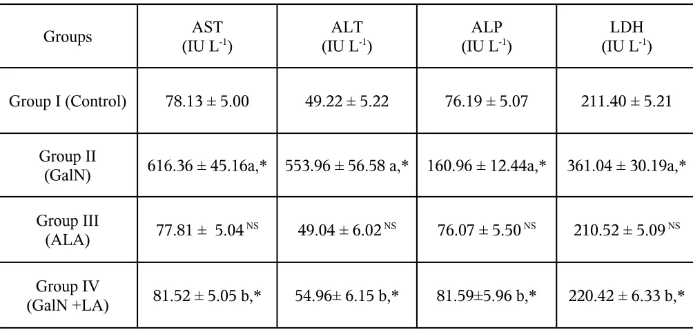
Effect of Alpha lipoic acid on marker enzymes
In the present study, intraperitoneal administration of a single dose of GalN (500
mg/kg body weight) induced severe biochemical changes as well as oxidative injury in
liver tissue. There was a significant (P < 0.05) rise in the levels of serum marker enzymes
asparate transaminase (AST), alanine transaminase (ALT), alkaline phosphatase (ALP)
and lactate dehydrogenase (LDH) in the serum of GalN administered (Group II) rats as
compared to that of Group I control rats (Table I). The administration of ALA to Group
IV animals restored the levels of these enzymes to near normalcy (P < 0.05) as compared
to those Group II GalN-injected rats. In ALA alone administered rats (Group III) versus
controls, no significant changes were observed.
Effect of Alpha lipoic acid on antioxidant enzymes
Activities of endogenous antioxidant enzymes such as superoxide
dismutase(SOD), catalase(CAT), glutathioneperoxidase(GPx) and glutathione
reductase(GR) was significantly (P < 0.05) lower in the liver tissue of Group II
GalN-injected rats as compared to that of Group I normal control rats (Table II). The observed
reduction in the activities of GPx, GR in GalN-induced oxidative condition might be due
to decreased availability GSH. In the present study, the treatment of Group IV rats with
ALA, significantly (P < 0.05) reversed all these GalN-induced alterations in the activities
of antioxidant enzymes (SOD, CAT, GPx and GR) to a near normal status. The normal
rats receiving ALA alone (Group III) did not show any significant change when
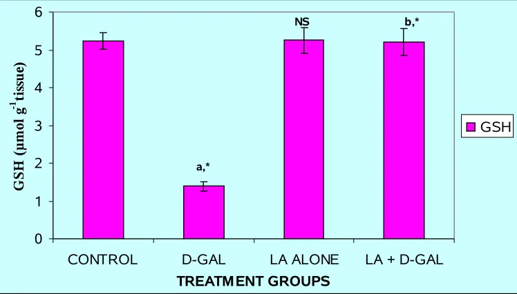
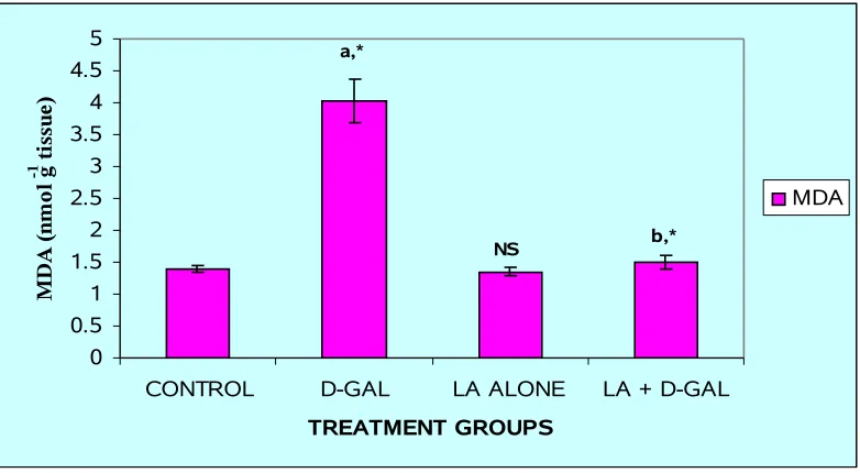
Effect of Alpha lipoic acid on GSH and lipid peroxidation levels
Intraperitoneal injection of GalN induced a significant (P < 0.05) increase in the
level of LPO (Fig. 7), which was paralleled by significant (P < 0.05) reduction in the
level of GSH (Fig. 6) in the liver tissue of Group II animals as compared to normal
controls. GSH plays an important role in the regulation of variety of cell functions and in
cell protection from oxidative injury. Depletion of GSH results in enhanced LPO and due
to this there is an increase in GSH consumption (38), as observed in the present study. In
this study, the pretreatment with ALA (Group IV) significantly (P < 0.05) counteracted
the GalN-induced LPO and restored the level of GSH to near normal level in Group IV
rats as compared to that of Group II animals.
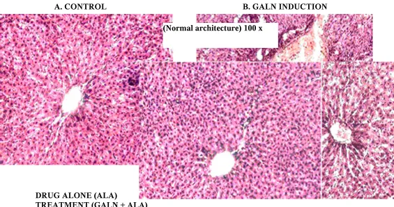
Histopathological examination
The hepatoprotectivity effect of ALA was confirmed by histopathological
examination of the liver tissue of control and treated animals. Control liver shows normal
architecture. The histological architecture of GalN treated liver sections showed
hepatocytes with centre lobular necrosis and inflammation. The histological of normal rat
received ALA alone group does not show any structural differences when compare with
control. Treatment group - administration of ALA (50 mg/kg) reduces the necrosis and
inflammation had been produced by GalN induced toxicity which has been showing its
DNA Fragmentation studies
The effect of GalN on DNA damage in the rat liver and the protection
rendered by alpha lipoic acid is presented in (Fig.9) The formation of DNA fragmentation
occurs in GalN induced group (By Agarose Gel Electrophoresis) and alpha lipoic acid
[image:44.612.63.565.290.529.2]pretreatment significantly suppress the DNA damage induced by GalN.
TABLE I: EFFECT OF ALPHA LIPOIC ACID AND D-GALACTOSAMINE ON THE
ACTIVITIES OF LIVER MARKER ENZYMES IN SERUM
Groups AST
(IU L-1) (IU LALT-1) (IU LALP-1) (IU LLDH-1)
Group I (Control) 78.13 ± 5.00 49.22 ± 5.22 76.19 ± 5.07 211.40 ± 5.21
Group II
(GalN) 616.36 ± 45.16a,* 553.96 ± 56.58 a,* 160.96 ± 12.44a,* 361.04 ± 30.19a,*
Group III
(ALA) 77.81 ± 5.04 NS 49.04 ± 6.02 NS 76.07 ± 5.50 NS 210.52 ± 5.09 NS
Group IV
(GalN +LA) 81.52 ± 5.05 b,* 54.96± 6.15 b,* 81.59±5.96 b,* 220.42 ± 6.33 b,*
Results are expressed as mean ± S.D. for 6 rats. Comparisons are made between: a- Group I and
Group II; b-Group II and Group IV. NS- Group I and Group III.
TABLE II: EFFECT OF ALPHA LIPOIC ACID AND D-GALACTOSAMINE ON
THE ACTIVITIES OF LIVER ENZYMIC ANTIOXIDANTS
Results are expressed as mean ± S.D. for six rats. Units - SOD: U/mg protein, CAT: U/mg
protein, µmoles H2O2 consumed min−1 mg−1 protein. GPx: U/mg protein, µmoles GSH oxidized
min−1 mg−1 protein. GR: nmoles NADPH oxidized min−1 mg−1 protein .Comparisons are made
between: a-Group I and Group II; b-Group II and Group IV. NS- Group I and Group III.
[image:45.612.113.487.358.571.2]* Statistically significant (P < 0.05); NS – non-significant.
Fig.6: LEVELS OF GSH IN THE LIVER OF THE EXPERIMENTAL ANIMALS
Results are given as mean ± S.D. for six rats. Comparisons are made between:
a-Group I and GroupII; b-Group II and Group IV. NS- Group I and Group III
* Statistically significant (P < 0.05); NS – non-significant.
0 1 2 3 4 5 6
FIG.7: LEVELS OF MDA IN THE LIVER OF THE EXPERIMENTAL ANIMALS
Results are given as mean ± S.D. for six rats. Comparisons are made between:
a-Group I and Group II; b-Group II and Group IV. NS- Group I and Group III
* Statistically significant (P < 0.05); NS – non-significant
0 0.5 1 1.5 2 2.5 3 3.5 4 4.5 5
CONTROL D-GAL LA ALONE LA + D-GAL
TREATMENT GROUPS M D A ( n m o l g
-1 t
FIG. 8 - HISTOLOGICAL EXAMINATION OF LIVER IN EXPERIMENTAL
RATS
A. CONTROL B. GALN INDUCTION
DRUG ALONE (ALA)
TREATMENT (GALN + ALA)
FIG. 9 – EFFECT OF ALPHA LIPOIC ACID AND D-GALACTOSAMINE IN DNA FRAGMENTATION
Mar ker DNA N.C T.C 50 mg/kg
(GalN toxicity shows the centre lobular necrosis and Inflammation) 100 x
(Normal architecture) 100 x
(ALA alone does not shows any changes while compare with control tissue) 100 x
[image:47.612.201.342.566.781.2]1 LANE – MARKER DNA
2 LANE – NORMAL CONTROL (Normal DNA BAND)
3 LANE – TOXIC CONTROL (Fragmented DNA BAND)
VI. DISCUSSION
GalN induced oxidative stress in the rat liver produces the imbalance between
biochemical processes leading to formation of reactive oxygen species and the cellular
antioxidant cascade, causes molecular damage that can lead to a critical failure of
biological functions and ultimately cell death (39,40 ,41).
At the course of liver injury, the transport function of hepatocytes is
distressed which can result in the leakage of plasma membrane, thereby causing an
increase in serum marker enzymes like AST, ALT, ALP and LDH (42, 43). GalN
administration in rats disrupts the membrane permeability of the plasma membrane
causing leakage of the enzymes from the cell, which leads to elevation in levels of marker
enzymes (44). In this study the rise in AST, ALT, ALP and LDH levels induced by GalN
administration was significantly reduced by ALA pretreatment suggesting that its
hepatoprotectivity activity might be due its effect against cellular leakage and loss of
functional integrity of the cell membrane in hepatocytes.
LPO distorted the levels of some free radical scavengers which indirectly reveals
the oxidative stress in liver (45).LPO involves the direct reaction of oxygen and lipid to
form radical intermediates and to produce semi stable peroxides, which in turn damage
the enzymes, nucleic acids, membranes and proteins (46). LPO is thought to be an
important mechanism of liver injury and malondialdehyde (MDA) is one of its end
products and measurement of MDA can be used to assess lipid peroxidation levels (47).
It has been well documented that GalN administration produces the MDA-protein
ALA scavenges hydroxyl radicals; H2O2, singlet oxygen, superoxide radicals and peroxyl
radicals thereby preventing the free radicals mediated LPO (49). In this study ALA
pretreatment diminish the LPO end products, and protect the membrane damage caused
by GalN. Earlier reports have shown that LPO induced by GalN is concomitant with
GSH depletion (50).
GSH is the intracellular non-protein sulphydryl compound that supply as both
nucleophile and an effective reductant by interacting with various electrophilic and
oxidized compounds and its depletion results in increased defenselessness of cell to
oxidative stress (51, 52). The consequence of decrease in cellular GSH levels is an
increase of free radical intermediates leading to oxidative stress and potent cellular
damage. The reduced form of GSH becomes readily oxidized to oxidized glutathione
(GSSG) with the GPx on interacting with free radicals (53, 54). In the present study
pretreatment of the rats with ALA significantly increased the concentration of GSH. It
had been known that the ALA play an important role in improving the GSH status
through the mechanism of increasing the cystine availability, because the cystine is the
rate-limiting factor in GSH biosynthesis. ALA system induces the cystine uptake and
thereby increasing GSH synthesis. In our present investigation pretreatment with ALA
aggravates the hepatic GSH level which is in accordance with the previous studies (55).
A major defense mechanism in the human system involves the antioxidant
enzymes including SOD, CAT and GPx which convert active oxygen molecules into
terminators (56, 57). CAT is the key component of the antioxidant defense system,
inhibition of this protective mechanism leads to free radicals induced cellular damage.
The observed decrease in the activity of CAT in oxidative stress is consistent with earlier
studies (58, 59, and 60). ALA is able to increase glucose uptake in vitro (61). Enhanced
glucose uptake by cells serve as a fuel for both the pentose phosphate shunt and oxidative
phosphorylation, thus bringing up the cellular levels of nicotinamide adenine dinucleotide
phosphate (NADPH) and nicotinamide adenine dinucleotide (NADH) and thereby
enhances the activity of CAT in stress condition (62). SOD is an enzyme extensively
used as a biochemical indicator of pathological states associated with oxidative stress. At
the first step of the defense system against oxidative stress, it catalyzes dismutation of the
super oxide anion into hydrogen peroxide. H2O2 is one of the most active oxygen species,
at the second step of the antioxidant defense system; GPx and CAT independently
degrade H2O2 to water. Several lines of evidence indicated that SOD activity should
therefore produce an excess H2O2 that must be efficiently neutralized by either GPx or
CAT (63, 64). Our data indicated that GalN induced oxidative stress causes a significant
inhibition of SOD and CAT. The possible reason for this finding could be the decrease of
this enzyme caused by enhanced lipid peroxidation in the stress condition (65). On the
other hand, declined SOD and CAT activity in liver tissues of GalN treated rats was
brought back to a normal level with administration of ALA. The GPx which is coupled
with GSH, together with catalase a major cellular reducer of hydrogen peroxide (66). The
stress-induced decrease in the activity of GPx documented in our study is corroborated by
earlier investigations (58, 59). GPx activity in the liver of stressed rats was also brought
![[Bis[tris(3,5 dimethylpyrazolyl)methane]nickel(II)][tetrachloronickelate(II)]–methanol–water (1/1/1)](data:image/gif;base64,R0lGODlhAQABAIAAAP///wAAACH5BAEAAAAALAAAAAABAAEAAAICRAEAOw==)