R E S E A R C H A R T I C L E
Open Access
Multiple-locus variable-number tandem-repeat
analysis (MLVA) genotyping of human
Brucella
isolates in Malaysia
Bee Yong Tay
1*, Norazah Ahmad
1, Rohaidah Hashim
1, Jama
’
ayah Mohamed Zahidi
1, Kwai Lin Thong
2,
Xiu Pei Koh
2and Azura Mohd Noor
1Abstract
Background:Brucellosis is one of the most common zoonotic diseases worldwide. It can cause acute febrile illness in human and is a major health problem. Studies in human brucellosis in Malaysia is limited and so far no genotyping studies has been done onBrucellaisolates. The aim of the study was to determine the genetic diversity amongBrucella species isolated from human brucellosis, obtained over a 6-year period (2009–2014).
Methods:In this study, the genotypic characteristics of 43 humanBrucella melitensisisolates were analysed using multiple-locus variable-number tandem-repeat analysis (MLVA) which consisted of eight minisatellite loci (panel 1) and eight microsatellite loci; panels 2A (3 microsatellite loci) and panel 2B (5 microsatellite loci). Two humanBrucella suisisolates were also investigated using the MLVA assay.
Results:Using panel 1 (MLVA8), two genotypes namely genotype 43 and 44 were obtained from the 43B. melitensis isolates. Using the combination of panels 1 and 2A loci (MLVA11), two genotypes were obtained while using the complete panels 1, 2A and 2B, nine genotypes were obtained. The polymorphisms in using the complete panels (MLVA16) were observed in three loci from panel 2B, which showed a diversity index higher than 0.17. AllB. melitensis isolates were closely related to the East Mediterranean group. ForB. suisisolates, only genotype 6 and genotype 33 were obtained using panel 1 and MLVA11 respectively.
Conclusion:In conclusion, the results of the present study showed a low genetic diversity amongB. melitensisandB. suisisolates from human patients. Based on the MLVA16 assay,B. melitensisbelonging to the East Mediterranean group is responsible for the vast majority ofBrucellainfections in our Malaysian patients. To our knowledge, this is the first genotyping study of humanBrucellaisolates in Malaysia.
Keywords:Brucellosis, MLVA, Genotype
Background
Brucellosis, a disease caused by the bacteria of genus
Brucellais a re-emerging zoonosis worldwide. This dis-ease is highly prevalent in many regions, particularly in Central Asia, the Middle East, the Mediterranean region, Africa and Latin America [1]. Brucellosis pose a major health concern in humans with initial clinical symptoms reported as fever, myalgia, malaise and may later develop into chronic disease affecting multiple organs [2]. In the
livestock industry, brucellosis cause significant repro-ductive losses in animals and subsequently causes great
economic losses [3]. Infection of Brucella sp. from
ani-mals to humans can occur by consumption of infected unpasteurised milk and dairy goods, inhalation of in-fected aerosolized particles and through direct contact with infected animal parts [4].
Currently eleven species are recognized in theBrucella
genus [5–9], four of which are known to infect humans.
They are Brucella melitensis, Brucella abortus, Brucella
suis and Brucella canis [4]. In addition, Brucella
inopi-natawas recovered from a human patient [8]. Bacterial
typing has seen a shift towards molecular identification * Correspondence:[email protected]
1
Bacteriology Unit, Institute for Medical Research, Jalan Pahang, 50588 Kuala Lumpur, Malaysia
Full list of author information is available at the end of the article
using multiple-locus variable-number tandem repeat analysis (MLVA) as tools which is widely used in the
study of Brucella species and other bacterial species of
medical importance [10–13]. Based on the approach of
high mutation rate in tandem DNA repeats loci, this im-portant methodology has proven its discriminatory power
in the study ofBrucellaspecies with high homogeneity and
the ability to distinguish isolates of various geographic ori-gins [10, 11]. MLVA typing has been useful in outbreak and epidemiological trace-back investigations and in confirming
laboratory or foodborne acquired infections [14–18].
Malaysia, a small country located in Southeast Asia has reported cases of animal brucellosis but the percent-age is lower compared to its neighbouring countries [19]. The presence of brucellosis in the Malaysian livestock population has occurred for many decades although the seroprevalence is relatively low [20]. In a recent study con-ducted from year 2000 to 2009, the seroprevalence of bru-cellosis among goats was detected throughout Malaysia but at generally low seroprevalences. The seroprevalence of brucellosis among goats was 0.91 % and among farms was 7.09 % [21]. The seroprevalence of bovine brucellosis among cattle population in Malaysia was 21.8 % [22]. No current data on the seroprevalence of porcine brucellosis
is available except for data from 1955–56 which reported
low titres of brucellosis among tested pig sera [20]. In Malaysia, occupations associated with animals or animal related materials such as livestock workers, veterinarians and laboratory workers are most likely to be infected by brucellosis [23, 24]. Human brucellosis cases in Malaysia also occurred through consumption of unpasteurized goat milk [25]. In a study among sus-pected human brucellosis cases, the seropositivity of brucellosis was only 5.4 % which suggests that the seroprevalence of brucellosis among individuals who have contact with infected animals in Malaysia is low [26]. According to Ministry of Health Malaysia, less than five cases were reported in government hospitals between the year 2000 and 2008 [27]. From year 2009 onwards, human brucellosis cases continued to be re-ported and most were sporadic cases. However, in the year 2011, an outbreak occurred in Penang, a state in the northern part of Malaysia with more than 30 cases being reported [28]. There are limited studies on human brucellosis in Malaysia and to date no genotyping
stud-ies on the human Brucella isolates have been reported
in Malaysia.
In this study, the MLVA assay was used to investigate the genetic diversity of both outbreak and sporadic human
Brucella isolates collected between the year 2009 and 2014 to determine the genotype of isolates causing human brucellosis in Malaysia. To the best of our knowledge, this is the first genotyping study conducted in Malaysia for
humanBrucellaisolates.
Methods Ethics statement
This study was carried out with the approval of Medical Research & Ethics Committee, Ministry of Health Malaysia in accordance to the International Conference of Harmonization Good Clinical Practice Guidelines (KKM/NIHSEC/08/0804/P11-666).
Brucella isolates and DNA preparation
A total of 45 isolates from human brucellosis cases in
the year 2009–2014 were investigated. All samples were
taken as part of standard care. Thirty-five isolates were outbreak cases occurring in the year 2011 while ten iso-lates were sporadic cases occurring between 2009 and 2014. All isolates were identified at the genus level based on classical identification tests such as Gram staining, positive oxidase and catalase tests, urease activity, dye sensitivity, agglutination with monospecific antisera for A and M antigens (Animal Health Veterinary Laboratories Agency, Weybridge, UK) and hydrogen sulfide production
as described [29]. Forty –three isolates were identified as
Brucella melitensisand two isolates were identified as
Bru-cella suis. Total genomic DNA from these isolates were extracted using the DNeasy Blood and Tissue Kit (Qiagen, Hilden, Germany) and the DNA concentrations were mea-sured using NanoDrop Spectrophotometer (Nanodrop
Technologies, Wilmington, USA).Brucella melitensis16 M
was used as the control strain.
Polymerase chain reaction (PCR) amplification and MLVA
The sixteen sets of primers used in this MLVA assay were based on previously described methods [10, 11]. The 16 loci are categorized in three panels namely panels 1, 2A and 2B. Panel 1 composed of eight minisatellite loci (Bruce06, Bruce08, Bruce11, Bruce12, Bruce42, Bruce43, Bruce45, Bruce55) are used for species identification. Panel 1 also provides subspecies classification. The remaining three microsatellite loci in panel 2A (Bruce18, Bruce19, Bruce21) and five microsatellite loci in panel 2B (Bruce04, Bruce07, Bruce09, Bruce16, Bruce30) are used for further subspecies differentiation. All forward primers for each pri-mer set except for Bruce07 and Bruce19 were synthesized with fluorescence dyes (FAM, HEX, or TAMRA). For Bruce07 and Bruce19, the primers were synthesized with either FAM or HEX as the reverse primer.
Monoplex PCR was performed in a total volume of
50μl containing 10 ng of DNA, 0.3μM of each primers
and 25 μl of My Taq Mix, 2x (Bioline, London, UK).
with a total volume of 10 μl containing 8.7 μl HI-DI formamide (Applied Biosystems, Lincoln City, USA),
0.3 μl GeneScan 1200 LIZ size standard (Applied
Bio-systems, Lincoln City, USA) and 1 μl of diluted
ampli-fied products at 95 °C for 5 min using an Eppendorf Mastercycler (Eppendorf AG, Hamburg, Germany). The denatured products were further resolved by capil-lary electrophoresis on an ABI 3730xl DNA analyzer (Applied Biosystems, Lincoln City, USA). Fragment sizes obtained were then analyzed using the GeneMapper software version 4.0 (Applied Biosystems, Lincoln City, USA).
MLVA data analysis
The copy number for each locus was managed as a charac-ter dataset using BioNumerics version 6.0 (Applied-Maths, Belgium). The clustering analysis was based on the categor-ical coefficient and Unweighted Pair Group Method Using Arithmetic Averages (UPGMA) method. The genotypes identified were compared with the isolates in the Bru-cella2012 MLVA database [30]. The genetic diversity and confidence interval of each locus were calculated using the Hunter-Gaston Diversity Indices (HGDI) via an online tool V-DICE available at the HPA website [31].
Results
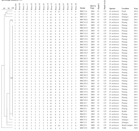
Genotyping of humanBrucella melitensisisolates using MLVA16 assay
The 43B. melitensisisolates were shown to have genetic
similarity coefficient ranging from 75.5 to 100.0 %
(Fig. 1). Thirty-four isolates(cluster A2) shared the same
MLVA16 genotype. Cluster A1 comprising two isolates differs at one locus (one repeat unit difference at the highly variable panel 2B locus Bruce04). The genetic similarity coefficient between cluster A1 and A2 is 93.8 %. These isolates were part of the outbreak strains that occurred in Penang in year 2011. Interestingly, iso-late BRC7/11 which was from a patient from the state of Kedah, was shown to have 100.0 % similarity coefficient with the outbreak strains. This case was previously thought to be a sporadic case. The remaining isolates were from sporadic cases with similarity coefficient ran-ging from 75.5 to 87.5 %.
Using panel 1, two genotypes were observed namely geno-type 43 (42 isolates) and genogeno-type 44 (one isolate). The ge-notypes 43 and 44 belonged to the East Mediterranean group [11]. Further analysis with panel 1 and 2A revealed two genotypes, where the majority of the isolates were geno-type 125 (42 isolates) followed by genogeno-type 109 (1 isolate). Using the complete MLVA assay (panels 1, 2A and 2B), nine genotypes were obtained namely MLVA type M01 to MLVA type M09.
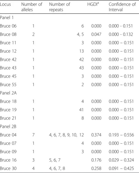
The increase in number of genotypes when using MLVA16 was the result of the high polymorphism
displayed in panel 2B where Bruce04, Bruce16 and Bruce30 showed the highest discriminatory markers, with a diversity index of > 0.17 harbouring 3, 4 and 7 al-leles respectively (Table 1). Among the three markers, Bruce04 shows the highest polymorphism. Bruce07 and Bruce09 with relatively small alleles (4 and 3 repeat units respectively) do not vary in the present collection. All panel 1 and panel 2A showed no diversity except Bruce08 of panel 1 which showed slight diversity, har-bouring 2 alleles.
Genotyping of humanBrucella suisisolates using MLVA16 assay
Two B. suis isolates from two sporadic cases showed
81.3 % in similarity (Fig. 2). Both B. suis isolates
belonged to genotype 6 with isolate BRC3/09 showing
closest relation to the B. suisbiovar 1 group. The other
B. suis isolate (BRC1/12) was not categorized in any group. MLVA11 shows isolate BRC3/09 and BRC1/12 belonged to genotype 33. Using the complete MLVA16
assay, two genotypes were obtained from the twoB. suis
isolates with the numbering of MLVA type M10 to MLVA type M11.
Discussion
This is the first human brucellosis investigated using
MLVA as the genotyping tools on 45 human Brucella
isolates in Malaysia. Human brucellosis cases in this study were reported in seven states namely Perlis, Perak, Penang, Kedah (northern region), Selangor, Kuala Lumpur (federal territory) and Negeri Sembilan (central region). All the states were located in Peninsular Malaysia (Fig. 3). No human brucellosis cases were reported in the states of Sabah and Sarawak (East Malaysia). In our study, Penang had the highest reported cases of infection in humans by
B. melitensisfollowed by the state of Selangor.
Our findings most likely could be supported by a serosurveillance study conducted from 2000 to 2009
showing prevalence and distribution of zoonotic B.
melitensis in goats which is higher in Peninsular Malaysia as compared to East Malaysia [21]. From the
same study, it was also found that infection ofB.
meli-tensisin goats is widespread among farms, affecting all 13 states and federal territories in Malaysia. The state of Perlis had the highest seroprevalence followed by Penang and Melaka. A recent study confirmed the presence of brucellosis in livestock by the isolation of
B. melitensis in seropositive goats in Peninsular Malaysia [32]. All the findings from these animal bru-cellosis studies are of importance in the epidemiology of human brucellosis in Malaysia.
Several studies have shown that B. melitensis is the
most isolated Brucella species in human cases [16, 18].
accounts for most of the cases reported in this study. Our data reveal that human brucellosis in Malaysia is highly-related to caprine infection as suggested by the clinical history of the patients.
Isolate BRC2/12 from a sporadic case reported in year 2012 shared similar MLVA profile as the 33 outbreak cases occurred in year 2011. This sporadic case was re-ported in the state of Penang and clinical history showed the patient consumed unpasteurised goat milk and the case is unrelated to the outbreak cases. Interestingly, in the outbreak cases, it was discovered that 33 patients consumed unpasteurised goat milk sourced from the
same farm. All 33 isolates showed identical MLVA pro-files (MLVA type M05) with good correlation with the epidemiological data. The MLVA assay also revealed that isolate BRC7/11 reported in Kedah, a state situated in the northern region was linked to the 32 cases reported in Penang indicating an outbreak from a common source of infection. However two of the isolates (BRC30/ 11 and BRC31/11) which were reported during similar outbreak period showed slight difference from the 33 outbreak isolates.
Both isolates BRC30/11 and BRC31/11 differed from the 33 isolates (MLVA type M05) at the bruce04
[image:4.595.59.535.100.547.2]microsatellite locus from panel 2B by a difference of one repeat unit. Clinical history of isolate BRC30/11 indi-cated that the patient consumed unpasteurised goat milk during the outbreak period. Clustering analysis showed that isolates BRC30/11 and BRC31/11 were 93.8 % re-lated to the 33 isolates of MLVA type M05, indicating that they are closely related. The two isolates did not share the same MLVA type which could be the result of mutation events in the course of outbreak. A study in Turkey also noted the same finding where two of the isolates yielded different MLVA profile from the out-break isolates [18].
High polymorphism is associated with some of the most variable loci in panel 2B (Bruce 04, Bruce16 and
Bruce30) as observed in this study. The three highly variable loci of panel 2B were also observed in Turkish and Chinese isolates [16, 18]. MLVA16 assay has also been shown to have the ability to differentiate a new exposure from a relapse case in the same patient [11]. In this study, isolates BRC26/11 and BRC33/11 from the outbreak were isolated from the same patient be-fore treatment and after two months of completion of treatment respectively. Both isolates showed identical MLVA profile (MLVA type M05) implying that the pa-tient most likely had a relapsed infection with the
same strain of B. melitensis. To confirm this, whole
genome sequencing analysis may provide a more definitive answer.
Isolate BRC5/10 was grouped in panel 1 genotype 44 and no clinical history was given for the patient who hails from Perlis, a state situated in the northern region of Malaysia. The panel 1 of MLVA16 assay indicated the isolate belonging to genotype 44 (1-4-3-13-3-2-3-2)
clus-tered in the Brucella melitensis of East Mediterranean
group. The panel 1 genotype 44 has been reported in Turkey and also present in Israel [11, 18].
B. melitensishas previously been clustered into three major lineages comprising the East Mediterranean,
West Mediterranean and American group [11]. B.
abortus and B. suis have no prior groupings. Our
re-sults suggest that theB. melitensisisolates in Malaysia
most likely form a homogenous group belonging to the East Mediterranean group. The data also showed
that human Brucellaisolates in Malaysia comprised of
panel 1 genotypes 6, 43 and 44. Genotype 43 was also observed in other parts of the world including Turkey
and China [16, 18]. Panel 1 genotypes 49 and 51 of
Bru-cella melitensis in the Western Mediterranean group which are common in Italy [11] were not detected in this study.
B. suisespecially of biovars 1 and 3 are the causative agent of swine brucellosis in human and are
com-monly sporadic [4]. In our study,B. suis infection also
[image:5.595.57.290.100.389.2]occurred among human patients. Both were sporadic cases reported in Penang and Negeri Sembilan. Isolate BRC1/12 originated from a single patient who works Table 1Number of alleles and HGDI values of 43 isolates
Locus Number of alleles
Number of repeats
HGDIa Confidence of Interval
Panel 1
Bruce 06 1 6 0.000 0.000 - 0.151
Bruce 08 2 4, 5 0.047 0.000 - 0.132
Bruce 11 1 3 0.000 0.000–0.151
Bruce 12 1 13 0.000 0.000–0.151
Bruce 42 1 42 0.000 0.000–0.151
Bruce 43 1 43 0.000 0.000–0.151
Bruce 45 1 3 0.000 0.000–0.151
Bruce 55 1 2 0.000 0.000–0.151
Panel 2A
Bruce 18 1 4 0.000 0.000–0.151
Bruce 19 1 41 0.000 0.000–0.151
Bruce 21 1 8 0.000 0.000–0.151
Panel 2B
Bruce 04 7 4, 6, 7, 8, 9, 10, 12 0.374 0.193–0.556
Bruce 07 1 4 0.000 0.000–0.151
Bruce 09 1 3 0.000 0.000–0.151
Bruce 16 3 5, 6, 7 0.176 0.029–0.324
Bruce 30 4 4, 6, 7, 8 0.258 0.091–0.425
a
Hunter–Gaston Diversity Index
M11 6
2 3 6 11 3 1 5 2 4 38 9 5 6 7 11 3 BRC3/09 33 B. suis Negeri Sembilan 2009
percentage similarity (%)
100 95 90 85 Br uce06 Br uce 08 Br uce 11 Br uce 12 Br uce 42 Br uce 43 Br uce 45 Br uce 55 Br uce 18 Br uce 19 Br uce 21 Br uce 04 Br uce 07 Br uce 09 Br uce 16 Br uce 30
2 3 6 11 3 1 5 2 4 38 9 5 5 11 9 3
Strain BRC1/12 MLVA Type M10 Pa ne l 1 6 P a nel 1+ 2A Species B. suis Location Penang Year 2012 33
[image:5.595.52.538.616.697.2]as a palm estate farmer and the case was reported in Penang. Even though he was not occupationally-involved in animals, he may have some contact with a close family member who was working in a pig slaughter farm. When queried upon the Brucella2012 database, the genotype did not cluster to any particular group and the isolate belonged to panel 1 genotype 6. Another isolate which
was typed asB. suiswas BRC3/09 which was identified in
a patient in the state of Negeri Sembilan in the central region of Peninsular Malaysia. Isolate BRC3/09 also belonged to panel 1 genotype 6. Panel 1 genotyping identified the profile of both isolates as genotype 6
(2-3-6-11-3-1-5-2) which is closely related to B. suis
bio-var 1 group. The two B. suisisolates yielded MLVA11
genotype 33.
The presence of brucellosis in Malaysia was first
docu-mented in animals with the isolation of Brucella abortus
in 1950 [33],Brucella suisin 1960s [33] andB. melitensis
in 1994 [23]. Subsequently, brucellosis was detected in
humans in 1980 by the isolation of B. suisbiotype 3 [34]
and in 1994 by the isolation of B. melitensis [23]. The
detection ofB. melitensisandB. suisamong Malaysian
population decades ago still affect the current popula-tion today as shown in our data. One of the reasons of the continuous brucellosis infection in human popula-tion in Malaysia could be due to the animal reservoir. It is suspected that the annual mass importation of goats from endemic countries and importation of various breeding stocks of swines from Europe into
Malaysia that causes the persistence of brucellosis in
the animal population [19–21].
Our present study consists of 45 human isolates only. It is expected that more local isolates will be continu-ously studied to determine presence of new genotypes in the country, subsequently allowing for the genotyping results made accessible for querying and comparison to
other Brucella isolates from wider geographical origins.
Combined with new methodologies of whole genome sequencing, data obtained from analyses of MLVA11 genotype 125 from our present collection and from dif-ferent parts of the world will be an important approach
to the molecular epidemiological studies of brucella
throughout the country as well as globally.
Conclusions
In conclusion, the results of the present study showed a
low genetic diversity among B. melitensis and B. suis
isolates from human patients. The genotypes found in Malaysia comprise mainly of panel 1 genotypes 43 and
44, grouped in B. melitensis of East Mediterranean
while panel 1 genotype 6 belonged to the B. suisbiovar
1 group. Panel 1 genotype 43 made up the most com-mon genotype circulating in this country as observed in both outbreak and sporadic isolates. All the three panel 1 genotypes were confined to Peninsular Malaysia only since no cases of human brucellosis have been reported in Sabah and Sarawak (East Malaysia).
[image:6.595.57.540.88.355.2]Competing interests
The authors declare that they have no competing interests.
Authors’contributions
TBY performed the overall work and drafted the manuscript. NA managed the project and reviewed the manuscript. RH and AMN performed bacterial identification of the isolates. TKL provided the analysis tools, analyzed data and improving the manuscript. KXP provided technical help of analysis tools, analysis and interpretation of cluster analysis. JMZ prepared the DNA samples. All authors read and approved the final manuscript.
Acknowledgements
We would like to thank the Director General of Health Malaysia for permission to publish this article. This study was supported by the Ministry of Health Malaysia Grant JPP_IMR 11–035. We also thank Siti Hawa Hamzah for providing the materials for this study.
Author details
1Bacteriology Unit, Institute for Medical Research, Jalan Pahang, 50588 Kuala
Lumpur, Malaysia.2Institute of Biological Science, Faculty of Science, University of Malaya, 50603 Kuala Lumpur, Malaysia.
Received: 15 October 2014 Accepted: 21 May 2015
References
1. Pappas G, Papadimitriou P, Akritidis N, Christou L, Tsianos EV. The new global map of human brucellosis. Lancet Infect Dis. 2006;6:91–9. 2. Pappas G, Akritidis N, Bosilkovski M, Tsianos E. Brucellosis. N Engl J Med.
2005;352:2325–36.
3. Bamaiyi PH, Abd-Razak NS, Zainal MA. Seroprevalence and economic impact of eradicating zoonotic brucellosis in Malaysia: a case study of Melaka state of Malaysia. Veterinary World. 2012;5:398–4.
4. Corbel MJ. Epidemiology. In: Corbel MJ, Elberg SS, Cosivi O, editors. Brucellosis in humans and animals. Geneva: WHO Press; 2006. p. 13–21. 5. Osterman B, Moriyón I. International Committee on Systematics of
Prokaryotes Subcommittee on the taxonomy of Brucella: minutes of the meeting. Int J Syst Evol Microbiol. 2006;56:1173–5.
6. Foster G, Osterman BS, Godfroid J, Jacques I, Cloeckaert A.Brucella cetisp. nov. andBrucella pinnipedialissp. nov. forBrucellastrains with cetaceans and seals as their preferred hosts. Int J Syst Evol Microbiol. 2007;57:2688–93. 7. Scholz HC, Hubalek Z, Sedláček I, Vergnaud G, Tomaso H, Al Dahouk S, et al.
Brucella microtisp. nov., isolated from the common voleMicrotus arvalis. Int J Syst Evol Microbiol. 2008;58:375–82.
8. Scholz HC, Nöckler K, Göllner C, Bahn P, Vergnaud G, Tomaso H, et al.
Brucella inopinatasp. nov., isolated from a breast implant infection. Int J Syst Evol Microbiol. 2010;60:801–8.
9. Whatmore AM, Davison N, Cloeckaert A, Al Dahouk S, Zygmunt MS, Brew SD, et al.Brucella papionissp. nov., isolated from baboons (Papiospp.). Int J Syst Evol Microbiol. 2014; doi:10.1099/ijs.0.065482-0
10. Le Flèche P, Jacques I, Grayon M, Al Dahouk S, Bouchon P, Denoeud F, et al. Evaluation and selection of tandem repeat loci for aBrucellaMLVA typing assay. BMC Microbiol. 2006;6:9.
11. Al Dahouk S, Le Flèche P, Nöckler P, Jacques I, Grayon M, Scholz HC, et al. Evaluation of Brucella MLVA typing for human brucellosis. J Microbiol Methods. 2007;69:137–45.
12. van Belkum A. Tracing isolates of bacterial species by multilocus variable number of tandem repeat analysis (MLVA). FEMS Immunol Med Microbiol. 2007;49:22–7.
13. Lindstedt BA, Torpdahl M, Vergnaud G, Le Hello S, Weill FX, Tietze E, et al. Use of multilocus variable-number tandem repeat analysis (MLVA) in eight European countries. Euro Surveill. 2013;18(4):pii=20385.
14. Ferreira AC, Chambel L, Tenreiro T, Cardoso R, Flor L, Dias IT, et al. MLVA16 typing of Portuguese human and animalBrucella melitensisandBrucella abortusisolates. PLoS One. 2012;7:e42514.
15. Garofolo G, Di Giannatale E, De Massis F, Zilli K, Ancora M, Cammà C, et al. Investigating genetic diversity ofBrucella abortusandBrucella melitensisin Italy with MLVA-16. Infect Genet Evol. 2013;19:59–70.
16. Jiang H, Fan MG, Chen JD, Mi JC, Yu RP, Zhao HY, et al. MLVA genotyping of Chinese humanBrucella melitensisbiovar 1, 2 and 3 isolates. BMC Microbiol. 2011;11:256–7.
17. Lucero NE, Tenenbaum M, Jacob NR, Escobar GI, Groussaud P, Whatmore AM. Application of variable number of tandem repeats typing to describe familial outbreaks of brucellosis in Argentina. J Med Microbiol. 2010;59:648–52. 18. Kiliç S, Ivanov IN, Durmaz R, Bayraktar MR, Ayaşlioğlu E, Uyanik MH, et al.
Multiple-locus variable-number tandem-repeat analysis genotyping of human
brucellaisolates from Turkey. J Clin Microbiol. 2011;49:3276–83.
19. Bamaiyi PH, Hassan L, Khairani-Bejo S, Zainal AM. Updates on brucellosis in Malaysia and Southeast Asia. Malaysian J Vet Res. 2014;5:71–82.
20. Bahaman AR, Joseph PG, Siti KB. A review of the Epidemiology and Control of Brucellosis in Malaysia. Jurnal Veterinar Malaysia. 2007;19:1–6.
21. Bamaiyi PH, Hassan L, Khairani-Bejo S, ZainalAbidin M, Ramlan M, Adzhar A, et al. The prevalence and distribution ofBrucella melitensisin goats in Malaysia from 2000 to 2009. Prev Vet Med. 2015; doi:10.1016/ j.prevetmed.2015.02.001
22. Anka MS, Hassan L, Adzhar A, Khairani-Bejo S, Mohamad RB, Zainal MA. Bovine brucellosis trends in Malaysia between 2000 and 2008. BMC Vet Res. 2013;18(9):230.
23. Kementerian Pertanian dan Industri Asas Tani Malaysia. Penyakit Melitensis. In: No Dokumentasi: PVM 1(5):1/2010. Protokol Veterinar Malaysia. 2015. http://www.dvs.gov.my/documents/10157/5c0aa392-708d-4f94-bdcc-80ff39647523. Accessed 18 February 2015.
24. Hartady T, Saad MZ, Khairani Bejo S, Salisi MS. Clinical human brucellosis in Malaysia: a case report. Asian Pac J Trop Dis. 2014;4:150–3.
25. National Poison Centre: Raw goat milk sicken boys. http://www.prn.usm.my/ poison_news.php?news_id=6116. (2015). Accessed 23 February 2015. 26. Jama’ayah MZ, Heu JY, Norazah A. Seroprevalance of brucellosis among
suspected cases in Malaysia. Malays J Pathol. 2011;33:31–4.
27. Ministry of Health Malaysia. Guidelines for the Diagnosis, Management, Prevention and Control of Human Brucellosis in Malaysia. 2014. http:// www.moh.gov.my/images/gallery/Garispanduan/Human_Brucellosis.pdf. Accessed 22 July 2014.
28. Mardiyah Z, Laina M, Naziehah MD, Tosiah A:Brucellaspp. di dalam susu kambing. http://los.moh.gov.my/ebook/laporantahunan2011/files/assets/ basic-html/page162.html (2013). Accessed May 2013.
29. Corbel MJ, Banai M. Genus I. Brucella. In: Brenner DJ, Krieg NR, Garrity GM, Staley JT, Boone DR, Vos PD, Goodfellow M, Rainey FA, Schleifer KH, editors. Bergey’s Manual of Systematic Bacteriology. New York: Springer; 2005. p. 370–86.
30. Brucella2012 MLVA database. http://mlva.u-psud.fr. 2014. Accessed 25 July 2014. 31. VNTR Diversity and Confidence Extractor. 2014.
http://www.hpa-bioinformatics.org.uk/cgi-bin/DICI/DICI.pl. Accessed 25 July 2014. 32. Bamaiyi PH, Hassan L, Khairani-Bejo S, Zainal Abidin M, Ramlan M, Krishnan
N, et al. Isolation and molecular characterization ofBrucella melitensisfrom seropositive goats in Peninsula Malaysia. Trop Biomed. 2012;29:513–8. 33. Kementerian Pertanian dan Industri Asas Tani Malaysia. Penyakit Brusela suis
dalam babi. In: No Dokumentasi: PVM 1(6):1/2011. Protokol Veterinar Malaysia. 2015. http://www.dvs.gov.my/documents/10157/27f59410-3ab9-4244-8596-f12b95b3420a. Accessed 10 March 2015.
34. Heng NH, Joseph PG. Eradication of brucellosis in cattle in Malaysia and its public health significance. Kajian Veterinar. 1986;18:255–60.
35. Daniel Dalet. Malaysia. 2015. http://d-maps.com/
carte.php?num_car=5391&lang=en. Accessed 3 March 2015.
Submit your next manuscript to BioMed Central and take full advantage of:
• Convenient online submission
• Thorough peer review
• No space constraints or color figure charges
• Immediate publication on acceptance
• Inclusion in PubMed, CAS, Scopus and Google Scholar
• Research which is freely available for redistribution
![Fig. 3 Map of Malaysia showing states with reported case of human brucellosis (adapted from Google maps [35])](https://thumb-us.123doks.com/thumbv2/123dok_us/8367379.316901/6.595.57.540.88.355/malaysia-showing-states-reported-human-brucellosis-adapted-google.webp)
