This is a repository copy of A comparison of friction behaviour for ex vivo human, tissue engineered and synthetic skin.
White Rose Research Online URL for this paper: http://eprints.whiterose.ac.uk/134399/
Version: Accepted Version
Article:
Bostan, LE, Taylor, ZA orcid.org/0000-0002-0718-1663, Carré, MJ et al. (3 more authors) (2016) A comparison of friction behaviour for ex vivo human, tissue engineered and synthetic skin. Tribology International, 103. pp. 487-495. ISSN 0301-679X
https://doi.org/10.1016/j.triboint.2016.07.023
Copyright (c) 2016 Elsevier Ltd. All rights reserved. Licensed under the Creative Commons Attribution-Non Commercial No Derivatives 4.0 International License
(https://creativecommons.org/licenses/by-nc-nd/4.0/).
[email protected] https://eprints.whiterose.ac.uk/
Reuse
This article is distributed under the terms of the Creative Commons Attribution-NonCommercial-NoDerivs (CC BY-NC-ND) licence. This licence only allows you to download this work and share it with others as long as you credit the authors, but you can’t change the article in any way or use it commercially. More
information and the full terms of the licence here: https://creativecommons.org/licenses/
Takedown
If you consider content in White Rose Research Online to be in breach of UK law, please notify us by
1
A Comparison of Friction behaviour for
ex vivo
Human, Tissue Engineered
and Synthetic Skin
L.E. Bostan1, Z.A. Taylor2, M.J. Carré2, S. MacNeil1, S.E. Franklin3, R. Lewis*2
1Department of Materials Science and Engineering, The Kroto Research Institute, North
Campus, University of Sheffield, Broad Lane, Sheffield, S3 7HQ, United Kingdom
2 Department of Mechanical Engineering, The University of Sheffield, Mappin Street,
Sheffield S1 3JD, United Kingdom
3 Philips Research, Eindhoven, The Netherlands
*corresponding author: [email protected]
Abstract
Skin tribology is complex and in situ behaviour of skin varies considerably between test subjects. The main influencing factor, elasticity, varies due to structural and moisture differences. To find a more reliable test platform, for the first time, synthetic and biological (tissue engineered) substitutes were compared to ex vivo skin, epidermis and dermis.
Friction initially increased with rising hydration, before decreasing beyond a threshold for all samples. Friction for Synthetic skin and dermis increased at a similar rate to the other samples, but from a different starting point, and friction dropped at lower hydration. Tissue engineered skin could provide a reliable test platform, but the synthetic skin could only be used if the offset in the data is accounted for.
1 Introduction
Human skin tribology is a very diverse and complicated area of study. This is largely due to the complex structure of skin, its non-linear material properties and the fact that the surface condition and texture has influence (e.g. presence of fingerprint ridges or hair). It is also open to the environment and comes into contact with many different materials. It is very important to understand skin friction in particular though as this plays a critical role in many tasks of daily living [1] and leisure activities [2, 3] that involve gripping, feeling and manipulating objects; it dictates levels of comfort and how we feel about a product [4] and it affects how consumer products and medical devices interact with the skin as well as driving adverse effects such as skin reddening and irritation. It is also an important factor in assessing how well topical cosmetic and medical skin treatments are performing [5].
2
all have different ridge patterns [9]. The ridges also lead to a further friction mechanism occurring as the ridges interact with surface texture features, known as “interlocking” [10-13]. The changes in skin elasticity mainly result from variability in skin structure (Liu et al., 2015) [14] and different moisture levels [15, 16].
Quantifying the Young’s Modulus of skin is not a straight forward process and examination of the literature gives a wide spread of data. Techniques such as tensile testing [17, 18], indentation [19, 20] and suction methods [21] are used. Data varies across the different measurement techniques for the same skin sample. The effective Young’s Modulus in skin being loaded by a probe in a tribological test will also vary with applied load and also with probe diameter [22].
Young’s modulus varies across all the different layers of skin with the stratum corneum being several orders of magnitude stiffer than measurements taken on the forearm (largely because of the soft underlying tissue) [23]. It also varies, as mentioned above, with moisture level. Tensile tests have shown a decrease from ≈1 GPa at 50% RH [17] to a value of ≈3 MPa in water [18]. Notably, while a great deal of data exists for forearm skin and ex vivo skin and different skin layers, little data exists for skin substitutes, especially engineered skin.
As a result of the variability observed during in situ skin friction testing and in skin properties such as elasticity, a great deal of attention is being paid to finding an alternative material to use as a test platform. There are two main options available: biological and synthetic substitutes.
Biological substitutes can be generated using tissue engineering. The main application for these processes is treatment of severe burns. When a person’s body is severely burned, they may not have enough healthy skin remaining for healing to occur through skin cell regeneration within their own skin. In this case, tissue engineered skin provides a potential substitute. A lot of progress has been made regarding the construction of tissue engineered skin from a biological point of view [24, 25], but it has not been tested from a mechanical and tribological perspective.
Tissue engineered skin construction involves very complex processes and it is not accessible for every laboratory, thus, an alternative approach is to use a synthetic skin substitute. Development of synthetic skin substitutes has largely been driven by the need for a material for use in training of medical practitioners and a large range is available [26]. These materials are optimised for their mechanical properties, however, rather than their tribological response. Applications where friction has been a strong consideration have been in assessing skin textile interactions for improvement of comfort [27, 28] and topical skin formulations [29] or in understanding blistering [30, 31] and the development of artificial “sensing” fingers [32] or robot fingers [33, 34].
The aim of the work outlined in this paper was to determine whether biological and synthetic skin substitutes behave like ex vivo human skin in tribological tests. This was primarily to determine their viability as test platforms for assessing skin interactions with materials used to manufacture medical devices, consumer products and other objects that come into contact with skin. A particular focus was assessment of friction in changing moisture conditions as this has been identified as a weakness of some synthetic substitutes.
2 Experimental Details
3
dermis after incubation in sterile 1 M NaCl for 18 h at 37°C) and SynDaverTM, a synthetic skin.
2.1 Specimen Preparation
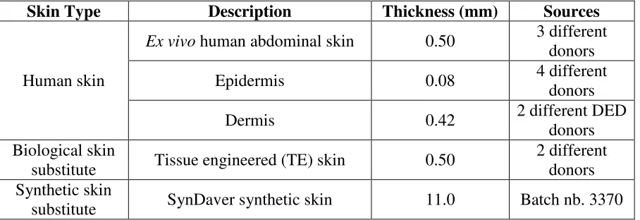
The skin specimens used in the study are summarised in Table 1. Human skin samples were collected from consenting patients undergoing abdominoplasty. This type of surgery involves the removal of excess tissue from the middle and lower abdomen from patients with loose or sagging skin after pregnancy or major weight loss. All tissue was banked and used on an anonymous basis under Human Tissue Authority Research Tissue Bank Licence Number 12179. The samples were stored in a standard physiological solution (phosphate buffered saline, PBS) with antimicrobial agents at 4°C, for no longer than 12 hours before processing. Sample thicknesses were measured using micro-callipers or histology images.
Skin Type Description Thickness (mm) Sources
Human skin
Ex vivo human abdominal skin 0.50 3 different
donors
Epidermis 0.08 4 different
donors
Dermis 0.42 2 different DED
donors Biological skin
substitute Tissue engineered (TE) skin 0.50
2 different donors Synthetic skin
[image:4.595.73.527.275.431.2]substitute SynDaver synthetic skin 11.0 Batch nb. 3370
Table 1. Sample description and variability of sources
The TE skin was prepared as previously described [35]. The human skin was incubated in 1M sodium chloride at 37°C for 12 hours before the epidermis was removed using forceps, leaving behind the de-cellularised de-epidermised dermis (DED). The epidermal samples were stored in PBS with antimicrobial agents at 4°C, and the DED samples were stored in tissue culture media (10% Green media) [36].
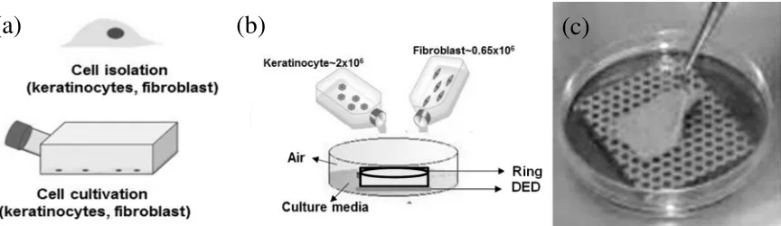
The DED was used to make TE skin constructs (as shown in Figure 1) as described previously [36]. The cells, human keratinocyte and fibroblasts, were cultured (Figure 1a) until they reached 80% confluency. They were then seeded onto DED samples using stainless steel rings (Figure 1b). The rings were removed after 3 days and the tissue engineered constructs (dermis plus cells) were elevated to the air-liquid interface by transferring them to a stainless steel grid in a petri dish culture plate (Figure 1c).
Cultures were maintained for 14 days at 37°C and 5% CO2 and the Green media was replaced every three days.
The TE skin structures were examined using histology (see Figure 2), to confirm that they had formed in an appropriate way. The histology sections showed that there was an underlying dermis with a fully reconstructed epidermis. Keratinocytes grown at the air‑liquid
4
[image:5.595.83.515.158.283.2]Because the TE skin constructs are made from DED (equivalent of dermis) on the top of which cells were seeded (keratinocytes and fibroblasts), the DED was checked to see if it had a fully reconstructed stratified epidermis (i.e., the differentiated cells seeded on the top of DED)).
Figure 1. Preparation of Tissue Engineered (TE) Skin: (a) Cell Isolation; (b) Cell Seeding; (c) Air-Liquid Interface
5
Figure 2. Hematoxylin and Eosin (H&E) Stain of: (a) Human Skin; (b) TE Skin; (c) Epidermis; (d) Dermis (the samples were fixed with 3.7% formaldehyde; after dehydration,
the specimens were embedded in paraffin, and sectioned into 4 µm thickness pieces) (E – Epidermis; D – Dermis)
The synthetic substitute skin was purchased from SynDaver™ (model T-PLA-A-0010). It is made up of layers to mimic the skin (epidermis and dermis, 1 mm; fat tissue, 5 mm, and muscular tissue, 5 mm). It is typically used by medical professionals to practice surgery. This synthetic tissue is a patented SynTissue™ brand synthetic human tissue. It is claimed to be experimentally designed on the basis of physical tests performed on actual living tissue [37].
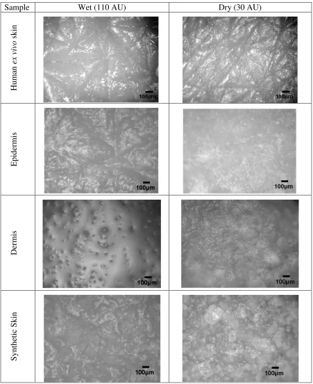
All the samples were kept in a hydrated state until testing: PBS was used for storage of human skin, epidermis and dermis (4°C), Green’s media for tissue engineered skin (37°C) and distilled water in the case of synthetic skin (room temperature). Prior to testing, all samples were allowed to stabilise at room temperature for one hour. Surface images of each sample are shown in Figure 3 in dry and wet conditions (Corneometer measurements of 30 AU and 110 AU respectively – see Section 2.2 for details). TE skin and ex vivo skin looked very similar (as would be expected) so only one image is included. The surface of the ex vivo skin looks very similar to that used by Adams et al. [15]. Much more water is present on the surface of the “wet” dermis and synthetic skin.
6
Sample Wet (110 AU) Dry (30 AU)
H
um
an
ex
v
iv
o
s
ki
n
E
pi
de
rm
is
D
er
m
is
S
ynt
he
ti
c S
ki
[image:7.595.71.522.69.621.2]n
7
2.2 Friction Testing
In order to provide a stable base for friction testing and improve the consistency of the sample thicknesses (Table 1), the synthetic substitute skin was used as a base layer for the dermis, human skin and TE skin tests. This was glued onto sand paper attached to the rig. The epidermis was tested too, in order to provide a comparison that might validate the use of synthetic skin as a base layer. For these studies different skin donors were tested (Table 1).
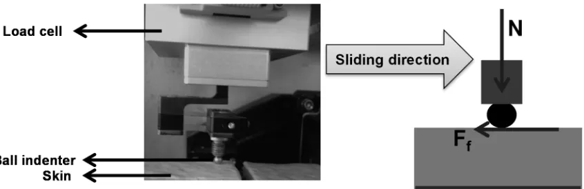
[image:8.595.85.508.314.451.2]Friction tests were carried out using a CETR-UMT 2 (Bruker) tribometer. The configuration used was a normally loaded probe (a 4 mm diameter stainless steel ball) being driven across the sample in a linear reciprocating fashion. The tests were completed at three different normal loads (5, 10 and 30 mN) at an average speed of 0.5 mm/s for six sliding cycles with a stroke of 10 mm (one cycle is twice the stroke). The normal load was stabilised for 20 s before testing. Figure 4 shows the measurement system which is composed of a double load cell (one for normal load and one for friction force). The kinetic coefficient of friction was calculated as the ratio between the friction force and the normal force. For each sample at least two repeats were carried out.
Figure 4. Measurement System for Friction Force (N – Normal Load; Ff – Friction Force)
A Corneometer 825 (Courage-Khazaka Electronics) was used to measure hydration levels (as described previously by Palma et al. [38] and Waranuch et al. [39]). This instrument has a handheld probe and measures moisture based on the capacitance of skin due to its properties as a dielectic medium. The probe sensor area is 49 mm² and the measurement depth is 10-20
8
Number of Friction Tests, Normal Load Applied and Hydration Level Range (AU)
Sample Description 30 mN 10 mN 5 mN
Human skin
Ex vivo human
abdominal skin (HL: 60-110)
17 35 16
Biological skin substitute
Tissue
engineered skin (HL: 30-100)
27 30 24
Skin epidermis (HL: 40-110)
27 26 22
Skin dermis (HL: 40-100)
10 17 12
Synthetic skin substitute
SynDaver synthetic skin (HL: 20-120)
[image:9.595.73.526.73.285.2]69 42 38
Table 2. Description of Friction Tests Carried out
3 Results and Discussion
3.1 Effect of Hydration Level on Skin Friction and Proposed Mechanisms for Observed Behaviour
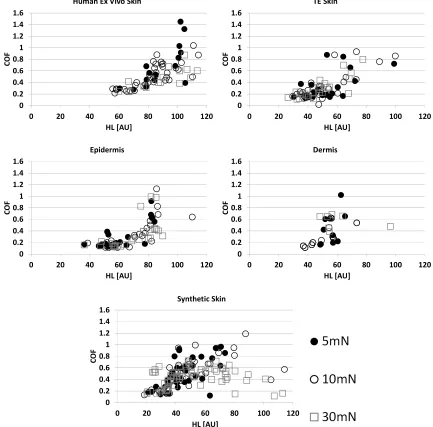
[image:9.595.85.511.436.711.2]The graphs in Figure 5 show the friction data measured across a range of hydration levels for each material at the three loads used. The spread in the data is high, but in line with friction measurements taken at different hydration levels by Hendriks & Franklin [10].
Figure 5. Coefficient of Friction (COF) versus Hydration level (HL) for the Five Specimens at Loads of 5 mN, 10 mN and 30 mN
0 0.2 0.4 0.6 0.8 1 1.2 1.4 1.6
0 20 40 60 80 100 120
C O F HL [AU] 5 mN 0 0.2 0.4 0.6 0.8 1 1.2 1.4 1.6
0 20 40 60 80 100 120
C O F HL [AU] 10 mN 0 0.2 0.4 0.6 0.8 1 1.2 1.4 1.6
0 20 40 60 80 100 120
C
O
F
9
As hydration levels increase for each of the materials, the friction generally rises. In some cases past a certain point the friction then decreases. This behaviour is similar to that seen in previous studies [15, 16]. It is also noticeable that the friction levels are lowest at the highest applied load. The effects of load will be explored in more detail in the next section, but aside from the effect load has on its own it will also influence the levels of moisture in the interface, for example at higher loads more liquid may be squeezed out of the specimen.
The ex vivo skin, TE skin and epidermis all give a similar response. The synthetic skin and
dermis friction values, however, while having a similar gradient to the other specimens, are slightly offset to the left, i.e., the values climb at the same rate from a lower “dry” hydration level. The friction levels of the synthetic material and the dermis also drop at a lower hydration level. This is most noticeable at the highest applied load of 30mN.
As hydration levels increase, a number of possible mechanisms exist that can explain the increasing friction levels [16]. The most dominant though is the plasticisation/softening of the skin as it absorbs the applied water which leads to a greater contact area between the probe and the skin and hence greater adhesive friction occurring [15, 16] (see Figure 6). It might be expected that the ex vivo skin, TE skin and epidermis would respond in the same way, due to this mechanism, when hydrated as they have similar material properties.
The dermis and the synthetic skin have a lower modulus than the other skin specimens (unpublished data) which could explain the higher friction levels that they give for the same hydration condition (which means that they look off-set from the other specimens). Initially as hydration is increased their elasticity changes at a similar rate, but it appears to be able to absorb less water than the skin samples as its friction dips earlier. At the point the friction drops it is likely that enough liquid is present for partial film of water to form (see Figure 6). Some evidence for this is shown in Figure 3, where much more liquid is visible on the fully wetted dermis and synthetic skin than the other specimens.
10
Figure 6. Proposed Hydration Mechanisms (contact area increase as hydration increases leads to rising friction coefficient, but eventual film formation leads to a friction drop)
It should be noted that, as shown in Figure 7, there were no major differences in measuring the hydration level before and after the friction tests as observed for ex vivo skin specimen 1 tests at 30 mN. This was the same for the other specimens.
Figure 7. Differences between Hydration Level before and after each Friction Test for ex vivo Skin at 30 mN
0 20 40 60 80 100 120
0 30 60 90 230 260 290 350 390 420 450
H
L
[A
U
]
Time (min)
[image:11.595.133.458.514.719.2]11
3.2 Effect of Normal Force on Friction
In order to demonstrate the effect of changing normal load, the friction data from Figure 5 has been replotted in Figure 8.
[image:12.595.74.507.193.620.2]There is some evidence of a slight reduction in friction coefficient as load increases for the ex vivo human skin. This has been observed in previous skin friction testing [22]. The fact this trend is not so clear for the other materials may be due to the lack of data at higher hydration levels.
Figure 8. Coefficient of Friction versus Hydration Level for three different Normal Loads for the Five Specimen Types
3.2.3 Patient Variability
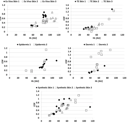
Measuring the coefficient of friction of human skin presents several challenges concerning consistency and repeatability of results. As mentioned earlier, one of the major issues is human variability. As stated in Table 1, ex vivo skin and skin used for epidermis and dermis preparation and for the TE skin came from a variety of donors. In Figure 9, the results are
0 0.2 0.4 0.6 0.8 1 1.2 1.4 1.6
0 20 40 60 80 100 120
C
O
F
HL [AU] Human Ex Vivo Skin
0 0.2 0.4 0.6 0.8 1 1.2 1.4 1.6
0 20 40 60 80 100 120
C O F HL [AU] TE Skin 0 0.2 0.4 0.6 0.8 1 1.2 1.4 1.6
0 20 40 60 80 100 120
C O F HL [AU] Epidermis 0 0.2 0.4 0.6 0.8 1 1.2 1.4 1.6
0 20 40 60 80 100 120
C O F HL [AU] Dermis 0 0.2 0.4 0.6 0.8 1 1.2 1.4 1.6
0 20 40 60 80 100 120
C
O
F
12
[image:13.595.80.515.122.546.2]plotted by donor and the different samples of the synthetic skin are compared (it should be noted that these were a different set of tests to those shown in Figures 5 and 8. As can be seen there is very good consistency between donors.
Figure 9. Plots of Patient/Sample Variability for Tests at 10mN
4 Skin Friction Modelling
Skin friction modelling using the approach proposed by Adams et al. [15] for in vivo forearm skin, was carried out to see whether similar trends were seen and to provide a comparison of
in vivo and ex vivo results.
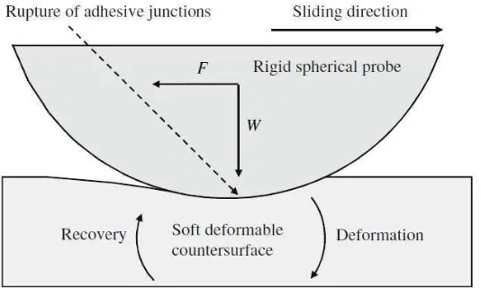
As mentioned earlier above, total friction force can be defined as a two term model (Equation 1) [40]. For a situation where a spherical probe is sliding across a skin surface, the adhesion is the result of interfacial shear, and deformation is the result of energy dissipation by sub-surface visco-elastic deformation (see Figure 10 and Equation 1).
0 0.2 0.4 0.6 0.8 1 1.2
0 20 40 60 80 100 120
C
O
F
HL [AU]
Ex Vivo Skin 1 Ex Vivo Skin 2 Ex Vivo Skin 3
0 0.2 0.4 0.6 0.8 1 1.2 1.4 1.6
0 20 40 60 80 100 120
C
O
F
HL [AU]
TE Skin 1 TE Skin 2 TE Skin 3
0 0.2 0.4 0.6 0.8 1 1.2
0 20 40 60 80 100 120
C
O
F
HL [AU] Epidermis 1 Epidermis 2
0 0.2 0.4 0.6 0.8 1 1.2 1.4 1.6
0 20 40 60 80 100 120
C
O
F
HL [AU] Dermis 1 Dermis 2
0 0.2 0.4 0.6 0.8 1 1.2 1.4
0 20 40 60 80 100 120
C
O
F
HL [AU]
13
Figure 10. A Schematic of a Rigid Spherical Probe Sliding on a Soft Counter-surface (from Adams et al. (2007))
= + (1)
The deformation component, defined originally by Greenwood and Tabor [41] and experimentally validated by Adams et al. [15] is:
= / / / (2)
where β is the visco-elastic hysteresis loss fraction, R is the probe radius, ν is the Poisson’s ratio, E is the effective Young’s modulus of the skin and W is the normal force. This is usually ignored, however, as its contribution to the overall friction force has been shown to be very low for this contact configuration at relatively low loads on skin [15, 22]. For the testing outlined in this work using a 4 mm diameter probe and using values from the literature for skin properties: β= 0.24 and ν = 0.49 [42] (Johnson et al., 1993); E = 100kPa [22] and W = 30 mN, Fdef = 0.017, an order of magnitude lower than the total dry skin friction force expected.
The interfacial, or adhesive, component is given by:
= (3)
where A is the contact area and τ is the interfacial shear strength. Despite the roughness seen on the ex vivo skin samples shown in Figure 4, it is anticipated that the reduction in elastic modulus and the swelling that occurs when skin is wet would cause it to be smooth within a probe contact [15]. This means that a Hertz approximation for contact area might be appropriate:
= ∗
/
(4)
It has been noted in work on thin organic films that the interfacial shear strength is related to the mean contact pressure, p, as follows [43]:
= ∀+ #∃ (5)
where τ0 is the intrinsic interfacial shear strength, α is a pressure coefficient and p = W/A.
14
[44] adhesion model for smooth contacts to take account of wetted skin where the stratum corneum has plasticised and become very soft:
= ∀ ∗
/ /
+ # (6)
The friction coefficient can then be calculated by dividing by W:
% = ∀ ∗
/ /
+ # (7)
[image:15.595.124.470.265.497.2]Using E = 40 kPa (value for wet skin used by Adams et al. [15]) and ν = 0.49, area, A, was calculated, using Equation 4, and then values for p were derived for each of the loads used in the current testing, 5, 10 and 30 mN. Using average friction force data for the ex vivo skin samples at 100 AU (for wet) and 40 AU (for dry) to calculate values for τ, a plot was created to derive values for τ0 and α, as shown in Figure 11.
Figure 11. Interfacial Shear Strength versus Pressure for ex vivo Skin
The values of α and τ0, 0.1164 and 5.1 kPa respectively, compare well with the values derived for wet forearm skin derived by Adams et al. [15]. This is a promising result and may indicate that ex vivo skin treated in the right way can give behaviour representative of in-vivo skin. There are a number of reasons why the behaviour would be slightly different, including the difference in temperature and the storage medium used for the ex vivo tissue.
In work of Adams et al., wet values were compared to literature values for dry glassy organic polymers, for which values for τ0 of a few MPa have been noted, which are several orders of magnitude higher, and α values of 0.1 to 1 [45]. The assumption was made that skin values would be similar in dry conditions, and that the addition of water therefore reduces the interfacial shear. However, the friction coefficient rises with increasing hydration levels. The only explanation for this must be an increase in the contact area. In that case the assumption of a Hertzian contact area is probably invalid, at least for the wet case. This would not be a surprise as during the tests in the current study the probe seemed to compress into the material quite heavily and there was a bow wave seen (similar to that observed in forearm testing by Kwiatkowska et al. [22]), which would increase the contact area.
y = 0.1164x + 5.1024 y = 0.148x + 1.6964
0 1 2 3 4 5 6 7
0 5 10 15 20 25
In te rf a ci a l S h e a r S tr e n g th , τ (k P a )
Contact Pressure, p(kPa) Wet Skin
15
Using the same approach to calculate α and τ0 for friction tests in dry conditions (around 60 AU, see Figure 5) using E = 100 kPa and ν = 0.49 gives values of 0.148 and 1.696 kPa respectively (see Figure 11). If it is assumed that the contact area is non-Hertzian, Fadh can be given by Equation 8:
= ∀ + # (8)
Assuming that α and τ0 stay the same for wet skin as calculated for dry and taking the average value of Fadh for 5 mN at 100 AU, A can be determined and is 2.7 times higher than the Hertzian approximation. This seems plausible.
If it is assumed that E for “dry” TE skin, dermis and synthetic skin are proportionally lower by the same amount as the shear moduli shown in Figure 9, and the values of α and τ0 are the same, the friction forces can be calculated as: 0.46, 0.49 and 1.21. These seem reasonable approximations when compared with the experimental values shown in Figure 9.
Equation 9, for the minimum film thickness, hc, in a sliding IEHL (Isoviscous Elasto-Hydrodynamic Lubrication), contact (where sufficient pressure is generated in the converging region of a contact for the surfaces to become separated by a thin film of liquid) was derived by Johnson et al. (1997) for the case when ν = 0.49:
ℎ∋ = 1.43,−./∀.0 1∀. ∀2 ∀. ∀. (9)
where η is the lubricant viscosity and V is the sliding velocity. This equation was used to obtain and approximate relationship for the coefficient of friction µIEHL [46]:
%3 45 = 1.51,−./∀. 01 ∀. 2 ∀. 7 ∀. (10)
Putting in values of, hc = 34 nm, which represents the minimum fluid film thickness required to fully separate the surfaces. This is considerably lower than the roughness of the surfaces used. For full film separation therefore, at the conditions used the surface roughness of the skin specimens were much lower than its unloaded state. µIEHL = 0.0025, which is
Predicted µIEHL for a smooth surface contact (using values of E = 40kPa and η = 1 mPas for tests with the 2 mm radius probe used in this work at 0.0005 m/s and 5 mN) was 0.0025 whih is lower than that seen in the tests, so it is likely that rather than full film separation mixed or boundary lubrication was occurring. This is supported by the calculation of the EHL film thickness (34 nm) which is less than the surface roughness.
5 Conclusions
This work was aimed at determining whether biological and synthetic skin substitutes behave like ex vivo human skin in tribological tests. This was primarily to determine if they could be used as a reliable test platform in tribological studies to assess skin interactions with materials used to manufacture medical devices, consumer products and other objects that come into contact with skin. The work has provided the first tribological assessment of tissue
engineered skin.
16
Acknowledgements
This research was carried out within the framework of the European Commission FP7 project UNITISS, FP7-PEOPLE-2011-IAPP/286174.
References
[1] Lewis, R., Menardi, C., Yoxall, A., Langley, J., 2007, “Finger Friction: Grip and Opening Packaging”, Wear, Vol. 263, pp1124-1132.
[2] Tomlinson, S.E., Lewis, R., Ball, S., Yoxall, A., Carre´, M.J., 2009, “Understanding the Effect of Finger-Ball Friction on the Handling Performance of Rugby Balls”, Sports
Engineering, Vol. 11, No. 3, pp109–118.
[3] Lewis, R., Carré, M.J., Abu Bakar, A., Tomlinson, S.E., 2012, “Effect of Surface Texture, Moisture and Wear on Handling of Rugby Balls”, Tribology International, Vol. 63, pp196-203.
[4] Chen, X., Barnes, C.J., Childs, T.H.C., Henson, B., Shao, F., 2009, “Materials Tactile Testing and Characterisation for Consumer Product Affective Packaging Design”,
Materials Design,Vol. 30, No., 10, Vol. 4299-4310.
[5] Sivamani, R. K., Goodman, J., Gitis, N. V. and Maibach, H. I., 2003a, “Friction coefficient of skin in real-time”, Skin Research and Technology, Vol 9, pp235–239.
[6] Derler, S., Gerhardt, L.-C., 2012, “Tribology of Skin: Review and Analysis of
Experimental Results for the Friction Coefficient of Human Skin”, Tribology Letters, Vol 45, pp1-27.
[7] Tomlinson, S.E., Lewis, R., Carré, M.J., 2007, “Review of the Frictional Properties of the Finger-Object Contact when Gripping”, Journal of Engineering Tribology,
Proceedings of the IMechE Part J, Vol. 221, pp841-850.
[8] Sivamani, R.K., Goodman, J., Gitis, N.V., Maibach, H.I., 2003b, “Coefficient of Friction: Tribological Studies in Man - An Overview”, Skin Research and Technology, Vol. 9, pp227-234.
[9] Liu, X., Lewis, R., Carré, M.J., Matcher, S.J., 2012, “Feasibility of using Optical Coherence Tomography to Study the Influence of Skin Structure on Finger Friction”,
Tribology International, Vol. 63, pp34-44.
[10] Hendriks, C.P., Franklin, S.E., 2010, “Influence of Surface Roughness, Material and Climate Conditions on the Friction of Human Skin”, Tribology Letters, Vol., 37, No. 2, pp361-80 373.
[11] Tomlinson, S.E., Carré, M.J., Lewis, R., Franklin, S.E., 2011a, “Human Finger Contact with Small, Triangular Ridged Surfaces”, Wear, Vol. 271, pp2346-2353.
[12] Derler, S., Preiswerk, M., Rotaru, G.M., Kaiser, J.P., Rossi, R.M., 2015, “Friction Mechanisms and Abrasion of the Human Finger Pad in Contact with Rough Surfaces”, Tribology International, Vol. 89, pp119-127.
17
[14] Liu, X. Gad, D., Lu, Z., Lewis, R., Carré, M.J., Matcher, S.J., 2015, “The Contributions of Skin Structural Properties to the Friction of Human Finger-Pads”, Journal of
Engineering Tribology, Proceedings of the IMechE Part J, Vol.229, pp294-311.
[15] Adams, M. J., Briscoe, B. J., Johnson, S. A., 2007, “Friction and Lubrication of Human Skin”, Tribology Letters, Vol. 26, No. 3, pp239-253.
[16] Tomlinson, S.E., Lewis, R., Liu, X., Texier, C., Carré, M.J., 2011b, “Understanding the Friction Mechanisms between the Human Finger and Flat Contacting Surfaces in Moist Conditions”, Tribology Letters, Vol. 41, pp283-294.
[17] Park, A.C., Baddiel, C.B., 1972a, “Rheology of Stratum Corneum I: A Molecular Interpretation of the Stress-Strain Curve”, Journal of the Society Cosmetic Chemists, Vol. 23, pp3-12.
[18] Park, A.C., Baddiel, C.B., 1972b, “The Effect of Saturated Salt Solutions on the Elastic Properties of Stratum Sorneum”, Journal of the Society Cosmetic Chemists, Vol. 23, pp471-479.
[19] Geerligs, M., van Breemen, L., Peters, G., Ackermans, P., Baaijens, F., Oomens, C.W.J., 2011, “In vitro Indentation to Determine the Mechanical Properties of Epidermis”, Journal of Biomechanics, Vol. 44, pp1176–1181
[20] Pailler-Mattei, C., Bec, S., Zahouani, H., 2008, “In vivo Measurements of the Elastic Mechanical Properties of Human Skin by Indentation Tests”, Medical Engineering and Physics, Vol. 30, pp599–606.
[21] Hendriks, F.M., Brokken, D., van Eemeren, J., Oomens, C.W.J., Baaijens, F.P.T., Horsten, J.B.A.M., 2003, “A Numerical-Experimental Method to Characterise the Non-Linear Mechanical Behaviour of Human Skin”, Skin Research and Technology, Vol. 9, pp274-283.
[22] Kwiatkowska, M., Franklin, S.E., Hendriks, C.P., Kwiatkowski, K., 2009, “Friction and Deformation Behaviour of Human Skin”, Wear, Vol. 267, pp1264-1273.
[23] van Kuilenburg, J., Masen, M.A., van der Heide, E., 2013, “Contact Modelling of Human Skin: What Value to use for the Modulus of Elasticity?”, Journal of
Engineering Tribology,Proceedings of the IMechE, Part J, Vol. 227, pp347–359.
[24] MacNeil, S., 2007, “Progress and Opportunities for Tissue Engineered Skin”, Nature, Vol. 445, pp874-880.
[25] MacNeil, S., 2008, “Biomaterials for Tissue Engineering of Skin”, Materials Today, Vol. 11, No., 5, pp26-35.
[26] Franklin, S.E., Baranowska, J., Hendriks, C.P., Piwowarczyk, J., Nachman, M., 2016 “Comparison of the Friction behaviour of Occluded Human Skin and Synthetic Skin in Dry and oist Conditions”, in press, Tribology Transactions.
[27] Cottenden, D.J., Cottenden, A.M., 2013, “A Study of Friction Mechanisms between a Surrogate Skin (Lorica soft) and Nonwoven Fabrics”, Journal of the Mechanical
Behaviour of Biomedical Materials, Vol. 28, pp410–426
18
[29] Skedung, L., Buraczewska-Norin, I., Danwood,N., Rutland, M.W., Ringstad, L., 2016, “Tactile Friction of Topical Formulations”, Skin Research and Technology, Vol. 22, p46-54.
[30] Guerra, C., Schwartz, C. J., 2011, “Development of a Synthetic Skin Simulant Platform for the Investigation of Dermal Blistering Mechanics”, Tribology Letters, Vol. 44, pp223-228.
[31] Guerra, C., Schwartz, C.J., 2012, “Investigation of the Influence of Textiles and Surface Treatments on Blistering using a Novel Simulant”, Skin Research and Technology, Vol. 18, pp94-100.
[32] Shao, F., Childs, T.H.C., Henson, B., 2009, “Developing an Artificial Fingertip with Human Friction Properties”, Tribology International, Vol. 42, pp1575-1581.
[33] Tiezzi, P., Lotti, F., Vassura, G., 2003, “Polyurethane Gel Pulps for Robotic Fingers”,
Proceedings of IEEE International Conference on Advanced Robotics, Coimbra,
Portugal, 30 Jun-3 Jul, 2003.
[34] Cutkosky, M.R., Jourdain, J.M., Wright, P.K., 1987, “Skin Materials for Robotic Fingers”, Proceedings of the 1987 IEEE International Conference on Robotics and
Automation, Los Alamitos, USA, March, pp1649–1654.
[35] Chakrabarty, K., Dawson, R., Harris, P., Layton, C., Babu, M., Gould, L., Phillips, J., Leigh, I., Green, C., Freedlander, E., MacNeil, S., 1999, “Development of Autologous Human Dermal-Epidermal Composites based on Sterilized Human Allodermis for Clinical Use”, British Journal of Dermatology, Vol. 141, pp811–23.
[36] Desphande, P., Ralston, D.R., MacNeil, S., 2013, “The Use of Allodermis prepared from Euro Skin Bank to Prepare Autologous Tissue Engineered Skin for Clinical Use”,
Burns, Vol. 39, pp1170-1177
[37] Syndaver, 2015, http://syndaver.com/shop/syntissue/muscular-tissue-plate/, accessed 27/10/15.
[38] Palma, L., Tavares, L., Monteiro, C., Bujan, M.J., Rodrigues, L.M., 2012, “Diet Water seems to Influence Skin Hydration and Biomechanics”, Proceedings of the World
Congress of the International Society for Bioimaging of the Skin (ISBS), Copenhagen
Denmark, 28-30 November 2012.
[39] Waranuch, N., Maphanta, S., Wisuitiprot, W., 2012, “Effect of Microparticles
Containing Green Tea Extract on Facial Skin Improvement”, Proceedings of the World
Congress of the International Society for Bioimaging of the Skin (ISBS), Copenhagen
Denmark, 28-30 November 2012.
[40] Bowden, F.P., Tabor, D., 1954, Friction and Lubrication of Solids, Oxford University Press, London.
[41] Greenwood, J.A., Tabor, D., 1958, “The Friction of Hard Sliders on Lubricated Rubber: The Importance of Deformation Losses”, Proceedings of the Physical Society of
London, Vol. 71, pp989-1001
[42] Johnson, S., Gorman, D.M., Adams, M.J., Briscoe, B.J., 1993, “The Friction and Lubrication of Human Stratum Corneum”, Proceedings of the 19th Leeds-Lyon
Symposium on Tribology, p663.
19
[44] Wolfram, L.J., 1983, “Fricion of Skin”, Journal of the Society Cosmetic Chemists, Vol. 34, pp465-476.
[45] Briscoe, B.J., Smith, A.C., 1980, “The Shear Properties of Thin Organic Films”, in
Reviews on the Deformation Behaviour of Materials, Vol. 3, ed. Feltham, P., Scientific
Publications, Israel, pp151-191.
[46] Johnson, S., Adams, M.J., Arvanitaki, A., Briscoe, B.J., 1997, “Film Thickness Measurements in Elastohydrodynamically - Lubricated Elastomeric Contacts”,