0022-538X/85/100012-07$02.00/0
Copyright X 1985, American Society for Microbiology
Differences
in
Cell-to-Cell
Spread
of
Pathogenic
and
Apathogenic
Rabies Virus
In
Vivo and In
Vitro
BERNHARDDIETZSCHOLD,l* TADEUSZ J. WIKTOR,1 JOHN Q. TROJANOWSKI,2 RODERICK I. MACFARLAN,'WILLIAM H. WUNNER,' M. J. TORRES-ANJEL,' ANDHILARY KOPROWSKI1 The Wistar Institute of Anatomy andBiology,1 andDepartment of PathologyandLaboratory Medicine, Division of
Neuropathology, University
of Pennsylvania
MedicalSchool,2
Philadelphia, Pennsylvania
19104 Received 19February1985/Accepted 11 June1985Pathogenic parental rabies virus and apathogenic variant viruswereshownto differ in theirabilitytoinfect neuronsinvivo and neuroblastoma cellsinvitro. Afterintracerebral inoculation,thedistribution ofinfected neuronsin the brainwassimilar for bothviruses,but the rate ofspreadthroughoutthebrain,the numberof infectedneurons,and thedegree of cellular necrosisweremuchlower in thecaseofapathogenicvirus. After adsorption to mouse neuroblastoma cells, apathogenic virus was less rapidly internalized than pathogenic virus, and cell-to-cell spread of apathogenic variantvirus wascompletely prevented bythe addition ofrabies virus-neutralizingantibody, whereas the spread of pathogenicviruswas notaffected.
The replication ofrabies virus in vivo is almost entirely
restricted to nerve tissue, and neurotropism is the main
feature ofrabies infection. Afterintramuscular inoculation, rabies virus replicates first in the nearby striated muscle
cells,then enters the sensory nerve onunmyelinated neural
endings in motor nerve endplates or neuromuscular or
neurotendinal spindles, andspreadsalong peripheral nerves up tothe spinal cord into the brain (10). Unlike manyother virus infections, inwhich neutralizingantibody limits virus
spread, rabies virus infection spreads from cell to cell in
tissue culture despite a continuous overlay of antirabies serum (16),and ithas beenpostulated thatthisescapefrom neutralization may reflect the mechanisms ofspread during natural infection (16). The basic mechanism(s) involved in
the pathogenicity of fixed rabies viruses and factors that determinemanifestation ofthedisease after infectionare not well understood but most likely involve multiple factors, includingtheimmunological competenceofthe host.
Recent investigations with apathogenic rabies variant vi-ruses indicate that the glycoprotein of rabies viruses is a
major determinant of virus pathogenicity (3, 4). The
anti-genic variant(RV194-2) selectedfor itsresistance to
neutral-izing monoclonal antibody 194-2exhibitedamodified
patho-genicity inadultmice (4), achange which corresponds to a
single amino acid substitution oftheglycoprotein molecule
atamino acid
position
333(Arg
-- IleorGln) (4).
Thisaminoacid substitution at position 333 was also identified in the
apathogenic fixed rabies virusstrains Flury HEPand Kelev (W. H. Wunner and C. L. Smith, unpublished data), which
also resisted neutralization by monoclonal antibody 194-2.
Thus,theamino acidsubstitution atposition 333 appears to be a molecular marker of the apathogenic phenotype. The
majorbiologicaldifference in the in vivo behavior of
patho-genic and apathogenic rabies viruses is the extent of virus spread in the central nervous system, the apathogenic vi-rusesbeing significantly less neuroinvasive.
Inthe presentstudy, we compared the pathogenic parental rabies virus and the biochemically and biologically well
characterized apathogenic RV194-2 variant virus for their
ability
toinfectneuronal cells in vivo and to spread from cell to cell in atissueculture system that accuratelyreflects the*Correspondingauthor.
in vivo behavior ofpathogenic and apathogenic fixed rabies virus.
MATERIALS AND METHODS
Cells. Baby hamsterkidney cell line BHK-21 (14) and C 1300 clone NA mouse neuroblastoma (NA) cells (9) were propagated in Eagle minimal essential medium supplemented with 10% fetal calf serum.
Viruses. Clone-purified challenge virus standard CVS-11 (6) and ERA (1)parental stocks were produced in BHK-21 cell monolayers infected at amultiplicity of infection(MOI) of 0.1 PFU per cell and incubated at 33°C for 4 days in the presence ofEagle minimal essential mediumsupplemented with 0.2% bovine serum albumin.
Variant strains expressing anantigenically altered glyco-protein were selected from CVS-11 and ERA virus stocks after treatment with monoclonal antibodies and cloning of
neutralization-resistant mutants as described previously
(18). The neuropathogenicity of variants for adult mice was evaluated based on survival after intracerebralinoculation of 5- to 6-week-old female ICR mice. The nonpathogenic vari-antsof CVS-11 and ERAviruseswere selected with mono-clonal antibody 194-2(4).
Virus infectivity titration. Monolayers of hamster CER cells were infected with 0.1 ml of virus at serial fivefold dilutions and incubated for 60 min toallowvirus adsorption, overlaid withnutrient agarosemedium(17),andincubatedat
35°C for 4 days. Plaques were counted after removal of agarose and staining withcrystal violetsolution.
Radioimmunoassay.Serialtwofold dilutionsof inactivated virus (50 ,ug per well) in sodium carbonate-bicarbonate
buffer(pH 9.5) were air dried on soft plastic 96-well plates (Dynatech Laboratories, Inc.) and incubated for 1 h with
phosphate-buffered saline (PBS) containing 10% agamma horse serum. Monoclonal antibody 509-6, specific for rabies virus glycoprotein, and antibody 502-2, specific for rabies
nucleocapsid antigen, were added as ascites (25 ,ul) at a 1:1000 dilution to a series of wells to which antigen was adsorbed. Plates wereincubatedfor 1 h at 37°Cand washed threetimeswith PBS.
125I-labeled
goatanti-mouseimmuno-globulin (25 ,ul; specific activity, 0.5
j,Ci/,g)
containing30,000cpm was added to each well. Plates were incubated at
37°Cfor 1 h andwashedthree times with PBS. The bottoms 12
on November 10, 2019 by guest
http://jvi.asm.org/
SPREAD OF RABIES IN VIVO AND IN VITRO 13
of the wells were then cut with anincandescent wire, and
radioactivity in the wellswasmeasured ina gamma counter.
Thehighest dilution of antigen giving 10% ofthe maximum countsperminute overthebackground was considered the
endpoint of titration.
Immunohistopathology. Six-week-old ICR female mice wereinjected in the frontal lobe ofthe cerebrum with 25 ,ul of CVS-11 orCVS RV194-2 virus
(104
PFU)witha 27-gaugeneedle. At the indicated times, four animals were
anesthe-tized and perfused with periodate-lysine-paraformaldehyde fixative (8). Brains wereremoved, kept in the same fixative
for 24 h, paraffin embedded, and sectioned by standard
procedures. A peroxidase-anti-peroxidase technique (15) was used to demonstrate rabies virus antigen. Briefly,
deparaffinized
brain tissue sections were treated for 24 h with rabbit anti-rabies nucleocapsid serum diluted 1:200.Thisantibody detected CVS-11 and CVS RV194-2antigens with similarsensitivity. Sectionswerethenincubated for30 minwithgoat anti-rabbit immunoglobulin G (Cappel Labo-ratories) diluted 1:100 and for30min with peroxidase-anti-peroxidase complex diluted 1:600 (Cappel Laboratories). Slideswerethenwashed with0.5 MTris hydrochloride(pH
7.6) and stained with diaminobenzidine tetrachloride (type II; Sigma Chemical Co.) as described previously (15) and
counterstained withMayer's
hematoxylin.
Immunofluorescent antibody staining. Viral antigen in
in-fected cells was detected by the direct immunofluorescent
antibody
staining technique. Cells werewashed with PBS, fixed for 30 min in cold acetone, and air dried. Cells werethen incubated with fluorescein isothiocyanate-labeled rab-bitpolyclonal antinucleocapsid antibody for30min, washed in PBS and distilledwater, and examined under UV illumi-nation. The anti-rabies nucleocapsid antibody used was
equally
efficient indetecting
CVS-11
and CVS RV194-2viruses.The percentageof infected cellswasdetermined by
counting
atleast 1,000 cells.Isolationof virus from infected mouse brain. At 2, 3, and 4
days postinfection (p.i.), three animals infected with RV194-2virusweresacrificed, and the brainswereremoved andhomogenized. The virus wasthen isolated on BHK-21
cell cultures as previously described (19). The antigen
makeup
of this virus has been determined(4). RESULTSImmunohistochemical and histological studies. Pathogenic
parentCVS virus andapathogenic variant viruswere
exam-inedfor their
ability
to infect neurons in vivo. Brain tissue from mice infectedintracerebrally
withpathogenic
orapathogenic
viruswasexaminedhistologically
at3, 5, and7days p.i.
Theimmunoperoxidase staining technique
wasused to demonstrate the presence of rabies
antigens
atthecellular level
(Fig. 1).
Frozen sections of infected mousebrains stained
by
direct immunofluorescence withanti-rabiesnucleocapsid
serum or anti-rabiesglycoprotein
serum alsogavesimilar results
(data
notshown).
At 3
days p.i.
withapathogenic virus,
viralantigen
wasidentified
in neurons caudal to theinjection
site in theneocortexand the
hippocampus
of bothhemispheres.
How-ever, in thecerebellum,
which does notproject
directly
tothe neocortex, there was no evidence of immunoreactive virus
(Fig. 1A).
Incontrast,immunoperoxidase-stained
cer-ebellum sections ofmouse brain infected with
pathogenic
virus revealedstrong evidence ofviral
antigen
in thewhite matter(Fig.
1D,
see areamarkedby star).
At 5
days
p.i.
in theapathogenic
virus-infectedtissue,
viral
antigen
was detected in thecerebellum,
mostly
con-fined to axons in the white matter (Fig. 1B), although an
occasional neuron in the cerebellar cortex also contained
viral antigen. The intensity of staining and the number of neuronswith immunoreactiveviral protein in the neocortex and the hippocampus also increased compared with those
found after 3 days (data not shown). Similarly, affected neurons were also recognized in the thalamus and the hypothalamus. Atthistime, in the pathogenic virus-infected
cerebellum, infection already progressedto a larger number
ofPurkinje cells (Fig. 1E). Also, in contrast to the
patho-genic virus infection,adefiniteleptomeningitis was observed
in mouse brains with apathogenic virus (data not shown).
The infiltrate was composed of mononuclear cells, and
similar infiltrates were observed around blood vessels as
they entered the brain parenchyma. This could not be
demonstrated throughout the infection with pathogenic
vi-rus.
By 7 days p.i. in the apathogenic virus-infected mouse
brain, immunoreactive viral antigen was detected
through-outthebrain, includingthebrain stem. CerebellarPurkinje
neurons containing immunoreactive viral antigen were
readily identifiedbut were not numerous atthis time (fewer
than 20 per section) (Fig. 1C). Most neurons, however,
includingthoserevealing viral antigen, appeared healthy. In the pathogenic virus infection, neurons were much more
intensely stained, and many appeared to be undergoing necrosis. In addition, the neurophil became vesicular or
rarified, consistent with intracerebral edema and suggesting thatmortality resulted from brain swelling.
These results of comparative studies indicate that both
apathogenic
andpathogenic viruses spread within the brain in similar pathways; however, the spread of apathogenic virus was slower, as evidenced by the amount of viralantigen
detectedatdifferent timesexamined. This difference in therate ofspread ofpathogenicversusapathogenic viruswas a consistent observation in all four mice examined at eachofthedifferent timepoints. Virus isolated3and4days p.i. from brains of apathogenic CVSRV194-2 virus-infected mice retainedtheantigenic phenotype of RV194-2 virus, i.e., resistance to neutralization by monoclonal antibody 194-2. No virus could be recovered from CVS RV194-2 virus-infected mouse brains 2 days p.i. Therefore, the limited
spread is not due to aphenotypic reversion that has been
observed aftervirus passage innewborn mousebrain(4).
In vitro studies with pathogenic and apathogenic rabies
viruses. The cell-to-cell
spread
of virus was studied witheitherNAcells
(C
1300cloneNA)(Fig.
2B)orBHK-21cells(Fig.
2A).Monolayers
ofNAorBHK-21cellswereinfectedat an MOI of0.1 PFU per cell with either
pathogenic
orapathogenic variantvirus. At 1 hp.i., antirabies serum,at a finaldilution of1:100 and
corresponding
to aneutralization index of7.6,wasaddedtotheinfectedcells,
andat24 hp.i.,
less than10% ofBHKand NAcells infected with
pathogenic
and
apathogenic virus,
respectively,
containedrabies-specific antigen,
as demonstratedby
fluorescentantibody
staining.
In BHK-21cells infectedwitheitherpathogenic
orapathogenic
variant CVSvirus,
the infectionspread
in the presence ofneutralizingantibody
touninfectedneighboring
cells until 100%of the cells were infectedat96 h
p.i.
(Fig.
2A). In NA cells infected with
pathogenic
CVS virus(Fig.
2B), thekinetics of virus
spread
in the presence ofneutral-izing antibodywassimilartothat in BHK-21 cells.
However,
in NA cells infected with the
apathogenic
variant virusderived fromCVS
(Fig. 2B)
orERAvirus(data
notshown),
no spread of infection was observed in the presence ofvirus-neutralizing antibody.
VOL.56, 1985
on November 10, 2019 by guest
http://jvi.asm.org/
14 DIETZSCHOLD ET AL. J. VIROL.
A D
A*;~~~~~~~~~~~~~~~~~
'i :~t.t
~~4: se,
i N9.A 1.X:P~
'644'svRtS;W'S'!z;t't'-.j*4
*o ~'g f .~tAC4;A,izt,,. - - *-* tsr*¢
4 0i! r
to-~.
*+
3-
* * :#4.;~*N
th,½
Is
s0¼2:4::t
r
r-~
*S3tA A t @ %~~
at
.7).- stt.;;P
g-
2;At
*¢;tr"8<''
| [image:3.612.79.537.74.666.2]-~~~~~~~~A£~~~~~~~~~~~~~~
;~~~~~~~
A '.?$4; i .\. Vt7i
* v<A
e".a
T~~~~
z*~~~>4g'.A.A- S~~~~~~~~~~~~~fi 11
4 *
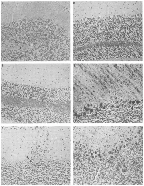
FIG.1micrPho
M,.1.*-- $ t4C
A
4'
A,
4
~
*
n
A~~~~~~~~~~~~~~ -~~~~~~~~
J.-J714~AOA~ ~ ~
sections.The mice weresacnificedonday 3 (A andD),5(B andE),or 7 (Cand F) after aneocortical (frontal lobe) injection of either virus. Viralnucleocapsid antigen was detected as described in Materials andMethods.Theimmunostained patterns of the cerebellum exemplify the differences in the behavior of these viruses.At 3days,no evidence of spread of theapathogenic virus to the cerebellum was seen (see star), whileimmunoreactive pathogenic virus was present mainly in the white matter(D).At5 and 7days,Purkinje neurons (large arrows indicate perikarya; small arrowsindicate dendrites) were the predominant class of cerebellar neurons in which immunoreactive pathogenic (E and F) orapathogenic(B andC)virus was found. Thedifferencebetween the two viruses in thecerebellumandelsewherewasprimarilyquantitative; many more Purkinje cells contained immunoreactive pathogenic virus at early survival times compared with the apathogenic virus. All sectionswerelightlycounterstained withhematoxylin. Magnification, x184.
T ,t 1--
on November 10, 2019 by guest
http://jvi.asm.org/
SPREAD OF RABIES IN VIVO AND IN VITRO 15
24 48 72 96 0
B
24 48 72 96
[image:4.612.130.493.73.260.2]Hours after infection
FIG. 2. Cell-to-cellspreadof pathogenic CVS-11 virusand apathogenicCVS RV194-2 variant virus in BHK cells (A) and mouse NA cells (B). Monolayers ofBHKcells or mouse NA cells were infected at an MOI of 0.1 PFU per cell with either pathogenic CVS-11 virus(@)or apathogenic CVSRV194-2virus(0).Aftertheadditionof antirabies serumat 1 h p.i., infected cells were examined at 24, 48, 72, and 56 h p.i. for thepresence of rabies antigen by fluorescent antibody staining technique.
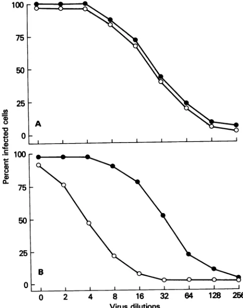
We thentested the susceptibility of BHK-21 and NA cells to infection by pathogenic and apathogenic variant virus.
The titersforboth viruses, as determined by plaque assay in
CERmonolayers, wereidentical (107.6 PFU/ml). Cellswere
infected with twofold dilutions of virus, and at 24 h p.i. the percentage ofinfected cells was determinedby the
fluores-100
r
75L
50h
25F
o 100
A
I
75F
SOh-centantibody technique. Ateachdilutionof pathogenicand apathogenic virus, a similar number of BHK cells was
infected (Fig. 3A). However, eightfold more apathogenic
variant virus than pathogenic virus was needed to infect a comparable number of NA cells(Fig. 3B).
Figures 4 and 5 illustrate the kinetics of infectious virus
production and the kinetics of viral protein synthesis,
re-spectively,in NA cells.Cells were infected at anMOI of0.1, PFU per cell, and at 1 h p.i., the cells were treated with antirabies serumfor 1 htoneutralize nonadsorbed virus. In NAcells, 100 times more pathogenic virus than apathogenic
virus was produced by 12 h p.i. (Fig. 4). At 36 h p.i., the yieldsof pathogenicandapathogenic viruses were equal. To assess viral protein synthesis, the amount of nucleocapsid
protein and glycoprotein present in cell lysates was deter-mined by radioimmunoassay with monoclonal antibodies
specific for N or G protein. Similar to the production of
virus, the synthesis of G and N protein at 12 h p.i. was
significantly lower in NA cells infected with apathogenic virus than in NA cells infected with pathogenic virus (Fig. 5A and B).
Previous studies (12, 20) revealedno differences in
com-petitive inhibition by the parent virus and RV 194-2
apathogenic virus for binding of radiolabeled virus to BHK-21 or NA cells. Therefore, we investigated whether differences in the kinetics of virus internalization might
account for the different rates of infecting NA cells with
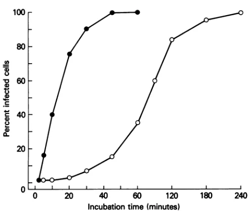
pathogenic andapathogenic virus. Monolayers of NA cells were incubated with 10 PFU of virus per cell for various
times, and noninternalized virus was neutralized by the addition of antirabies serum. Thecells werethen examined at 18 h p.i. for the presence of viral antigen by fluorescent antibodystaining. The number of infected cellswas similar
B
0 2 4 8 16 :
[image:4.612.55.300.411.721.2]Virus dilutions
FIG. 3. Susceptibility ofBHK-21 (A)and NA(B)cellsto infec-tion by pathogenic CVS-11 or apathogenic CVS RV194-2 virus. Cells were infected with twofold dilutions ofpathogenic (0) or I apathogenic(0)virus. Thetiterfor both undiluted viruseswas107.6 32 64 128 256 PFU/ml. At 24 h p.i., the percentage of infected cells was
deter-minedbythefluorescentantibodytechnique.
100r
A0
U)
a)
.)
4) a1)
40~ 75 F
50o
25F
0
Cl)
0
a)
0) a).
c
2C.
a)
0-25
-0 VOL.56, 1985
on November 10, 2019 by guest
http://jvi.asm.org/
16 DIETZSCHOLD ET AL.
lOOr
80H
U)
0)
*0
0
40
0) 0!
60
-40 F
20 F
0 20 40 60 120
Incubation time (minutes)
0 12 24 36 48 60 72 84 FIG. 6. Kineticsof internalization of pathogenicCVS-11 (@)and
[image:5.612.315.560.69.278.2]Time (hours) apathogenic CVS RV194-2 (0) virus. Monolayers of NA cellswere incubatedwith 10 PFUpercellofpathogenicorapathogenic virus
FIG. 4. Kinetics ofproduction of infectious virus in NA cells. for various time periods. At the indicatedtimes, antirabies serum NA cells were infected at an MOI of 0.1 PFU per cell with was added to the culture medium to neutralize noninternalized pathogenic CVS-11 (S)orapathogenicCVS RV194-2(0)virus. At virus. At 18 hp.i. the cellswereexamined for thepresenceofviral 1 hp.i., cellsweretreatedwithantirabiesserumfor 1 htoneutralize antigen byfluorescentantibody technique.
noninternalized virus. Antiserum was removed, cells were
incu-bated for the indicated times at 37°C, and the amount of virus released into thetissueculture mediumwasdeterminedby plaque
assay. 30% ofapathogenic virus-infected NA cells contained viral
antigen, and 90% of the cells were infected when antibody
wasadded at180min
p.i. (Fig.
6).
at12and 18 hp.i.; however, theamountof viral antigenper
cell was greater at 18 h. When rabies virus-neutralizing DISCUSSION
antibody was added at 30 min p.i., viral antigen could be
demonstrated in 90% of NA cells infected with pathogenic Theimmunohistologicaldatademonstratequalitative sim-virusbut only in 10% of NA cells infected with apathogenic ilarities in the infection of the centralnervous systemtissue virus. Whenneutralizing antibody wasaddedat60min p.i., after intracerebral inoculation of mice with the pathogenic
(A
CM .)
* 100
10 A
0 12 24 36 60 0 12 24 36 60
[image:5.612.72.293.72.309.2]Incubation time (hours)
FIG. 5. Synthesisofglycoprotein(A) andnucleocapsid protein(B)inNAcells infected with pathogenic CVS-11(-)orapathogenicCVS RV194-2(0)virus. NA cells wereinfectedatanMOI of0.1 PFUpercell and, 1 h p.i., treated with antirabiesserumfor1 htoneutralize noninternalizedvirus.Antiserum-containing tissue culture mediumwasreplaced by fresh medium, and cellswereincubated fortheindicated
times at 37°C and lysed with 1% Nonidet P-40. The amount ofglycoprotein or nucleocapsid protein in the lysate was determined by
radioimmunoassay.
7F
6I
a
5
0
0
U-2
4
0-2 ,
F
1
180 240
J. VIROL.
8
F
on November 10, 2019 by guest
http://jvi.asm.org/
[image:5.612.153.472.499.676.2]SPREAD OF RABIES IN VIVO AND IN VITRO 17
CVS parent or apathogenic variant (RV194-2) virus. Differ-ences in mouse brain infected with these viruses were mainly quantitative. The pathogenic virus spread more rapidly from the neocortex to the cerebellum and infected more neurons than did the apathogenic virus. It is possible that neural mechanisms controlling virus transport (10) determine the rates ofdissemination of rabies virus and that such mecha-nismsare affectedby the specific amino acid at position 333 of the viralglycoprotein molecule. This may be significant in determining lethality of the virus.
Our results on the spread of apathogenic virus in the centralnervous systemafter direct inoculation into the brain are similar to those described by
Kucera
et al. (7), who demonstrated a reduced ability of apathogenic virus to spread to the central nervous system after inoculation of virus into the anterior chamber of the eye.The observation that neuronal cells in vivo exhibit differ-ences in susceptibility to pathogenic and apathogenic virus can be correlated with the behavior of these viruses in cultured NA cells, which retain several characteristics of neurons (2, 11, 13). Whereas no differences between patho-genic and apathopatho-genic rabies virus infection were observed in BHK-21 cells, NA cells were much more susceptible to pathogenicthan to apathogenic virus. Only 15% of NA cells were infected with apathogenic virus when antirabies serum was added 45
min
after virus adsorption (Fig. 6). Antirabies serum added at the same time to NA cells infected with pathogenic virus failed to prevent complete infection of the cells. Since competitionbinding
experiments did not reveal any differences in the attachment of pathogenic and apathogenic virus to NA cells (20), it seems likely that the pathogenic virus is more rapidly internalized than apathogenic virus.Most interesting are the observeddifferencesin cell-to-cell spread between pathogenic and apathogenic viruses which occur in NA cells in the presence of antirabies serum, although these experiments do not reveal the particular mechanisms involved. Possible reasons for these observed differences include: (i) altered fusion function of the glyco-protein due to the point mutation at residue 333, although no differences were observed in pH-dependent fusion ofcells
infected with apathogenic versus pathogenic rabies virus (unpublished observations); (ii) site-specific proteolytic
cleavage involving arginine at position 333 in thepathogenic virus glycoprotein which may induce virus activation similar to that found in parainfluenza virus (5), although no evidence for any cleavage mechanisms has been found to support this hypothesis; and (iii) differences in affinity for putative recep-tors in viral attachment to cells in culture. Previously de-scribed binding studies involving competition for virus at-tachment to saturable cellular receptor sites have suggested that these two viruses compete forthe same specific binding site (20). In the present studies, the number ofvirusparticles of apathogenic virus required to infect an equal number of NA cells was 10 times greater than thenumber ofparticlesof pathogenic virus required, suggesting that more of the apathogenic virus particles, which were apparently capable of binding to cell surface molecules, were incapable of entering the cell. It is possible that either the apathogenic virus interacts with the cell surface receptor in a manner that is different from that of pathogenic virus, resultingin aless efficient receptorfunction, or the two virus types attach to different species of cell surface receptors, one ofwhich is less efficient for internalization of virus. The utilization of different receptor molecules on the NA cell membrane surface by pathogenic and apathogenic rabies virus would
explain the differences in their infectionratesandspreading
patterns
bothin vivo and in vitro. Wearecurrently isolating receptor molecules from NA cellsand
brain tissue which havebinding specificityfor pathogenic or apathogenicrabies virus or both. These experiments should reveal whetherdifferencesinvirulencebetween pathogenic and apathogenic rabies virus are determined by the nature of the receptor responsible for virus-host cell binding in vivo or in vitro.
ACKNOWLEDGMENTS
This work was supported by Public Health Service grants
AI-09706,AI-18562,NS-16723, and NS-00762 and by a grant from the R. J. Reynolds Foundation.
We thank MarinaHoffman for carefully readingthe manuscript. LITERATURE CITED
1. Abelseth, M. K. 1964. An attenuated rabies vaccine for domestic animals produced in tissue culture. Can. Vet. J. 5:279-286.
2. Augusti-Tocco, G., and G. Sato. 1969. Establishment of func-tionalclonallines of neurons from mouse neuroblastoma. Proc. Natl. Acad. Sci. USA 64:311-315.
3. Coulon, P., P. Rollin, M. Aubert, and A. Flamand. 1982. Molecular basis of rabies virus virulence. I. Selection of avirulent mutants of the CVS strain with anti-G monoclonal antibodies. J. Gen. Virol. 61:97-100.
4. Dietzschold, B., W. H. Wunner, T. J. Wiktor, A.D. Lopes, M. Lafon, C. Smith, and H. Koprowski. 1983. Characterization of an antigenic determinant of the glycoprotein which correlates with pathogenicity of rabies virus. Proc. Natl. Acad. Sci. USA 80:70-74.
5. Garten, W., T. Kohama, and H.-D. Klenk. 1980. Mutational changes of the protease susceptibility of glycoprotein F of Newcastle disease virus: effects on pathogenicity.J.Gen. Virol. 50:135-147.
6. Kissling, R. E. 1958. Growth of rabies virus in non-nervous tissue culture. Proc. Soc. Exp. Biol. Med. 98:223-225. 7. Ku&era, P., M. Dolivo, P. Coulon, and A. Flamand. 1985.
Pathways of the early propagation of virulent and avirulent rabies strains from the eye to the brain. J. Virol. 55:158-162.
8. McLean, I. W., and P. K. Nolsane. 1974. Periodate-lysine-paraformaldehyde fixative: a new fixative for immunoelectron microscopy. J. Histochem. Cytochem. 22:1077-1083.
9. McMorris, F. A., and F. H. Ruddle. 1974. Expression of neuronal phenotypes in neuroblastomacell hybrids. Dev. Biol. 39:226-246.
10. Murphy, F. A. 1977. Rabies pathogenesis. Arch. Virol. 54:279-297.
11. Nelson, P. G., J. H. Peacock, andJ. Aniano. 1971. Responses of neuroblastoma cells to iontophoretically applied acetylcholine. J. Cell. Physiol. 77:353-362.
12. Reagan, K. j.,and W. H. Wunner. 1984. Early interactions of rabies virus with cell surface receptors in nonsegmented nega-tive strand viruses, p. 387-392. In D. H. L. Bishop andR. W. Compans (ed.), Nonsegmented negative strand viruses. Aca-demic Press, Inc., New York.
13. Schubert, D., S. Humphreys, C. Baroni,andM. Cohn. 1969. In vitro differentiation of a mouse neuroblastoma. Proc. Natl. Acad. Sci. USA 64:316-323.
14. Stoker, M., andI. MacPherson. 1964. Syrian hamster fibroblast cell line BHK-21 and its derivates. Nature (London) 203:1355-1357.
15. Taylor, C. R. 1978. Immunoperoxidase techniques. Arch. Pathol. Lab. Med. 102:113-121.
16. Wiktor, T. J. 1966. Dynamicsof rabies virus infection in tissue culture. International Symposium on Rabies, Tallores, 1965. Symp. Ser. Immunobiol. Stand. 1:65-80.
17. Wiktor, T. J. 1973. Tissue culture methods in rabies virus
VOL.56, 1985
on November 10, 2019 by guest
http://jvi.asm.org/
investigation. W.H.O. Monogr. Ser. 23:101-123.
18. Wiktor, T. J., andH. Koprowski. 1980. Antigenic variants of rabies virus. J. Exp. Med. 152:99-112.
19. Wiktor,T. J., R.I.Macfarlan, C. M. Foggin,and H.Koprowski. 1984. Antigenic analysis of rabies and Mokola virus from
Zimbabwe using monoclonal antibodies. Dev. Biol. Stand. 57:199-211.
20. Wunner, W. H., K. J. Reagan, and H. Koprowski. 1984. Characterization of saturable binding sites for rabies virus. J. Virol. 50:691-697.