Copyright ©1985, American Societyfor Microbiology
Cells that
Constitutively Express the Herpes Simplex Virus
Immediate-Early
Protein ICP4 Allow Efficient Activation of Viral
Delayed-Early Genes in
trans
ROY H. PERSSON, SILVIA BACCHETTI, ANDJAMES R. SMILEY* PathologyDepartment, McMaster University, Hamilton, Ontario, Canada L8N 3Z5
Received 13 November1984/Accepted 11 January 1985
Tostudytheroleof herpes simplex virustype1immediate-earlyproteinsin thetranscriptional activation of herpessimplex virusgenes, weisolatedstably transformedcellsexpressing herpes simplexvirus type 1ICP4, an immediate-early protein known from previous studies to be necessary for delayed-early and late transcription.Thesecells efficiently expressedsixdelayed-early herpes simplexvirusgenesintroducedby viral superinfection,in theabsence ofdenovoviralprotein synthesis. In contrast, thedelayed-earlygeneencoding
alkalineexonuclease andthe lategeneencoding the capsid proteinVP5wereexpressedatmuch lowerlevels. Expression ofa second late gene, that for glycoprotein C, was undetectable under the same experimental conditions. Theseresults suggestthatmany,butnotall, delayed-earlygenes areefficiently activated by ICP4; in addition, they demonstrate that although the late gene for VP5 is detectably activated by ICP4, its full expression requires additional factors.
Herpes simplex virus (HSV) genes are expressed in three
sequentialwavesduring lyticinfections, largely as a result of regulation at the level of transcription (20). Although viral genes are transcribed in the nucleus of the infected cell by thehost RNA polymerase 11 (14), virally coded proteins are
known to play a major role in controlling the transition
between transcriptional phases. Because most of the HSV genes are controlled by separate promoters (for a review, seeE. K. Wagner in B. Roizman,ed., The Herpes Viruses, Vol. 3, in press), this transcriptional control is presumably mediated by the differential interactions ofalimited number
ofregulatorswithalarge numberofcis-acting sites.Thefive immediate-early (or a)genes are the first HSV genes to be expressed; they are also the only ones transcribed when viralprotein synthesis is prevented (1, 10, 27, 34, 35, 42-44). The product ofone ofthese genes (ICP4) is necessary for boththeinitial activationand the continuedtranscriptionof thedelayed-early (or
P)
and late (or y)viral genes (34, 41).Indeed, ICP4 is the onlyHSVproteinknown at present to be involved in the positive control of transcription. The role, if any, oftheremaining four immediate-early proteins in viral
geneactivation remains unknown.
The results of Honess andRoizman (20) and Watson and
Clements (41)areconsistent withthe idea thatexpressionof one or moreofthefiveimmediate-early proteins sufficesfor the full activation of delayed-early genes and that ICP4 is necessary for this activation. The scheme of Honess and
Roizman (20) also suggests that the efficient expression of
late genes requires the action of delayed-early proteins in addition toimmediate-earlyproteins.Resultsofmore recent studies havesuggested that the late genesareheterogeneous in their transcriptional requirements, with some being
ex-pressed at low levels at early times and when viral DNA
replication is blocked (for example, the gene encoding the
major capsid protein VP5 [15; Wagner, in press]), whereas othersareexpressed only aftertheonsetof DNAreplication (for example, glycoprotein C [gC; 12; Wagner, in press]). Because theformer class of late genes (the "leaky late" or
*Correspondingauthor.
13-y)
arefirstexpressed
atearly
times,
itseemspossible
thattheyaredirectly activated byimmediate-early proteins.
Toexamine theroleof ICP4in the activation of delayed-early and late genes, we transfected a cloned HSV type 1 (HSV-1) DNAfragmentencodingICP4 and possibly ICP47 into mouse cells and report here on the isolation of stably transformed cell lines that constitutively express these im-mediate-early proteins. These cells supported efficient
acti-vationofavariety ofdelayed-early genes underconditions
thatprecludede novoviralprotein synthesis.An exception within thisclass was the geneforalkaline exonuclease. We concludethat,atmost,onlytwoof thefiveimmediate-early proteins are required for the activation of the majority of delayed-early genes. By contrast, theleakylate gene encod-ing thecapsid proteinVP5 wasexpressedonlyat alow level,
and expression of the late gene for gC was not detected.
These results suggest thatalthoughimmediate-earlyproteins suffice forthe detectableactivation ofsomeleakylate genes, their efficient expression requires additional factors. The sameappearstobetruefortheactivation oftightlate genes.
MATERIALSAND METHODS
Cellsandvirus. Verocells and thymidine kinase-deficient
(tk-), adenine phosphoribosyl transferase-deficient (aprt-)
LTAcells were maintained inax-minimal essential medium
(GIBCO Laboratories) containing 10% fetal bovine serum. Cell lines cotransformed by pRHP6 and PN17BS3 were isolatedby transfecting subconfluent 60-mm plates of LTA cells with 100 ng of each plasmid DNA and 10 ,ug of
high-molecular-weight LTA cell carrierDNA perml by the cal-ciumphosphatecoprecipitation technique (18) as described
previously (15). AfterHAT selection (40), individual colo-nieswerepicked withacloningcylinderand
expanded
intocelllines. HSV-1
(strain
KOS) waspropagated
andtitrated on Verocells.Labelingandimmunoprecipitation ofproteins.Infected(20
PFU percell,unlessindicatedotherwise)oruninfectedcells were labeled for the indicated times in methionine-free
mediumsupplemented with 2% dialyzedfetalbovine serum and 25to40,uCiof
[L-35S]methionine
(1100to1400Ci/mmol;
414
on November 10, 2019 by guest
http://jvi.asm.org/
Amersham Corp.) per ml. Where indicated, cycloheximide (50 ,ug/ml) was added 45 min before infection, maintained
until6 h postinfection, and then replaced with actinomycin D(10,ug/ml). After labeling, the cells were scraped from the dishes, washed three times with phosphate-buffered saline, andlysed by sonicationin RIPAbuffer (50 mM
Tris-hydro-chloride [pH 7.2], 0.15 M NaCl, 0.1% sodium dodecyl sulfate, 1% sodium deoxycholate, 1% Triton X-100) plus 1 mM phenylmethylsulfonyl fluoride (Sigma Chemical Co.) and0.11 trypsin inhibitor unit of aproteinin (Sigma) per ml. The lysateswere clarified by centrifugation (27,000 x gfor 15min). For immunoprecipitations, 0.5 ml of lysate and 10 ,ul of ascitic fluid containing the appropriate monoclonal antibody were incubated at 4°C on a rotating wheel for 2 to 8 hinthe presenceof protein A-Sepharose beads (Pharmacia
Fine Chemicals). After the beads were washed three times
withRIPAbuffer,the precipitated proteins were released by
heating at 100°C for 3 min in sample buffer and were
electrophoresed overnight at 80 to 90 V in 9%
poly-acrylamide gels by the method of Laemmli (23). The gels were prepared for fluorography by the method of Bonner and Laskey (8). The following monoclonal antibodies were used to detect specific HSV proteins: 58S (ICP4) and 19S
(gC) (38); 74S (p40) (M. Zweig, personal communication); Ql (alkalineexonuclease) (5); II 481B-2 (gE) (25); 18 m B3 (gD) (3); A4-2 (ICP8), 13 a A5(ribonucleotidereductase), B2 (gB)anda-E-10 (control) ( M. J. Eveleghand S. Bacchetti, unpublisheddata). ICP47wasdetected after immunoprecipi-tation with the IE12/76 antiserum(30).
Si nuclease mapping. Total cytoplasmic RNA was
pre-pared from infected(10 PFU percell)oruninfected cellsby
the method of Berk and Sharp(7). Where indicated, cyclo-heximide (50 ,ug/ml) wasadded 45 minbeforeinfection and maintained continuously. Uniquely end-labeled,
single-strandedhybridization probeswereprepared fromwild-type thymidine kinaseand VP5 DNAfragments with
[-y-32P]ATP
(NewEngland NuclearCorp.)and T4polynucleotide kinase
(Bethesda Research Laboratories, Inc.), as described by
Maxam and Gilbert (29). RNA (10 ,ug) was used for each
hybridization reaction. Conditions forhybridization andS1
nuclease digestionwere aspreviously described (15).
RESULTS
Isolation of cells expressing ICP4. An XhoI fragment of
HSV-1 DNA present in plasmid pRHP6 was used in these
studiesas a source ofthe ICP4gene
(Fig.
1). Although thisfragment bears only one complete transcription
unit,
thatwhich encodes ICP4 mRNA, it also contains the complete protein coding sequence for ICP47 (43) and sequences
encodinga portion of the amino terminus of ICPO(27). To obtain cells which constitutively express ICP4,we
cotrans-fected mouse Ltk- aprt- (LTA) cells (31) with pRHP6and
pN17BS3 (Fig. 1B). pN17BS3 bears arecombinant gene in which the HSV-1 thymidine kinase structural sequence is
under the control of the promoter of the leaky-late gene
encoding the major capsid protein VP5 (15). The rationale forusing this recombinant geneasthe selected markerwas twofold. First, because the recombinantgene lacks part of the sequences present in the thymidine kinase and VP5
transcription units, we were subsequently able to discrimi-nate between transcripts arising from pN17BS3 and the wild-type HSV genome in superinfected cells (see below).
Second,wehavepreviouslyfound that the VP5 promoter of the VP5-thymidine kinase hybrid gene is not used in
unin-fectedcells;instead, transcription initiatesataweakinternal promoter within the thymidine kinase structural sequence
ARRANGEMENT ICP27
00
x x ~
-ICPO ICP4 ICP47 ICP22 ICP4
10Kb
Insorted at Xho I sit, ofpMK 16
pRHP6
0 Em
B i
el
TKStructure
VP5 Promoter RNA
E la
Isequence
FIG. 1. Structure ofplasmids used. (A) Plasmidcontaining the ICP4 gene. The figure diagrams the locations of the five HSV-1 immediate-earlygenesonthe ISLarrangement of theviralgenome (see textforreferences).The maplocation ofthe 10-kilobase (kb) XhoIfragmentusedas a sourceof the ICP4gene isindicated. Black bars indicate the location of the ICP47 coding sequences and the approximate location of the ICPO coding sequences. pRHP6 was
derivedby cloningtheXhoIfragmentintotheXhoI site ofpMK16 (22). (B) PlasmidcontainingtheVP5-thymidinekinasehybridgene. pN17BS3(15) bearsthe VP5gene promoterfusedtothethymidine kinase (TK) structural gene sequence. The fusion links the VP5 promoterelements upstreamof -10tothe nontranslated leader of thymidinekinaseat +56. After activationby HSVgene products, transcription driven from the VP5 promoter initiates just
down-streamfrom the fusion site in the thymidine kinase nontranslated leader sequences.
(15), resulting in an extremely low
frequency
ofwild-type
thymidine kinase-transformed colonies. However, the VPS promoter present in theresulting
transformants is activated after infectionwithHSV,
withdelayed-early
kinetics.There-fore,
wethought
itpossible
that the presence ofacotrans-fected ICP4genewould activatetheVPS promoter,
resulting
in an increased
frequency
ofthymidine
kinasetransform-ants. The increase inthe number of
thymidine
kinase-posi-tive colonies obtained in
cotransfections,
ascompared
withthat obtained with the
VPS-thymidine
kinase genealone,
varied in several trials from none at all to
approximately
50-fold(data
notshown).
Atpresentweareunabletoexplain
this
variability.
Fourthymidine kinase-positive
colonies isolated from a trialshowing
a 50-fold elevation in thefrequency
ofthymidine
kinase transformantswerescreenedfor
expression
of ICP4by
immunoprecipitation
of[35S]
methionine-labeled cell extracts with the 58S monoclonal
antibody
specific
for ICP4(38).
All fourcoloniesproduced
readily
detectablequantities
ofthe ICP4protein
(Fig.
2A).
One ofthese celllines, Z4,
continued toproduce
ICP4at a constant rateduring
11 months of continuous culture. Inaddition,
Z4 cells were found to expressbarely
detectablelevelsof ICP47
(Fig.
2C). BecausethepRHP6
plasmid
does notcontain thecomplete
ICP47transcription
unit,
expres-sionofthisprotein evidently
was madepossible through
theacquisition
of a functionalpolyadenylation
signal during
transfection. Resultsof
preliminary
experiments,
to be dis-cussedbelow, however,
havesuggested
thatICP47
per se does notplay
a role in the activation of the viral genesanalyzed.
Therefore,
in the remainder of this report we assume that thetranscriptional
activation of HSV genes observed in Z4cells is dueto ICP4 alone.on November 10, 2019 by guest
http://jvi.asm.org/
[image:2.612.322.560.69.243.2]M
2
3
4
5
6
7
WON
e _ o-S
U
3zX2
M
1
23 456
f. + +
B
200
I.
am. a
97 *
69
18.4.
12.33-+}
- + _ + _1 2 3
FIG. 2. Sodium dodecyl sulfate-polyacrylamide ge resis of[35S]methionine labeled proteins immunoprec lysates oftransformed cellsby anti-ICP4oranti-ICP4 (A)Screening of cell lines forICP4synthesis. Protein cipitated from cell lysates by a monoclonal antibodi ICP4wereloaded in lanesmarked +. Proteinsprecipi cifically from the same lysates by an inactive unrela wereloaded in lanes marked -. Lysates 2 through 5 fourcell lines established by cotransfection. Lysates fromHSV-infectedand mock-infectedcells, respectiv is from control N17BS3 cells. The plate is a
comi
following differentexposures of the samegel:18 h(lys (M) [marker]; and 20days (the remainder). Labeling (infected cells) or 16 h. The numbers on the sides molecular weights (x 10-3) of
'4C-labeled
marker pi England Nuclear) loaded inthe lane marked M.(B)Sta inZ4cells. Lanes 1 and 4 contain protein immunoprec Z4cells at the end of a 3-h labeling period; lanes 2Eprotein fromZ4cellsaftera3-hlabelingand asubsequi
and lanes 3 and 6 contain proteinfromZ4 cells after and asubsequent 8-hchase in the presence of 50 ,ug mideper ml.Symbolsare asin (A).(C)DetectionofIC
The initiation site of the thymidine kinase-related tran-M A scriptspresent in twoofthesecotransformed cell lineswas located by Si nuclease mapping witha probe derived from thewild-type thymidine kinase gene. The results (datanot
shown) demonstrate that both lines expressed
thymidine
kinase RNA driven from the VP5 promoter; this result is consistent with the activation of this promoterby ICP4. An 200 alternative explanation is that the VP5 promoter was
acti-vated by linkageto the ICP4 enhancer element (11, 24, 28) during transfection. However, data presented in the next section more directly demonstratethat the ICP4 present in
w 9 7 these cells issufficienttopartially activatethe VP5 promoter in trans.
trans-activation of the thymidine kinase and VP5 genes in Z4 cells. To determine whether the ICP4 in Z4 cells was 69 sufficient to activate thethymidine kinase and VP5 genes in trans, we employed the following experimental design: Z4 and control nonexpressor N17BS3 cells (15) were
superin-fected with HSV-1 in the presence of cycloheximide to preventviralprotein synthesis. Underanalogous conditions incells not expressing ICP4, only immediate-early mRNAs are transcribed (1, 10, 26, 42). The accumulation of viral mRNA was then assessed by Si nuclease analysis of in-fected cellular RNA with probes specific for
transcripts
arisingfrom theviral genome. For these experiments to be meaningful, it was first necessary to show that the ICP4 protein present in Z4 cells was stable during thecyclohexi-mideblock. This was found to be the case. Prelabeled ICP4 was not detectably degraded during an 8-h chase in the presence ofcycloheximide (Fig. 2B).Thisresult is in accord with previousdata on the stability of the ICP4 protein (45). We found that blocking de novo viral protein synthesis with cycloheximidecompletelysuppressedthe accumulation of viral thymidine kinase transcripts in N17BS3 cells (Fig.
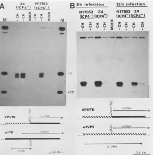
3A). In contrast,ICP4+ Z4 cells supported the accumulation ofsubstantialquantities of thymidine kinaseRNAunder the same experimental conditions. Indeed, the level of
thymi-dine kinase RNA found in cycloheximide-blocked Z4 cells wassimilarto thatobserved in unblocked N17BS3 cellsand was about one-half ofthat observed in unblocked Z4 cells. Results of this experiment suggest that theICP4 present in Z4cells issufficienttoactivate theexpression ofathymidine kinase gene present on a superinfecting viral genome to levels of expression approximating those observed in a normal infection. In marked contrast to the results with
thymidine kinase, only marginal activationof the leaky-late
l electropho- VP5 gene was observed in similar experiments (Fig. 3B).
ipitated from Infected Z4 cells accumulated detectable, butlow,levelsof 47 antibodies. VP5 RNA in the presence of the protein synthetic block,
simmunopre- whereas no VP5 transcripts were detected in the control y
specific
for N17BS3 cells. We estimatethat cycloheximide-blocked Z4 tatednonspe- cellsaccumulated at most 1/20th ofthe levels ofVP5 RNAEted
antibody
found inunblocked cells. Asdescribed in detailbelow,these1 and 6
are
resultssupport
ourprevious suggestion
that the VP5pro-sely.
rl.,Lvsate
L.at 77 moteris firstactivatedby
immediate-early
proteins
andthat posite of theate1);6days
3 wasfor 4h refer to the roteins (New ibility of ICP4 :ipitatedfrom and 5 contain ent8-hchase;
a3-h labeling of cyclohexi-'P47synthesis
inZ4cells. Proteinsimmunoprecipitatedfrom celllysates byarabbit antiserum directedagainstthepresumptive C-terminus ofICP47(30) wereloadedin lanesmarked+.Proteinsprecipitatednonspecifically fromthesamelysates bynormalrabbitserumwereloaded in lanes marked-.Lysate 1, control LTA cells(31);lysate 2,Z4cells; lysate 3, LTA cells infected with 40 PFU of HSV-1 (KOS) per cell and labeled 1 to 3hpostinfection. Labelingin allcaseswasfor 2 h with 75,uCi of[35S]methionineperml. Numberstothe leftarein kilobases. Gels in(A) and(B)were9%polyacrylamide;thegel in(C)wasa5
to12%gradient gel.
on November 10, 2019 by guest
http://jvi.asm.org/
[image:3.612.61.298.71.518.2]A (IC_P_4_
M sv 0
...
N17B53
(IC P4_)
uJ V 0
E M
I,
*. :E ~~4Mw
B
Sh infectionN17BS3 Z4 (IC P47) (1CP4+)
12h infection N17BS3 Z4
(iCPp4)(ICP4+)
_ -_ -- _
a
v0
-1:~~~~~~~~~~~~: *~~~~~~~~~~~~~~~~2
VP5JTK ->
wtTK -, ->
WtTKK
VP5/TK §1 ->
-il
__RNA
--wtVP5 ,T\r.RNA _ _
r77 ~
__\\\\s\\\\\\\\\\M-FIG. 3. Si nuclease mapping of viral thymidine kinase (TK) and VP5 transcripts induced by ICP4. (A) Thymidine kinase RNA. A single-stranded probe labeled at theBgIlIsite at +56 in the wild-type thymidinekinase gene and extending to the EcoRI site at -80 was hybridizedtocytoplasmic RNA prepared from Z4 and N17BS3 cells 8 h after infection with 10 PFU of HSV-1 per cell. Where indicated, cycloheximide (CH; 50p.g/ml) wasadded 45 min before infection and maintained continuously. RNA prepared from mock-infected cells (MOCK)wasused asacontrol. After S1 nuclease treatment, the digestion products were sized on an8% polyacrylamide sequencing gel. Markers (M)weregenerated bycleaving the probe fragment withTaqI (-1) and PstI (+16). (B) VP5 RNA. A single-stranded probe labeled at anRsaIsiteat +158in the wild-type VP5 gene and extending to aHhaIsite at-159 was hybridized to cytoplasmic RNA prepared either 8or12 h afterinfection with HSV-1. Labels are as in (A).Si nuclease digestion products were sized on a6% polyacrylamide sequencing gel. Kb, kilobases.
its expression is subsequently boosted by the increased template copy number resulting from viral DNA replication, aprocess that requires delayed-early gene products (15).
Effects of ICP4 on expression of other HSV genes. The
results presented above suggest that a delayed-early gene was efficiently activated by ICP4, whereas a late gene was
activated only marginally. To test the generality of this
finding,wesurveyedtheeffects of ICP4on theexpressionof a variety of viral genes. Insted of directly scoring for the RNAs derivedfrom these genes, we used an indirect
trans-lational assay. After allowing mRNA to accumulate in the presence of cycloheximide for 6 h, the protein synthetic
block was removed. Further transcription of viral mRNA was prevented by the addition of actinomycin D, and the accumulated mRNAs were allowed to be translated in vivo in the presence of
[35S]methionine.
After 5 h, the resulting protein products were analyzed by gel electrophoresis (Fig. 4). The total complement of virally induced proteins wasexamined by directly analyzing the labeled extracts, and a numberofspecific proteinsweredetected by
immunoprecipi-tationwith monoclonal antibodies.
The results support the general conclusions made above. IncontrolN17BS3cells, reversal of the cycloheximide block was followed by detectable translation of only a limited number of viral-specific proteins, as expected for cells
blocked in the immediate-early phase of infection. The
proteinsthat we couldconfidently identify includeICP4 and
ICPO,bothbelongingto theimmediate-earlyclass. In marked contrast, reversal of the cycloheximide block in Z4 cells resulted in thetranslation ofat least 5 additional
polypept-ides thatwereeasily detectable in total cellextracts
(arrow-heads, Fig. 4). Among these five polypeptides, ICP6 (a
delayed-early protein associated with ribonucleotide reduc-tase activity [2]) and ICP8 (the major viral DNA binding protein [33]) could be identified on the basis oftheir
elec-trophoretic mobility. Immunoprecipitations confirmed the presence ofICP6 and ICP8 and demonstrated the
accumu-lation ofreadily detectable quantities of gB, gD, andgE as well. Alloftheseproteins previously have been classified as
belongingtothedelayed-early class(Wagner, inpress). We
consistently found that the levels of ICP6 obtained after reversal of the cycloheximide block were similar to those obtained in unblockedcells, whereasthe levelsof ICP8, gB, gD, and gE were lower. Even with this reduced level of
translation, ICP8 remainedoneofthe mostintenselylabeled
proteinsvisible in the total cell extracts. These data suggest that many delayed-early genes are efficiently activated by
the ICP4present in Z4 cells.
Unblocked Z4 and N17BS3 cells contained at least five
easily
detectable viralproteins
that did not accumulate tou v u i U U Us
+ + E + +
on November 10, 2019 by guest
http://jvi.asm.org/
[image:4.612.153.465.66.382.2]200.
lysates
fCP6
ICP8 gB gD gE alk.exo. p40 gCa b cd a b c d a b c d a b c d a b cd a b c d a b c d a b c d a b c d
_ as
_,.
z--
_97o
69>
1.4 *a
_mm
.me.
9I!,
46* -_1.
30h
--FIG. 4. Translational analysis ofmRNAs accumulating in cycloheximide-treated Z4 cells. Cycloheximide-treated (lanes aand b) or untreated(lanesc andd) ICP4 expresser Z4 cells (lanes band d)orcontrolN17BS3cells(lanesa and c)wereinfected for6h andsubsequently allowedtotranslate accumulated transcripts for 5 h in medium containing10 ,ugofactinomycinD and40 ,uCi of[35S]methionine perml. Relative amountsofindividualgeneproducts synthesized werevisualized byfluorography of sodium dodecyl sulfate-polyacrylamide gels. Total cell lysates are shown in the four lanes at left. The remainder of the figure shows material immunoprecipitated with monoclonal antibodies specific fortheindicated HSV-1 proteins. Arrowheads betweenlanes a and bforthe lysates indicate proteins translated from mRNAsinduced in cells containing ICP4 butnotinduced incontrolcells. Similarly, dots between lanes b andcindicate proteins translated from mRNAs induced in untreated cells but notinduced in cells treated withcycloheximide, regardless ofthe presenceof ICP4. Thefigure isacompositeof thefollowing differentexposures: 22 h(lysates), 1 h(ICP6andgB), 4 h(ICP8),4 days(gD andp40),12 h (gE and alkaline exonuclease [alk. exo.]) and 3 days (gC). Numbers at left indicate the molecular weights of"C-labeled marker proteins (NewEngland Nuclear)(x103).
high levels in cycloheximide-blocked Z4cells (Fig. 4).
Pre-sumably, these proteins aretheproducts of viralgenesthat arenot efficiently activated by ICP4. The largest and most
abundant ofthese five proteins is the majorcapsid protein
VP5, the mRNA of which was shown above to be only
marginally activatedin Z4 cells. Immunoprecipitations
iden-tified three other proteins that fall into this class: viral
alkaline exonuclease, nucleocapsid protein p40, and gC. Bothp40and gC arelateproteins, soitisnotsurprisingthat
they behave similarly to VP5 in this assay. However, we were surprised to find that alkaline exonuclease did not
accumulate in Z4 cells after reversal ofthe cycloheximide block, because it has been classified as a delayed-early protein (13). As discussed further below, this result may
imply that the delayed-early genes fall into more than one class with respect totheirtranscriptional requirements.
DISCUSSION
Intheexperimentsdescribedhere,weattemptedto assess the role ofa limited subset of the HSV-1 immediate-early proteinsin thetranscriptional activation ofavariety ofHSV genes. Our strategy was to first isolate stably transformed celllinesexpressingICP4 and thentoexamineexpressionof HSV genes introduced into these cells by viral superinfec-tion under conditions preventing de novo viral protein synthesis. As mentioned above, Z4 cells also express the
immediate-early protein ICP47. That this protein plays no
major role in the activation of the viral genes studied,
however, is suggested by preliminary, butessentially simi-lar, results obtained with cells transformed by a plasmid
encodingICP4 and ICP22(insteadofICP47).Althoughitcan be argued that ICP22 and ICP47 might substitute for each
other when in combination with ICP4,weconsider thistobe unlikely. Therefore, in the ensuing discussion we assumed that all of the observedeffects result from the action ofICP4 alone. The specific questions that we have posed are as follows: (i)to what extent does ICP4 activate the transcrip-tion ofdelayed-early genes? and (ii) does ICP4 alsodirectly activateleakylate genes?
Our experimental approach of using stably transformed linesexpressingICP4 waspreferred foranumberofreasons overthe alternative strategyofcotransfectingcells withthe ICP4gene, alongwithsuspected targetsof ICP4action,in a transient assay and scoringfortargetgeneactivation. First, even under the best conditions, onlya small proportion of acutely transfected cells express the introduced markers, limiting the signal associated with a positive response. Second, it is not obvious a priori that HSV genes are controlledby a singleregulatory event. In the present case wewereable toquantifythe response of the target genes to thesuspectedregulatoryfactorbycomparingthesignalwith thatobtained in viral infections. Based onthiscomparison, weconclude thatICP4 iscapable ofnearlyfullactivation of atleast two delayed-early genes, those encoding thymidine kinase andICP6,butonlymarginallyactivates the late gene for VP5. It would have been difficult, ifnot
impossible,
to arrive at this conclusion by a transient expression assay.Third, during transfection, originally unlinked sequences becomephysicallylinked(31).Becauseavarietyof viral and cellular genes have been shown to be associated with
cis-acting DNA sequences that potentiate the
activity
of linked promoters in the absence of thetrans-acting factorsordinarily required for their activity (4, 19, 21), the trans action of a suspected regulator can only be definitively
i
on November 10, 2019 by guest
http://jvi.asm.org/
[image:5.612.100.524.69.287.2]established when the possibility of recombination during transfection is excluded. Several studies have documented the fact that chromosomally integrated HSVsequences are not normally available forrecombination with a superinfec-ting viral genome (9, 37). Consequently, we are confident that the observed effects are due to activation in trans.
Although our approach has a number ofadvantages, we relied on several assumptions that might limit the interpre-tation of our results. First, we assumed that all of the observed effects are due to the expression of the ICP4 protein in Z4 cells. Second, all of the assays fortheeffects of preexisting ICP4 were carried out by viral superinfection, a condition that unavoidably introduces virion structural pro-teins into the cells. Results of several studieshave suggested that one or more virioncomponentspositivelyregulateHSV immediate-earlypromoters (6, 11, 28). Although there is no evidence that these virioncomponents alsodirectly contrib-ute to the control ofother HSV genes, this possibility has not yet been excluded. Consequently, atpresent we cannot eliminate a role for virion structural proteins in delayed-early gene expression. Third, ourexperiments have directly and indirectly measured the levels ofviral mRNAs accumu-lating in the presence of cycloheximide. We assumed that these levels reflect the rate of transcription ofthe genes in question andthat the rate oftranslationofthe accumulated mRNAs reflects, at least grossly, theirabundance. Finally, we assumed that the observed effects ofcycloheximide on viral transcription result from its inhibition of viral protein
synthesis, a view that seems well supported.
Given these assumptions, the following conclusions are warranted. First, the ICP4 protein present in Z4 cells has a dramatic effect on the expression of many delayed-early
HSV geneswhen thesecells are superinfected with HSV-1. The synthesis of six delayed-early mRNAs was resistant to the effects ofcycloheximide inthese cells, yielding
substan-tial levels of these mRNAs under conditions that otherwise prevent theirdetectable accumulation. At least two of these mRNAs, those encoding thymidine kinase and ICP6, were induced to levels close to those obtained in a normal
infection. Weconclude thatICP4 is sufficientforthe efficient activation of thedelayed-early genes tested, with the excep-tion of the geneencoding alkalineexonuclease. This conclu-sion is reinforced by the fact that similar results were obtained regardless of the presence ofICP47 (or ICP22) in addition toICP4 (unpublished data). As acorollary to this, we also conclude that the remaining HSV immediate-early
proteins (ICPO andICP27)donot play anessentialrole in the activation of these delayed-early genes. However, it is possible that these proteins contribute to the expression of thealkalineexonucleasegene. Because the accumulationof alkaline exonuclease mRNA occurs with delayed-early ki-netics (13) and is notinhibited whenviral DNAreplication is blocked, delayed-early proteins are unlikely to influence its expression. Our finding that the exonuclease gene was not
efficiently activated under the same conditions in which six otherdelayed-earlygenes were activated provides evidence
that delayed-early genes do not form a homogeneous class with respect to theirtranscriptional requirements, a
conclu-sionthat is reminiscent of the findings ofPereiraetal. (32). Itwillbe interestingtodetermine how many other
delayed-early genes fall into the same class as the alkaline
exonu-clease gene and to identify the additional factor(s) required
fortheir efficient expression.
Thesecondmajorconclusionthat we have reached in this study is that immediate-early proteins are sufficient for the
detectable activation of the leaky-late gene encoding VP5.
However, thisgeneisexpressed athighlevelsonly after the
action of additional
factors,
so that theexpression obtained incycloheximide-blocked Z4cellsisonlyasmallfraction ofthatobtained in unblocked cells. On the basis of the kinetics
of activation ofaVP5-thymidine kinase hybrid gene bythe
productsofsuperinfecting HSV,wehave previouslyargued
(15) that immediate-earlyproteinsalone sufficeforthe initial
activation ofthe VP5 promoter and that thehigherlevels of expressionobtainedduringthe latephase of infectionresult from the effects of template amplification. The results
ob-tained in this study support thishypothesis. We do notyet
know whether the late genes for p40 and gC are also
marginally activated by ICP4. The answer to this question will require adirectanalysis oftheirtranscripts.
The presence ofICP4 in Z4cells supportsthe extraordi-narilyefficient expressionofmanyHSVdelayed-early genes, yetthis striking alteration in theability ofthe cells toutilize HSV promoters does not noticeably affect their viability.
Presumably, then, the ordered and regulated expression of the cellular genes in Z4 cells isnot dramatically perturbed.
However, results of recent studies on an analogous protein
encoded by pseudorabies virus (19, 21) suggest that such a
protein is able to activateboth transfected cellular
P-globin
genes and adenovirus genes. There is also evidence that HSV ICP4 itself activates a transfected cellular ,B-globin
gene (16). The problem then, is to account for these seem-ingly nonspecific effects on non-HSV genes, while at the sametime accountingfor thelackof a lethal alteration in the
expression ofresident cellular genes. There aretwo alterna-tive hypotheses toreconcile thesedata. Thefirst, which has also been proposed by Everett (16), holds that ICP4
specif-ically activates extrachromosomal genes, regardless of se-quence. This hypothesis accounts for the effects on acutely transfected adenovirus and ,-globin genes and also for the activation of HSV genes borne on a superinfecting viral genome. However, it does not explain thefact that chromo-somally integrated HSV genes are also efficiently activated
bytheimmediate-early proteins supplied byasuperinfecting
viral genome (see, for example, references 37 and 39). The alternative extreme position holds that ICP4 acts in a
se-quence-specific fashion, activating onlythose genes bearing
specific ICP4 recognition sequences. This hypothesis, al-though possible, is difficult to reconcile with the lack of obvious sequence homology among the promoters of de-layed-early HSV genes (Wagner, in press) and with the effects of the pseudorabies ICP4 analog on adenovirus and cellular genes. Consequently, we are at present unable to arrive at a simple model to explain the effects of ICP4. Possibly, they involve both a generalized activation of the transcription ofextrachromosomal genes and amorespecific activation of specialized HSV promoters. Because ICP4 represents one of the proteins that has been most clearly demonstrated to becapable ofmodifying the transcriptional specificity ofeucaryoticcells, clarification ofits mechanism
of actionis likely to be of general interest.
We expect that Z4 cells will prove to be useful in this
exercise,providing an HSV analog of 293 cells (17) (permis-sive cells expressing adenovirusEMA).
ACKNOWLEDGMENTS
This research was funded by grants from the National Cancer Institute of Canada and the Medical Research Council of Canada. S.B. is aResearch Associate and J.R.S. is a Research Scholarofthe National Cancer Institute of Canada. R.H.P. holds a studentship from the Alberta Heritage Foundation for Medical Research.
The expert technical assistance of Helen Rudzroga is gratefully
on November 10, 2019 by guest
http://jvi.asm.org/
acknowledged. Wethank W. E. Rawls and F. L. Graham forhelpful
comments on the manuscript; R. McKinnon, W.-C. Leung, and D. R. Helinski for providing plasmids; and K. L. Powell, P. G. Spear and H. S. Marsden for supplying theQl andII 481B-2ascitic fluids and theIE12/76 antiserum, respectively. Wearealsograteful
toC. M. Preston for donating the pGX157 plasmid encoding ICP4 and ICP22. Finally, we areparticularly indebted to M. Zweig who providedus with the anti-ICP4 antibody 58Saswellas the74S and
19S asciticfluids.
LITERATURE CITED
1. Anderson, K. P., R. H. Costa,L.E.Holland,and E. K. Wagner.
1980.Characterization of herpes simplex type1 RNApresentin the absence of denovoprotein synthesis. J. Virol. 34:9-27.
2. Bacchetti, S., M. J. Evelegh, B. Muirhead, C. S. Sartori, and
D. Huszar. 1984.Immunological characterization of herpes sim-plex virustype 1and 2 polypeptide(s) involved in viral ribonu-cleotide reductase activity. J. Virol. 49:591-593.
3. Balachandran, N., D. Harnish, W. E. Rawls, and S. Bacchetti.
1982.Glycoproteins of herpes simplex virustype2asdefined by
monoclonal antibodies. J. Virol. 44:344-355.
4. Banerji,J.,S. Rusconi, andW. Schaffner. 1981. Expression ofa
,-globin gene is enhanced by remote SV40 sequences. Cell
27:299-308.
5. Banks, L., D. J. R. Purifoy, P. J. Hurst, R. A. Killington, and
K. L. Powell. 1983. Herpes simplex virus non-structural
pro-teins. lV. Purification of the virus-induced deoxyribonuclease andcharacterization of the enzymeusing monoclonal
antibod-ies. J. Gen. Virol. 64:2249-2260.
6. Batterson, W., and B. Roizman. 1983. Characterization of the
herpes simplex virion-associated factor responsible for the
induction ofotgenes. J. Virol. 46:371-377.
7. Berk, A. J., andP. A.Sharp. 1977. Sizing and mappingof early
adenovirus mRNAs by gel electrophoresis of S1 endonuclease
digested hybrids. Cell 12:721-732.
8. Bonner, W. M., and R. A. Laskey. 1974. A film detection
method for tritium-labeled proteins and nucleic acids in
poly-acrylamide gels. Eur. J. Biochem. 46:83-88.
9. Campione-Piccardo, J., and W. E. Rawls. 1981. Inability to rescueviralgenesfrom human cellsbiochemically transformed by herpes simplex virus type 2 DNA. Can. J. Microbiol. 27:1123-1128.
10. Clements, J. B., J. McLauchlan, and D. J. McGeoch. 1979.
Orientation of herpes simplex virus type 1 immediate early mRNAs. Nucleic Acids Res. 7:77-91.
11. Cordingley,M. J., M. E. M. Campbell, and C. M. Preston. 1983.
Functionalanalysis ofaherpes simplex virus type 1promoter:
identification of far-upstream regulatory sequences. Nucleic
AcidsRes. 11:2347-2365.
12. Costa, R. H., G. Cohen, R. Eisenberg, D. Long, and E. K.
Wagner. 1984. Direct demonstration that the abundant
6-kilobaseherpes simplex virus type 1 mRNAmapping between 0.23 and0.27map unitsencodes the major capsid protein VP5. J. Virol. 49:287-292.
13. Costa, R. H., K. G. Draper, L. Banks, K. L. Powell, G. Cohen,
R.Eisenberg, and E. K. Wagner. 1983. High-resolution charac-terization of herpes simplex virus type 1 transcripts encoding alkaline exonuclease and a 50,000-dalton protein tentatively
identifiedas acapsidprotein. J. Virol. 48:591-603.
14. Costanzo, F., G. Campadelli-Fiume, L. Foa-Tomasi, and E.
Cassai. 1977. Evidence that herpes simplex virus DNA is transcribed by cellular RNA polymerase B. J. Virol. 21:996-1001.
15. Dennis, D., and J. R. Smiley. 1984. Transactivation ofa late
herpes simplex viruspromoter. Molec. Cell. Biol.4:544-551.
16. Everett, R. D. 1983. DNA sequences required for regulated
expression of the HSV-1 glycoprotein Dgenelie within 83 bp of
the RNAcapsites. Nucleic AcidsRes. 11:6647-6666.
17. Graham, F.L., J. R.Smiley,W.C. Russell, and R. Nairn. 1977.
Characteristics ofahuman cell line transformed by DNA from
humanadenovirus5. J. Gen. Virol. 36:59-72.
18. Graham, F. L., and A. J.vander Eb.1973.Anewtechniquefor
the assay of the infectivity of adenovirus 5 DNA. Virology 52:456-467.
19. Green, M. J., R. Treisman, and T. Maniatis. 1983. Transcrip-tional activation of cloned P-globin genes by viral immediate-early gene products. Cell 35:137-148.
20. Honess, R. W., and B. Roizman.1974. Regulationof herpesvirus macromolecular synthesis. I. Cascaderegulation of the synthe-sisof threegroups of viral proteins. J. Virol. 14:8-19. 21. Imperiale, M. J., L. T. Feldman, and J. R. Nevins. 1983.
Activation of gene expression by adenovirus and herpesvirus regulatory genes acting intrans and by acis-acting adenovirus enhancer element. Cell 35:127-136.
22. Kahn, M., R. Kolter, C. Thomas, D. Figurski, R. Meyer, E. Remaut, and D. R. Helinski. 1979. Plasmid cloning vectors derived from plasmids ColEl, R6K, and RK2. Methods Enzymol. 68:268-280.
23. Laemmli, U. 1970. Cleavage of structural proteins during the assembly of the head of bacteriophage T4. Nature (London) 227:680-685.
24. Lang, J. C., D. A. Spandidos, and N. M. Wilkie. 1984. Tran-scription regulation of a herpes simplex virus immediate early gene is mediated through an enhancer-type sequence. EMBOJ. 3:389-395.
25. Lee, G. T.-Y., M. F. Para, and P. G. Spear. 1982. Location of the structural genes forglycoproteinsgD and gE and forother polypeptidesin the Scomponent ofherpes simplex virus type 1 DNA. J. Virol. 43:41-49.
26. Leung, W.-C., K. Dimock, J. R. Smiley, and S. Bacchetti. 1980. Herpes simplex virus thymidine kinase transcripts are absent from both nucleus and cytoplasm during infection in the pres-ence ofcycloheximide. J. Virol.36:361-365.
27. Mackem, S., and B. Roizman. 1980. Regulation of herpesvirus macromolecular synthesis: transcription-initiation sites and do-mainsofagenes. Proc. Natl. Acad. Sci. U.S.A.77:7122-7126. 28. Mackem, S., and B. Roizman. 1982. Differentiation between cx promoter and regulator regions of herpes simplex virus 1: the functional domains and sequence of a movable ax regulator. Proc. Natl. Acad. Sci. U.S.A. 79:4917-4921.
29. Maxam, A. M., and W. Gilbert. 1980. Sequencing end-labeled DNA with base-specific chemicalcleavages.MethodsEnzymol. 65:499-560.
30. Palfreyman, J. W., J. B. Maclean, E. Messeder, and R. C. Sheppard. 1984. Successful use ofoligopeptidesasimmunogens in the preparation of antisera toimmediate-early geneproducts of herpes simplex virus type 1. J. Gen.Virol. 65:865-874. 31. Pellicer, A., D.Robins,B. Wold, R. Sweet, J.Jackson, I. Lowy,
J. M. Roberts, G. K. Sim, S. Silverstein, and R. Axel. 1980. Altering genotype and phenotype by DNA mediated gene transfer. Science209:1414-1422.
32. Pereira, L., H. M. Wolff, M. Fenwick, and B. Roizman. 1977. Regulation of herpesvirus macromolecular synthesis. V. Prop-erties ofox polypeptides made in HSV-1 and HSV-2 infected cells. Virology 77:723-749.
33. Powell, K. L.,andD. J.Purifoy. 1976. DNA binding proteins of cells infected by herpes simplex viruses types 1 and 2. Inter-virology 7:225-239.
34. Preston, C. M. 1979. Control of herpes simplex virus type 1 mRNA synthesis in cells infected with wild-type virus or the temperature-sensitive mutant tsK. J. Virol. 29:275-284. 35. Rixon, F. J., and J. B. Clements. 1982. Detailed structural
analysis of two spliced HSV-1 immediate-early mRNAs. Nu-cleic AcidsRes. 10:2241-2256.
36. Roberts, J. M., and R. Axel. 1982. Geneamplification and gene correction insomatic cells. Cell 29:109-119.
37. Sandri-Goldin, R. M., A. L. Goldin, L. E. Holland, J. C. Glorioso, and M. Levine. 1983. Expression of herpes simplex virus , and y genes integrated in mammalian cells and their inductionby anoxgeneproduct. Mol. Cell. Biol.3:2028-2044. 38. Showalter, L. D., M.Zweig, and B. Hampar. 1981. Monoclonal
antibodies to herpes simplex type 1 proteins including the immediate-early protein ICP4.Infect. Immun. 34:684-692. 39. Smiley, J. R., H. Swan, M. M. Pater, A. Pater, and M. E.
Halpern. 1983. Positive control of the herpes simplex virus
on November 10, 2019 by guest
http://jvi.asm.org/
thymidine kinase gene requires upstream DNAsequences. J. Virol. 47:301-310.
40. Szybalski, W., E. H. Szybalska, and G. Ragni. 1962. Genetic studies with human cell lines. Cancer Inst. Monogr. 7:75-89. 41. Watson, R. J.,andJ. B. Clements.1980. Aherpes simplex virus
type 1 function continuously required for early and late virus RNAsynthesis. Nature (London)285:329-330.
42. Watson, R. J., C. M. Preston, and J. B. Clements. 1979. Separation and characterization of herpes simplex virustype 1 immediate-early mRNAs. J. Virol. 31:42-52.
43. Watson, R. J., and G. F. Vande Woude.1982. DNAsequenceof
animmediate-earlygene(IE mRNA-5) of herpes simplextype1. Nucleic Acids Res. 10:979-991.
44. Whitton, J. L., F. J. Rixon, A. J. Easton,and J. B. Clements. 1983. Immediate-early mRNA-2 of herpes simplex virusestypes
1and 2 isunspliced: conservedsequencesaround the 5' and 3' termini correspondtotranscription regulatory signals. Nucleic Acids Res. 11:6271-6287.
45. Wilcox, K. N., A. Kohn, E.Sklyanskaya, and B. Roizman. 1980. Herpes simplex virus phosphoproteins. I. Phosphate cycleson
and offsome viralpolypeptides and canalter their affinity for
DNA.J. Virol.33:167-182.

![FIG. 2.resislysates Sodium dodecyl sulfate-polyacrylamide ge of [35S]methionine labeled proteins immunoprec of transformed cells by anti-ICP4 or anti-ICP447](https://thumb-us.123doks.com/thumbv2/123dok_us/1406395.93574/3.612.61.298.71.518/resislysates-sodium-polyacrylamide-methionine-labeled-proteins-immunoprec-transformed.webp)