0022-538X/88/114096-08$02.00/0
CopyrightC) 1988, American Society for Microbiology
Characterization of Major Recognition Sequences
for
a
Herpes
Simplex
Virus Type 1
Origin-Binding Protein
ANDREW KOFFANDPETERTEGTMEYER*
DepartmentofMicrobiology, State University of New YorkatStony Brook, Stony Brook, New York 11794-8621 Received 3 June1988/Accepted 30 July 1988
To investigate early initiation events in the replication of herpes simplex virus type 1, we analyzed
interactions of proteinsfrominfected cellextractswith thesmall originofherpes simplexvirustype 1
(orisl).
Using the mobilityshiftassay, wedetectedtwoorigin-specific bindinginteractions. We characterized themore
prominent interactiononbothstrandsof theDNAduplexwithDNaseIprotectionandmethylationinterference assays.Protein binding protects 17basesof DNAoneach strandfromDNaseI. Thesesequences arelocated
atthe leftend ofthe centralpalindromeandareshiftedfourbases relativetooneanother. On thebasisof the DNaseprotectionpattern,webelievethis proteintobe relatedtotheorigin-bindingproteindefinedbyEliaset al.(P. Elias,M. E.O'Donnell,E.S.Mocarski, andI.R. Lehman, Proc. Natl. Acad.Sci. 83:6322-6326,1986).
OurDNase I footprintshowsbothstrongand weakareasofprotection. Theregions strongly protectedfrom DNase I align with the essential contact residues identified by interference footprinting. Methylation interference defines a small binding domain of 8 base pairs: 5'-GTTCGCAC-3'/3'-CAAGCGTG-5'. This
recognitionsequence containstwoinverted5'-GT(T/G)CG-3' repeatswhich sharea2-baseoverlap; thus, the
origin-binding protein probably binds tothe invertedrepeatsas a dimer.
Herpes simplex virus type 1 (HSV-1) is an attractive
modelforDNA replicationbecause the virus encodesmost
oftheproteins required forDNAsynthesis.Anoutline ofthe
eventsin the replication of HSV-1 suggests mechanisms by which replication may be initiated. A portion of viral DNA is
foundascircles withinthenucleiofinfected cells(9, 13, 19).
Soonafter the onset of viral DNA replication, a number of
different forms of viral DNA can be detected. Late in
infection,ashift topredominantly large, rapidly sedimenting
formsofDNA occurs(1, 13). Theselarge structures suggest that, at least late in infection, the DNA replicates asarolling
circle. The mechanism ofthe transition from a circle to a
rolling circlehas not beendefinedexperimentally. Intheory,
thegeneration ofarollingcircle wouldrequireanickin one
strandofthe DNAduplex.Thenick,in turn, would provide
a 3' hydroxyl that could act as aprimer for leading strand
synthesis.Theeventsleadingtorollingcirclereplicationare
HSV-1origin specific (21, 22, 24).
Analysis ofdefective interfering viralparticles (8)
identi-fies two regions of HSV-1 DNA that contain origins. One
origin
(orisl)
islocated in the "c" repeatflanking the shortunique segment, and the other (oriLl) is located in an
internal region of the long unique segment. Recently, a transientreplication assaywas used to study HSV-1origins (21, 22, 24, 26). This procedure allowed the mapping of
functional origin sequences. The origins of HSV-1 (22, 24,
28), HSV-2(14), and varicella-zoster virus (VZV) (23) have
been sequenced andcompared. All of the originscontain a
large palindrome centered on an alternating AT sequence
motif. The extent of the palindromic sequences and the
length of the AT motifare variable. Alignment of the four
origins of HSV-1 and HSV-2 indicates that extensive
con-servation exists leftward from the center of symmetry.
Sequences to the right of the center of symmetry are less
homologous. Comparison of the HSV origins with the VZV
origindemonstratesconservation in the left end, but not in
therightend, of thepalindrome. Within the leftregion, there
* Correspondingauthor.
is an 11-base-pair (bp) sequence, 5'-CGTTCGCACTT-3',
thatiscompletely conserved in all fiveorigins. This conser-vation implies that the sequence is essential for efficient
originfunction.
HSV-1 encodes many proteins directly and indirectly involved in viral DNA metabolism. Seven open reading frames encode all of the viral proteins directly involved in viral DNAreplication(29). Fourof these have been matched with the major DNA-binding protein (ICP8), the viral DNA polymerase, a DNA polymerase accessory protein
(65KDBP),
and theorigin-binding protein (OBP) (18a, 29). Inaddition, primase activity has also been found in extracts
from infected cells (12). Genetic and biochemical studies
indicate that many of the replication proteins interact in a
multiprotein complex (12, 16, 18, 25). The OBP, purified from a nuclear extract, protects 18 bases of DNA from DNaseI (6). These sequencespartially overlaptheleft arm of thepalindromein
orisl.
Recently,OBPwasalso shownto bind to sequences overlapping the right arm of thepalin-drome inaconcentration-dependent manner(5).
In this study, we demonstrate that the OBP recognition
sequence isasmall inverted repeat. This inverted repeat is
composed oftwo5-base recognition elements that have an
overlap of 2 bases. DNase I protection, methylation inter-ference, and sequence analysis show that the recognition sequence is asymmetric. These findings suggest that the OBP is involved in anasymmetric event attheorigin.
MATERIALS ANDMETHODS
Cells and virus. Verocells were maintained as described by Welleret al. (27). Stocks of HSV-1 strain F weregrown inVerocellsasdescribedbyRoizman andSpear (20). Virus stocks were titered by infecting cells in Dulbecco modified
Eagle medium and 1%fetal calfserum. Afteradsorption of
thevirus,cellswere incubated for4hin Dulbeccomodified
Eagle medium with
5%
fetal calf serum. Plaques were counted aftera72-h incubation in medium containing0.5%
pooled humanimmune serum.
Plasmid constructions. All cloning procedures were per-4096
on November 10, 2019 by guest
http://jvi.asm.org/
5'--AAGCTTGCATGCCTGCAGGTCCAGATCTGAGCTT
TTCGAACGTACGGACGTCCAGGTCTAGACTCGAA]
Sma I Xa T
V2-. UA1L ZA-. LR2-LI&aLL ZIQLE CCATGGGTCGACCGGATCCCCGGGTACCGAGCTCGAATTC
GGTACCCAGCTGGCCTAGGGGCCCATGGCTCGAGCTTAAG-pHAK
Q ,- ..;60....
pLAT
FIG. 1. HSV-1 originsequencesand flankingpolylinkersequences of the pHAK clone.Orisl sequencesare numberedas describedby
Stow andMcMonagle (24), and restriction sitesareshown abovethepolylinkersequences.Theposition of the centralpalindromeis indicated byarrowsbetween theDNAstrands. The line above theupperstrandrepresentstheOBPDNase Ifootprint describedbyEliasetal.(6).Bars
underthesequenceidentify regions oforislthatwereused in the analyses described in thispaper. pHAK(_) containsacompleteorigin,
andpLAT( Ezi) hasatruncated origin.
formed asdescribed by Maniatis et al. (15), and allenzyme
conditions were those recommended by the manufacturer.
Origin sequences were cloned into a vector derived from pUC18 by replacement ofthe HincIl sitewithaBglIIsiteby linker insertion. This vector was called pUC18-B. Two
origin-containing fragments were derived from the pOR-S series, constructed by Deb and Doelberg (3), after we
replaced theHindIII site with aBgIII linker. Thecomplete
originwasderivedfrompOR-S.Atruncatedorigin, inwhich sequences rightward of the AT motif were deleted, was
derived from pOR-S-1. The BglII-to-BamHI origin-con-taining fragments ofpOR-S and pOR-S-1 wereinserted into the corresponding sites of pUC18-B to create pHAK and pLAT, respectively (Fig. 1). Plasmids were maintained in Escherichia coliHB101, and DNA was purified fromCsCl gradientsafterisolation bythemethodofBirnboimandDoly (2).
Protein extraction. Vero cells in 30 150-cm2 flasks were
infectedwithHSV-1(strain F)atamultiplicity of infection of 10. Infected cells were collected 16 h after infection by vigorously shaking the flasks. Cellswerecentrifugedatalow
speed and washed with 20mMHEPES
(N-2-hydroxyethyl-piperazine-N'-2-ethanesulfonic acid)-sodium hydroxide (pH
7.6)-0.5 mM dithiothreitol(DTT)-150mMNaCl. Cellpellets
were suspended in 2 ml of lysis buffer (20 mM HEPES-sodium hydroxide [pH 7.6], 0.5mM DTT, 0.5 mM phenyl-methylsulfonyl fluoride [PMSF], 2 ,ug ofleupeptin per ml) and broken witha tight-fitting pestle ofaDounce
homoge-nizer on ice. All subsequent procedures were done at a
temperature of4°C or less. The cell homogenate was
ad-justedto10%glycerol (vol/vol) andcentrifugedat100,000 x g for 1 h. The supernatant fraction was adjusted to 35% ammonium sulfate andgentlymixed for 45min. The suspen-sion wasthen centrifuged at30,000 x g for 1 h. Thepellet
was suspended in 1.5 ml of protein storage buffer(20 mM HEPES-sodium hydroxide [pH 7.6], 0.5mM DTT, 0.5 mM PMSF, 0.5 mM disodium EDTA [pH 8.0], 10% [vol/vol] glycerol). Supernatant and pellet fractions were dialyzed
against proteinstorage bufferand stored at -70°C. Protein
was quantitated by using a protein assay kit (Bio-Rad
Laboratories).
Gel mobilityshiftbindingassay. The target fragment
con-taining
orisl
wasmadebydigestingpHAKwithHindIIIandEcoRI,and the 5'overhangswerefilledwith [a-32P]dATPby theKlenowfragmentofDNApolymerase.The labeled DNA fragment was purified by electrophoresis through a 50 mM
HEPES-sodium hydroxide (pH 7.6)-5%polyacrylamide gel at 10V/cm. Probe DNA was elutedby soakingthegelslice in0.5 M ammoniumacetateand 1 mM disodium EDTA(pH 8.0) overnight at 42°C and was subsequently precipitated with ethanol. DNAwas suspended at0.1 ,ug/ml of 10 mM Tris hydrochloride-1 mM disodium EDTA (pH 8.0). End-labeled DNA(0.1 ng)wasincubated with variousamountsof protein and poly(dI-dC) poly(dI-dC) (indicated in figure legends)inbindingbuffer(50mMHEPES-sodiumhydroxide [pH 7.6], 0.1 mM disodium EDTA [pH 8.0], 5 mM MgCl2, 0.5 mMDTT,100 mMNaCI).Protein-DNAcomplexeswere
allowedto form for 30minon ice. Justpriorto electropho-resis, samplebuffer(50mM HEPES-sodiumhydroxide [pH 7.6], 0.5 mM DTT, 80% glycerol, 0.1%bromophenol blue, 0.1% xylene cyanol) was added to one-fourth of the final volume. The sample wasapplied to a50 mM MOPS
(mor-pholinopropanesulfonic acid)-sodium hydroxide (pH 7.6)-5% polyacrylamide gel and electrophoresed at a constant voltage of10 V/cmfor 2.5 h. Gelsweredried on Whatman 3MM paper,and autoradiographywasdone by using inten-sifier screensat -70°C.
DNase Ifootprinting. pHAK (1 ,ug)was3'labeledateither theHindlIl orBamHI endaspreviously described (4).The origin-containingfragmentwasethanolprecipitatedand
sus-pendedin10 mMTris-hydrochloride-1 mM disodium EDTA (pH 8.0) ata concentration of200,000 cpm/,ul for use in a
bindingreaction. Approximately 55 p.gofproteinin the 35% ammonium sulfateextract wasincubated with the DNA in the presence of 8 pugofpoly(dI-dC) poly(dI-dC) in a stan-dard binding reaction. DNase I (0.4 U) was added, andthe tube was incubated on ice for 1 min. The DNase I was
stopped by adjustingthebindingreactionto50mM disodium
570 587 6
~~~
~~~~~63
r 4 9540 5 0 5 0 570 580 590 6 0 6 0 6 0 N1 649 6 6
GGCCGCCG5GTAAAAGAAGTGAGAAC
kGCGTTCGCACTTCGTCCCAATATATATATATTATTAGGGCGAAGTGCGAGCACTGG ;CGW-J.qw
CCGGCGGCCCATTTTCTTCACTCTTGCGCTTCGCAAGCGTGAAGCAGGGTTATATATATATAATAATCCCGCTTCACGCTCGTGACCGCGGCCGGGGCCC
-P ql v
on November 10, 2019 by guest
http://jvi.asm.org/
[image:2.612.51.549.78.279.2]EDTA(pH 8.0) andbytheaddition of 10 ,ug of salmon sperm
DNA. Samplebuffer wasadded, andfreeandprotein-bound
DNAs were separated by usingthe gel mobility shift assay
described above except that 10 mM disodium EDTA was
addedtothe gel andrunning buffer. Thegelwasexposedat
4°C for 4 h, and appropriate fragments were excised and
eluted. DNAwasconcentrated by ethanolprecipitationand
suspended in 10
[lI
of95% formamide sample buffer.Sam-ples in equivalent counts per minute, of the bound and
unbound fractions wereloaded onto a7 M urea-10%
poly-acrylamide gelandelectrophoresed at55°C.Gelsweredried
and exposed to X-ray film by using intensifier screens at
-70°C. Autoradiographs were then analyzed by
densito-metryon anUltroScan XL system(LKB Instruments,Inc.). Methylation interference. Origin fragments were end
la-beled asdescribed forDNase Ifootprinting. Methylation of
the DNAwas performedasdescribed by Wrightetal. (30), except that the sodium cacodylate and MgCl2 buffer was
replaced with binding buffer. The dimethyl sulfate (DMS)
wasquenched by the addition of50
RI
of1.0 M,-mercap-toethanol and 1.5 M sodiumacetateandby ethanol
precip-itation.The DNAwasethanolprecipitatedthreemoretimes
and was suspended in 10 mM Tris-hydrochloride-1 mM
disodium EDTA (pH 8.0) at 200,000
cpm/,ul.
The bindingreactionwas performedunder the sameconditions used for
DNase I
footprinting.
Free and bound fractions of DNA were resolved by using the gel mobility shift assay. DNAwas isolated from thegel fragment, precipitated with
etha-nol, and broken at the modifiedG residuesas describedby Maxam and Gilbert (17) for the G > A reaction. After the
final lyophilization, the DNAwassuspended and treatedas
described in DNase Ifootptinting. RESULTS
Gel mobility shift analysis of protein-DNA interactions at
orisl.
Todefineprotein-DNA interactionsatorisl,
weusedthe gel mobility shift assay (11). In initial
experiments,
weincubated an unfractionated infected cell extract with the
HindIII-to-EcoRI
origin containing
restrictionfragment frompHAK.Weexpectednumerousprotein-DNAinteractionsto
retard the migration ofthe target fragment to a variety of
positions
in thegel during electrophoresis. To reducenon-specific protein-DNA interactions, we added 1 ,ug of
com-petitor poly(dI-dC)
poly(dI-dC). When 5 ,ug ofcrude ex-tractfrom HSV-1-infected Vero cells(S100) wereincubatedwith the probe, we detected four distinct protein-DNA
interactions (Fig. 2). Theseinteractionsarenumbered 1to4
on the basis oftheir
proximity
to the free DNA. None ofthesefour interactions was evident when extracts of
unin-fected cells were used in this assay (data not shown).
Increasing
the amount of S100 from infected cells in thebindingreaction caused the DNAatallpositionsto
accumu-lateatthe topof thegel. Weinterpretedthisfindingto mean
thatmultiple proteinsboundtotheDNAbecause of thehigh
protein concentrationused inbinding.
To simplify these
interactions,
we separated the S100extract intotwofractionsby precipitation with 35% ammo-nium sulfate. When 5 ,ug ofproteinin the 35% ammonium sulfate supernatant fraction was used in the mobility shift assay,theprobewasretardedtopositions 2, 3,and4butnot
to position1. Increasedamountsof the supernatant fraction
caused allof the DNAtoremainatthe top of thegel.Protein in the precipitate fraction of the
35%
ammonium sulfatereactionretarded DNAmigrationprimarilytoposition 1and
tothe topof the
gel.
Less DNAwasshiftedtopositions 2, 3,Prol
Bo
tein: 0 Si00 Supernatant Precipitate
pg: 0 5 10 20 5 10 20 5 10 20
und
14 ~ -
-2 - -_
-1 - -Rzo - - _W_wN
[image:3.612.325.562.72.180.2]_ _
Ulo
FIG. 2. Gel mobility shift assay for OBPs. Proteins, in crude extracts (S100) or 35% ammonium sulfate fractions of the crude extracts from infected cells, were bound to theorisl probeinthe presence of 1 jig of poly(dI-dC) poly(dI-dC). Bound and free DNAs were separated by polyacrylamide gel electrophoresis. The positions of the bound and free DNAs are indicated on the left. Lines connect similar DNA positions induced by the different extracts.
and 4. Thus, the fractionated proteins increased the accu-mulation of DNA in positions 1 to 4, and we had sufficient material for analysis of these protein-DNA interactions.
Specificity of protein-DNA interactions. We used competi-tion analysis to determine which of the mobility shift posi-tions were induced by HSV-1 origin recognition proteins. A variety of competitor DNAs were added to the labeled origin DNA fragment, described previously for the binding reac-tion, and were incubated with the 35% ammonium sulfate precipitate fraction. Control, nonorigin DNAs included lin-ear copolymers of poly(dI-dC) poly(dI-dC) and form I pUC18-B DNA. As specific competitors for origin DNA sequences, we used form I pHAK and pLAT DNAs. The wild-type plasmid, pHAK, contains all of the origin se-quences in the radiolabeled probe. The truncated plasmid, pLAT, has origin DNA but lacks the sequences to the right of the AT segment (Fig. 1). To reduce nonspecific interac-tions, twice the usual amount of poly(dI-dC) poly(dI-dC) was added to all of thebinding reactions. The competitors were added in a 20,000-fold excess of total DNA. The specific competitors also had a 100-fold excess of origin sequences. Fractionation of DNA-binding proteins and an
increase in the amount of total competitor DNA in the
binding reaction allowed theresolution of two components atposition 2. Figure 3 shows theeffects of competitor DNAs
onDNA binding by proteins in the 35% ammonium sulfate
precipitate fraction.
All of thecompetitors reduced theDNA-bindingreactions that result in the retention of DNA at the top of the gel (compareFig. 2and 3). Neither the addition ofpUC18-Bnor
theaddition ofpoly(dI-dC) poly(dI-dC) DNAs reduced the
binding interactions at positions 1 and 2b. In contrast,
pHAK and pLAT DNAs strongly interfered with these interactions (Fig. 3). We did notidentify anyorigin-specific interactions in the 35% ammonium sulfate supernatant frac-tion under these binding conditions (data not shown). We conclude thatonly the interactionsatposition1and 2bwere
origin specific, because no otherprotein-DNA interactions
were dramatically affected by the addition of the origin-containing competitors. pLAT competed slightly less effec-tively than pHAK forposition 1-inducing protein. Perhaps, theregion deleted in pLAT has aminorrole in the
binding
of theprotein that causes the retardation ofDNA toposition 1. We did not analyze position 2b further for two reasons. First, under these conditions, positions 2a and 2b were not wellresolved. Second, when these positionswereresolved,on November 10, 2019 by guest
http://jvi.asm.org/
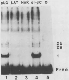
pUC LAT HAK dl-dC 0
I
*?
2b
2a
Free
1 2 3 4 5
FIG. 3. Identification oforigin-specific interactions by competi-tionanalysis. The ammonium sulfate-precipitated fraction of protein (10 p.g) wasadded tothe labeledHindIII-EcoRI originprobe from pHAK in the presence of2 ,ugofpoly(dI-dC) poly(dl-dC). Addi-tional competitor DNA (2 ,ug) was added to the samples: lane 1, formIpUC18-B (pUC);lane2, formIpLAT; lane 3, formIpHAK; and lane 4,linear poly(dI-dC) poly(dI-dC). Lane5containedonly the origin probe. Free and bound DNAs were separated by gel electrophoresis. Boundspecies arenumberedontheright.
the amountofDNA atposition 2b was insufficient to allow
adequate analysis. The remainder ofthis study focuses on
the more prominent binding interaction atposition 1. DNaseIfootprinting analysis. Weused DNase I footprint-inganalysis (10) to define the sequences covered by protein
at position 1. We incubated the 35% ammonium sulfate
precipitate fraction with a singly end-labeled,
origin-con-taining restriction fragment frompHAK(Fig. 1) andallowed
protein-DNA complexes to form. DNA was subsequently
cutwithDNase Iunderconditions which generated lessthan one nick per molecule ofDNA.
We
separated the bound from the free DNAby using gelmobility shift electrophore-sis. Free DNAandDNAatposition 1were eluted fromthemobility shift gel and run on a denaturing polyacrylamide
gel.
Protein protected nucleotides 571 through 587 on the upper strand
from
DNase I (top of Fig. 4). The DNase Ifootprint ofthe upperstrandwasverysimilartothefootprint
of the partially purified OBP described by Elias et al. (6),
exceptthatourfootprintwasonenucleotide shorteron the 5'end. Weinterpret thisto meanthat theposition 1-inducing
protein is probably the OBP. Inthe present study(Fig. 4),
OBP protected regions in this domain to different extents.
DNase protection from nucleotides 571 through 577 was
strong, while protection from nucleotides 578 through 587
wasweaker. Onthe lowerstrand, the OBP protected
nucle-otides567through583fromDNaseI(bottom
of
Fig.4). Thisregion was the same length as the footprint on the upper
strand but shifted4 bases to the left. Protection of nucleo-tides 577 through580was strongerthanprotection ateither end of the footprint. We suggest that the areas ofstronger DNaseprotectiononbothstrandsarelikelytorepresentthe
moreimportant recognition sequencesfor OBP.
Identification ofOBP contact sites. We used methylation
interference todefine the DNA recognition sequenceofthe
OBP. We treated the end-labeledorigin probewith DMSto
methylate the N7 position of a single guanine per DNA
molecule. After incubation of the modified probe with the
35% ammonium sulfate precipitate fraction, the free and
bound DNAs were separated by using the mobility shift
assay.We eluted free DNA and DNAatposition1from the geland treated them withpiperidinetobreak theDNA atthe
modifiedpositions. The cleavage productswereidentifiedon a denaturing polyacrylamide gel. This DMSassay depends ontheability of modificationsatcertainpositionstointerfere withprotein binding.Modifications of these positions would be underrepresented in the bound DNA fraction and over-represented in the free DNA fraction.
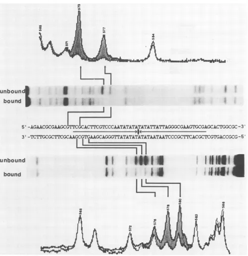
Figure 5 shows DMS interference footprints for both strands of the origin DNA. Most guanine modifications appeared with equal frequency in the bound and free frac-tions. These positions are unlikely to be important in protein recognitionand binding. On theupperstrand,nucleotidesat positions 573 and 577 were virtually absent in the bound DNA and were overrepresentedin thefree DNA (topof Fig. 5). On the lower strand, guanines at positions 576, 578, and 580interfered with binding (bottom of Fig. 5). Position 576 played a minor role in the formation of the OBP-DNA complex because it was only slightlyunderrepresented in the bound DNA. Significantly, all interfering guanines occur in an 8-bp segment of DNA fromnucleotides 573 through 580. Furthermore, the nucleotide contactsidentified by methyla-tioninterference align closely with the areas of strong DNase I protection on both strands. Consequently, we think that the OBP recognizes the sequence 5'-GTTCGCAC-3'/3'-CAAGCGTG-5'.
DISCUSSION
Elias et al. (6) originally showed that a protein partially purified from extracts of HSV-1-infected cells binds to the left arm of the large palindrome in the
orisl.
More highly purified OBP also binds to a site on the right arm ofthepalindrome withalowerefficiency (5). In the presentstudy,
we have identified two
orisl-specific
protein-DNA interac-tions by a mobility shift assay and have characterized the moreprominent interaction withhigh-resolutionfootprinting techniques. Because our DNase -I footprint closely resem-bles that identified by Elias et al. on the left arm of thepalindrome (6), we assume that we are describing arelated
interaction. In the present study, we have defined the
nucleotide recognitionsequencesfor OBPwithintheDNase
footprintdomainbyusingmethylationinterferenceanalysis.
DNase I footprints of origin DNA show that the OBP protects17 bases ofboth DNA strands. The two footprints
overlap but are four nucleotides out of register. In our
studies,the extentofDNaseprotection withinthe footprint
areais notuniform; small regions of the DNAare strongly
protected fromDNase, while other regions are only partly protected. Perhaps, the strong areas of protection corre-spondtoclosecontacts between DNA and protein, and the weaker protection represents steric hindrance of DNase
overadjacentareas.Thesevariationsin theextentofDNase
protectionare notevident in thefootprints of Eliasetal. (6).
We do not know whether the origin-binding activity ofour
preparation represents a single protein or a complex of
proteins.Complexedproteins could influencetheaffinityand the pattern ofDNA binding by OBP. This possibility may explain the small differences in the DNase protection pat-terns reported in this and in previous studies.
Other
subtlevariationsin thepreparation of extracts or in thefootprinting
conditions could also explainthe differences.
The DNase Ifootprintdomain and flankingareascontain many repeatmotifs(Fig.6A). To determinewhich,if any, of these motifs are involved in protein binding, we used a methylation interference analysis on both strands of the
DNA. Methylationevents cause stronginterference of OBP
binding within the areas strongly protected from DNase I.
J
on November 10, 2019 by guest
http://jvi.asm.org/
[image:4.612.111.232.77.218.2]4 ....
*
I
II
ii
N
IHI
,illill
11111
5'
-AGAACGCGAA{GCGT¶FCGCACTtGTCCCAATATATATATMTAITAGGGAAGTGCGAGCAC¶B3GCGC
-3'3'
-WTP;CGCTNGCAAGCGTGAAGCAGGGCTAT
AAA
TlCCG
ACCGCWGTACCGCG-5
LI
1
|lll--- _
- ---
'''''''''-'--' -' '''
I
I
I
0 a
WI
H..I
1.'.
H
H11
I
I
I
0
N
Nv
Pi
0
N-IU,
N4
rX
m
0
rI
I
C Vt
0
0
in
In e
N N
in in
WI 0
In)
C,
0
a
in
FIG. 4. DNase Ifootprints ofOBPbound toboth DNA strandsofthe HSV-1origin. The HindIII-BamHI fragmentfrompHAK was labeledateitherend and usedastheprobe. Protein (55jig)from the35% ammonium sulfateprecipitatewasaddedtotheorigin probeinthe presenceof8 jigofpoly(dI-dC) poly(dI-dC).Afterprotein-DNAcomplexeshadformed,thebinding reactionsweretreatedwith DNase I. Freeand bound DNAswereresolvedby gelelectrophoresis. ElutedDNAfractionswereanalyzedbyelectrophoresis throughadenaturing gel. Position markers(notshown)weregenerated bythe Greaction-sequencing technique describedbyMaxamandGilbert(17). Apartial sequenceof
orisl
is shown in the centerofthefigure. Thecentral palindrome isindicated by arrows. Autoradiograms anddensitometer tracings ofthefootprinted DNAs areadjacent tothe appropriate DNA strands. Shadingbetween thedensitometertracings of boundand unboundDNAshighlights areas protected by OBP.The area ofinterference is confined to an 8-bp 5'-GTTCG
CAC-3'/3'-CAAGCGTG-5' sequence overlapping the left
arm of the palindrome. All of the guanines in this region appear to bind the OBP. This 8-bp segment corresponds
exactlyto twoinverted andoverlapping repeatedsequences
butnot to anyother groupofrepeatedsequences
(Fig. 6A).
The DNA repeat sequence is
5'-GT(T/G)CG-3'.
Figure 6B shows that this inverted repeat motif occurs in the samevnboun
bound
*I*1*I
ZI
I
j.i
I
unbound
bound
IPWI ..- -...W
II
I
..
on November 10, 2019 by guest
http://jvi.asm.org/
[image:5.612.70.568.68.597.2]AN ORIGIN-BINDING PROTEIN OF HSV-1 4101
I1
in
unbound3
bound
I
I
In
I
5'
-AGAACGCGAAGCGMGCAC'IXG¶[CCAkTATATATATA¶TA¶1AGGGCGAAGTflCGAGCACTGGCGC
-3'3'
-7¶'Ic¶CGCT1rGCAAGCGTaAACGCTA
...sII
ACCGC¶ACGCTCGTGACCGCG
-5II
III
mis
IiL
..K1_.
iHINN;
1
4
a co
in,
?i
N
[image:6.612.56.557.71.589.2]in
FIG. 5. Methylation interferencefootprintsof OBPonbothDNAstrands. TheHindIII-BamHIfragmentfrompHAKwaslabeledateither end and modifiedby DMS foruse astheorigin probe. Protein(55
pug)
fromthe35%ammonium sulfate precipitatewasaddedtotheorigin probein the presence of 8 ,ug ofpoly(dI-dC) poly(dI-dC).Free and bound DNAswereseparatedby gel electrophoresis.Eluted DNAswerebrokenatthemodified residues withpiperidineandanalyzed by electrophoresisthroughadenaturing gel.Thefigureisarrangedasdescribed in Fig.4.
overlapping form at the left end of all sequenced
alpha-herpesvirus origins. With the exception of VZV, the
re-peatedsequencesarealsopresentatthe
right
endofherpes-virusorigins.The twobases thatflanktherepeated sequence arealso well conservedat both ends ofherpesvirusorigins. Theseflankingnucleotides are either bothcytosinesorboth adenines. The presence of cytosine or adenine residues appears tobe related to the nucleotide at position 3 in the
coresequence. Flankingbases and the baseatposition3are either allpurinesorallpyrimidines.Thebindingsitesonthe
right end of both
orisl
andoris2
have variations from theconsensus sequence at position 2, and
orisl
also has a mismatchatthe5'-flanking nucleotide(underlined inFigure 6B).The mismatched bases inorisl
mayexplain
thebinding
of OBP to both ends of the HSV-1
origin
with different affinities (5).unbound
bound
VOL.62, 1988
on November 10, 2019 by guest
http://jvi.asm.org/
A
*
0
5' GAAGTGAGAACGCGAAGCGTTCGCACTTCGTCCCAAT
< 1
) II
CCACTCTTGCGCTTCGCAAGCGTGAAGCAGGGTTA 5'
00
B
Location Sequence12345
ORISI
left5
CGTTCG
C3'
ORISI
right AGTGCG
A_ _
ORILI
leftC
GTTCG
C
A GTGCG A
A
GTGCG
AORILI
right
CGTTCG
CORISZ
left C GTTCG CA GTGCG A
ORIS2
right AGTGCG
AC GCTCG C
ORIL2
left CGTTCG
CA GTGCG A
ORIL2
right
C GTTCG CC GTTCG C
VZV ORIS
left A GTGCG AConsensus 5
A
GTGCG
A 3'
or
5' C GTTCG C 3' FIG. 6. The mechanism of OBP bindingtothe HSV-1 origin. (A) Identification of therepeatmotifresponsible for OBP binding. The
arrowsbetween the oris strands locate repeatedsequences in the protein-binding region. The highlighted arrows indicate the core
pentanucleotides for OBP binding. The solid bar below the upper
strandindicates the DNaseprotection domain of OBP reported by Elias etal. (6). The barsonthe outside of the DNAstrands show
areas ofDNase protection determined in thepresent study. Solid segmentsindicatestrongprotection, andopensegmentscorrespond toweakprotection. Circles identify methylated guanines that inter-fere with protein binding in our studies. Solid and open circles indicate strong and weak interference, respectively. Dotted lines align the interfering bases with the invertedrepeats.(B) Comparison of this sequence with presumptive binding sites in other alpha-herpesviruses. The tablearrangesthebinding motifs identified in the leftarmof theorisl palindromewith homologous motifs inthe left
andrightarms of theorigins from HSV-1, HSV-2, and VZV. The
corepentanucleotide is numbered 1 to5, and flanking nucleotides
are shown. A consensus sequence includes the seven nucleotides
shown. Twononequivalent copies of the consensus sequence ap-pear at each binding site. Base changes from the consensus se-quence areindicatedby underlining.
The use of overlapping inverted repetitions as binding motifs inorigins of replication is not unique. The T antigens of simian virus 40 and polyomavirus both bind to the pentanucleotide 5'-GAGGC-3' in a palindrome at the center of their origins of replication (7). Each arm of thepalindrome contains two pentanucleotides oriented as direct repeats with 1-bp spacing between them. The arms of the palin-dromes are arrangeddifferently in these two viruses. Simian virus 40 has a 1-bp separation between the inverted repeats, while the inverted repeats of polyoma virus overlap by two
base pairs. This alternative arrangement of polyoma
se-quences indicates that recognition sequences for protein subunits can overlap inreplication origins.
Our findings suggest that the herpesvirus OBP binds to the inverted repeat motif as a dimer. In every putative binding
site, thereis an asymmetry of the repeats at position 3 and
within the flanking bases. This asymmetry could be impor-tantfor subsequent protein function. OBPs have two poten-tialroles in the initiation of replication. One possible role is to assemble a preinitiation complex at the origin. In this case,theasymmetrical bindingsitecould impart directional-ity to a bound complex of proteins. The other possibildirectional-ity is that the OBP couldparticipate in an origin initiation event; the asymmetry of the OBP-binding site could focus origin events to one strand of origin DNA. For example, the
initiation of a rolling circle may require the nicking of one
andonly one strand of duplex DNA. Our findings add to the framework of information that can be used for the design of
genetic and biochemical approaches to these functional
problems.
ACKNOWLEDGMENTS
Thisinvestigationwassupported by Public HealthServicegrants CA-18808,CA-38146, and5-T32CA-09176 awarded bytheNational CancerInstitute,Departmentof Health andHumanServices.
ADDENDUM
Pureproteinencodedby the UL9 gene of HSV-1 (18a) has
the same methylation interference pattern as the OBP
de-scribedinthisstudy(KoffandTegtmeyer,unpublisheddata;
P. D. Olivo, N. J. Nelson, and M. D. Challberg,
unpub-lisheddata). Thus, OBP is the product ofthe UL9 gene. LITERATURECITED
1. Ben-Porat, T., and S. A. Tokazewski. 1977. Replication of herpesvirus DNA. II. Sedimentation characteristics of newly synthesized DNA.Virology 79:292-301.
2. Birnboim, H.C., andJ. Doly. 1979. Arapid alkaline extraction procedure for screening recombinant plasmid DNA. Nucleic AcidsRes. 7:1513-1523.
3. Deb, S., and M. Doelberg. 1988. A67-base-pair segmentfrom theori-Sregion of herpes simplex virustype1 encodes origin function. J. Virol.62:2516-2519.
4. DeLucia,A.L.,B.A.Lewton, R.Tjian, and P. Tegtmeyer. 1983. Topography of simian virus 40 A protein-DNA complexes: arrangement ofpentanucleotide interaction sitesattheoriginof replication. J.Virol. 46:143-150.
5. Elias, P., andI. R. Lehman.1988.Interaction oforiginbinding protein with anoriginofreplicationof herpes simplexvirus 1. Proc. Natl.Acad. Sci. USA 85:2959-2963.
6. Elias, P., M. E.O'Donnell,E. S. Mocarski, and I. R. Lehman. 1986. A DNAbinding protein specific foranorigin of replication ofherpes simplex virus type 1. Proc. Natl. Acad. Sci. USA 83:6322-6326.
7. Fields,B. N.1985.Replicatior.ofpapovaviruses,p. 393-410. In B. Fields(ed.), Virology.RavenPress,New York.
8. Frenkel, N., H. Locker, and D. A. Vlazny. 1980. Studies of
on November 10, 2019 by guest
http://jvi.asm.org/
[image:7.612.70.289.93.513.2]defective herpes simplex viruses. Ann. N.Y. Acad. Sci. 354:347-370.
9. Friedmann, A., J. Shlomai, and Y. Becker. 1977. Electron microscopy ofherpes simplex virus DNA molecules isolated frominfectedcells by centrifugation in CsCldensity gradients. J. Gen. Virol. 34:507-522.
10. Galas, D. J., and A. Schmitz. DNAase footprinting: a simple method for the detection ofprotein-DNA binding specificity. Nucleic AcidsRes. 5:3157-3170.
11. Garner, M. M., and A. Revzin. 1981. A gel electrophoresis methodfor quantifyingthe binding of proteins tospecificDNA regions: application to components of the Escherichia coli lactose operon regulatory system. Nucleic Acids Res. 9: 3047-3060.
12. Holmes, A. M., S. M. Wietstock, and W. T. Ruyechan. 1988. Identification and characterization ofa DNA primaseactivity present in herpes simplex virustype 1-infected HeLacells. J. Virol. 62:1038-1045.
13. Jacob, R.J., and B. Roizman. 1977. Anatomyofherpes simplex virusDNA. VIII. Properties of the replicating DNA. J. Virol. 23:394-411.
14. Lockshorn, D., and D. A. Galloway. 1986.Cloning and charac-terization oforiL2, alarge palindromicDNAreplication origin ofherpessimplex virustype 2. J.Virol.58:513-521.
15. Maniatis, T., E. F. Fritsch, and J. Sambrook. 1980. Molecular cloning: alaboratorymanual. Cold SpringHarborLaboratory, ColdSpring Harbor, N.Y.
16. Marsden, H.S., M. E. M. Campbell, L. Haarr, M. C. Frame, D.S. Parris, M. Murphy, R. G. Hope, M. T. Muller, and C. M. Preston. 1987. The 65,000-Mr DNA-binding and virion trans-inducing proteins of herpes simplex virus type 1. J. Virol. 61:2428-2437.
17. Maxam, A. M., and W. Gilbert. 1977. A new method for sequencingDNA. Proc. Natl. Acad. Sci. USA 74:560-564. 18. O'Donnell, M. E., P. Elias, B. E. Funnell, and I. R. Lehman.
1987.Interaction between theDNApolymeraseandthe single-strandedDNA-binding protein (infected cell protein 8) of herpes simplex virus 1.J. Biol. Chem. 262:4260-4266.
18a.Olivo, P.D., N. J. Nelson, and M. D. Challberg. 1988. Herpes simplex virus DNA replication: the UL9 gene encodes an
origin-binding protein. Proc. Natl. Acad. Sci. USA
85:5414-5418.
19. Poffenberger, K. L., and B. Roizman. 1985. A noninverting genomeof aviableherpes simplex virus 1: presenceof head-to-tail linkages in packaged genomes and requirements for circu-larization afterinfection. J. Virol. 53:587-595.
20. Roizman, B., and P. G. Spear. 1968. Preparation ofherpes simplex virus of high titer.J. Virol.2:83-84.
21. Stow, N. D. 1982.Localization ofanorigin ofDNAreplication within theTRS/IRSrepeated region of the herpessimplex virus type 1 genome. EMBOJ. 1:863-867.
22. Stow, N. D. 1985.Mutagenesis ofa herpessimplexvirus origin of DNAreplication andits effect on viralinterference.J. Gen. Virol.66:31-42.
23. Stow, N. D., and A. J. Davison. 1986. Identification of a varicella-zostervirus origin ofDNAreplication andits activa-tionby herpessimplex virustype 1 geneproducts.J.Gen. Virol. 67:1613-1623.
24. Stow, N. D., and E. C. McMonagle. 1983. Characterization of theTRS/IRSorigin ofDNAreplication of herpessimplex virus type 1.Virology 130:427-438.
25. Vaughan, P. J., L. M.Banks, D. J. M. Purifoy, and K. L. Powell. 1984. Interactions between herpes simplex virus DNA-binding proteins.J.Gen. Virol. 65:2033-2041.
26. Vlazny, D. A., and N. Frenkel. 1981. Replication ofherpes simplex virus DNA: localization ofreplication recognition sig-nals within defective virus genomes. Proc. Natl. Acad. Sci. USA78:742-746.
27. Weller, S. K., D. P.Aschman, W. R. Sacks,D. M. Coen,and P. A.Schaffer. 1983.Geneticanalysis oftemperature sensitive mutantsofHSV-1: the combineduseofcomplementation and physical mappingfor cistronassignment.Virology 130:290-305. 28. Weller, S. K., A.Spadaro,J. E.Schaffer,A. W.Murray, A. M. Maxam, and P. A. Schaffer. 1985. Cloning, sequencing, and functionalanalysisoforiL,aherpes simplexvirustype 1origin of DNAsynthesis. Mol. Cell. Biol. 5:930-942.
29. Wu, C. A., N.J. Nelson, D. J.McGeoch,andM. D.Challberg. 1988. Identification ofherpes simplex virus type 1 genes
re-quired for origin-dependent DNA synthesis. J. Virol. 62:435-443.
30. Wright, P. J., A. L. DeLucia, and P. Tegtmeyer. 1984. Se-quence-specificbinding of simian virus40 Aproteintononorigin and cellularDNA. Mol.Cell. Biol. 4:2631-2638.