Copyright © 2002, American Society for Microbiology. All Rights Reserved.
Ultrastructural Localization of the Herpes Simplex Virus Type 1 U
L
31,
U
L
34, and U
S
3 Proteins Suggests Specific Roles in Primary
Envelopment and Egress of Nucleocapsids
Ashley E. Reynolds,
1Elizabeth G. Wills,
1Richard J. Roller,
2Brent J. Ryckman,
2and Joel D. Baines
1*
Department of Microbiology and Immunology, Cornell University, Ithaca, New York 14853,1and
Department of Microbiology, University of Iowa, Iowa City, Iowa 522422
Received 1 April 2002/Accepted 4 June 2002
The wild-type UL31, UL34, and US3 proteins localized on nuclear membranes and perinuclear virions; the
US3 protein was also on cytoplasmic membranes and extranuclear virions. The UL31 and UL34 proteins were
not detected in extracellular virions. US3 deletion caused (i) virion accumulation in nuclear membrane
invaginations, (ii) delayed virus production onset, and (iii) reduced peak virus titers. These data support the herpes simplex virus type 1 deenvelopment-reenvelopment model of virion egress and suggest that the US3
protein plays an important, but nonessential, role in the egress pathway.
Herpes simplex virus type 1 (HSV-1) virions contain a linear double-stranded DNA genome of approximately 152 kb that is packaged into an icosahedral capsid shell. An amorphous teg-ument layer surrounds the capsid and is, in turn, surrounded by an envelope composed of a host-derived lipid bilayer studded with viral integral membrane proteins. After the viral genome is replicated and packaged into capsids within the nucleus, assembled nucleocapsids acquire a primary lipid envelope by budding through the inner nuclear membrane (INM) into the space located between the inner and outer leaflets of the nu-clear envelope (25, 33). Whereas the derivation of the primary envelope from the INM is widely accepted, the route of transit of the nascent virions from the perinuclear space to the extra-cellular space is more controversial. An overview of the key players in herpesvirus egress and a comparison of the salient features of the two proposed envelopment models have been recently published (8, 25).
A single-step model of herpesvirus envelopment was pro-posed for the prototypical alphaherpesvirus HSV-1 (6, 18, 35, 44). This model proposes that enveloped virions move through the endoplasmic reticulum (ER) and the Golgi apparatus in transport vesicles with concomitant modification of primary virion glycoproteins. The single-step envelopment model is supported by the observations that (i) enveloped particles within vesicles can be readily detected by electron microscopy and in fracture label studies (35, 44) and (ii) virion egress and virion-associated glycoprotein processing are both inhibited in cells treated with the ionophore monensin (18). On the other hand, neither of these observations can exclude the alternative deenvelopment-reenvelopment model. Such a model is sup-ported by mounting ultrastructural and biochemical evidence (3, 10, 13, 14, 30, 37, 41, 46, 50) and has been proposed for HSV-1, other alphaherpesviruses such as varicella-zoster virus
(VZV) and pseudorabies virus (PrV), and betaherpesviruses such as human cytomegalovirus. In this model, primary envel-opment occurs by budding through the INM but the primary envelope surrounding the perinuclear virion is lost, presumably by fusion with the outer lamellae of the nuclear envelope. In a second step, reenvelopment occurs by wrapping of the nucleo-capsid and its associated tegument with a lipid bilayer origi-nating from a membranous cytoplasmic organelle bearing viral glycoproteins previously modified by transit through the nor-mal secretory pathway. It has been proposed that the second envelope is derived from membranes that normally reside within the trans-Golgi network or other Golgi membranes (3, 11, 24, 47, 50).
Several proteins have been implicated in the initial budding of herpesvirus nucleocapsids at the INM, including the HSV UL11, UL31, and UL34 proteins, along with glycoprotein K, a
protein necessary for envelopment in nondividing cells (1, 15, 16). Studies done in our laboratories previously demonstrated that the UL31 and UL34 gene products of HSV-1 form a
complex that is targeted to the nuclear rim and is essential for optimal primary envelopment of nucleocapsids (32, 34). Simi-lar results have been obtained upon analysis of the UL31 and
UL34 homologues of PrV (10, 21).
The UL31 gene product is a nuclear matrix-associated,
nucleotidylylated phosphoprotein that, in association with the UL34 gene product, localizes to the nuclear rim of
HSV-1-infected cells (2, 4,32, 48). The UL34 gene product is a nuclear
membrane-associated phosphoprotein with a predicted type II integral membrane topology. Also, UL34 protein is a substrate
for the HSV-1 US3-encoded kinase (9, 28, 29, 34, 36, 49). As
demonstrated by Reynolds et al. (32),US3 kinase is required
for even distribution of the UL31 and UL34 proteins around
the nuclear rim of wild-type-infected cells. In addition, the US3-encoded kinase has been proposed to play a role in
pro-tecting HSV-1-infected cells from virus-induced apoptosis (17, 23). In the absence of the PrV US3 protein homologue, large
numbers of enveloped virions appear to accumulate within invaginations of the nuclear membrane (22, 45). These data led
* Corresponding author. Mailing address: Department of Microbi-ology and ImmunMicrobi-ology, Cornell University, Veterinary Medical Center (VMC) C5 131, Ithaca, NY 14853. Phone: (607) 253-3385. Fax: (607) 253-3384. E-mail: [email protected].
8939
on November 8, 2019 by guest
http://jvi.asm.org/
to the deduction that the US3-encoded kinase is also important
for the efficient deenvelopment of nascent virions that occurs upon fusion of the virion envelope with the outer nuclear membrane (ONM).
The goal of this study was to determine the localization of the HSV-1 US3, UL31, and UL34 proteins in infected cells at
the ultrastructural level. Consistent with the deenvelopment-reenvelopment model of virion egress, UL31 and UL34
pro-teins were observed to associate with perinuclear virions but not with extracellular virions. The localization of the HSV-1 US3-encoded kinase in infected cells and the phenotype of
cells infected with the US3-null mutant virus provide support
for the hypothesis that one of several potential roles of US3
kinase is to promote efficient egress of virions from the nucleus into the cytoplasm.
The cell lines used for this study were previously described (31, 43). The wild-type HSV-1(F) virus and US3 mutants
R7037 and R7039 (provided by Bernard Roizman, University of Chicago) have been previously characterized (7, 29). R7037 contains a deletion of portions of the US3 and US4 open
reading frames (ORFs), and R7039 contains a deletion of portions of the US2 and US3 ORFs. The construction and
growth properties of HSV-1(F) UL34-null mutant vRR1072
(tk⫹) have been described previously (34). U
L31-null mutant
virus R5132, also provided by Bernard Roizman, has been described previously (5). Both vRR1072 (tk⫹) (U
L34-null
mu-tant virus) and R5132 (UL31-null mutant virus) were
propa-gated on stably transfected, complementing cell lines as de-tailed previously (32).
The following protocol was utilized for all immunogold elec-tron microscopy. Vero cells were infected at a multiplicity of infection (MOI) of 5 and maintained at 37° C until harvesting at 14 to 18 h postinfection (hpi). The viral inoculum used for each preparation was diluted in 199V medium (199 medium supplemented with 1% newborn calf serum, penicillin, and streptomycin [43]). Harvested cells were pelleted by centrifu-gation and fixed with 4% formaldehyde and 0.25% glutaralde-hyde in 0.1 M sodium phosphate buffer (pH 7.4) for 30 min at 25°C and then for 90 min at 4°C. Fixed cells were washed three times for 10 min per wash in phosphate-buffered saline (PBS) at 4°C, dehydrated with increasing ethanol concentrations at 4 and⫺20°C, and embedded stepwise at⫺20°C with increasing concentrations of LRWhite (Electron Microscopy Sciences, Fort Washington, Pa.). The samples were then polymerized under UV light at⫺35°C overnight.
UL31 protein is predominantly localized to the nuclear
membrane of HSV-1(F)-infected cells.Thin sections were pre-pared for immunogold electron microscopy as described above and probed with UL31 protein-specific rabbit polyclonal
anti-serum that was prepared as described previously and diluted 1:2 (31, 32). Donkey anti-rabbit immunoglobulins conjugated with 12-nm-diameter colloidal gold particles were incubated with the thin sections for 1 h (electron microscopy grade 12-nm colloidal gold AffiniPure donkey anti-rabbit immunoglobulin G [IgG]; catalog no. 711-205-152; Jackson ImmunoResearch Laboratories, Inc., West Grove, Pa.). Excess antibodies were removed by washing with PBS-Tween 80–1% fish gelatin. Post-labeling fixation was performed with 2.5% glutaraldehyde in 0.1 M phosphate buffer for 10 min and followed by rinsing with distilled water. Sections were counterstained with 2% uranyl acetate and lead citrate, coated with Formvar (0.5% Formvar in ethylene dichloride; Ladd Research Industries, Williston, Vt.), and examined with a Philips 201 transmission electron microscope. Conventionally rendered negatives of electron mi-croscopic images were scanned with ScanWizard Pro PPC 1.02 software (Microtek, Redondo Beach, Calif.), and digital im-ages were generated with Adobe Photoshop 5.0 software.
The distribution of UL31 protein in cells infected with
var-ious viral strains analyzed by transmission electron microscopy (TEM) is summarized in Table 1. Statistical analysis of the means and standard errors of the means presented in Table 1 was done by PROC UNIVARIAT utilizing SAS (Statistical Analysis Systems).
Colloidal gold beads representing the localization of UL31
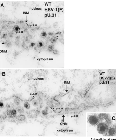
protein were detected lining the INM and, to a lesser extent, the ONM of cells infected with HSV-1(F), as seen in Fig. 1A and B. The UL31 gene product was also associated with
envel-oped viral particles located between the lamellae of the nuclear envelope. Ten randomly selected whole-cell sample sections were counted, and approximately two-thirds of the gold beads were associated with parts of the nuclear rim (in the INM, in the ONM, between leaflets in the perinuclear space, and within cytoplasmic and nuclear sites directly adjacent to the nuclear membrane leaflets) or with viral particles that associated with these sites. Approximately 1/5 of the gold particles were asso-ciated with the central nucleoplasm, and approximately 1/10 of the particles were localized free in the cytosol, on cytoplasmic structures, or in association with cytoplasmic or extracellular virions (Fig. 1C).
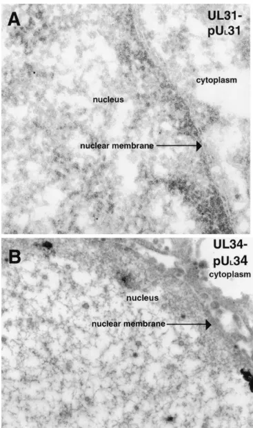
In cells infected with the UL31 deletion virus and harvested
[image:2.587.42.544.85.156.2]at time points comparable to those of experiments with wild-type virus-infected cells (Fig. 2A), very few viral particles were
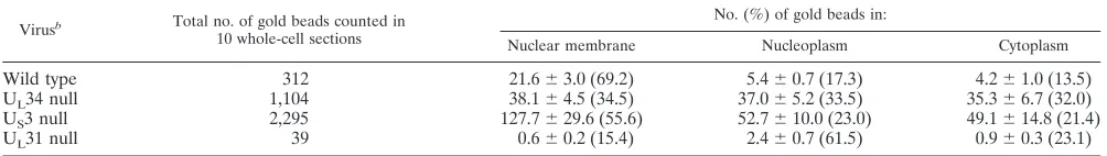
TABLE 1. Summary of the distribution of UL31 protein determined by immunogold analysis of thin sectionsa
Virusb Total no. of gold beads counted in
10 whole-cell sections
No. (%) of gold beads in:
Nuclear membrane Nucleoplasm Cytoplasm
Wild type 312 21.6⫾3.0 (69.2) 5.4⫾0.7 (17.3) 4.2⫾1.0 (13.5)
UL34 null 1,104 38.1⫾4.5 (34.5) 37.0⫾5.2 (33.5) 35.3⫾6.7 (32.0)
US3 null 2,295 127.7⫾29.6 (55.6) 52.7⫾10.0 (23.0) 49.1⫾14.8 (21.4)
UL31 null 39 0.6⫾0.2 (15.4) 2.4⫾0.7 (61.5) 0.9⫾0.3 (23.1)
aThe total numbers of gold beads in 10 randomly selected whole-cell sections of wild-type virus [HSV-1(F)], U
L31-null virus-, UL34-null virus-, and US3-null
virus-infected cells were quantified. The mean number of UL31 protein-specific gold beads and the standard error of each mean are indicated for three regions of the
cells. The following formula was used to calculate the percentage of gold beads in a given region of the cell infected with the indicated virus: (number of gold bead
particles per region of 10 whole-cell sections/total number of gold bead particles in 10 whole-cell sections)⫻100⫽% of gold beads per region.
bTen samples of each virus were tested
8940 NOTES J. VIROL.
on November 8, 2019 by guest
http://jvi.asm.org/
FIG. 1. Digitally scanned electron micrograph of thin sections of wild-type (WT) HSV-1(F)-infected Vero cells harvested and fixed 14 to 18 hpi. The nucleus, INM, ONM, and cytoplasm are labeled in panels A and B. Gold beads associated with the UL31 protein (pUL31) are demarcated with arrowheads. Panel C demonstrates extracellular virions devoid of pUL31-specific immunogold label. For reference, in the electron micro-graphs presented here, HSV-1 nucleocapsids are approximately 120 nm in diameter and gold beads are 12 nm in diameter. Original magnification of panels A to C,⫻45,000.
on November 8, 2019 by guest
http://jvi.asm.org/
FIG. 2. Scanned digital electron micrograph of UL31-null mutant infected thin sections of Vero cells (A) or UL34-null mutant virus-infected thin sections of Vero cells (B) harvested and fixed late in infection (14 to 18 hpi). In panel A, thin sections were probed with UL31-specific rabbit antisera. The cytoplasm, nuclear membrane, and nucleus are all labeled. Original magnification of panel A,⫻45,000. In panel B, thin sections were probed with UL34 protein-specific IgY antibodies. Original magnification of panel B,⫻45,000.
8942 NOTES J. VIROL.
on November 8, 2019 by guest
http://jvi.asm.org/
located outside of the nucleus, in contrast to the appearance of cells infected with HSV-1(F). This indicated that the UL31
protein, while not absolutely essential for egress of nascent virions from the nucleus, greatly facilitated the process. Upon staining of sections of cells infected with the UL31 deletion
virus with the UL31 protein-specific rabbit polyclonal
anti-serum, it was apparent that nonspecific staining with the UL31
polyclonal antisera was minimal, yielding a mean value of ap-proximately four gold beads per cross section of an entire cell (averaged from 10 randomly selected sections counted). Wild-type virus-infected cells had, on average, a mean of approxi-mately 31 gold beads per whole-cell cross section (averaged from 10 random whole-cell sections counted).
The difference in the quantity of gold beads detected at the nuclear membranes between cells infected with the wild-type virus and the UL31-null mutant was particularly striking.
Whereas the mean number of gold particles at the nuclear membranes of wild-type virus-infected cells was approximately 22, in a sample size of 10 whole-cell sections selected at ran-dom, the nuclear membrane of UL31-null virus-infected cells
had a mean of only 0.6 gold bead per section (averaged from the 10 random cell sections counted). Thus, we deduced that the immunoreactivity detected in HSV-1(F)-infected cells at the nuclear membrane was specifically attributable to the pres-ence of the UL31 gene product.
The UL34 gene product is detectable on the INM and ONM
of cells infected with HSV-1(F).Thin sections of cells infected with HSV-1(F) were prepared as described above and reacted with an affinity-purified chicken IgY antibody directed against the UL34 protein diluted 1:50 in 1% cold bovine serum
albu-min-PBS (32). Donkey anti-chicken IgY antibodies conjugated with 12-nm-diameter colloidal gold particles were incubated with the sections probed with the UL34 protein-specific
chicken IgY antibody for 1 h (electron microscopy grade 12-nm colloidal gold AffiniPure donkey anti-chicken IgY; cat-alog no. 703-205-155; Jackson ImmunoResearch). Stained sec-tions were prepared as described above and subsequently ex-amined by TEM. Representative results are shown in Fig. 3. Immunostaining with the UL34 antibody was considerably
more intense than was UL31 protein-specific immunostaining,
and hundreds of beads were visible in a given section of a whole cell (Fig. 3A). Unlike the UL31 protein, where the
ma-jority of the gold beads were localized on the INM, colloidal gold particles representing the localization of the UL34 protein
were detected in approximately equal amounts on the INM and ONM, as shown in Fig. 3C. Additional immunoreactivity specific for the UL34 protein was detectable on perinuclear
viral particles (Fig. 3B). As was the case with the UL31 protein,
UL34 protein-specific immunoreactivity was not observed in
association with structures in the cytoplasmic compartment, cytoplasmic viral particles, or virions in the extracellular space (Fig. 3D). As a negative control, UL34-null mutant
virus-in-fected cells were harvested at time points comparable to those of the wild-type virus-infected cells and incubated with the UL34 antibodies (Fig. 2B). Minimal background staining was
detected by immunogold analysis, and we therefore concluded that the staining seen in wild-type HSV-1(F)-infected cells incubated with anti-UL34 IgY antibodies was specific for UL34
protein epitopes. It is noteworthy that by immunogold analysis, neither the UL31 nor the UL34 protein was detectable in
as-sociation with cytoplasmic or extracellular viral particles. Sim-ilarly, the PrV-encoded homologues of these proteins are not present at detectable levels on intracytoplasmic or extracellular particles but are readily detectable on perinuclear particles (10, 21). Previous studies demonstrated that low levels of the HSV-1 UL34 protein are detectable in virions purified from
cytoplasmic extracts (29). This observation is consistent with our immunogold analyses inasmuch as cytoplasmic virion prep-arations would be expected to contain some virions purified from the perinuclear space that contain the UL34 protein.
Although our studies do not necessarily rule out the possibility that the UL31 and UL34 proteins are present in extracellular
virions, the very strong immunoreactivity associated with nas-cent viral particles, compared with the virtual absence of im-munoreactivity in extracellular particles, indicates that the two particle types, perinuclear and extracellular, differ significantly in UL31 and UL34 protein content. Such observations provide
strong evidence that during egress of viral particles, the initial, INM-derived envelope containing the integrated UL34 protein
is removed and reenvelopment provides a novel envelope lack-ing (or containlack-ing drastically decreased levels of) the UL34
protein. These data are most consistent with a model in which the membrane acquired during primary envelopment is lost by fusion with the ONM and deenveloped viral particles are re-leased into the cytoplasm, where the particles are wrapped in a new envelope derived from the Golgi apparatus or another membranous organelle.
The presence of both the UL31 and UL34 proteins on virus
particles located between the lamellae of the nuclear mem-brane is the first evidence supporting our previous hypothesis that the UL31/UL34 protein complex becomes incorporated
into virions upon budding at the INM (32). We hypothesize that the UL31/UL34 complex at the INM engages
nucleocap-sids, causing them to accumulate at the nuclear envelope and subsequently undergo primary envelopment. When budding of nucleocapsids through the INM occurs, nascent virions labeled with the UL31 and UL34 proteins accumulate in the
perinu-clear space, as shown in Fig. 1 and 3. We predict that, in the absence of either protein, nucleocapsids will not be effectively retained at the nuclear rim and, consequently, envelopment of nucleocapsids will not occur efficiently.
UL31 protein localization in cells infected with the UL34
deletion virus.As described previously (32), the UL34 protein
plays a crucial role in maintenance of the UL31 protein at the
nuclear rim of wild-type virus-infected cells. This was con-firmed by immunogold analyses. In cells infected with the UL34 deletion virus, the level of UL31 protein-specific
immu-noreactivity was approximately evenly distributed among the nuclear membrane, the nucleoplasm, and the cytoplasm. The results are summarized in Table 1.
The UL34 protein is not strictly associated with the nuclear
rim in UL31-null mutant virus-infected cells.Thin sections of
cells infected with the UL31-null mutant were also reacted with
the UL34 protein-specific antisera, and bound antibody was
detected by reaction with gold bead-conjugated anti-chicken antisera. Examination of the samples by TEM revealed that UL34 protein-specific immunoreactivity localized primarily at
the INM and ONM. Unlike the appearance of cells infected with the wild-type virus, UL34 protein-specific
immunoreactiv-ity was also associated with regions of the cytoplasm in a
on November 8, 2019 by guest
http://jvi.asm.org/
FIG. 3. Scanned digital electron micrograph of thin sections of wild-type(WT) virus-infected Vero cells harvested and fixed late in infection. Thin sections were incubated with UL34 protein-specific chicken IgY antibody. The nucleus, nuclear membrane, and cytoplasm are labeled in panel A and, in panels B and C, the INM and ONM are indicated with arrows, as are the cytoplasm and nucleus. Panel D shows extracellular virions probed with UL34 antisera devoid of gold label. Regions labeled with UL34 protein-specific antisera are demarcated with arrowheads labeled pUL34. Original magnification of panels A to D,⫻45,000.
8944
on November 8, 2019 by guest
http://jvi.asm.org/
largely perinuclear distribution (data not shown). A minor portion of the total detectable UL34 protein immunoreactivity
was observed within the nucleoplasm (data not shown). Previous studies by Reynolds et al. (32) characterizing the distribution of the UL31 protein in UL34-null mutant
virus-infected cells by indirect immunofluorescence assay (IFA) also demonstrated that the UL31 protein is mislocalized from the
nuclear rim and is localized primarily in the central nucleus and, to a lesser extent, in the cytoplasm.
The model of UL31 and UL34 localization and function
previously proposed by Reynolds et al. (32) was largely based on IFA data. The ultrastructural information gained from the present study demonstrates, for the first time, that both of these HSV-1 proteins associate with the INM, among other structures at the nuclear membrane. Analyses of the cells in-fected with the UL31- and UL34-null mutants indicate that
whereas each protein has the capacity to target the nuclear rim region in the absence of the reciprocal protein, association in the INM is optimized in the presence of both proteins in infected cells. Given the previous observation that UL31 and
UL34 are sufficient to target one another to the nuclear rim in
the absence of other HSV-1 proteins (32), it is likely that cellular proteins also contribute to the localization of the UL31/UL34 protein complex at the INM. Given the nuclear
matrix association of the UL31 protein (4), it is reasonable to
hypothesize that such proteins might include lamins or lamin receptors that normally localize on the nucleoplasmic face of the INM.
The data are consistent with our previously proposed model (32), which was based on the lamin B receptor localization paradigm (38, 39, 40). Briefly, the UL34 protein integrates
itself into the ER membrane in a type II orientation, diffuses laterally along the lipid bilayer into the ONM, which is con-tinuous with the ER, diffuses past the nuclear pore complex (NPC) with its N-terminal domain in the lateral channel of the NPC, and moves to the INM, where the bulk of the protein resides in the nucleoplasm. In HSV-1(F)-infected cells, the INM-bound UL34 protein encounters the UL31 gene product,
which is targeted to the nucleus by virtue of an N-terminal nuclear localization signal (51). When the UL34 protein
inter-acts with the UL31 protein, a complex of the proteins is formed
that is predicted to be stably anchored to the nuclear mem-brane through the transmemmem-brane domain of the UL34 gene
product and the nuclear matrix association of the UL31
pro-tein.
Subcellular localization of the US3-encoded kinase.We have
previously reported that the US3-encoded kinase and its
sub-strate, the UL34 protein, colocalize extensively in the absence
of other viral factors (32). The subcellular localization of the US3-encoded kinase in HSV-1-infected cells has not been
re-ported. Inasmuch as the US3 and UL34 proteins colocalize in
transiently transfected cells (32), it was hypothesized that the US3 and UL34 proteins would also colocalize in
wild-type-infected cells.
To test this hypothesis, HEp-2 cells were grown to approx-imately 70% confluence on sterile glass coverslips and infected at an MOI of 10 for 12 h at 37°C with HSV-1(F) or R7039. Infected cells were fixed for 15 min in 2% formaldehyde–PBS, washed three times in PBS, and permeabilized for 15 min in immunofluorescence (IF) buffer as previously described (32).
Cells were blocked for 1 h in IF buffer supplemented with 0.01% pooled human immunoglobulins, washed three times in PBS, reacted for 1 h with primary antibodies diluted in IF buffer, washed three times in PBS, and then reacted for 1 h with secondary antibodies diluted in IF buffer. Chicken anti-UL34 antibody was diluted 1:4,000, rabbit anti-US3 antibody
(26) was diluted 1:1,000, and donkey anti-chicken globulin-Texas Red conjugate and goat anti-rabbit immuno-globulin-fluorescein isothiocyanate conjugate were both di-luted 1:200. Immunostained cells were analyzed by confocal microscopy as previously described (32). The results are shown in Fig. 4.
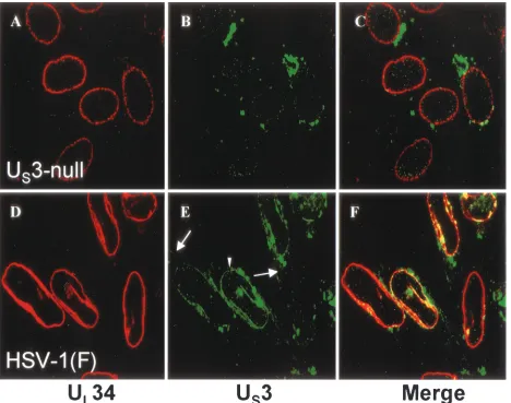
As previously demonstrated by Reynolds et al. (32), in cells infected with a US3-null virus, the UL34 protein was detected
in a punctate distribution at the nuclear envelope (Fig. 4A). This is in stark contrast to cells infected with HSV-1(F), where the UL34 protein adopted a more uniformly even distribution
at the nuclear envelope (Fig. 4D). In HSV-1(F)-infected cells, the US3-encoded kinase was detected at the plasma
mem-brane, in cytoplasmic structures, and at the nuclear envelope, where it colocalized with the UL34 protein (Fig. 4E and F).
Areas of colocalization of the two proteins appear yellow in these merged images. The localization of the US3 protein at
the nuclear rim (marked with a white arrowhead in Fig. 4E) and the plasma membrane (marked with a long white arrow in Fig. 4E) was never seen in cells infected with the US3-null virus
and is distinct from the largely cytoplasmic background fluo-rescence detected in cells infected with that virus, demonstrat-ing that these are sites of specific anti-US3 reactivity (Fig. 4B).
Some of the background fluorescence detected in Fig. 4B may be attributable to incomplete blocking of the virus-encoded Fc receptor (a complex of glycoproteins E and I [19]) despite the use of pooled human immunoglobulins as a blocking agent. The presence of the background fluorescence does not permit any conclusion to be drawn about US3 protein localization in
the cytosol or on cytoplasmic membranes as determined by IFA. These IFA data indicate that the US3 protein localizes to
the plasma membrane and the nuclear envelope, where it co-localizes with the UL34 protein (Fig. 4E and F). In view of the
previous report that the US3 and UL34 proteins colocalize in
transiently transfected cells (32), we therefore hypothesize that the US3-encoded kinase and its substrate, the UL34 protein,
may physically and stably interact.
While the relationship between the localization of the UL34
protein and that of the US3 protein in other
alphaherpesvi-ruses has not been addressed, studies of the localization of PrV and HSV-2 US3 homologues have detected them diffusely
dis-tributed throughout infected cells (12, 22).
The HSV-1 US3-encoded kinase localization in infected cells
and association with extracellular particles are markedly dif-ferent from the distribution of the UL31 and UL34 proteins.To
characterize the localization of the US3-encoded kinase at the
ultrastructural level, thin sections of cells infected with HSV-1(F) or the US3-null mutant R7037 were reacted with a US
3-specific rabbit polyclonal antiserum (supplied by Bernard Roizman) (26) diluted 1:10 in cold bovine serum albumin-PBS and bound IgG was detected as described for the UL31
pro-tein. Representative results are shown in Fig. 5.
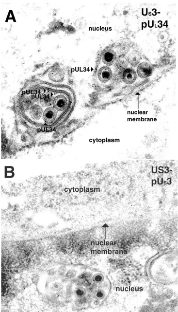
Like the distribution of the UL34 protein in
HSV-1(F)-infected cells, the US3 protein was associated with both
on November 8, 2019 by guest
http://jvi.asm.org/
lae of the nuclear envelope and with perinuclear viral particles as well. No obvious staining specific for the US3 protein was
detectable in the nucleoplasm of infected cells. Several key differences between the distribution of the US3 protein and
that of the UL31 or UL34 protein were noted. Unlike the
appearance of the UL31 or UL34 protein, gold beads
demar-cating the location of the Us3 protein were detected exten-sively within the cytoplasm. Cytoplasmic US3 protein was
de-tected in the cytosol and was associated with ribbon-like structures that resembled membranous organelles. In marked contrast to the UL31 and UL34 gene products, the US3 protein
was clearly associated with viral particles localized at the plasma membrane and extracellular viral particles, as shown in Fig. 5C. This observation is consistent with US3-specific
immu-noreactivity detected at the plasma membrane of wild-type-infected cells analyzed by IFA (Fig. 4E). As a negative control, thin sections infected with a US3-null HSV strain were stained
with the US3 antisera, and they exhibited negligible levels of
background immunostaining (Fig. 7B).
The UL31 and UL34 proteins are associated with nuclear
membrane invaginations in cells infected with a US3-null
vi-rus.Previous studies utilizing IFA demonstrated that the UL31
and UL34 proteins colocalize in punctate regions associated
with the nuclear rim in cells infected with HSV US3-null
mu-tant viruses. To characterize these structures at the ultrastruc-tural level, Vero cells were infected with the R7037 US3
mu-tant virus and subjected to immunogold TEM with antiserum directed against either the UL31 or the UL34 protein as
de-scribed above. Representative results are shown in Fig. 6 and 7. The morphology of the nuclear membrane in cells infected with the US3-null HSV strain differed markedly from that of
cells infected with HSV-1(F). Specifically, individual thin sec-tions of an entire cell typically contained approximately 5 to 10 clusters of one to several enveloped viral particles along the nuclear rim labeled with UL31 (examples are shown in Fig. 6)
and UL34 (an example is shown in Fig. 7A) protein-specific
[image:8.587.60.528.72.441.2]antibodies. The clustered viral particles were completely or partially surrounded by membranous structures. The lumen of
FIG. 4. Digital confocal images showing localization of the UL34 and US3 proteins in HEp-2 cells infected with HSV-1(F) or US3-null HSV-1. Subconfluent monolayers of HEp-2 cells were infected for 12 h with either US3-null mutant virus R7039 (A to C) or HSV-1(F) (D to F). Formaldehyde-fixed cells were immunostained with chicken anti-UL34 antibody that was detected with donkey anti-chicken IgG-Texas Red conjugate (red), and rabbit anti-US3 antibody was detected with goat anti-rabbit IgG-fluorescein isothiocyanate conjugate (green). The long white arrow indicates US3 protein detected at the plasma membrane, and the white arrowhead indicates US3 protein detected at the nuclear membrane. Areas of colocalization of the two proteins appear yellow in the merged images (C and F). Original magnification,⫻1,000.
8946 NOTES J. VIROL.
on November 8, 2019 by guest
http://jvi.asm.org/
FIG. 5. Digitally scanned electron micrograph of wild-type (WT) virus-infected Vero cells harvested and fixed late in infection. Thin sections were probed with US3 protein-specific rabbit polyclonal antisera. Bound antibody is indicated with arrowheads labeled pUS3. The nucleus is indicated, and the locations of the INM and ONM are indicated by double arrowheads in panel A. In addition to the subcellular structures labeled in panel A, the location of the cytoplasm is indicated in panel B. In panel C, the cytoplasm and plasma membrane (PM) are designated and extracellular virions labeled with pUS3 are shown. Original magnification of panels A to C,⫻45,000.
8947
on November 8, 2019 by guest
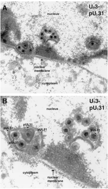
FIG. 6. Digitally scanned image of electron micrographs of US3-null mutant virus-infected cells harvested and fixed late in infection. Thin sections were probed with UL31 protein-specific rabbit polyclonal antisera. Regions labeled with gold beads are marked with arrowheads labeled pUL31. The nucleus, nuclear membrane, and cytoplasm are labeled in panel A, and the nucleus is designated in panel B. Original magnifications: A,⫻30,000; B,⫻45,000.
8948
on November 8, 2019 by guest
http://jvi.asm.org/
FIG. 7. Scanned digital electron micrograph of thin sections of US3-null mutant virus-infected cells incubated with anti-UL34 chicken IgY. Bound antibody is indicated with pUL34-labeled arrowheads, and the nucleus and cytoplasm are labeled. In panel B, thin sections of US3-null HSV-infected cells were incubated with US3 protein-specific rabbit polyclonal antisera. Original magnification of panels A and B,⫻45,000.
8949
on November 8, 2019 by guest
many of these membrane-bound packets of viral particles was continuous with the nuclear membrane and thus appeared to be an invagination of one or both lamellae of the nuclear envelope.
The UL31 gene product was located almost exclusively at the
nuclear membrane, as in wild-type virus-infected cells. How-ever, the appearance of UL31 immunostaining in cells infected
with the US3-null virus differed from that of cells infected with
the wild-type virus inasmuch as (i) the distribution largely localized within packets of viral particles at the perinuclear space and (ii) significantly more UL31 protein-specific
immu-noreactivity was detected in cells infected with the US3-null
virus. Specifically, a mean of approximately 230 gold particles was detected per section, as opposed to a mean of approxi-mately 31 gold particles per section of cells infected with HSV-1(F), as shown in Table 1.
In conclusion, cells infected with the US3-null mutant virus
contain abnormally large numbers of viral particles containing the UL31 and UL34 proteins, which are wrapped in one or
more layers of nuclear membrane. Very similar structures have been reported in cells infected with a US3-null PrV (22). The
discrete, nuclear envelope-associated foci of the colocalized UL31 and UL34 proteins detected by optical sectioning of
US3-null HSV-infected cells described previously (32) likely
correspond to the virion-containing membranous vesicles char-acterized by TEM in this study, given that the sizes and num-bers of vesicles detected by the two assays are comparable. The observation that many of the membranes surrounded several enveloped virions suggests that particles are delayed in their transit from the nucleus to the cytoplasm and ultimately, to the extracellular space. It is worth noting that the US3 kinase does
not appear to be required for budding of nucleocapsids through the INM and also is not required for association of the UL31 and UL34 proteins with nascent virions in the
perinu-clear space. One possibility consistent with these observations is that the US3 protein is necessary for proper regulation of the
deenvelopment of perinuclear virions at the ONM. It is also possible that the Us3-encoded kinase plays a direct or indirect role in facilitating virion transport.
Deletion of the US3 ORF impairs growth of HSV on HEp-2
cells.It has been reported that the US3-encoded kinase is not
essential for growth in tissue culture cells (27). This conclusion was based largely on an experiment in which Vero cells were infected at high and low MOIs with HSV-1(F) and at 48 hpi, the virus yields were determined to be similar for HSV-1(F) and two strains of US3-null HSV. Inasmuch as the US
3-en-coded kinase affects the localization of two essential proteins, we have extended that study by determining the single-step growth characteristics of US3-null HSV in HEp-2 cells.
Repli-cate confluent monolayers of Vero cells in 12-well dishes were infected at an MOI of 5 for 1 h at 4°C with HSV-1(F) or either of two independently isolated US3-null mutant viruses (R7037
and R7039). Each inoculum was then replaced with 37°C V medium (Dulbecco modified Eagle medium supplemented with 5% heat-inactivated calf serum) and incubated at 37°C for 2 h. To remove and inactivate residual virus, infected cells were washed once with 37°C citrate buffer (50 mM sodium citrate, 4 mM KCl [adjusted to pH 3.0 with HCl]) and incubated for 1 min in a second wash of the same buffer. Cells were then washed twice in 37°C V medium and incubated in 2 ml of V
medium for the remainder of the infection. At various times, infected cells were frozen at⫺80°C, subsequently thawed, and then sonicated with a Fisher Sonic Dismembrator at a power level 0 for 30 s to lyse the cells. The infectious virus titer was then determined on Vero cells by plaque assay. The data are shown in Fig. 8.
On HEp-2 cells, HSV-1(F) replication had entered the pro-ductive phase by 6 hpi and reached a plateau phase by 25 hpi, with a titer of approximately 107 PFU/ml. In contrast, the
US3-null virus strains did not initiate production of infectious
virus until after the 6-h time point and reached plateau titers at 25 hpi of only 6⫻105and 8⫻105PFU/ml (R7037 and R7039,
respectively). Moreover, the 48-hpi yield of HSV-1(F) was approximately 3.5 ⫻ 107 PFU/ml, compared with
approxi-mately 1⫻106PFU/ml for both U
S3-null strains. Each of the
US3-null viruses used has deletions that affect either the US2 or
the US4 (glycoprotein G) gene. While we cannot exclude the
possibility that mutations in US2 and US4 independently give
rise to indistinguishable defects in single-step growth, it seems most likely that the observed growth phenotypes of both vi-ruses are the result of their common failure to express US3.
These data indicate that, as in Vero cells, the US3 ORF is
dispensable for growth in HEp-2 cells. However, production of infectious US3-null progeny was slightly delayed and peak
ti-ters were decreased 10- to 30-fold compared with those of HSV-1(F). In the assay performed, a decreased viral yield could reflect a decrease in virus particle production, egress, or infectivity. However, the observation that deletion of US3
de-lays the onset of infectious virus production favors an impair-ment of virus particle production or egress over a simple de-crease in specific infectivity. Deletion of the US3 ORF also
results in an altered nuclear rim distribution of UL31 and UL34
compared to wild-type virus-infected cells (32). It is possible that the altered distribution of the UL34 and UL31 proteins,
[image:12.587.304.534.70.242.2]both of which are involved in viral assembly and egress, results in the growth defect associated with deletion of the US3 locus
FIG. 8. Single-step growth analysis of HSV-1(F) and two HSV US3-null mutant viruses on HEp-2 cells. Replicate cultures of HEp-2 cells were infected with HSV-1(F) or either of two US3-null mutant viruses (R7037 and R7039). At the indicated times, cells were har-vested and viral yield was determined by titration on Vero cells. Virus yield is expressed as PFU per milliliter of culture medium. Each datum point represents the mean of three independent experiments, and the error bars indicate the sample standard deviation.
8950 NOTES J. VIROL.
on November 8, 2019 by guest
http://jvi.asm.org/
in HSV. The increase in the total number of UL31
protein-specific gold beads detected in UL34-null mutant- and US3-null
mutant-infected cells compared with wild-type virus-infected cells shown in Table 1 may be (i) reflective of this proposed delay in the egress of virions or (ii) due to direct or indirect effects of the US3 protein on the UL31 or UL34 protein.
The data reported herein are consistent with reports con-cerning the US3 homologues of other alphaherpesviruses. It
has been proposed that the US3-encoded kinase of PrV is
involved in deenvelopment of perinuclear virions at the ONM (45), and US3 deletion mutants of PrV exhibit an
approxi-mately 10-fold reduction in viral yield in a cell type-dependent manner (20). Similar results have also been reported for US3
deletion mutants of bovine herpesvirus type 1 (42).
We thank Bernard Roizman of the University of Chicago for the UL31 and US3 deletion viruses and the US3 antisera. We are grateful to Jarek Okulicz-Kozaryn (Cornell University), the staff of the Cornell Integrated Microscopy Center, and Jean Ross (Central Microscopy Research Facility, University of Iowa) for technical support and assis-tance. We thank Robert Nurse (Department of Crop and Soil Sci-ences, Cornell University) for assistance with statistical analysis of the data.
These studies were supported by the University of Iowa, Public Health Service awards AI 41478 (R.J.R.) and GM 50740 (J.D.B.), National Research Service award F32 GM20448 (A.E.R.), and training grant AI 07533 to the University of Iowa (B.J.R.).
REFERENCES
1.Baines, J. D., and B. Roizman.1992. The UL11 gene of herpes simplex virus
1 encodes a function that facilitates nucleocapsid envelopment and egress
from cells. J. Virol.66:5168–5174.
2.Blaho, J., C. Mitchell, and B. Roizman.1994. An amino acid sequence shared by the herpes simplex virus 1 alpha regulatory proteins 0, 4, 22, and
27 predicts the nucleotidylylation of the UL21, UL31, UL47, and UL49 gene
products. J. Biol. Chem.269:17401–17410.
3.Browne, H., S. Bell, T. Minson, and D. W. Wilson.1996. An endoplasmic reticulum-retained herpes simplex virus glycoprotein H is absent from
se-creted virions: evidence for reenvelopment during egress. J. Virol.70:4311–
4316.
4.Chang, Y. E., and B. Roizman.1993. The product of the UL31 gene of herpes
simplex virus 1 is a nuclear phosphoprotein which partitions with the nuclear
matrix. J. Virol.67:6348–6356.
5.Chang, Y. E., C. Van Sant, P. W. Krug, A. E. Sears, and B. Roizman.1997.
The null mutant of the UL31 gene of herpes simplex virus 1: construction and
phenotype in infected cells. J. Virol.71:8307–8315.
6.Darlington, R. W., and L. H. Moss III.1968. Herpesvirus envelopment.
J. Virol.2:48–55.
7.Ejercito, P. M., E. D. Kieff, and B. Roizman.1968. Characterization of herpes simplex virus strains differing in their effects on social behaviour of
infected cells. J. Gen. Virol.2:357–364.
8.Enquist, L. W., P. J. Husak, B. W. Banfield, and G. A. Smith.1998. Infection and spread of alphaherpesviruses in the nervous system. Adv. Virus Res.
51:237–347.
9.Frame, M. C., F. C. Purves, D. J. McGeoch, H. S. Marsden, and D. P. Leader.1987. Identification of the herpes simplex virus protein kinase as the
product of the viral gene US3. J. Gen. Virol.68(Pt. 10):2699–2704.
10.Fuchs, W., B. G. Klupp, H. Granzow, N. Osterrieder, and T. C. Mettenleiter.
2002. The interacting UL31 and UL34 gene products of pseudorabies virus are involved in egress from the host-cell nucleus and represent components
of primary enveloped but not mature virions. J. Virol.76:364–378.
11.Gershon, A. A., D. L. Sherman, Z. Zhu, C. A. Gabel, R. T. Ambron, and M. D. Gershon.1994. Intracellular transport of newly synthesized
varicella-zoster virus: final envelopment in the trans-Golgi network. J. Virol.68:6372–
6390.
12.Goshima, F., T. Daikoku, H. Yamada, S. Oshima, T. Tsurumi, and Y. Nishiyama.1998. Subcellular localization of the US3 protein kinase of
her-pes simplex virus type 2. Arch. Virol.143:613–622.
13.Granzow, H., F. Weiland, A. Jons, B. G. Klupp, A. Karger, and T. C. Mettenleiter.1997. Ultrastructural analysis of the replication cycle of
pseu-dorabies virus in cell culture: a reassessment. J. Virol.71:2072–2082.
14.Granzow, H., B. G. Klupp, W. Fuchs, J. Veits, N. Osterrieder, and T. C. Mettenleiter.2001. Egress of alphaherpesviruses: comparative
ultrastruc-tural study. J. Virol.75:3675–3684.
15.Hutchinson, L., and D. C. Johnson.1995. Herpes simplex virus glycoprotein
K promotes egress of virus particles. J. Virol.69:5401–5413.
16.Jayachandra, S., A. Baghian, and K. G. Kousoulas.1997. Herpes simplex virus type 1 glycoprotein K is not essential for infectious virus production in actively replicating cells but is required for efficient envelopment and trans-location of infectious virions from the cytoplasm to the extracellular space.
J. Virol.71:5012–5024.
17.Jerome, K. R., R. Fox, Z. Chen, A. E. Sears, H.-Y. Lee, and L. Corey.1999. Herpes simplex virus inhibits apoptosis through the action of two genes, Us5
and Us3. J. Virol.73:8950–8957.
18.Johnson, D. C., and P. G. Spear.1982. Monensin inhibits the processing of herpes simplex virus glycoproteins, their transport to the cell surface, and the
egress of virions from infected cells. J. Virol.43:1102–1112.
19.Johnson, D. C., M. C. Frame, M. W. Ligas, A. M. Cross, and N. D. Stow.
1988. Herpes simplex virus immunoglobulin G Fc receptor activity depends
on a complex of two viral glycoproteins, gE and gI. J. Virol.62:1347–1354.
20.Kimman, T. G., N. De Wind, T. De Bruin, Y. de Visser, and J. Voermans.
1994. Inactivation of glycoprotein gE and thymidine kinase or the US3-encoded protein kinase synergistically decrease in vivo replication of
pseu-dorabies virus and the induction of protective immunity. Virology205:511–
518.
21.Klupp, B. G., H. Granzow, and T. C. Mettenleiter.2000. Primary envelop-ment of pseudorabies virus at the nuclear membrane requires the UL34 gene
product. J. Virol.74:10063–10073.
22.Klupp, B. G., H. Granzow, and T. C. Mettenleiter. 2001. Effect of the pseudorabies virus US3 protein on nuclear membrane localization of the
UL34 protein and virus egress from the nucleus. J. Gen. Virol.82(Pt. 10):
2363–2371.
23.Leopardi, R., C. Van Sant, and B. Roizman.1997. The herpes simplex virus 1 protein kinase US3 is required for protection from apoptosis induced by
the virus. Proc. Natl. Acad. Sci. USA94:7891–7896.
24.Loomis, J. S., J. B. Bowzard, R. J. Courtney, and J. W. Wills.2001. Intra-cellular trafficking of the UL11 tegument protein of herpes simplex virus type
1. J. Virol.75:12209–12219.
25.Mettenleiter, T. C.2002. Herpesvirus assembly and egress. J. Virol.76:1537– 1547.
26.Munger, J., A. V. Chee, and B. Roizman.2001. The US3 protein kinase
blocks apoptosis induced by thed120 mutant of herpes simplex virus 1 at a
premitochondrial stage. J. Virol.75:5491–5497.
27.Purves, F. C., R. M. Longnecker, D. P. Leader, and B. Roizman.1987. Herpes simplex virus 1 protein kinase is encoded by open reading frame US3
which is not essential for virus growth in cell culture. J. Virol.61:2896–2901.
28.Purves, F. C., D. Spector, and B. Roizman.1991. The herpes simplex virus 1
protein kinase encoded by the US3 gene mediates posttranslational
modifi-cation of the phosphoprotein encoded by the UL34 gene. J. Virol.65:5757–
5764.
29.Purves, F. C., D. Spector, and B. Roizman.1992. UL34, the target of the
herpes simplex virus US3 protein kinase, is a membrane protein which in its
unphosphorylated state associates with novel phosphoproteins. J. Virol.66:
4295–4303.
30.Radsak, K., M. Eickmann, T. Mockenhaupt, E. Bogner, H. Kern, A. Eis-Hubinger, and M. Reschke.1996. Retrieval of human cytomegalovirus gly-coprotein B from the infected cell surface for virus envelopment. Arch.
Virol.141:557–572.
31.Reynolds, A. E., Y. Fan, and J. D. Baines.2000. Characterization of the UL33
gene product of herpes simplex virus 1. Virology266:310–318.
32.Reynolds, A. E., B. J. Ryckman, J. D. Baines, Y. Zhou, L. Liang, and R. J. Roller.2001. UL31 and UL34 proteins of herpes simplex virus type 1 form a
complex that accumulates at the nuclear rim and is required for envelopment
of nucleocapsids. J. Virol.75:8803–8817.
33.Roizman, B., and A. E. Sears.1996. Herpes simplex viruses and their
repli-cation, p. 2221–2278.InB. N. Fields, D. M. Knipe, and P. M. Howley (ed.),
Fields virology, 3rd ed. Lippincott-Raven, Philadelphia, Pa.
34.Roller, R. J., Y. Zhou, R. Schnetzer, J. Ferguson, and D. DeSalvo.2000.
Herpes simplex virus type 1 UL34 gene product is required for viral
envel-opment. J. Virol.74:117–129.
35.Schwartz, J., and B. Roizman.1969. Concerning the egress of herpes simplex virus from infected cells: electron and light microscope observations.
Virol-ogy38:42–49.
36.Shiba, C., T. Daikoku, F. Goshima, H. Takakuwa, Y. Yamauchi, O. Koiwai, and Y. Nishiyama.2000. The UL34 gene product of herpes simplex virus type 2 is a tail-anchored type II membrane protein that is significant for virus
envelopment. J. Gen. Virol.81(Pt. 10):2397–2405.
37.Skepper, J. N., A. Whiteley, H. Browne, and A. Minson.2001. Herpes simplex
virus nucleocapsids mature to progeny virions by an envelopment3
deen-velopment3reenvelopment pathway. J. Virol.75:5697–5702.
38.Smith, S., and G. Blobel.1993. The first membrane spanning region of the lamin B receptor is sufficient for sorting to the inner nuclear membrane.
J. Cell Biol.120:631–637.
39.Soullam, B., and H. J. Worman.1993. The amino-terminal domain of the
lamin B receptor is a nuclear envelope targeting signal. J. Cell Biol.120:
1093–1100.
on November 8, 2019 by guest
http://jvi.asm.org/
40.Soullam, B., and H. J. Worman.1995. Signals and structural features in-volved in integral membrane protein targeting to the inner nuclear
mem-brane. J. Cell Biol.130:15–27.
41.Stackpole, C. W., and M. Mizell.1968. Electron microscopic observations on herpes-type virus-related structures in the frog renal adenocarcinoma.
Vi-rology36:63–72.
42.Takashima, Y., H. Tamura, X. Xuan, and H. Otsuka.1999. Identification of the US3 gene product of BHV-1 as a protein kinase and characterization of
BHV-1 mutants of the US3 gene. Virus Res.59:23–34.
43.Taus, N. S., B. Salmon, and J. D. Baines.1998. The herpes simplex virus 1
UL17 gene is required for localization of capsids and major and minor capsid
proteins to intranuclear sites where viral DNA is cleaved and packaged.
Virology252:115–125.
44.Torrisi, M. R., C. Di Lazzaro, A. Pavan, L. Pereira, and G. Campadelli-Fiume.1992. Herpes simplex virus envelopment and maturation studied by
fracture label. J. Virol.66:554–561.
45.Wagenaar, F., J. M. Pol, B. Peeters, A. L. Gielkens, N. de Wind, and T. G. Kimman.1995. The US3-encoded protein kinase from pseudorabies virus
affects egress of virions from the nucleus. J. Gen. Virol.76(Pt. 7):1851–1859.
46.Whealy, M. E., J. P. Card, R. P. Meade, A. K. Robbins, and L. W. Enquist.
1991. Effect of brefeldin A on alphaherpesvirus membrane protein
glycosy-lation and virus egress. J. Virol.65:1066–1081.
47.Whiteley, A., B. Bruun, T. Minson, and H. Browne.1999. Effects of targeting herpes simplex virus type 1 gD to the endoplasmic reticulum and trans-Golgi
network. J. Virol.73:9515–9520.
48.Yamauchi, Y., C. Shiba, F. Goshima, A. Nawa, T. Murata, and Y. Nishiyama.
2001. Herpes simplex virus type 2 UL34 protein requires UL31 protein for its relocation to the internal nuclear membrane in transfected cells. J. Gen.
Virol.82(Pt.6):1423–1428.
49.Ye, G. J., and B. Roizman.2000. The essential protein encoded by the UL31 gene of herpes simplex virus 1 depends for its stability on the presence of
UL34 protein. Proc. Natl. Acad. Sci. USA97:11002–11007.
50.Zhu, Z., M. D. Gershon, Y. Hao, R. T. Ambron, C. A. Gabel, and A. A. Gershon.1995. Envelopment of varicella-zoster virus: targeting of viral
gly-coproteins to the trans-Golgi network. J. Virol.69:7951–7959.
51.Zhu, H. Y., H. Yamada, Y. M. Jiang, M. Yamada, and Y. Nishiyama.1999. Intracellular localization of the UL31 protein of herpes simplex virus type 2.
Arch. Virol.144:1923–1935.
8952 NOTES J. VIROL.