A DISSERTATION ON
CLINICO - EPIDEMIOLOGICAL PROFILE, CD4
CORRELATION AND OUTCOME OF PAEDIATRIC HIV
M.D (BRANCH VII)
PAEDIATRIC MEDICINE
MARCH 2009
THE TAMILNADU DR.MGR.MEDICAL
UNIVERSITY
TAMILNADU
CHENNAI
CERTIFICATE
This is to certify that the dissertation titled “Clinico epidemiological profile,
CD4 correlation and outcome of paediatric HIV” submitted by Dr.M.Vaideeswaran
to the Faculty of pediatrics,The Tamilnadu M.G.R.Medical University,Chennai in
partial fulfillment of the requirement for the award of M.D.Degree (Pediatrics) is a
bonafide research work carried out by him under our direct supervision and guidance.
Dr.P.Amutha Rajeshwari,M.D.,DCH.,
Professor and Head of the Department,
Institute of Child Health & Research Centre, Madurai Medical College,
DECLARATION
I Dr.M.Vaideeswaran, solemnly declare that the dissertation titled “Clinico
epidemiological profile, CD4 correlation and outcome of paediatric HIV” has been
prepared by me.
This is submitted to the Tamilnadu Dr.M.G.R.Medical University, Chennai in
partial fulfillment of the rules and regulations for the M.D.Degree Examination in
Paediatrics.
Place: Madurai
Date: Dr.M. VAIDEESWARAN
ACKNOWLEDGEMENT
I am greatly indebted to my teacher, Prof. Dr.P.Amutha Rajeswari, Professor
and Head of the Department of Pediatrics, for her supervision, guidance and
encouragement while undertaking this study.
I would like to thank Prof. Dr.M.L.Vasanthakumari who guided me to a
great extent for her valuable suggestions and guidance.
I am also extremely grateful to my unit chief Prof. Dr.Chitra Ayyappan, for
the guidance which has helped me a lot in completing the work successfully.
I also would like to thank Prof. Dr. G. Mathevan, for his guidance and
suggestions.
I also thank all the members of the Dissertation committee for their valuable
suggestions. I gratefully acknowledge the help and guidance received from Asst.Prof.
Dr. M. Kulandaivel, Asst. Prof. Dr.M. Balasubramanian, Asst.Prof. Dr. Nataraja
rathinam at every stage of this study.
I am indebted to Prof. Dr.Uma, Head of the Department, Microbiology for her
support.
I thank the Dean and the members of the Ethical committee, Government Rajaji
hospital, Madurai for permitting me to perform this study.
Professor of Medicine, GRH, Madurai, for his valuable suggestions and guidance.
I would like to thank Mr. R. Balasubramanian, Statistician, Council of
Medical Research and Entomology who helped me a lot.
I also express my gratitude to all my fellow postgraduates for their kind
cooperation in carrying out this study and for their critical analysis.
Last but not the least, I am indebted to all the children enrolled in this study and
their parents without whose cooperation, this study would not have been possible.
CONTENTS
S.NO. TOPIC PAGE NO.
1. INTRODUCTION 1
2. REVIEW OF LITERATURE 4
3. AIM OF THE STUDY 23
4. MATERIALS AND METHODS 24
5. RESULTS 37
6. DISCUSSION 49
7. CONCLUSIONS 61
8. LIMITATIONS 62
9. RECOMMENDATIONS 63
INTRODUCTION
Acquired immunodeficiency syndrome is caused by infection with human
immunodeficiency virus type 1 and rarely type 2. The HIV pandemic has spread word
wide since 1981.The first paediatric case of AIDS was reported to the centre for disease
control and prevention in November 1982. Since then HIV infection in children
represents a major setback to child health all over the world
Statement of problem and need of the study:
India harbours world’s second highest number of HIV infected people (1). HIV
infection is increasingly becoming a prominent cause of childhood morbidity and
mortality in India. Presently, 2, 02,000 children are living with HIV/AIDS in India.
50% of these die within 2 years, constituting about 18% of 3.1 million AIDS deaths
every year (2). Despite the magnitude of the problem, there is paucity of data on
various issues in paediatric HIV infection from India. In spite of that it is now well
known that the epidemiological and clinical features differ greatly from country to
country. The epidemiological features depend upon the social and cultural practices of
those people which may again vary from region to region. Opportunistic infections
(OIs) are an important cause of morbidity and mortality in children infected with HIV.
organisms and parasites endemic in that country. However, few data are available
regarding the overall prevalence, incidence and immunologic correlates associated with
these diseases in the paediatric HIV population. We undertook this study to identify the
epidemiological, clinical background and its correlation with CD4 values and the
NATURAL HISTORY:
Perinatal transmission is the main mode of acquisition of HIV infection in
children and also via blood products and rarely homosexual or heterosexual in
adolescents. A bimodal distribution of disease progression is seen in vertically
transmitted HIV infection in children. Early onset of symptoms at younger than 12
months of age with rapidly progressive disease and high mortality is observed in 10-25
percent of infected infants. The majority of children with vertical transmission appear to
have later onset symptoms and a better prognosis, with median time to development of
AIDS of more than 5 years and survival of 6-9 years. Early age of disease onset is a
REVIEW OF LITERATURE
Most studies about paediatric HIV infection came from the developed countries.
No such large scale information is available in India. HIV infection being a global
emergency and spreading like a pandemic, the number of children affected with HIV
infection is rapidly increasing. Most of the studies related to clinical profile of HIV
came in the pre –ART era.
Highly active anti-retroviral therapy (HAART) has changed the face of
HIV/AIDS by leading to a dramatic decrease in HIV-related morbidity and mortality
among those with access to therapy (3). Until recently, HAART was not accessible to a
vast majority of the 5.1 million Indians living with HIV in India (4) primarily due to its
high cost. There was a significant decrease in the number of incident opportunistic
infections, especially tuberculosis, in patients on HAART.
The advent of potent antiretroviral drugs has enabled transformation of human
immunodeficiency virus (HIV) infection from a fatal to a chronic disease in developed
countries (5). However supportive care, appropriate prophylaxis and management of
infections are equally important. They are, in fact, the mainstay of therapy in countries
such as ours where antiretroviral therapy is ill affordable by most patients (6). The
introduction of HAART become more available, descriptions of treated disease will
afflicted by wasting, malnutrition, and chronic diarrhoeal disease. Appropriate
immunizations, prophylaxis with cotrimoxazole, and preventative measures such as
boiling water must be taken in order to avoid morbidity and mortality in children. Little
is known about the natural history of HIV disease in Indian children, and further
research is needed.
The proposed study evaluates the clinical profile including opportunistic infections in
relation to CD4 and determines the response to antiretroviral therapy.
Initial evaluation in HIV infected children includes detailed history to ascertain
the mode of transmission, symptoms related to HIV and HIV status of the family
members. The socioeconomic status and level of disease awareness in the caretakers
should also be enquired into. A detailed physical examination including anthropometry,
pulse oximetry, fundoscopy and neurodevelopment assessment should be carried out.
Subsequently if feasible, the immune status and viral burden should be
determined by estimation of CD4 counts and HIV RNA levels. Other laboratory tests
include a complete hemogram including platelet count, liver and renal function tests,
and X ray film of the chest, tuberculin test, stool examination, urine analysis and ECG.
Immunoglobulin levels, echocardiogram, computerized tomography (CT) of the chest
After clinical and immune categorization of the illness, treatment details are
PRE ART EVALUATION AND FOLLOW UP
S.No. Evaluation Interval
1. Complete history and physical examination
3 months
2. Review of systems 3 months
3. Development examination
< 1 year age
1-3 years
> 3 years
3 months
6 months
Annually
4. Chest radiography Annually
5. Laboratory tests
Complete blood counts
CD4 / CD8
LFT RFT Urine analysis 3 months 3 months 3 months
Baseline and as indicated
Annually
6. Mantoux test Annually
7. Referrals
Ophthalmology
Cardiology
Annually
Antiretroviral therapy:
Antiretrovirals can be classified as nucleoside reverse transcriptase inhibitors
(NRTIs), non-nucleoside reverse transcriptase inhibitors (NNRTIs) and protease
inhibitors (PIs). As many as 13 antiretroviral agents are currently FDA approved for
treatment of HIV infection (5). NRTIs were the first drugs to be used and are the least
expensive. NNRTIs are increasingly being evaluated in pediatric treatment protocols.
They are not associated with significant interactions with food; need to be taken once or
twice daily and freely cross the blood brain barrier. Protease inhibitors are very
efficacious but expense and side effects are major limiting factors. Details of commonly
used drugs are listed in Table 1. Certain new drugs including NRTIs such as Abacavir,
COMMONLY USED ANTIRETROVIRAL IN CHILDREN
Drug Dose Side effects Trade name Practical points
NRTIs Zidovudine (AZT, ZDV) Neonates-2 mg/kg 6 hourly children 90-180
mg/m2 6-8 hrly Anemia, myopathy Zidovir soln. 50mg/ml 100mg capsules, 300 mg tablets Good CNS penetration
Decrease dose if there is bone marrow suppression
Didanosine (ddI)
90mg/m3 12 hrly
neuropathy, pancreatitis,
abd. pain ,
diarrhea
Administer empty stomach, space administration with protease inhibitors
Lamivudine
(3TC) 4 mg /kg 12 hrly Pancreatitis, neuropathy, neutropenia
Lamivir soln 50 mg / 5 ml Tablets 150 mg
Prevents emergence
of zidovudine
resistance
Stavudine (d4T)
1mg / kg 12 hrly
Headache,GI upset, neuropathy
Stavir
capsule 30 mg, 40 mg
Should not be combined with AZT
NNRTIs
Nevirapine
(NVP) 120-200 mg/m3 12 hrly Skinstevenjohnson rash, syndrome
Nevimune
200 mg
tablets
Start with 120 mg/m2 for 2 wks and then increase to full dose if no side effect
Protease Inhibitors
Ritonavir 400mg/m2/12
hrly Badtaste,vomiting, nausea, diarrhea Norvir 80 mg / ml, capsules 100 mg
Start with 250 mg/m2 and then increase give with meals
Nelfinavir 20-30 mg / kg 8 hrly
Diarrhoea, abdominal pain
Give with meals
Indinavir 50mg/m2 8
hrly Nephrolithiasis Empty stomach, adequate hydration Saquinavir 50 mg / kg /
hrly
Diarrhoeal headache
capsules
Antiretroviral therapy provides significant clinical benefit in HIV infected
children with immunologic or clinical symptoms of HIV infection with substantial
improvements in neurodevelopment, growth, immunologic and/or virologic status (7).
The efficacy of antiretroviral therapy in asymptomatic children has not been
demonstrated. However given the pathophysiologic sequence of events in HIV
infection, early aggressive therapy may help in preservation of the immune system and
minimization of the risk of antiviral resistance. Moreover it has been found that infants
and children have higher levels of plasma virus and more rapid disease progression as
compared to adults (5).
Therefore the Working Group on Antiretroviral Therapy and Medical Management
of HIV-Infected Children has proposed the following indications for initiation of
antiretroviral therapy (8).
• Clinical symptoms associated with HIV infection (Categories A, B, C) • Evidence of immune suppression (Immune category 2 or 3)
• For asymptomatic children > 1 year with normal immune status the preferred approach is to offer therapy to all. Alternatively treatment may be deferred and
the virologic, immunologic and clinical status monitored. Therapy is initiated in
the following circumstances.
• High or increasing HIV RNA copy number.
• Rapidly declining CD4 count/CD4 % approaching immune category 2. • Development of clinical symptoms.
To simplify, the recent consensus is to use antiretrovirals in all HIV infected children
irrespective of clinical, immunologic and virologic status (5).
Regimen of anti retroviral drugs :
• Preferred regimen :two NRTI plus one PI
• Recommended dual NRTI combinations: ZDV+ddI, ZDV+3TC, d4T+ddI,
d4T+3TC,
• Protease inhibitors: RTV;NFV; IDV;SQV
• Alternate regimen:NVP+2NRTI
• Secondary Alternate regimen:2NRTI
As in adults, the combination of two NRTIs and one protease inhibitor is the
preferred regime and comprises what is known as Highly Active Antiretroviral Therapy
(HAART). Recommended NRTI combinations are zidovudine (ZDV) with didanosine
(ddI) or ZDV and lamivudine (3TC). Limited data are available for combinations of
stavudine (d4T) and ddI, d4T and 3TC, and ZDV and zalcitabine (ddC). Combinations
of d4T with ZDV and ddC with ddI/d4T/3TC should be avoided. The protease
inhibitors currently approved for infants and children are nelfinavir or ritonavir and
indinavir for those who can swallow capsules (8).
Nevirapine with two NRTIs is an alternative and cheaper regime. The
combination of only two NRTIs though of limited benefit can be offered when cost,
non-availability or toxicity precludes the use of protease inhibitors (8). This
combination results in significant clinical improvement, virologic and immunologic
recovery and improves the quality of life although for a short time (7). The risk of
resistance with such therapy in children is of a lesser concern, as they in usual
circumstances do not pass on the resistant strains to others.
Monotherapy with any drug should be employed only for prevention of perinatal
Monitoring and follow up:
On follow up the child should be evaluated for clinical improvement, compliance,
adverse drug reactions as well as for rise in the CD4 counts and suppression of HIV
RNA levels (Table 3). CD4 counts are estimated every three months. Plasma RNA
determinations 4 weeks after initiating therapy and then 3 monthly are advocated. With
a combination of two NRTIs a five-fold reduction of HIV RNA levels by 8-12 weeks is
expected (8). With the standard regime of 2 NRTIs and a protease inhibitor, a ten-fold
reduction is expected by 8-12 weeks and levels should be undetectable by 4-6 months
(8). However since perinatally infected children have high baseline RNA levels,
complete suppression is often not achievable unlike adults (5). In case of lack of
optimal response, compliance, dosing schedule and possible drug interactions should be
reviewed. If these are satisfactory and the lack of response is persistent, antiretroviral
resistance should be suspected. Molecular techniques to identify mutations for the
confirmation of resistance to antiretroviral drugs are under evaluation.
Madhivanan et al (9) and Merchant (10) and his colleagues in their studies
conducted in India in 2001 reported that, Vertical transmission is responsible for
between 67 and 87 per cent of paediatric HIV infection, with the majority of the
The clinical features of HIV infection in children are different from those in
adults. Perinatally infected children become symptomatic by five years of age. Failure
to thrive is the most common clinical condition associated with HIV infection in
children(9,10) Pulmonary and extrapulmonary tuberculosis was consistently the most
frequent opportunistic infection reported in two major studies in Indian children with
HIV. (9, 10)
Opportunistic infections (OIs) are an important cause of morbidity and mortality
in children infected with HIV. However, few data are available regarding the overall
prevalence, incidence and immunologic correlates associated with these diseases in the
paediatric HIV population.
CD4 T-lymphocyte is the immune system cell that HIV infects and destroys, and
the CD4 count roughly reflects the state of the immune system. Low CD4 T-cell counts
are considered to be a marker of the progression of HIV infection and AIDS, and have
been called the 'signature' of HIV (Balter 1997). Since HIV was first claimed to be the
cause of AIDS in 1984, the CD4 count has been widely used to make treatment and
diagnostic decisions, but the use of the CD4 count has been controversial, and
recommendations regarding how to use them have changed many times over the years.
CD4 estimation is the backbone of AIDS control program in developing nations. It has
risk of developing opportunistic infections.HIV weakens the immune system so that
opportunistic infections develop. Chakravarty et al, 2006 Indian study,(11)has showed
children with opportunistic infection have lower CD4 values as compared to children
without opportunistic infection . Ylitalo et al (12) h as also proved the same.
Chakravarthy et al., 2006 Indian study (11) has showed CD4 percentage declining with
progression in the WHO clinical stages of HIV infection.
The progression of disease is related to gradual disruption of lymph node
architecture leading to high levels of viremia and disappearance of CD4 cells
during later stages of disease. However, further studies of CD4 counts in relation
to antiretroviral therapy in children needs to be done.
Fox- Wheeler et al (13) in his studies has showed ,HIV is associated with malnutrition.
Similar observation has been reported in other studies as well, Shah and Bachou,
Uganda, 2006. (14,15 )
Agarwal, et al (16) in his studies conducted in Banaras Hindu university in 2005
has showed, CD4% correlated significantly with the deterioration of the WHO clinical
stages and increasing grades of protein energy malnutrition .
Protein energy malnutrition leads to depletion of CD4 counts, and this is
perhaps exacerbated by the presence of HIV infection. Protein energy
Anniek et al (17) in his studies compared paediatric HIV in African settings to
paediatric HIV in Western settings and to adult HIV in African settings. Several
differences in diagnostic, clinical, immunological and virological characteristics were
identified, as well as variations in the most influential factors for disease progression
and response to ART. Environmental factors may influence disease progression,
mortality, loss to follow-up, adherence and the need to adapt the regimen. The selected
African paediatric programs recorded a higher increase in median CD4-percent than the
selected Western paediatric programs and a higher increase in CD4-count than the
selected African adult programs. Compared to the adult programs, the African
paediatric programs had lower drop-out rates, higher reported adherence levels and
comparable mortality rates. The Western paediatric programs, however, had the lowest
mortality rates.
Pulmonary and extra pulmonary tuberculosis was consistently the most frequent
opportunistic infection reported in two major studies in Indian children with HIV.
Although frequencies differed, oral candidiasis, hepatosplenomegaly, recurrent
respiratory tract infection, Pneumocystis carinii pneumonia, chronic lung disease,
persistent generalized lymphadenopathy, chronic diarrhoea, pyrexia of unknown origin,
chronic hypertrophic parotitis, chronic otorrhoea, bacterial skin infection, and PPE
have also been reported in these studies.
Western cohorts where it is the most common AIDS diagnosis in infancy (18).
Pneumocystis jerovecii causes severe pneumonia in patients with AIDS. Occurrence of
PCP establishes the diagnosis of AIDS (19) and it is the most common AIDS-defining
illness in the developed world. In India, however, very low rates (0.7 to 7%) 4, of PCP
have been reported. Some reasons for this could be the predominance of other
pulmonary diseases like TB, and due to under diagnosis of incident cases.. According to
a large natural history study, Indian patients with PCP were 4.5 times more likely to die
than patients without PCP. Median survival after diagnosis of PCP in this study was 24
months.
Bacterial pneumonia was reported as an opportunistic infection in 1.8 per cent of a
large southern Indian cohort of HIV-positive patients. Similar to HIV negative individuals,
the most common causes of acute community acquired pneumonia, are encapsulated
bacteria, Streptococcus pneumonia and Haemophilus influenzae. Rates of bacterial
pneumonia can be up to 25-fold higher among HIV infected children than in the general
community. Although bacterial pneumonias can occur relatively early in the course of
Oral candidiasis occurs frequently in children with HIV infection; it has been
reported as one of the most common HIV-associated condition, occurring in up to 70 per
cent of cases. The pseudo membranous “white patches” variant of candidiasis is associated
with more severe immunosuppression than the erythematous, hyperplastic or angular
chelitis types.
Cryptococcal meningitis (CM) has been reported as the most common
opportunistic infection of the CNS of Indian children with HIV. Poor prognostic factors
for CM include positive blood cultures, altered mental status, CSF antigen titre above
1:1024, positive CSF India ink smear, CSF white cell count below 20cells/cu.mm, and
elevated CSF pressures. Gold standard diagnosis of CM requires demonstration of
organism in CSF.
TB meningitis in HIV children is different from HIV negative. Cognitive
dysfunction is more common, and pathological features demonstrate reduced and atypical
inflammatory responses, and extensive vasculopathy. There is absence of minimal
meningeal enhancement and absence of communicating hydrocephalus on computed
tomography (CT) scan in HIV-positive patients. As expected, mortality is higher in the
Chronic diarrhoea is a major problem in HIV infected children, affecting up to 76
per cent of those with AIDS. It is associated with a 3.3 fold increased risk of disease
progression. In reports from north, south and east India, Isospora belli and
Cryptosporidium parvum were the two most common causes of chronic diarrhoeal disease
in HIV infected children. Blastocystis hominis, Strongyloides stercoralis, Entamoeba
histolytica, Giardia lamblia, enteropathogenic Escherichia coli, Enterocytozoon bieneusi
and Campylobacter jejuni are other causative agents of diarrhoea in Indian patients with
HIV. There was no geographic pattern to the frequency of organisms.
A variety of ocular conditions associated with AIDS in India have been reported:
extensive blepharitis and spontaneous lid ulcer, extensive molluscum contagiosum CMV
retinitis, herpes simplex keratitis, bilateral papilloedema with cryptococcal meningitis. The
most common ophthalmic opportunistic infection in India is CMV retinitis, which almost
always occurs in patients with CD4 counts <50 cells.
With the availability of antiretroviral therapy at lower cost, the clinical profile of
HIV disease is now changing to include drug-related toxicities and immune reconstitution
syndrome.
Immune reconstitution is of increasing concern in the developing world as
HAART becomes more available in settings where opportunistic infections, especially
of the patient despite treatment. This could indicate a successful, though undesirable,
effect of HAART, or instead, treatment failure and subsequent progression of the
AIM OF THE STUDY
1. To study the clinical profile of pediatric HIV.
2. To correlate the clinical profile with CD4 values.
MATERIALS & METHODS
Setting
Study was conducted in Institute of child health and research centre-Government Rajaji hospital, Madurai.
Collaboration Departments
The study was done in collaboration with ART centre, Department of Microbiology, Department of Pathology , GRH,Madurai.
Ethical committee
Approval for the study was obtained from The Ethical committee of Govt. Rajaji Hospital.
Study design
Prospective cross sectional analytical study
Study period
Sample size
A total of 355 children which included all children enrolled in ART centre, Government Rajaji Hospital, Madurai.
Inclusion criteria:
1.
Newly diagnosed HIV positive children >18 months up to 15Years.
2. Children < 18 months with positive DNA PCR
Exclusion criteria:
1. Children previously on ART
The study was conducted prospectively from June 2006 to July 2008 (26 months)
and included children (aged 1 month to 15 years) admitted consecutively to the
pediatric wards with the diagnosis of HIV infection. Children were tested for HIV if
they had one or more of the following manifestations: prolonged unexplained fever,
chronic diarrhea, generalized lymphadenopathy, recurrent systemic infections,
for the HIV testing with appropriate pre and post-test counseling. ELISA testing for
HIV antibodies was performed for establishing the diagnosis. If detected to be positive,
confirmation was done by two more ELISA tests. Children less than 18 months of age
underwent confirmation by DNA-PCR testing. HIV status of the parents and siblings of
the affected children was also requested (after appropriate counseling).
Definitions of Clinical factors studied :
Asymptomatic :
No HIV related symptoms reported and no signs on examination.
Persistent generalized lymphadenopathy (PGL) :
Swollen or enlarged lymph nodes > 1 cm at two or more non contiguous sites, without known cause.
Unexplained persistent hepatosplenomegaly :
Enlarged liver and spleen without obvious cause
Papular pruritic eruptions :
Fungal nail infections :
Fungal paronychia (painful, red and swollen nail bed) or onycholysis (painless separation of the nail from the nail bed). Proximal white subungual onchomycosis is uncommon without immunodeficiency.
Angular cheilitis :
Splits or cracks on lips at the angle of the mouth with depigmentation usually responding to antifungal treatment but may occur.
Lineal Gingival Erythema : (LGE)
Erythematous band that follows the contour of the free gingival line ; may be associated with spontaneous bleeding.
Extensive wart virus infection :
Characteristic warty skin lesions ; small fleshy grainy bumps, often rough, flat on sole of feet (plantar warts), facial, more than 5% of body area or disfiguring.
Extensive molluscum contagiosum infection :
disfiguring.
Recurrent oral ulcerations (two or more in six months) :
Aphthous ulceration, typically with a halo of inflammatio and yellow grey pseudo membrane.
Unexplained parotid enlargement :
Asymptomatic bilateral swelling that may spontaneously resolve and recur, in absence of other known cause, usually painless.
Herpes zoster :
Painful rash with fluid filled blisters, dermatomal distribution, can be haemorrhagic on erythematous background, and can become large and confluent. Does not cross the midline.
Recurrent upper respiratory tract infection (URTI) :
pain and nasal discharge (sinusitis) or painful swollen eardrum (ortitis media), sore throat with productive cough (bronchitis) sore throat (pharyngitis) and barking croup like cough (LTB). Persistent or recurrent ear discharge.
Unexplained moderate malnutrition :
Weight loss : low weight for age, up to 2 standard deviations (SDs), not explained by poor or inadequate feeding and or other infections and not adequately responding to standard management.
Confirmed by documented loss of body weight of -2 SD, failure to gain weight on standard management and no other cause identified during investigation.
Unexplained persistent diarrhoea :
Unexplained persistent (14 days or more) diarrhoea (loose or watery stool, three or more times daily), not responding to standard treatment). Confirmed by stools observed and documented as unformed. Culture and microscopy reveal no pathogens.
Unexplained persistent fever (intermittent or constant, for longer than one month)
obvious foci of disease reported or found on examination. Malaria must be excluded in malarious areas. Confirmed by documented fever of > 37.5 C with negative blood culture, negative malaria slide and normal or unchanged CXR, and no other obvious foci of disease.
Oral Candida : (outside first 6-8 weeks of life)
Persistent or recurring creamy white to yellow soft small plaques which can be scraped off (pseudomembranous), or red patches on tongue, palate or lining of mouth, usually painful or tender (erythmatous form).Confirmed by microscopy or culture.
Oral hairy leukoplakia :
Fine small linear patches on lateral borders of tongue, generally bilaterally, which do not scrape off.
Lymph node TB :
Non acute, painless “cold” enlargement of lymphnodes, usually matted, localized to one region. May have draining sinuses. Response to standard anti-TB treatment in one month. Confirmed by histology or fine needle aspirate for Ziehl Neelsen stain/Culture.
Nonspecific symptoms, eg. Chronic cough, fever, night sweats, anorexia and weight loss. In the older child also productive cough and haemoptysis. Abnormal CXR. Response to standard anti – TB treatment in one month. Confirmed by positive sputum smear or culture.
Severe recurrent presumed bacterial pneumonia :
Cough with fast breathing, chest indrawing, nasal flaring, wheezing and grunting. Crackles or consolidation on auscultation. Responds to course of antibiotics. Current episode plus one or more in previous 6 months.
Confirmed by isolation of bacteria from appropriate clinical specimens (induced sputum, BAL, lung aspirate).
Acute necrotizing ulcerative gingivitis or stomatitis, or acute necrotizing ulcerative periodonitis.
Severe pain, ulcerated gingival papillae, loosenig of teeth, spontaneous bleeding, bad odour, and rapid loss of bone and or soft tissue.
pulmonary infiltrates present for more than two months with no response to antibiotic treatment and no other pathogen found. Oxygen saturation persistently < 90%. May present with corpulmonale and may have increased exercise induced fatigue. Characteristic histology.
Chronic HIV associated lung diseases (including bronchiectasis)
History of cough productive of copious amounts of purulent sputum (bronchiectasis only), with or without clubbing, halitosis, and crepitations and or wheezes on auscultation.
Confirmed by CXR may show honeycomb appearance (small cysts) and / or persistent areas of opacification and / or widespread lung destruction, with fibrosis and loss of volume.
Unexplained anaemia (<8g/dl) or neutropenia (<1000/mm3) or chronic thrombocytopenia (< 50000/mm3):No presumptive diagnosis. Diagnosed on laboratory testing, not explained by other non HIV conditions, or not responding to standard therapy with haematinics, antimalarials or antihelminthics as outlined in MCI.
Unexplained severe wasting, stunting or severe malnutrition (not adequately responding to standard therapy.)
feet, and or weight for height of 3 SDs as defined by WHO IMCI guidelines.
Confirmed by documented weight loss of > -3 SD +/- oedema
Pneumocystis pneumonia PCP :
Dry cough, progressive difficulty in breathing, cyanosis, tachypnoea and fever ; chest indrawing or stridor. (Severe or very severe pneumonia as in IMCI). Usually of rapid onset especially in inflants under six months of age. Response to high dose co-trimoxazole +/- prednisolone. Confirmed by : CXR typical bilateral perihilar diffuse infiltrates ; microscopy of induced sputum or BAL or NPA, or histology of lung tissue.
Recurrent severe presumed bacterial infection, eg. Empyema, pyomyositis bone or joint infection, meningitis but excluding pneumonia. Fever accompanied by specific symptoms of signs that localize infection. Responds to antibiotics. Current episode plus one or more in previous 6 months. Confirmed by culture of appropriate clinical specimen.
Oesophageal candidiasis (or candidiasis of trachea, bronchi or lungs). Chest pain and dysphagia (difficulty in swallowing), odynophagia (pain on swallowing food and fluids) or retrosternal pain worse on swallowing (food and fluids) responds to specific treatment. In young children, suspect particularly if oral candida observed and food refusal occurs and / or difficulties / crying when feeding. Confirmed by macroscopic appearance at endoscopy, microscopy of specimen from tissue or macroscopic appearance at bronchoscopy or histology.
Extrapulmonary / disseminated TB :
Systemic illness usually with prolonged fever, night sweats, weight loss. Clinical features of organs involved, eg. Sterile pyuria, pericarditis, ascites, pleural effusion, meningitis, arthritis, orchitis. Respods to standard anti TB therapy. Confirmed by positive microscopy showing AFB or culture of Mycobacterium tuberculosis from blood or other relevant specimen except sputum or BAL, biopsy and histology.
IAP (Indian Academy of Paediatrics) grading of malnutrition :
Grade I - 71-80% of expected weight for age
Grade II - 61-70% of expected weight for age
Grade III - 51-60% of expected weight for age
Clinical details were recorded including demo-graphic details, possible
modes of transmission, presenting symptoms, nutritional status and opportunistic
infections at the time of presentation. Opportunistic infections were diagnosed
using the standard protocol. Baseline CD4 lymphocyte counts were determined by
FACS count. Based on the clinical presentations, the children were categorized
into various WHO clinical stages (I to IV) (3). Weight for age was used to grade
them (IAP classification) for protein energy malnutrition (4). They were further
classified based on CD4% values in accordance with WHO classification of
immunodeficiency (3). CD4 cell count varies with age so CD4% was used to define
immunologic category (5). Antiretroviral therapy was started according to
National AIDS Control Organization (NACO) guidelines (3). Patients were
followed up every month and CD4 counts were repeated at 6 monthly interval.
Pulmonary tuberculosis was diagnosed on the basis of positive Manteoux test
(erythema and induration 5 mm), chest radiograph, screening of family members for
tuberculosis, nonresponse to conventional antibiotic therapy and good response to
antitubercular drugs. Tuberculosis was managed as per RNTCP guidelines.
cytology or excision biopsy while abdominal tuberculosis was diagnosed on the basis of
findings on ultrasonography of the abdomen and also barium studies.
HIV encephalopathy was diagnosed on the basis of clinical features, neuroimaging
findings, CSF (cerebrospinal fluid) studies and exclusion of other processes causing
similar clinical manifestations. HIV cardiomyopathy was recognized by virtue of the
color Doppler findings. Patients were treated symptomatically based on their clinical
presentation. Opportunistic infections were treated adequately and appropriate
prophylaxis was administered for prevention of recurrence of opportunistic infections.
(8).All exposed infants were prescribed cotrimoxazole prophylaxis from 6 weeks of
age. Older children were prescribed cotrimoxazole only if ART eligibility criteria were
met. First-line drugs include zidovudine or stavudine plus lamivudine plus nevirapine or
efavirenz. Where possible, children with hemoglobin levels less than 10 g/dL were not
prescribed zidovudine. Nevirapine was used preferentially in all children, while
efavirenz was reserved for those requiring concurrent treatment with rifampicin or not
tolerating nevirapine. After treatment initiation, patients were under observation for 2
weeks. At each visit, peer counselor & ART counselor performs an adherence
assessment. These adherence counselling sessions were also used to provide and
education regarding drug adverse effects.
worksheet at every follow up visit .Data were then analysed. One way ANOVA
(analysis of variance) test was used to compare the means and Fisher’s exact/ Chi
square test for categorical variables.
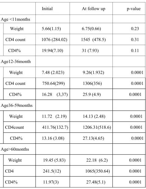
Changes in the weight, CD4 count, CD4% at the baseline and at 6 months
follow up visit were compared using paired t test.
RESULTS
During the study period, 355 children were screened positive for HIV
infection. Their demographic profile is given in Table I.
The mean age of first presentation was 4.5 years (0-15 years). Vertical
transmission was the assumed mode of transmission in 97% children in view of
maternal seropositivity. Blood transfusion was considered the cause of infection in
1 child (received transfusion from his distant relative). Multiple injections was
presumed mode in 2 children (history of injection at village hospitals).
Fever was the most common presenting symptom (42%), followed by cough
(38%), chronic diarrhea (26%), skin manifestations (26%), generalized
lymphadenopathy(22%) hepato-splenomegaly (22%) and other
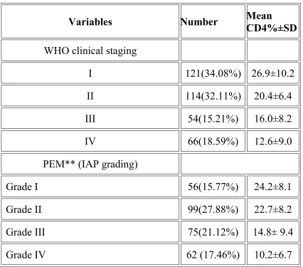
One third of the children were asymptomatic. CD4% declined with
deterioration of WHO clinical stages of the disease (Table II). Protein energy
malnutrition was present in 82% children. Mean CD4 percentage was 42.6%±10.7
in children without malnutrition (18%). Mean CD4 percentage was 18%±9.7 in
children with some degree of malnutrition (82%) The various grades of protein
energy malnutrition with their corresponding mean CD4 values are given in Table
III. The correlation of severity of malnutrition with decline in CD4 percentage was
significant (P<0.05).
One hundred and twenty one children (30%) had opportunistic infections at
the time of evaluation. Patients with opportunistic infections had lower CD4 values
(12.9%±9.4) as compared to patient without any opportunistic infections (28.4%
±8.6). Tuberculosis was the most common opportunistic infection. Disseminated
tuberculosis and pneumocystis pneumonia and CMV retinitis had the least CD4
values.
Pruritic skin lesions, anemia, hepatosplenomegaly and lymphadenopathy and
tuberculosis were commonly noticed in older children. Serious life threatening illness
like pyogenic meningitis, recurrent bronchopneumonia, pneumocystis pneumonia and
Tuberculosis was present in 54 cases. (pulmonary 21, extrapulmonary 33). Out
of 33 extra-pulmonary, 30 were TB lymphadenopathy,2 were TB meningitis, 1
Disseminated TB. Out of 54 Tuberculosis cases, manteaux was positive in 21 cases,
sputum positive for AFB in 2 cases. 5 out of 15 deaths were (33% mortality) due to
tuberculosis. (overall mortality 4.2% ).
134 children, in whom Antiretroviral therapy was started & followed up for
atleast 6 months, 119 were alive and on follow up.
There were 15 deaths. The median age at ART initiation was 54 months, with
6 children (9.9%) younger than 11 months, 27 (28.6%) aged 11 to 36 months, and
33 (36-59 months),68 children (60 months –15 yrs ).
Of those 134 ( WHO stage II – 18 , stage III- 54, stage IV -62), who started
on ART, the median age was 4.5 yrs, 81 were male and 53 were female. The mean
weight(SD)at the baseline was14.3 (6.64) and increased to 16.58 (7.19) at the last
follow up visit. The mean CD4 cell ( SD) percentage at ART initiation among the
119 children was 13.47(4.25) % and increased to 25.51(6.9)% at the last follow up.
Median Hemoglobin was 8.6 at the base line and 9.4 gm % at the last visit .
Fifteen children died over 2 years of follow-up. Two children failed to respond to
first line ART regimen and were considered as treatment failure. They were
In our study, Out of 134 children starting ART,102(76.11%) began stavudine plus
lamivudine plus nevirapine, 3(2.2%) began zidovudine plus lamivudine plus
nevirapine, 2 (1.49%) began zidovudine plus lamivudine plus efavirenz, and 27
(20.14%) began stavudine plus lamivudine plus efavirenz because 27 cases were
screened positive for tuberculosis initially. Of patients starting a
nevirapine-based regimen, 4 were switched to efavirenz ,because of allergy. Of patients
starting a Zidovudine-based regimen, 2 were switched to stavudine, as they
developed anemia.
Thirteen patients (9.7%) had adverse effects related to the ART. 4(2.9%) patient
had severe gastritis. (1 on Zidovudine and 3 on stavudine regimen) initially which
subsided with treatment. Two patients (1.4%) had hepatotoxicity which
succumbed due to TB meningitis. 2 patient (1.4%) had zidovudine (AZT) induced
anemia, 4 patients (2.9%) had Nevirapine (NVP) induced rash, 1 patient (0.7%)
had Stavudine (d4T) induced Peripheral neuropathy.
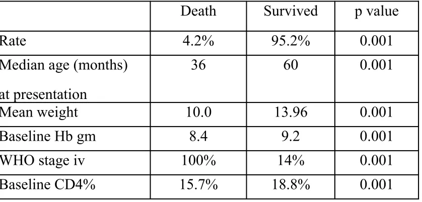
Out of 355 children, 15 (4.2%) died,. Children who died were younger compared
to those who survived, (median age, 36 months vs 60 months; P < .001), had lower
mean weight (10.0 vs 13.96 P < .001), had lower baseline median hemoglobin
concentration (8.4 g/dL vs 9.2 g/dL; P < .001), were more likely to be WHO clinical
vs18.8%) compared to those who survived. Mortality was associated with
younger age, lower weight-for-age, anemia WHO stage IV and CD4 cell
depletion
TABLE I
DEMOGRAPHIC PROFILE OF HIV INFECTED CHILDREN
Variables Number
Mean CD4%
±SD
WHO classification immunodeficiency
Not significant Mild Advanced Severe
Gender
Male 199(56.05) 18.8 52(26.1) 64(32.1) 43(21.6) 40(20.1)
Female 156(43.94) 21.5 60(38.4) 45(28.8) 25(16.0) 26(16.6)
Age in years
0<1 yr 45( 2.67) 21.0±2.69 21(46.6) 20(44.4) 2(4.4) 2(4.4)
1 <3yr 95(26.76) 21.6±7.6 44 (46.31) 24(25.26) 14(14.73) 13(13.68)
3 <6yr 92(25.91) 18.9± 8.2 34 (36.95) 25(27.17) 16(17.39) 17(18.47)
6<9yr 41(11.54) 17.2± 9.5 11(26.82) 4 (9.7) 17(41.46) 9 (21.95)
9 <12yr 48(13.52) 15.8± 6.9 15 (31.25) 11(22.91) 14(29.16) 8(16.66)
TABLE II
CD4 CORRELATION WITH CLINICAL PROFILE OF HIV INFECTED
CHILDREN
Variables Number Mean CD4%±SD
WHO clinical staging
I 121(34.08%) 26.9±10.2
II 114(32.11%) 20.4±6.4
III 54(15.21%) 16.0±8.2
IV 66(18.59%) 12.6±9.0
PEM** (IAP grading)
Grade I 56(15.77%) 24.2±8.1
Grade II 99(27.88%) 22.7±8.2
Grade III 75(21.12%) 14.8± 9.4
Oppurtunistic infections Number Mean CD4%±SD
Anaemia Hb %
> 14
12 – 14
10 – 12
8 - 10
<8 10 (2.8%) 36(10.1%) 72 (20.3%) 232(65.3%) 5 (1.4%) 36±6.2 32±6.2 26±8.2 24±8.4 12.0±3.4
Tuberculous lymphadenopathy : 30(8.4%) 26±8.6
lymphadenopathy 78(21.9%) 32±7.2
Thrombocytopenia 1(0.2%) 12
papullar pruritic eruptions : 53(14.9%) 36±11.2
viral warts : 20(5.6%) 28±8.6
Molluscum contagiosum 22(6.1%) 11±5.4
Seborrhoeic Dermatitis 20 (5.6%) 26±6.2
Scabies 17(4.7%) 26±7.4
Chicken pox 14(3.9%) 16±5.6
Herpes zoster 7(1.9%) 24±7.4
Ulcerative gingivitis/periodontitis 28(7.8%) 14±6.2
Oral candidiasis 18(5.0%) 9±7.2
Chronic /Recurrent Diarrhoea 91(25.6%) 22±8.4
Hepatosplenomegaly 79(22.2%) 21±8.1
Cryptosporidiasis 16(4.5%) 11±6.2
Pulmonary Tuberculosis 21(5.9%) 16±7.4
Bacterial Pneumonia 30(8.4%) 18±8.4
Pneumocystis carinii pneumonia 2(0.5%) 3.5±0.5
Lymphoid interstitial pneumonia 1 (0.2%) 15.5
Chronic lung disease 3(0.8%) 12+2
CSOM/Otorrhoea 46(12.9%) 25±5.5
Recurrent Upper Resp. Infection 96(27.0%) 24±6.4
Tuberculous Meningitis 2(0.5%) 8±2.0
Pyogenic Meningitis 2(0.5%) 12±2.0
HIVEncephalopathy/Developmentaldelay 10(2.8%) 10±2.5
CMV Retinits 2(0.5%) 5±0.5
Table-III
COMPARISON OF CD4 BEFORE AND AFTER ART
Before
treatment
(n=134)
At followup
(n=119)
p- value
Mean CD4
count
427(306) 1095(470) 0.001
Mean CD4 % 13.47(4.25) 25.5(6.90) 0.001
Table-4
AGE WISE CHANGE IN WEIGHT AND CD4 COUNT AFTER ART
Initial At follow up p-value
Age <11months
Weight 5.66(1.15) 6.75(0.66) 0.23
CD4 count 1076 (284.02) 1545 (478.5) 0.31
CD4% 19.94(7.10) 31 (7.93) 0.11
Age12-36month
Weight 7.48 (2.023) 9.26(1.932) 0.0001
CD4 count 750.64(299) 1306(356) 0.0001
CD4% 16.28 (3,37) 25.9 (4.9) 0.0001
Age36-59months
Weight 11.72 (2.19) 14.13 (2.48) 0.0001
CD4count 411.76(132.7) 1206.31(518.6) 0.0001
CD4% 13.16 (3.08) 27.13(4.65) 0.0001
Age>60months
Weight 19.45 (5.83) 22.18 (6.2) 0.0001
CD4 241.5(12) 1065(350.64) 0.0001
Table – 5
AGE WISE MORTALITY
AGE GROUP
(n) 6 months
Survival
Mortality (%)
11 months 6 50% 3 (50%)
11 to 35 months 27 71% 8 (29.6%)
36-59 months 33 97% 1 (3.0%)
60 mon –15 yr 68 95.6% 3 (4.4%)
Table -6
FACTORS ASSOCIATED WITH MORTALITY
Death Survived p value
Rate 4.2% 95.2% 0.001
Median age (months)
at presentation
36 60 0.001
Mean weight 10.0 13.96 0.001
Baseline Hb gm 8.4 9.2 0.001
WHO stage iv 100% 14% 0.001
[image:51.612.84.503.408.609.2]DISCUSSION
This prospective cross-sectional study has analysed the clinical profile in
relation to CD4 status in HIV infected children attending our ART centre.
Clinical features in HIV-infected children in our study had some similarities and
[image:52.612.70.536.362.715.2]few differences from the previous Indian studies(20,21,22,23,24,25,26).
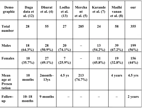
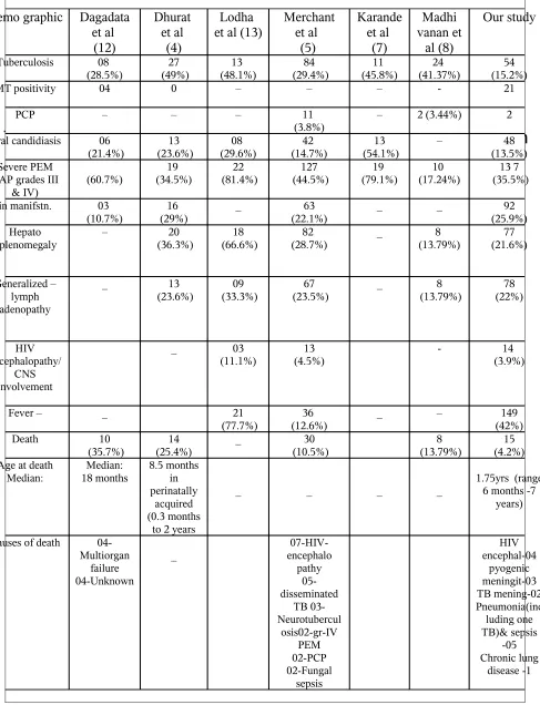
Table 6and 7 compare demographic data and clinical features of our study with
those of previously reported Indian studies.
Demo
graphic data et Daga al. (12)
Dhurat
et al. (4) Lodhaet al. (13)
Mercha nt et al. (5)
Karande
et al. (7) Madhi vanan et al. (8)
our
Total number
28 55 27 285 24 58 355
Males 18
(64.3%) (50.9%)28 (74.1%)20 – (54.2%)13 (67.2%)39 (56%)199
Females 10
(35.7%) (49.1%)27 (25.9%)7 – (45.8%)11 (32.8%)19 (44%)156
Mean age at Presen tation 10 months 2month– 13ys
4.5 ys 213 (74.7%)
4 years 4.5 yrs
Follow-up
10–18 months
Table 7-Comparison of demographic data of pediatric HIV infection
COMPARISION OF CLINICAL PROFILE WITH OTHER STUDIES
Demo graphic Dagadata et al (12) Dhurat et al (4) Lodha et al (13)
Merchant et al (5) Karande et al (7) Madhi vanan et al (8) Our study Tuberculosis 08
(28.5%) (49%)27 (48.1%)13 (29.4%)84 (45.8%)11 (41.37%)24 (15.2%)54
MT positivity 04 0 – – – - 21
PCP – – – 11
(3.8%) – 2 (3.44%) 2
Oral candidiasis 06
(21.4%) 13 (23.6%) 08 (29.6%) 42 (14.7%) 13 (54.1%) – 48 (13.5%) Severe PEM
(IAP grades III
& IV) (60.7%)
19
(34.5%) (81.4%)22 (44.5%)127 (79.1%)19 (17.24%)10 (35.5%)13 7
Skin manifstn. 03
(10.7%)
16 (29%)
_ 63
(22.1%)
_ _ 92
(25.9%) Hepato
Splenomegaly – (36.3%)20 (66.6%)18 (28.7%)82 _ (13.79%)8 (21.6%)77
Generalized – lymph adenopathy
_ 13
(23.6%) (33.3%)09 (23.5%)67 _ (13.79%)8 (22%)78
HIV encephalopathy/
CNS involvement
_ 03
(11.1%) (4.5%) 13 - (3.9%)14
Fever – _ 21
(77.7%) (12.6%)36 _ – (42%)149
Death 10
(35.7%) (25.4%) 14 _ (10.5%)30 (13.79%)8 (4.2%)15
Age at death Median: Median: 18 months 8.5 months in perinatally acquired (0.3 months
to 2 years
_ _ _ _ 1.75yrs (range 6 months -7
years)
Causes of death
04-Multiorgan failure 04-Unknown
_ encephalo 07-HIV-
As in the previous series, perinatal transmission was the most common mode of
transmission in our study. HIV acquired via sexual abuse has been reported previously
(21) but was not encountered by us.
Daga et al(25)reported the presence of known HIV infection in one/both parents
in 7 cases.
Growth retardation is a common feature in HIV infected children. Weight gain
has been used as a parameter in assessing the improvement in clinical status in many
studies(14). Somatic growth has shown to be affected in patients with HIV infection
(28). 82% of our children had some degree of PEM (Grade I to IV of IAP). 137
children (39%) in our series had severe malnutrition (grade III/IV) and 155 others
(43%) had mild malnutrition (grade I/II). This may be caused by various factors such as
poor nutrition, neglect, poverty, repeated infections, etc. Malnutrition has been reported
to be the most common manifestation in HIV-infected children (21,22,23).
Hepatomegaly was seen in 22% of our cases and was one of the most common
manifestations in our study. Hepatomegaly can be caused by the replication of the HIV
within the reticuloendothelial system and early onset lymphadenopathy and
hepatomegaly in the first 3 months of life is associated with rapid disease progression
(22) and in 36% of the cases studied by Dhurat et al. (21).
Generalized lymphadenopathy was seen in 22% of our patients and may have
been the result of viral infections (such as Epstein-Barr virus or cytomegalovirus),
opportunistic infections, and mycobacterial infections apart from being caused by the
HIV infection (28). Persistent generalized lymphadenopathy has been seen in 23.5% of
cases by Merchant et al. (22).
In the present study, diarrhea was the presenting manifestation in 22% children,.
Infections causing diarrhea in HIVinfected children include rotavirus, Shigellae,
Campylobacter, E. coli, cryptosporidiosis, isosporiasis, cytomegalovirus and atypical
mycobacteria (22,28). Cryptosporidiosis was reported in 16 cases.Chronic/recurrent
diarrhea has been seen in 15 to 43% cases in various studies 22,23,25 )
We encountered tuberculosis in 54 cases (15%). (pulmonary 21, extrapulmonary 33). Out
of 33 extra-pulmonary, 30 were TB lymphadenopathy, 2 were TB meningitis ,1
Disseminated TB. Out of 54 Tuberculosis cases, manteaux was positive in 21 cases,
sputum positive for AFB in 2 cases. Pulmonary tuberculosis occurred between wide
range of CD4 values ranging from 2% to 30%. TB meningitis occurred with low CD4
values (4% to 12%).
Tuberculosis in various forms—pulmonary and extrapulmonary has been reported
Mantoux (tuberculin) test as it may be falsely negative in patients with HIV (25). Only
four of eight children diagnosed to have tuberculosis had positive Mantoux test in the
study by Daga et al. (25). Twenty one patients had positive Mantoux test in our study.
As in other studies, lack of culture facilities have made it difficult for us to study the
presence of atypical mycobacteria and resistance pattern in HIV-infected children (22).
Merchant et al. reported 84 cases (29.4%) with tuberculosis in a cohort of 285 cases; of
whom 48 had pulmonary lesions, 21 had disseminated tuberculosis, 8 had tubercular
lymphadenopathy and 7 had neurotuberculosis (22). Dhurat et al. have reported
pulmonary tuberculosis in 16 cases, 9 extrapulmonary (of whom 4 had pulmonary
tuberculosis as well; extrapulmonary sites: abdominal-4, neurotuberculosis- 2 and
lymphadenopathy-3 cases) (21). A similar spectrum of tubercular manifestations was
seen in our study as well.
3 cases of Bronchiectasis were encountered in our study (4,5,9),as reported in
earlier studies. Bronchiectasis occurred when the CD4 is around 12% (severe
immunosuppression).
Pneumocystic carinii pneumonia was less common in our study. Pneumocystis
carinii pneumonia (PCP) was present in two cases. Merchant et al. have reported 11
cases of PCP (22). A low incidence of PCP has been reported among our children .
Whether the apparent differences in the occurrence of P. carinii pneumonia reflect
geographic variation in the prevalence of the organism needs to be explored.
Lymphoid interstitial pneumonia was seen in only one patient in our study.
Dermatological manifestations were common in our study(26%). Papular pruritic
eruptions (15%) were the most common skin lesion encountered in our study.
Seborrheic dermatitis(5.6%), scabies(4.7%), drug eruptions, and skin lesions associated
with nutritional deficiencies, herpes zoster(2%), molluscum contagiosum(6%), viral
warts(5.6%) were also noted. PPE occurred in CD4 ranging from (25 to 36%).
Mollucum contagiosum had the lowest CD4 (11%) compared to all other cutaneous
conditions. Skin lesions including herpes zoster (19 cases), chronic nonspecific
dermatitis (20 cases), scabies (18 cases), pyoderma (15 cases), molluscum contagiosum
(3 cases) and chronic paronychia (2 cases) have been reported by Merchant et al. (22).
Similarly, Dhurat et al. have reported seborrheic dermatitis (6 cases), chicken pox (4
cases; hemorrhagic in 2 cases) and herpes zoster (2 cases) (21).
Anemia was seen in 66% of cases in our study. The causes may include bone
marrow changes consistent with anemia of chronic disorders, nutritional deficiency
(folic acid or vitamin B12), adverse effects of medications and peripheral destruction of
erythrocytes (28). Anemia has been noted in symptomatic patients in the study by
Dhurat et al. (21) (32%) and Merchant et al. (22). Thrombocytopenia was reported in
one case.
in other studies (28). Merchant et al. had 13 cases with HIV encephalopathy in their
series (22). 3% of our patients had HIV-encephalopathy. The neuroimaging features
(cerebral atrophy, infarction, calcification) were also consistent with the diagnosis of
encephalopathy in these patients. Clinical and neuroimaging features of
encephalopathy were similar to the studies reported earlier (22,29). Presence of
HIV-encephalopathy is associated with poor outcome for survival (22,29).
Cardiomyopathy was seen in one case in the present study. One case of immune
reconstitution syndrome was encountered and managed appropriately.
12 cases of parotitis were encountered in our study. All oral conditions occurred
in CD4 range of moderate immuno deficiency (15-25%), except oral candidiasis which
occurred in CD4 values of severe immunodeficiency.
As in some of the previous studies, no cases with renal manifestations and
malignancies were reported in our study (21,22,28)
CD4 count was used as a measure of Immunological outcome in previous
studies.(31,32)WHO & national guidelines advise that clinicians use CD4 cell
percentage for assessment of children younger than 60 months and absolute CD4
cell count for children 60 months or older. Significant improvement in the CD4
count and CD4% in response to ART illustrated in our study is in accordance with
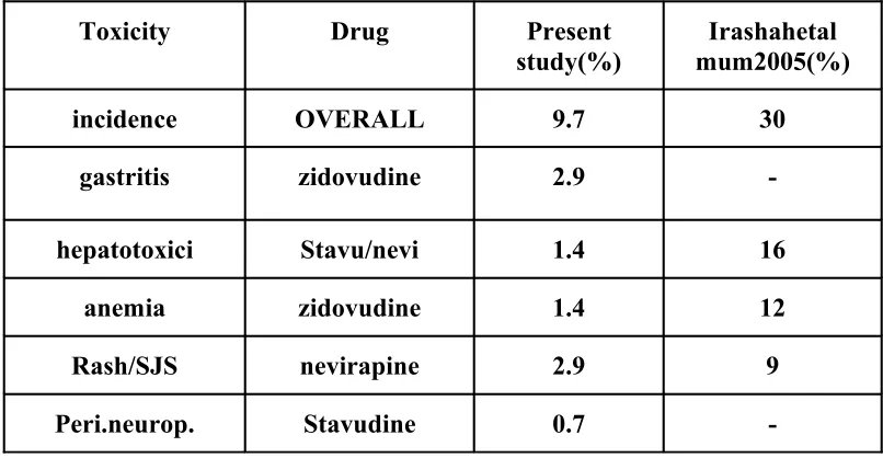
Thirteen patients (9.7%) had adverse effects related to the ART. 4(2.9%)
patient had severe gastritis.(1 on Zidovudine and 3 on stavudine regimen) initially
which subsided with treatment. Two patients (1.4%) had hepatotoxicity which
succumbed due to TB meningitis. 2 patient (1.4%) had zidovudine (AZT) induced
anemia, 4 patients (2.9%) had Nevirapine (NVP) induced rash, , 1 patient (0.7%)
had Stavudine (d4T) induced Peripheral neuropathy. Hepatotoxicity was far less
in the present study in view of close monitoring and careful selection of cases. This
is in contrast to previous Indian study which reported high incidence of
[image:59.612.89.492.452.661.2]hepatotoxicity.(33)
Table -9
COMPARISION OF DRUG TOXICITIES WITH OTHER INDIAN STUDY
Toxicity Drug Present study(%)
Irashahetal mum2005(%)
incidence OVERALL 9.7 30
gastritis zidovudine 2.9
-hepatotoxici Stavu/nevi 1.4 16
anemia zidovudine 1.4 12
Rash/SJS nevirapine 2.9 9
-Out of 355 children, 15 (4.2%) died,. Children who died were younger compared
to those who survived, (median age, 36 months vs 60 months; P < .001), had lower
mean weight (10.0 vs 13.96 P < .001), had lower baseline median hemoglobin
concentration (8.4 g/dL vs 9.2 g/dL; P < .001), were more likely to be WHO clinical
stage IV (100% vs 14%, P < .001), had lower baseline CD4 values (15.7%
vs18.8%) compared to those who survived. Mortality was associated with
younger age, lower weight-for-age, anemia WHO stage IV and CD4 cell
depletion
Mortality has been variably reported to be 10.5% (22), 25.4% (21) and 35.7%
(25) in various studies. Fifteen of our patients (4.2%) died in our study. These
differences in figures may reflect the differences in the clinical manifestations, early
versus late presentations, duration of follow-up, presence of opportunistic infection,
appropriate intervention with antiretroviral therapy, availability of ancillary and
CONCLUSION
HIV disease in children has a diverse range of manifestations in multiple
organ systems.
Fever is the most common initial presentation in children followed by
cough and diarrhoea.
Pulmonary tuberculosis is the most common opportunistic infection
followed by oral candidiasis.
Meningitis, CMV retinitis, Pneumocystis carinii pneumonia,oral
candidiasis, grade IV malnutrition, WHO class IV symptoms correlate with
very low CD4%.
Treatment with ART results in improvement in CD4%.
Mortality is associated with younger age, lower weight-for-age, anemia,
LIMITATIONS
Viral assays which remains the most sensitive indicator of response to
antiretroviral therapy could not be done because of cost concerns.
Protease inhibitor-based therapy remains very expensive and unaffordable to our
RECOMMENDATIONS
To suspect and recognize symptoms & signs of HIV early in infancy to start
ART earlier.
Early institution of ART in infancy to reduce underfive mortality due to HIV.
In the new era of generic HAART, physicians must be trained to identify and
manage the toxicities associated with HAART .
Interventions should focus on both child and caregivers and must address social
problems that adversely affect adherence.
Physicians and health care professionals should be taught about Universal
BIBLIOGRAPHY
1. Joint United Nations program on HIV/AIDS(UNAIDS)/WHO. AIDS epidemic
update. 2006. Available from: URL:http://www.un aids.org/en/
Publications/default.asp. Accessed October 1, 2007.
2. Joint United Nations Program on HIV/AIDS (UNAIDS) /WHO. AIDS epidemic
update. 2004. Available from: URL:http://www.unaids.org/en/ Publications/default.asp.
Accessed October 1, 2007.
3. Palella FJ, Delaney KM, Moorman AC, Loveless MO, Fuhrer J, Satten GA, et al.
Declining morbidity and mortality among patients with advanced HIV infection. NEJM
1998; 338 : 853-60.
4. National AIDS Control Organization. A note on HIV estimates 2003:
http://www.naco.nic.in/indianscene/ esthiv.htm accessed on August 17, 2004.
5. Palumbo PE. Antiretroviral therapy of HIV infection in children. Pediatr Clin North
Am 2000; 47: 155-169.
6. Lodha R, Singhal T, Jain Y, Kabra SK, Seth P, Seth V. Pediatric HIV Infection in a
7. Englund JA, Baker CJ, Raskino C et al. Zidovudine, didanosine, or both as the initial
treatment for symptomatic HIV-infected children. N Engl J Med 1997; 336: 1704-12
8. Centers for Disease Control and Prevention. Guidelines for the use of antiretroviral
agents in pediatric HIV infection. MMWR 1998; 47(RR-4): 1-44.
9. Madhivanan P, Mothi SN, Kumarasamy N, Yepthomi T, Venkatesan C, Lambert JS,
Solomon S. Clinical manifestations of HIV infected children. Indian J Pediatr
2003;70:615–620.
10. Merchant RH, Oswal JS, Bhagwat RV, Karkare J. Clinical profile ofHIV infection.
Indian Pediatr 2001;38:239–246.
11. Chakravarty J, Mehta H, Parekh A, Attili SV, Agarwal NR, Singh SP, et al. tudy on
clinico-epidemiological profile of HIV patients in eastern India. J Assoc Physicians
India 2006; 54: 854- 857.
12. Ylitalo N, Brogly S, Hughes MD, Nachman S, Dankner W, Van Dyke R, et al. Risk
factors for opportunistic illnesses in children with human immunodeficiency virus in
the era of highly active antiretroviral therapy. Arch Pediatr Adolesc Med 2006; 160:
778-787.
13. Fox- Wheeler S, Heller L, Salata CM, Kaufman F, Loro LM, Gilsang V, et al.