0022-538X/95/$04.0010
Copyrightq1995, American Society for Microbiology
Hepatitis Delta Virus Mutant: Effect on RNA Editing
T.-T. WU,1V. V. BICHKO,1W.-S. RYU,1S. M. LEMON,2
ANDJ. M. TAYLOR1*
Fox Chase Cancer Center, Philadelphia, Pennsylvania 19111-2497,1and University of North Carolina,
Chapel Hill, North Carolina 27599-70302
Received 30 March 1995/Accepted 25 July 1995
During the replication cycle of hepatitis delta virus (HDV), RNA editing occurs at position 1012 on the 1679-nucleotide RNA genome. This changes an A to G in the amber termination codon, UAG, of the small form of the delta antigen (dAg). The resultant UGG codon, tryptophan, allows the translation of a larger form of the
dAg with a 19-amino-acid C-terminal extension. Using HDV cDNA-transfected cells, we examined the editing potential of HDV RNA mutated from G to A at 1011 on the antigenome, adjacent to normal editing site at 1012. Four procedures were used to study not only the editing of the A at 1012, but also that of the new A at 1011: (i) nucleotide sequencing, (ii) a PCR-based RNA-editing assay, (iii) immunoblot assays, and (iv) immunoflu-orescence. Five findings are reported. (i) Even after the mutation at 1011, editing still occurred at 1012. (ii) Site 1011 itself now acted as a novel RNA-editing site. (iii) Sites 1011 and 1012 were edited independently. (iv) At later times, both sites became edited, thereby allowing the synthesis of the large form of thedAg (dAg-L). (v) Via immunofluorescence, such double editing became apparent as a stochastic event, in that groups of cells arose in which the changes had taken place. Evaluation of these findings and of those from previous studies of the stability of the HDV genomic sequence (H. J. Netter et al., J. Virol. 69:1687–1692, 1995) supports both the recent reevaluation of HDV RNA editing as occurring on antigenomic RNA (Casey and Gerin, personal communication) and the interpretation that editing occurs via the RNA-modifying enzyme known as DRADA.
RNA editing of hepatitis delta virus (HDV) RNA is essen-tial during viral infection to create an open reading frame for one of the two proteins that are essential for virus replication (4, 20, 28). In particular, editing allows the translation of a second form of the delta antigen (dAg), the only protein en-coded by HDV (27). This larger form,dAg-L, has an additional 19 amino acids (aa) at the C terminus (Fig. 1B) relative to the small form,dAg-S (Fig. 1A). While the originaldAg-S is es-sential for genome replication (15),dAg-L is also biologically relevant, in that it is needed for virus assembly (7, 24).dAg-L is isoprenylated at a unique cysteine 4 aa in from the novel C-terminus region (Fig. 1B) (11). IfdAg-L is mutated so that isoprenylation cannot occur, dAg-L will not function in the assembly of virion particles (19). It has been demonstrated that dAg-L is also a dominant negative inhibitor of genome repli-cation as supported bydAg-S (8), but it is not established wheth-er such inhibition is an essential part of the virus life cycle.
An RNA-editing site is unquestionably located at position 1012, but there may be other editing positions as well (22). The immediate target for the editing at position 1012 has been reported to be the genomic strand (3, 28), although more recent studies argue, to the contrary, that the target is the anti-enome (2). The mechanism of HDV RNA editing remains unclear. As will be explained, it could be carried out by the recently cloned RNA-modifying enzyme known as DRADA (14), which seems to be involved in the editing of the genomes of other RNA viruses (5) and also of certain cellular RNAs (6). The following studies actually began out of an attempt to design a mutated HDV that could not makedAg-L even after the normal RNA-editing event at 1012. With this in mind, the G at 1011 on the antigenome was changed to A (Fig. 1C). Thus, editing at 1012 would not only replace the ochre
termi-nation codon, UAA, with an opal terminator, UGA. As doc-umented here, we found that 1012 was still edited, and to our surprise, 1011 now became a new and independent editing site. Some HDV RNAs were even edited at both 1011 and 1012, thereby replacing the ochre terminator with tryptophan, UGG, and allowing synthesis ofdAg-L (Fig. 1D).
For most of the studies reported here, we actually used not the single mutant (Fig. 1C) but a double mutant (Fig. 1E) with an additional mutation, a nearby deletion at position 979. This deletion was used as a nucleotide sequencing marker. For this double mutant, even if thedAg-L reading frame were opened by editing at 1011 and 1012, the 8 aa at the normal C terminus would be replaced by 9 aa in another reading frame, to pro-duce a modified form ofdAg-L,dAg-L9(Fig. 1F). As reported here, we did in fact observe sequence changes consistent with editing at both 1011 and 1012. Therefore, to exclude any pos-sible effect of this mutated C-terminal extension on editing, we repeated certain experiments, as indicated, with the single mu-tant, and the results were not significantly different.
MATERIALS AND METHODS
Plasmids.Plasmid pSVL(D3) was constructed by insertion of a trimer of unit-length cloned HDV cDNA (wild type) (16) into expression vector pSVL (Pharma-cia), as described previously (15). A single mutation at nucleotide (nt) 1011 (Fig. 1C) and a double mutation at nt 1011 and 979 (Fig. 1E), were introduced via synthetic oligonucleotides. The single mutant was inserted as a partial dimer into the pSVL vector pDL538 (18). For the double mutant, a dimer of the modified monomer, permuted at the unique EcoRI site (16), was inserted into the pSVL vector.
Antibodies.Antibody specific for bothdAg-S anddAg-L was raised in rabbits by inoculation ofdAg prepared in Escherichia coli. Rabbit antibody rab-LP3 was derived against the peptide DILFPADPPFSPQSC, representing the carboxy terminus ofdAg-L (residues 197 to 211) and was shown to recognizedAg-L but notdAg-S (26). Mouse monoclonal antibody 3G3, recognizing both forms of dAg, was a generous gift from Michael Lai (13).
Transfection procedure.Huh7 cells (21) were seeded in plastic 35-mm-diam-eter tissue culture wells (Costar) with or without an inserted 12-mm-diam35-mm-diam-eter glass coverslip. Lipofectamine (Bethesda Research Laboratories) was used to transfect subconfluent cells in the presence of Opti-MEM (Bethesda Research Laboratories), as recommended by the manufacturer (12). Briefly, 12ml of Lipofectamine at 2 mg/ml and 2mg of plasmid DNA were each diluted into 0.5 ml of Opti-MEM. These two dilutions were combined, incubated for 30 min at * Corresponding author. Mailing address: Fox Chase Cancer
Cen-ter, 7701 Burholme Ave., Philadelphia, PA 19111-2497. Phone: (215) 728-2436. Fax: (215) 728-3616. Electronic mail address: jm_taylor@w470. fccc.edu.
7226
on November 9, 2019 by guest
http://jvi.asm.org/
room temperature, and applied to cells that had been washed once with Opti-MEM. After 5 h of incubation at 378C, the transfection mixture was removed and the cells were grown in Dulbecco’s modified Eagle’s medium supplemented with 10% fetal calf serum.
RNA extraction and Northern (RNA) analysis.As previously described (9), total RNA was extracted from transfected cells and, after glyoxalation, was submitted to electrophoresis in 1.5% agarose gels, followed by RNA blotting to detect genomic HDV RNA sequences. Genomic full-length HDV RNA probe was synthesized in vitro in the presence of [a-32
P]UTP and used as previously described (9). Quantitation and image processing were achieved with a Fuji imager (Fujix BAS1000).
Reverse transcription.For reverse transcription, we used a primer that bound to genomic RNA (nt 1301 to 1267, according to the nucleotide sequence of Kuo et al. (16). The RNA template (0.3mg) was copied, by using avian myeloblastosis virus reverse transcriptase (Life Sciences, St. Petersburg, Fla.), for 1.5 h at 428C. The product was incubated with RNase A (160mg/ml, 20 min, 378C) and then treated with pronase and sodium dodecyl sulfate, followed by extraction with phenol and then twice with ether, followed by precipitation with ethanol.
PCR and cDNA sequencing.The product of reverse transcription was sub-jected to PCR amplification with Taq polymerase and a Techne dry-block ther-mal cycler (20). The primers spanned nt 1217 to 1183 and 701 to 735 (16), and the reaction was carried out with 37 cycles of amplification. The PCR products were then gel purified and cloned with a pGEM-T vector system (Promega). The positive colonies were selected by a blue-white screening system and Perfectprep plasmid DNA kit (5 Prime 3 Prime) was used for preparation of plasmid DNA. The DNA sequence was determined by automatic sequencing (Applied Biosystems).
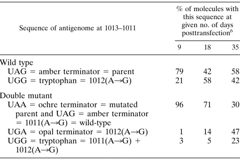
PCR and editing assay.As described elsewhere (10), the product of reverse transcription was subjected to nested PCR amplification with Vent polymerase (New England Biolabs) and an Idaho Technology thermal cycler. One of the final pair of primers was labeled at the 59end with biotin so that the product could be bound to superparamagnetic beads, denatured, and reconverted to double-stranded DNA with reverse transcriptase and a primer 59labeled with32P. The pro-duct released from the beads by digestion with SalI was tested for susceptibility to further digestion with NcoI and NlaIII. As shown in Table 2, susceptibility to these enzymes was specific for editing at positions 1011 and/or 1012 on the HDV genome. The digestions were assayed by electrophoresis of the products into an acrylamide gel followed by quantitation of radioactivity with a Fuji imager.
Immunoblot analysis.Transfected cells were lysed, and protein samples were subjected to electrophoresis in 15% polyacrylamide gels by the method of Lae-mmli (17). After electrotransfer of proteins to a nitrocellulose filter,dAgs were detected by using rabbit anti-dantibodies and125
I-labeled protein A (DuPont). Quantitation and image processing were achieved with a Fuji imager.
Immunofluorescence.Monolayer cultures on glass coverslips were fixed and stained, as previously described (1). Triple staining of cells was achieved by using a mixture of mouse monoclonal anti-dantibody 3G3 and rabbit anti-peptide antibody rab-LP3, specific fordAg-L, as primary antibodies and by addition of 2 mg of 49,6-diamidino-2-phenylindole (DAPI [Sigma]) per ml to stain the cellular DNA. Goat anti-rabbit immunoglobulin G polyclonal antibody conjugated with rhodamine (Boehringer) and goat anti-mouse immunoglobulin G plus immuno-globulin M conjugated with fluorescein (Sigma) were used as secondary anti-bodies. After being mounted, the samples were viewed with a Zeiss Axiophot microscope with a340 objective and specific filter blocks.
RESULTS
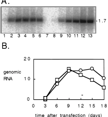
Replication of the mutant HDV genome.As presented in the introduction, the following studies involved examining the con-sequences for RNA editing at position 1012 on HDV of alter-ing the adjacent nucleotide at 1011. We compared the wild-type genome (Fig. 1A) with what we call the single mutant, which was modified at 1011 (Fig. 1C), or the double mutant, which was both modified at 1011 and deleted at 979 (Fig. 1E). At the outset, it was necessary to determine the effect of the mutations on the ability of the genome to replicate. Figure 2A shows a detailed comparison of replication for the double mutant relative to the wild type. Cultures of Huh7 cells were transfected with either wild-type or double mutant HDV cDNAs, and at regular intervals thereafter, RNA was extracted and examined by Northern analysis for HDV genomic RNA sequences (Fig. 2A). Quantitation of these data (Fig. 2B) in-dicated that replication of the wild type reached a maximum at 9 days after transfection and then progressively declined. The extent of replication of the double mutant was as good as or better than that of the wild type. Similar results were obtained with the single mutant (data not shown).
Nucleotide sequence analysis. We next used reverse tran-scription-PCR, cloning, and sequencing to determine the ac-tual sequence changes that occurred during replication of both the wild type and the double mutant. We used HDV RNAs extracted from cells at various times after transfection. In par-ticular, we studied region 1016 to 945 of the antigenome, as indicated in Fig. 1.
A total of 92 clones were sequenced, with results as summa-rized in Table 1. Table 1 shows that for the wild type, the only changes detected were at position 1012. Consistent with nor-FIG. 1. Changes on antigenomic HDV RNA as a result of mutagenesis and
editing. Shown are the nucleotide and derived amino acid sequences for the 39-terminal region (nt 1016 to 945) of the open reading frames on antigenomic HDV RNAs. (A and B) Wild type before and after editing at 1012. (C and D) Single mutant at 1011 before and after editing at both 1011 and 1012. (E and F) Double mutant, changed at 1011 and deleted at 979, both before and after editing at 1011 and 1012. In panel F, the deletion at nt 979 creates a frameshift and hence an altered amino acid sequence relative to panels B and D.
FIG. 2. Northern analysis of genomic HDV RNA in transfected cells. Cells were transfected with wild-type HDV cDNA (lanes 1 to 6), mock transfected (lane 7), or transfected with mutated HDV cDNA (lanes 8 to 13). Samples were taken at days 3 (lanes 1 and 8), 6 (lanes 2 and 9), 9 (lanes 3 and 10), 12 (lanes 4 and 11), 15 (lanes 5 and 12), and 18 (lanes 6 and 13) after transfection and were subjected to Northern analysis. (A) Radioactivity as detected with a Fuji imager. The mobility of the 1.7-kb genomic RNA is indicated at the right side. (B) Summary of the quantitation of the HDV RNA in transfected cells, expressed in arbitrary units per constant number of cells. Superimposed are the data for the wild-type (squares) and mutated (circles) HDV genomes.
on November 9, 2019 by guest
http://jvi.asm.org/
[image:2.612.327.541.77.308.2]mal HDV editing, these changes were of A3G on the antige-nome, and the frequency of changes increased with time after transfection. The wild-type sequences progressively thus changed from the amber termination codon to the tryptophan codon, thereby allowing the synthesis ofdAg-L.
However, for the double mutant, as summarized in Table 1, we detected not only changes at 1012 but also changes at 1011. These latter changes corresponded to A3G on the anti-genome. Therefore, such changes occurred as if we had cre-ated a second site of editing adjacent to the normal one. It is important to note that at the early times, by this sequencing assay, we only detected changes at either location but not at both; that is, the two sites were independently susceptible to the nucleotide change. The initial sequence was the ochre termination codon, UAA. With time, some sequences changed A to G at 1012 or 1011, to create the opal or amber terminator, respectively. Later, by 35 days, we even found that 30% of the sequences were changed at both locations, so as to create the tryptophan codon, thereby allowing the synthesis of the mod-ified form ofdAg-L, as predicted in Fig. 1F.
RNA-editing assay.A limitation of the nucleotide sequenc-ing method described above was sensitivity; for example, to detect nucleotide changes less frequent than 10%, it would be necessary to sequence much more than 10 clones. To solve this problem, we made use of a more sensitive editing assay (10). PCR amplification of RNAs edited at 1011 and/or 1012 can be detected via the creation of sites for the restriction enzymes
NcoI and NlaIII. As summarized in Table 2, we readily
de-tected editing at 1012 on the wild-type genome. For the double mutant, it was now possible to detect single and double editing as early as days 9 and 18. While these assays were more sen-sitive than those obtained by the sequencing, for the double mutant they were unable to distinguish between unedited UAA and singly edited UAG. Overall, the results of the two methods were either complementary or in agreement. The biggest discrepancy between the two methods was at day 9 for the double mutant being converted to UGA; sequencing of 11 clones indicated 18% (Table 1), while the value deduced by the subtraction of two sets of restriction enzyme data was 1% (Table 2). Our interpretation is that this apparent discrepancy is consistent with the experimental errors of the two methods.
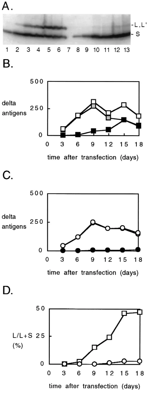
Immunoblot analysis of dAg. We next used immunoblot analysis in order to detect and quantitate the forms of dAg synthesized in the transfected cells. For transfections with the wild type (Fig. 3, lanes 1 to 6) we detected both dAg-S and dAg-L. As expected, the amount ofdAg-L increased with time and became equal to that ofdAg-S at day 18.
For the transfections with the double mutant (lanes 8 to 13), we also detecteddAg-S, but in addition, there was a species with the mobility ofdAg-L. On the basis of the initial sequence (Fig. 1E), the nucleotide sequencing data (Table 1), and the RNA-editing assays (Table 2), we made the preliminary inter-pretation that this band wasdAg-L9, just as predicted in Fig. 1F. Consistent with this interpretation, this band anddAg-L, but notdAg-S, were detected with antibody rab-LP3, a poly-clonal antibody specific for a peptide sequence unique to the C-terminal extension ofdAg-L but also partially present in the predicted extension of the putative protein dAg-L9(data not shown).
[image:3.612.63.557.87.198.2]The kinetics of appearance of these dAg species for the transfections with the wild type and double mutant are shown in Fig. 3B and C, respectively. The levels ofdAg-S showed an initial increase for 9 days, followed by a decline. Such results were very similar to those observed for the levels of RNA replication (Fig. 2B). The differences arise in terms ofdAg-L
TABLE 1. Sequence changes during HDV replication, as detected by nucleotide sequencinga
Sequence of antigenome at nt 1013–1011b % of molecules with this sequence at given no. of days posttransfection c
9 18 35
Wild type
UAG5amber terminator5parent 90 (9/10) 50 (5/10) 64 (7/11)
UGG5tryptophan51012(A3G) 10 (1/10) 50 (5/10) 36 (4/11)
Double mutant
UAA5ochre terminator5mutated parent 64 (7/11) 75 (15/20) 3 (1/30)
UAG5amber terminator51011(A3G)5wild type 18 (2/11) 10 (2/20) 30 (9/30)
UGA5opal terminator51012(A3G) 18 (2/11) 15 (3/20) 37 (11/30)
UGG5tryptophan51011(A3G)11012(A3G) ,9 (0/11) ,5 (0/20) 30 (9/30)
aCells were transfected as described in the legend to Fig. 2 with either the wild-type or double-mutant genome. At the indicated times after transfection, RNA-PCR,
cloning, and sequencing of individual clones were performed.
bThe table shows only sequences for antigenomic sequence 1013 to 1011, the position of the normal termination codon fordAg. Changes in other regions, with the
exception of nt 979 on the double mutant, were not detected.
cShown is the percentage of RNA molecules with the indicated sequence. In parentheses is the number of clones of this sequence/total number of clones examined.
TABLE 2. Sequence changes during the HDV replication, as detected by editing assaysa
Sequence of antigenome at 1013–1011
% of molecules with this sequence at given no. of days posttransfectionb
9 18 35
Wild type
UAG5amber terminator5parent 79 42 58 UGG5tryptophan51012(A3G) 21 58 42 Double mutant
UAA5ochre terminator5mutated parent and UAG5amber terminator 51011(A3G)5wild-type
96 71 30
UGA5opal terminator51012(A3G) 1 14 47 UGG5tryptophan51011(A3G)1
1012(A3G)
3 5 23
aCells were transfected as for Fig. 2 and Table 1, with either the wild-type or
double-mutant genome.
bAt the indicated times posttransfection, RNA-PCR, followed by two editing
assays, was used to determine the fraction of RNA molecules with a given sequence. The first assay was to determine the percentage of molecules with a newly created NcoI site (CCATGG), consistent with UGG at 1011 to 1013. For the double mutant, the second assay was for an NlaIII site (NCATGN), consis-tent with both UGA and UGG. Thus, subtraction of the result of the first assay (%UGG) from that of the second (%UGA 1UGG) yields %UGA. Also, subtraction of the result of the second assay from 100% yields %UAA1UAG.
on November 9, 2019 by guest
http://jvi.asm.org/
[image:3.612.316.555.488.646.2]and dAg-L9. These differences can be seen in absolute terms (Fig. 3B and C) but more clearly in terms of the percentage of totaldAg that is eitherdAg-L ordAg-L9(Fig. 3D). For the wild type, this percentage increased, and by day 15, it reached a plateau of about 50%. In contrast, for the double mutant, the percentage progressively increased, but even by day 18 had not even reached 3%. A comparable value was obtained for the single mutant (data not shown).
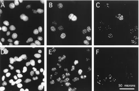
Immunofluorescent detection of dAg. The appearance of dAg-L anddAg-L9was further studied by immunofluorescence of transfected cells. Figure 4 shows the data at 9 days, which corresponds to the peak of genome replication anddAg-S expres-sion. It is important to note that at this time, the wild-type and double-mutant genomes have replicated to approximately the same extent (Fig. 2B). Coverslips were triple stained: with DAPI to stain nuclear DNA, with an antibody againstdAg to detect totaldAg, and with an antipeptide antibody (rab-LP3) to detect only large forms ofdAg (eitherdAg-L ordAg-L9).
For transfection with wild-type cDNA (Fig. 4A to C), about 40% of the DAPI-staining cells (Fig. 4A) were positive for both total dAg (Fig. 4B) anddAg-L (Fig. 4C). Positive cells were detected as small colonies. In these cells, the distribution of total antigen was nuclear. More specifically, there was diffuse nucleo-plasmic staining, along with a variable number of discrete dots. dAg-L anddAg-L9 showed staining of the same dots, but the amount of diffuse nucleoplasmic staining was relatively smaller.
Cells transfected with the double-mutant cDNA gave a sim-ilar staining pattern, with one important exception. Only a fraction, about 30%, of the cells in a clone that were positive for total antigen (Fig. 4E) were also positive for the large form (Fig. 4F). This observation was made under conditions in which the sensitivity of the assay for the large form was in-creased relative to that for the total—that is, when the positive signals in panel F were stronger than those in panel E. It appears, therefore, that only in a subpopulation of mutant-transfected cells do the mutations occur that are needed to produce dAg-L9. From an examination of other colonies of positive cells, the same heterogeneity was found to be the predominant situation. Less abundant were two other types of HDV-positive colonies. The first, was one in which each cell was positive for both total antigen and for the large form. The second, was one in which none of the cells that were positive for total antigen were positive for the large form. Since nuclear dots were detected in these cells by using antibody to total antigen, it means that the large form was not necessary for accumulation of antigen into such dots.
[image:4.612.58.296.71.706.2]The data presented above were collected on day 9. At earlier times, the large forms were more difficult to detect, especially for the single and double mutants. With the wild type, the large form was detectable by day 3, while with the mutants, detection was first possible at day 6. Even though immunofluorescence lacks quantitation, the method can have a greater sensitivity relative to an immunoblot; the largedAgs were detected 3 days earlier with immunofluorescence than with an immunoblot. At day 9, there was no detectable difference between the double mutant and the single mutant (data not shown).
FIG. 3. Immunoblot analysis to detectdAgs in transfected cells. Cells were transfected exactly as described for Fig. 2. Samples were subjected to immuno-blot analysis, and are indicated as in Fig. 2. (A) Radioactivity as detected with a
Fuji imager. The mobility of thedAg-S (S) anddAg-L (L, L9) forms of thedAg are indicated on the right. (B and C) Summary of quantitation, in arbitrary units per constant number of cells, of totaldAg (open symbols),dAg-S (shaded symbols), anddAg-L anddAg-L9forms (solid symbols). (B and C) Transfections with wild-type (squares) and mutated (circles) HDV genomes, respectively. (D) Fraction (percentage) of the large forms (dAg-L anddAg-L9) relative to the total dAg. Data are superimposed for transfections with wild-type (squares) and mu-tated (circles) HDV genomes.
on November 9, 2019 by guest
http://jvi.asm.org/
DISCUSSION
The present study was begun as an attempt to interfere with the consequences of HDV RNA editing at position 1012. We made a base change of G-A on the antigenome at the adjacent nucleotide, 1011. The expectation was that editing at 1012, even if it occurred on such a mutated genome, would still not allow the synthesis of the large form of thedAg (Fig. 1C). We found that the mutated genome not only replicated as well as the wild type (Fig. 2), but it revealed the following important new information about HDV editing. (i) Editing of A3G still occurred at 1012 (Tables 1 and 2). (ii) The novel A at 1011 was also changed to G, as if it became a new target for editing. (iii) Editing occurred independently at both 1011 and 1012. (iv) At later times after transfection, molecules edited at both sites were detected. Con-sistent with this double editing, we detected by immunoblotting the appearance of species ofdAg elongated at the C terminus (Fig. 3), just as predicted from the mutant sequence (Fig. 1D and F). (v) Finally, by immunofluorescence for this C-terminal exten-sion, we detected novel species in transfected cells (Fig. 4). It was as if cells transfected with the initial sequence were going through cell divisions and progressively accumulating replication-compe-tent HDV genomes that were doubly edited.
Our study has to be compared with a previous report by Casey et al. who studied the effects of a number of single-base mutations on the ability of HDV RNA to undergo editing at position 1012 (3). One such mutant was identical to the single mutant studied here (Fig. 1C). At 13 days after cDNA trans-fection, they examined thedAg species by immunoblotting and
used an RNA-editing assay to detect changes at 1012. They did not detect the largedAg-L; this is not surprising in that we found the amount ofdAg-L to be,2% relative todAg-S (Fig. 3B). Also, they used NlaIII to detect editing at 1012 and concluded the level was,30% of that achieved for the wild type. Our data agree with this in that we detected 4% and 19% editing at days 9 and 18, respectively (Table 2). However, our results differ in two impor-tant ways. First, when we assayed editing at even longer times, 35 days, the editing was as much as 70%. Second, we found that the mutated 1011 created a new site for editing.
[image:5.612.64.553.71.392.2]For some time it has been known that during replication of HDV RNA, the change, referred to as RNA editing, takes place at position 1012. Casey et al. from an analysis of many replication-competent genomes, subjected to prior single-base mutations at and around the editing site, concluded that the substrate for editing was the genomic RNA (3). Zheng et al. subsequently came to the same conclusion by a different ap-proach; they studied editing on nonreplicating HDV RNAs as expressed in cells and also as incubated in various nuclear extracts (28). It has not been possible to reproduce the results of Zheng et al. (data not shown), and furthermore, Casey and Gerin have more recently obtained better evidence to support the interpretation that editing occurs on the antigenomic RNA (2). That is, the change is equivalent to an A-G. Their new data have reopened the possibility (20) that HDV is edited post-transcriptionally by a recently cloned RNA-modifying activity, currently named DRADA, for double-stranded RNA-acti-vated adenosine deaminase (14). DRADA is a nuclear activity FIG. 4. Immunofluorescent detection ofdAgs in transfected cells. At 9 days after transfection with wild-type (A to C) or mutated (D to F) HDV cDNA, coverslip cultures were assayed for DAPI staining (A and D), totaldproteins (B and E), anddAg-L anddAg-L9(panels C and F). The scale is indicated in panel F. Panels A to C are assays of one field of cells; panels D to F are of another. Images were digitized with a Umax scanner and processed with Adobe Photoshop software.
on November 9, 2019 by guest
http://jvi.asm.org/
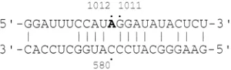
present in all animal cells (25). It acts on regions of complete or partial double-stranded RNA (6) and can convert adenosine to inosine (5). It is known to act on certain cellular RNAs and even viral RNAs (5, 6). In the case of a replicating RNA like HDV, this could explain the observed replacement of A-G. As represented in Fig. 5, the antigenome of wild-type HDV at and around the adenosine at 1012 can be predicted to fold into a region with some base pairing. Recently, Polson and Bass have reported in vitro studies of the RNA substrate requirements for optimal DRADA activity (23). They have examined not only the extent of base pairing but also the importance of nucleotides adjacent to the edited adenosine. The single striking rule that emerged concerns the 59neighbor: the frequency of editing is highest when this neighbor is A or U, much lower for C, and lowest for G. If we refer to 59-A or 59-U as satisfying the DRADA rule, then, as now summarized, the following three changes that occur on HDV RNA satisfy this rule: (i) the 1012 site of wild-type HDV, (ii) the 1012 site of HDV mutated at 1011, (iii) the 1011 site of mutated HDV. There are even additional examples. (iv) In a recent study of the stability of HDV during six consecutive passages in woodchucks, Netter et al. (22) changes corresponding to A3G on the antigenome not only at 1012 but also at 1005 and 1007, which were changes of at least comparable frequency. The latter two sites, which can be seen in Fig. 5, also fit the rule. (In Fig. 5, the three other A’s, at 1009, 1014, and 1020, do not fit the rule and were not detected as changing). (v) Finally, Netter et al. detected a total of 40 single-base changes. Of these, 32 (80%) were consistent with an A3G change on either the ge-nome or the antigege-nome, and 24 satisfy the 59-neighbor rule (22). In summary, the studies reported here for HDV replication in transfected cells and previous studies of HDV replication in an-imals are consistent with DRADA being the enzyme responsible for most of the nucleotide changes on replicating HDV RNA.
Is there competition between the replication of the edited and the unedited RNAs? Such competition could occur at two levels. First, it certainly may occur when HDV RNA is assem-bled into particles; in a subsequent infection of susceptible cells, genomes changed at 1012 would be unable to synthesize the dAg-S needed for replication. Second, from the study of Bichko et al. (1), we now know that even in the absence of assembly, HDV replication can continue after multiple rounds of cell division. Thus, at any time, a major feature in determining whether an HDV RNA species predominates could be replication compe-tence. Some species, either because of the location or extent of editing, could be at a disadvantage to other edited RNAs and could certainly be at a disadvantage relative to the wild type.
ACKNOWLEDGMENTS
This study depended on the assistance of Anita Cywinski and DNA Sequencing Facility and oligonucleotides provided by the DNA Syn-thesis Facility. We were also helped by Matt Bockol and Hans Netter.
We acknowledge the communication of unpublished data by John Casey and John Gerin. Constructive comments on the manuscript were given by David Lazinski, William Mason, and John Pugh.
J.M.T. was supported by grants AI-26522, AI-31927 and CA-06927, from the National Institutes for Health, and by an appropriation from the Commonwealth of Pennsylvania.
REFERENCES
1. Bichko, V., H. J. Netter, and J. Taylor. 1994. Introduction of hepatitis delta virus into animal cell lines via cationic liposomes. J. Virol. 68:5247–5252. 2. Casey, J., and J. Gerin. 1995. Personal communication.
3. Casey, J. L., K. F. Bergmann, T. L. Brown, and J. L. Gerin. 1992. Structural requirements for RNA editing in hepatitis delta virus: evidence for a uridine-to-cytidine editing mechanism. Proc. Natl. Acad. Sci. USA 89:7149–7153. 4. Casey, J. L., K. F. Bergmann, T. L. Brown, and J. L. Gerin. 1993.
Determi-nants of RNA editing in hepatitis delta virus. Prog. Clin. Biol. Res. 382:5–11. 5. Cattaneo, R. 1994. Biased (adenosine to inosine) hypermutation in animal
virus genomes. Curr. Opin. Genet. Dev. 4:895–900.
6. Cattaneo, R. 1994. RNA duplexes guide base conversions. Curr. Biol.4:134–136. 7. Chang, F. L., P. J. Chen, S. J. Tu, M. N. Chiu, C. J. Wang, and D. S. Chen. 1991. The large form of hepatitisdantigen is crucial for the assembly of hepatitisdvirus. Proc. Natl. Acad. Sci. USA 88:8490–8494.
8. Chao, M., S.-Y. Hsieh, and J. Taylor. 1990. Role of two forms of hepatitis delta virus antigen: evidence for a mechanism of self-limiting genome rep-lication. J. Virol. 64:5066–5069.
9. Fu, T.-B., and J. Taylor. 1993. The RNAs of hepatitis delta virus are copied by RNA polymerase II in nuclear homogenates. J. Virol. 67:6965–6972. 10. Fu, T.-B., T.-T. Wu, D. Lazinski, and J. Taylor. Molecular studies of hepatitis
delta virus RNAs. Methods in Mol. Genet., in press.
11. Glenn, J. S., J. A. Watson, C. M. Havel, and J. O. White. 1992. Identification of a prenylation site in the delta virus large antigen. Science 256:1331–1333. 12. Hawley-Nelson, P., V. Ciccaroni, G. Gebeyehu, and J. Jessee. 1993. Lipo-fectamine reagent: a new, higher efficiency polycationic liposome transfec-tion reagent. Focus 15:73–79.
13. Huang, S., and M. Lai. 1993. A unique conformation at the carboxyl termi-nus of the small hepatitis delta antigen revealed by a specific monoclonal antibody. Virology 193:924–931.
14. Kim, U., Y. Wang, T. Sanford, Y. Zeng, and K. Nishikura. 1994. Molecular cloning of cDNA for double-stranded RNA adenosine deaminase, a candidate enzyme for nuclear RNA editing. Proc. Natl. Acad. Sci. USA 91:11457–11461. 15. Kuo, M. Y. P., M. Chao, and J. Taylor. 1989. Initiation of replication of the human hepatitis delta virus genome from cloned DNA: role of delta antigen. J. Virol. 63:1945–1950.
16. Kuo, M. Y. P., J. Goldberg, L. Coates, W. Mason, J. Gerin, and J. Taylor. 1988. Molecular cloning of hepatitis delta virus RNA from an infected wood-chuck liver: sequence, structure, and applications. J. Virol. 62:1855–1861. 17. Laemmli, U. K. 1970. Cleavage of structural proteins during the assembly of
the head of bacteriophage T4. Nature (London) 227:680–685.
18. Lazinski, D. W., and J. M. Taylor. 1994. Expression of hepatitis delta virus RNA deletions: cis and trans requirements for self-cleavage, ligation, and RNA packaging. J. Virol. 68:2879–2888.
19. Lee, C.-Z., P.-J. Chen, M. Lai, and D.-S. Chen. 1994. Isoprenylation of large hepatitis delta antigen is necessary but not sufficient for hepatitis delta virus assembly. Virology 199:169–175.
20. Luo, G., M. Chao, S.-Y. Hsieh, C. Sureau, K. Nishikura, and J. Taylor. 1990. A specific base transition occurs on replicating hepatitis delta virus RNA. J. Virol. 64:1021–1027.
21. Nakabayashi, H., K. Taketa, K. Miyano, T. Yamane, and J. Sato. 1982. Growth of human hepatoma cell lines with differentiated functions in chem-ically defined medium. Cancer Res. 42:3858–3863.
22. Netter, H. J., T.-T. Wu, M. Bockol, A. Cywinski, W.-S. Ryu, B. C. Tennant,
and J. M. Taylor.1995. Nucleotide sequence stability of the genome of hepatitis delta virus. J. Virol. 69:1687–1692.
23. Polson, A., and B. Bass. 1994. Preferential selection of adenosines for modifi-cation by double-stranded RNA adenosine deaminase. EMBO J. 13:5701–5711. 24. Ryu, W.-S., M. Bayer, and J. Taylor. 1992. Assembly of hepatitis delta virus
particles. J. Virol. 66:2310–2315.
25. Wagner, R. W., and K. Nishikura. 1988. Cell cycle expression of RNA duplex unwindase activity in mammalian cells. Mol. Cell. Biol. 8:770–777. 26. Wang, J. G., J. Cullen, and S. M. Lemon. 1992. Immunoblot analysis
dem-onstrates that the large and the small forms of hepatitis delta virus antigen have different C-terminal amino acid sequences. J. Gen. Virol. 73:183–188. 27. Weiner, A. J., Q.-L. Choo, K.-S. Wang, S. Govindarajan, A. G. Redeker, J. L.
Gerin, and M. Houghton.1988. A single antigenomic open reading frame of the hepatitis delta virus encodes the epitope(s) of both hepatitis delta anti-gen polypeptides p24dand p27d. J. Virol. 62:594–599.
[image:6.612.94.262.73.124.2]28. Zheng, H., T.-B. Fu, D. Lazinski, and J. Taylor. 1992. Editing on the genomic RNA of human hepatitis delta virus. J. Virol. 66:4693–4697.
FIG. 5. Predicted rodlike folding of HDV antigenomic sequences at and around the editing site at 1012. The sequence, predicted base pairing, and numbering are as described by Kuo et al. (16). Note that the edited A at 1012, shown in boldface, is predicted to be adjacent to nt 580 on the other side of the rodlike structure. 1012 is thus not predicted to be base paired. Also, if 1011 is mutated to A, it is not predicted to be paired.