“ECHOCARDIOGRAPHIC EVALUATION OF PAPILLARY MUSCLE
FUNCTION IN ISCHEMIC MITRAL REGURGITATION”
Dissertation submitted for
D.M. DEGREE EXAMINATION
BRANCH II – CARDIOLOGY
MADRAS MEDICAL COLLEGE
AND
GOVERNMENT GENERAL HOSPITAL
CHENNAI – 600 003
THE TAMIL NADU DR.M.G.R MEDICAL UNIVERSITY
CHENNAI – 600 032
AUGUST 2007
CERTIFICATE
This is to certify that the dissertation entitled “ECHOCARDIOGRAPHIC EVALUATION OF PAPILLARY MUSCLE FUNCTION IN ISCHEMIC MITRAL REGURGITATION” is the bonafide original work of Dr.P.JAISANKAR, in partial fulfillment of the requirements for D.M. Branch-II (CARDIOLOGY) examination of THE TAMILNADU DR.M.G.R. MEDICAL UNIVERSITY to be held in August 2007.The period of post-graduate study and training was from August 2004 to July 2007.
THEDEAN PROFESSOR V.JAGANATHAN M.D., D.M.
MADRAS MEDICAL COLLEGE PROFESSOR AND HEAD OF THE DEPARTMENT OF CARDIOLOGY AND MADRAS MEDICAL COLLEGE
GOVERNMENT GENERAL HOSPITAL AND
CHENNAI - 600 003. GOVERNMENT GENERAL HOSPITAL CHENNAI - 600 003.
DECLARATION
I Dr.P.JAISANKAR, solemnly declare that this dissertation entitled,
“ECHOCARDIOGRAPHIC EVALUATION OF PAPILLARY MUSCLE FUNCTION IN ISCHEMIC MITRAL REGURGITATION” is a bonafide work done by me at the department of Cardiology, Madras Medical College and Government General Hospital during the period 2004 – 2007 under the guidance and supervision of the Professor and Head of the department of Cardiology of Madras Medical College and Government General Hospital, Professor V.Jaganathan M.D.D.M. This dissertation is submitted to The Tamil Nadu Dr.M.G.R Medical University, towards partial fulfillment of requirement for the award of D.M. Degree (Branch-II) in Cardiology.
Place : Chennai
Date:
Dr.P.JAISANKAR
ACKNOWLEDGEMENTS
A great many people made this work possible. I thank my Dean for allowing me to conduct this study.
My warmest respects and sincere gratitude to our beloved Prof V.Jaganathan Professor and Head of the Department of Cardiology, Government General Hospital, Chennai who was the driving force behind this study. But for his constant guidance this study would not have been possible.
My respectful thanks to Prof R.Alagesan for his constructive ideas, personal guidance and involvement in this study.
I am indebted to Prof. Geetha Subramanian,Prof A.Balaguru, Prof. B.Ramamurthy and Prof. P.Arunachalam without whom, much of this work would not have been possible.
I acknowledge Dr M.A.Rajasekar for the many useful comments he made during this project. In addition, I am grateful to, Dr. G.Gnanavelu, Dr.D.Muthukumar, Dr.G.Karthikeyan,
Dr.G.Palanisamy, Dr.P.S.Mohanamurugan Dr S.Venkatesan Dr.K.Meenakshi, Dr.G.Ravishankar, ,
Dr.Murthy, Dr.P.Justin Paul for tracing all those waveforms and guidance.
I am thankful to my wife DR.J.PRIYA for her support, understanding and for the help concerning the computer network.
CONTENTS
TITLE SHEET
CERTIFICATE
DECLARATION
ACKNOWLEDGEMENT
SERIAL
NO:
TITLE
PAGE NO:
1
INTRODUCTION
1-7
2
LITERATURE REVIEW
8-30
3
AIM & OBJECTIVE
31
4
METHODOLOGY
32-37
5
RESULT & ANALYSIS
39-48
6
DISCUSSION
49-55
7
CONCLUSION
56
BIBLIOGRAPHY
GLOSSARY OF ABBREVIATIONS AND ACRONYMS
SPECIMEN PROFORMA
MASTER CHART
“learn to heal”
INTRODUCTION
Ischemic mitral regurgitation is defined as mitral regurgitation due to coronary artery disease with structurally normal mitral valve leaflet and chordae. Some authors prefer to use the term as functional mirtal regurgitation (1). But functional mitral regurgitation can occur in idiopathic dilated
cardiomyopathy without coronary artery disease. So Ischemic mitral regurgitation and functional mitral regurgitation are not synonymous.
The incidence of coronary artery diseases in rural and urban population in India is reported to be between 14.8 per thousand to 65.4 per thousand(2). Patients with coronary artery diseases
during their course may develop complications such as arrhythmias, mechanical complications (ventricular septal rupture, Ischemic mitral regurgitation) and pump failure.
Ischemic mitral regurgitation occurs in approximately 20% of patients after myocardial infarction and 56% of patients with heart failure due to ischemic or non ischemic cardiomyopathy. (3, 4) Ischemic mitral regurgitation can occur in coronary artery disease both during
acute phase and chronic phase. Ischemic mitral regurgitation is more common in inferior wall myocardial infarction than anterior wall myocardial infarction.(5) There is a graded independent
associationbetween the severity of ischemic MR and the development of Heart failureafter myocardial infarction. Even mild ischemic MR is associatedwith an increase in the risk of heart failure. Ischemic
mitral regurgitation is an independent prognostic factor in patients with chest pain even without myocardial infarction. (6) Advancing age, female gender, multiple vessel coronary artery disease,
congestive heart failure, recurrent ischemia, large infarct size, and prior acute myocardial infarction are all risk factors for developing IMR.
Is the regurgitation of blood from left ventricle to left atrium during systole due to inadequate cooptation of mitral leaflet. Mitral regurgitation is caused by several diseases. In India Rheumatic fever is the leading cause of Mitral Regurgitation. As the incidence of RF is declining recently, Mitral Regurgitation due to other causes also contributes considerably.
Causes of Chronic Mitral Regurgitation;
(7)INFLAMMATORY Rheumatic heart disease Systemic lupus erythematosus Scleroderma
Degenerative
Myxomatous degeneration of mitral valve leaflets (Barlow click murmur syndrome, prolapsing leaflet, mitral valve prolapse)
Marfan syndrome Ehlers-Danlos syndrome Pseudoxanthoma elasticum
Calcification of mitral valve annulus INFECTIVE
Infective endocarditis STRUCTURAL
Rupture or dysfunction of papillary muscle
Dilation of mitral valve annulus and left ventricular cavity. HOCM (Hypertrophy obstructive cardiomyopathy )
Prosthetic Paravalvular leak. CONGENITAL
Mitral valve clefts or fenestrations
Parachute mitral valve abnormality in association with 1. Endocardial cushion defects
2. Endocardial fibroelastosis
3. TGA (D Transposition of great artery )
4. ALCOPA (Anomalous origin of left coronary from pulmonary artery )
ISCHEMIC MITRAL REGURGITATION (IMR)
Ischemic MR is a common complication of ischemic heart disease as mentioned above and it adversely affects the prognosis. (8, 9)
Mechanism of IMR
While acute MR secondary to rupture of the PM is well understood, the understanding of the development of IMR in ventricular dysfunction is not totally clear (1). Mitral annular dilatation may
leaflet overlap MR will develop. This mechanism is similar in patients with non ischemic cardiomyopathy who develop MR.10 The concept that MITRAL ANNULAR DILATATION causes
functional mitral regurgitation was created in 1956 by DR. Friedberg.11 But the first clinical study to
assess the mitral annular dilatation as the cause of IMR came from DR.BOLTWOOD et al in1983.12
When they sought to elucidate the mechanism of mitral regurgitation (MR) in dilated cardiomyopathy(DCM). One decade later BOLTWOOD was supported by DR SHENGQIN HE et al in 1997 and he concluded that annular dilatation increases regurgitation for any position of PM and creating clinically significant MR13
In contrary to above studies DR.YATAKA OTSUJI et al in 2002 said that Isolated Annular Dilation Does Not Usually Cause Important Functional Mitral Regurgitation. and he proved that in human studies.14.
Papillary Muscle Displacement and LV Geometry
Recent work in both animal and human studies has examined the role of global and regional ventricular dysfunction in PM displacement. When the Papillary muscles are displaced towards the LV apex, it causes tenting of the leaflet. Leaflet tenting pulls the tips of the mitral leaflets away from each other; resulting in less leaflet overlap and development of MR. This leaflet tethering is measured as leaflet tethering distance. All the studies in the past demonstrated consistent correlation of increased leaflet tethering distance with ischemic mitral regurgitation.
16. The impact of global dilatation was illustrated in a canine model of infarction where the pericardial
space was reduced by folding the pericardium over itself parallels to the long axis and suturing it to limit LV dilatation17. Then the left circumflex artery was ligated, producing ischemia of the
infero-posterior wall and medial PM. With restriction of LV dilatation, there was no MR. However when the LV was allowed to dilate, MR developed. Three-dimensional echocardiography demonstrated an increased tethering distance from the PM to the anterior annular ring. The tethering length correlated with LV sphericity.18
However, not all patients with global LV dilatation develop MR 19. In addition the
same amount of LV dysfunction is associated with varying degrees of MR.20 Kono et al demonstrated
that changes of LV geometry due to the regional hyperkinesias of the LV segment overlying the PM may play an important role in the development of IMR1. In a canine model of acute MI, they
demonstrated that changes in the configuration of the left ventricle can affect the position of one or both papillary muscle groups. Remodeling of the inferior basal wall caused apical displacement of the papillary muscles. This resulted in significant increase in the distance between the coaptation point of the mitral valve leaflets and the plane of the mitral valve resulting in IMR 1. In contrast, global models
In patients with LV dysfunction, Yia used quantitative echocardiography to determine the relationship between the degree of functional mitral deformation and local ventricular remodeling.20 they found that
the major determinant of effective regurgitant orifice (ERO) of IMR was the degree of systolic mitral tenting, and to a lesser extent, systolic mitral tenting was directly related to local remodeling that caused apical and posterior displacement of both PM. Lateral displacement of the PM was only weakly associated with the degree of IMR. The displacement of the PM was independent of LV global remodeling, Ejection Fraction, LV volume and sphericity.
The observation that ischemia worsened MR led to the term PM dysfunction”.21
However, an alternative hypothesis suggests that PM ischemia may in fact result in a decrease in MR by displacing the leaflets towards the annulus due to elongation of the ischemic PM.22, 23 This was
REVIEW OF LITERATURE
First, we will briefly review about the functional anatomy of mitral valve apparatus.
FUNCTIONAL ANATOMY OF MITRAL VALVE;
24, 25THE MITRAL APPARATUS is a complex - finely coordinated mechanism that can be deranged by a multiplicity of acquired and congenital disorders and requires competence for its functional integrity of six elements working in delicate concert.26 These anatomic elements are: (1) left
atrial wall, (2) annulus, (3) leaflets, (4) chordae tendineae, (5) papillary muscles, and (6) left ventricular wall.
The exact mode of closure of the normal mitral valve is still uncertain.27, 28 Left ventricular
systole begins with contraction of the papillary muscles. The vertical forces exerted by the contracting papillary muscles move the leaflets into apposition. As the intraventricular pressure rises, the free edges of the cusps firmly coapt, mutually supporting each other along a comfortable margin of their atrial surfaces, and firmly sealing the orifice. The remainder of each leaflet bulges like a parachute toward the left atrium.29 The annulus not only serves as a fulcrum for the leaflets, but during ventricular
systole decreases its circumferential size, thus reducing the area that the leaflets are required to bridge.30, 31 Even so, the surface area of the leaflets is about two and half times the area of the orifice,
the harmony of the valvular mechanism and render it incompetent. Coordinated interaction among these elements-which are interdependent- usually means that more than one, or even several, are deranged at any one
LEFT ATRIUM
Two contributions of the left atrium have been related to competence of the mitral valve: (1) contraction and relaxation 32, 33 and (2) atrial dilatation. Although atrial contraction and relaxation seem
to be capable of closing the mitral valve in man, the absence of such activity does not cause mitral regurgitation.34 Competence of the mitral apparatus depends in part on a normal sequence of ventricular
activation but not on a normal sequence of atrial and ventricular contraction. Loss of effective atrial contraction (atrial fibrillation, complete heart block, and others) does not necessarily result in regurgitation. 34 Left atrial enlargement itself can contribute to mitral regurgitation. Dilatation of the left
atrium does not affect the anterior leaflet since that leaflet is anchored to the root of the aorta. The posterior leaflet, however, can be directly affected.29 As the left atrium enlarges, its posterior wall is
displaced posteriorly and downward. Because of the continuity of the atrial endocardium and posterior mitral leaflet, this displacement exerts tension on the posterior leaflet.29 The displacement may prevent
that cusp from contacting its mate or may aggravate preexisting leaflet malapposition. Thus, as mitral regurgitation provokes left atrial enlargement, as Edwards and Burchell35 aptly put it, "mitral insufficiency begets mitral insufficiency” irrespective of the initiating causes.
MITRAL ANNULUS
of dilatation of the mitral ring. Nevertheless, the size of the ring does play a role in preserving competence of the mitral valve, albeit a role that relates less to dilatation than to failure to decrease its circumference during systole. The tissue at the basal attachments of the mitral leaflets is tough but pliable. Toughness opposes dilatation; pliability permits sphincter-like contraction of the annulus during systole, which reduces the area that the apposing leaflets must bridge by an estimated 20 to 50%.27,31 Left ventricular dilatation may exert an unfavorable effect on the annulus chiefly by applying
a distending pressure that opposes systolic annular contraction.
MITRAL LEAFLET
Proper closure of the leaflets represents an ultimate goal of the entire mitral mechanism. The areas of the two leaflets are nearly identical, but the basal to free edge length of the anterior leaflet is two or more times that of the posterior leaflet. Consequently, the anterior leaflet is more mobile, while the posterior leaflet fulfills a secondary or supporting role.”36 Although the mitral leaflets, like
other cardiac valves, have been looked upon as passive connective tissue structures, recent investigations have found within their substance striated muscle bundles to which an active function has been assigned.37. It is uncertain whether such muscle fibers have physiologic significance in
promoting closure of the valve.
PAPILLARY MUSCLE AND CHORDE TENDINAE
Functional anatomy of the normal papillary muscle:
24supply from the anterior descending branch of the left coronary artery. The posteromedial papillary muscle arises near the junction of the posterior wall of the left ventricle and the Interventricular septum and is supplied with blood either by tributaries from the circumflex artery or by posterior descending branches from the right coronary artery These small papillary arteries course longitudinally to the apex of the papillary muscle where they terminate in the arterioles, capillaries, venules, and small veins. At times these papillary arteries form arcuate anastomoses near the distal ends of the muscle. In the normal-sized heart, the long axis of the papillary muscle is oriented almost perpendicular to the atrioventricular ring. This orientation of the papillary muscles provides a mechanical advantage in that tension developed by the papillary muscles is applied almost perpendicular to the mitral valve leaflets. On the other hand, with ventricular dilatation the papillary muscles migrate laterally, so that tension developed by the papillary muscles is applied tangentially to the mitral leaflets. The greater the lateral displacement of the papillary muscles the greater the mechanical disadvantage. The function of the papillary muscles and chordae tendineae to restrain the movements of the mitral valve leaflets during ventricular systole is obvious. However, the dynamic nature of this function is not always appreciated. Normal mitral valve function depends upon the maintenance of the proper spatial relationships between the papillary muscles, the chordae tendineae, and the mitral valve leaflets throughout all phases of the cardiac cycle. During the isovolumetric phase of ventricular systole the rapid rise in intraventricular pressure causes the mitral valve leaflets to bulge towards the left atrium and to come into firm surface contact with each other, thus closing the atrioventricular orifice.38-41 The movement of the mitral valve
leaflets towards the atrium pulls the chordae tendineae taut. In the interest of completeness, it may be stated that there is evidence that the mitral valve leaflets come into apposition to close the atrioventricular orifice just before the onset of ventricular systole.42 Nevertheless, firm apposition of the
tendineae from each papillary muscle attach to the corresponding halves of both leaflets of the mitral valve. The papillary muscles and chordae tendineae must support the force acting upon the mitral valve, which is equal to the intraventricular pressure times the cross-sectional area of the atrioventricular orifice. In a previous report from this laboratory,43 we have estimated, on the basis of
certain theoretic considerations, that each papillary muscle of the left ventricle supports a total peak load of 19 tons during a 24 hour period for a heart rate of 70 beats per minute and an arterial blood pressure of 120/80 mm. Hg.
During the ejection phase of ventricular systole, the apex of the left ventricle moves towards the atrioventricuIar orifice. Since the moment-to-moment length of the chordae tendineae is fixed, the papillary muscles must shorten during systole to maintain the proper distance between the base of the papillary muscles and the atrioventricular orifice in order to prevent eversion of a portion of the mitral leaflets into the left atrium. It is of interest that motion pictures of mitral valve movement in the intact dog have demonstrated that the mitral valve leaflets move downward into the ventricle during ventricular systole rather than upward toward the atrium.44 Thus, contraction of the papillary muscles
takes up the slack which would have been created in the chordae tendineae as a result of the shortening of the distance between the apex of the left ventricle and the atrioventricular orifice during the ejection phase of ventricular systole. Furthermore, the papillary muscles must develop sufficient tension to overcome intraventricular pressure. This latter point is important since the tension in the free wall of the left ventricle decreases or remains constant during the ejection phase of systole45. Therefore, while
the muscle of the free wall of the left ventricle “loafs” during ventricular ejection, the papillary muscles must continue to develop more tension.
papillary muscle dysfunction and its manifestation. He enumerated the causes of PM dysfunction as follows;
Etiology of papillary muscle dysfunction
251. Circulatory insufficiency (ischemia) Angina pectoris
Infarction of papillary muscle Acute
Chronic (fibrosis)
Systemic circulatory disturbances (hypotension, erythrocytosis, anoxia, hematometakinesia, etc.)
2. Left ventricular dilatation Generalized
Localized (aneurysm)
3. Non ischemic atrophy of papillary muscle Senile
Associated with cachexia
4. Defective development of papillary muscle apparatus Congenitally long or short papillary muscle or chordae tendineae
Ectopic origin of papillary muscle Ectopic insertion of chordae tendineae 5. Endocardial disease
Endocardial fibroelastosis Endomyocardial fibrosis 6. Heart muscle disease
Inflammatory (myocarditis) Degenerative cardiomyopathy
Infiltrative (metastatic carcinoma, amyloidosis) 7. Neoplastic (primary tumor of myocardium)
Disturbances in the time course of papillary muscle activation and contraction
8. Rupture of papillary muscle or chordae tendinea
The most common causes of PM dysfunction is coronary insufficiency. Papillary muscle is vulnerable to ischemia because;24,25
Papillary muscle is subendocardial structures,
Papillary muscle are supplied by terminal branches of the coronary arteries, Papillary muscle is the thickest portion of the endocardium.
Papillary muscle develop large amounts of tension during ventricular systole,
These factors combine to render the papillary muscles particularly vulnerable to ischemia. The papillary muscles may become ischemic not only as a result of narrowing of the coronary arteries but also as a result of disease states associated with diminished coronary artery perfusion. Because of the large amounts of tension which must be developed by the papillary muscles during ventricular systole, they are easily damaged by ischemia. Furthermore, in some hearts the major blood supply to both papillary muscles of the left ventricle is derived from the same artery (circumflex branch of left coronary artery), so that the risk of simultaneous ischemia of both papillary muscles is increased.
consecutive hearts studied at necropsy46. During episodes of ischemia or following infarction of a
papillary muscle, the muscle is rendered completely or partially incapable of contraction. Providing that the heart is not enlarged, the normal spatial relationships between the elements of the mitral valve apparatus are maintained during isovolumetric contraction and the valve is competent However,
during ventricular ejection the slack created in the chordae tendineae by the apex-to-base movement of the left ventricle is not taken up because of the inability of the ischemic papillary muscle to shorten. Thus, a portion of each mitral valve leaflet everts into the left atrium and the valve becomes incompetent if the ischemia is only transitory, as during an episode of angina pectoris, clinical evidence of mitral valve incompetence rapidly subsides as the ischemic papillary muscle regains the ability to contract. On the other hand, following infarction of a papillary muscle, the clinical signs of mitral incompetence usually regress slowly as the papillary muscle gradually recovers. The changing characteristics of the murmur associated with papillary muscle dysfunction as a result of infarction of the muscle will be explained on this basis. It should be pointed out, however, that the sudden development of an apical systolic murmur after myocardial infarction is much more often due to papillary muscle dysfunction than to rupture of a papillary muscle or perforation of the interventricular septum. When ischemia and/or infarction results in diffuse scarring, degeneration, and atrophy of a papillary muscle, the retracted muscle pulls a portion of each mitral leaflet into the ventricle so that the valve is incompetent even during isovolumetric contraction. During the ejection phase of systole, the apex-to base movement of the left ventricle may permit better apposition of the mitral valve leaflets so that the degree of valve incompetence decreases.
In contrary to elaborate discussion of Papillary muscle dysfunction by previous author, in 1991
DR.SANJIVE KAUL et al,47did an experimental work with dogs and concluded that PMD and/or
Coaptation {IMLC}; the greater the degree of IMLC, the greater the MR. These findings suggest that the mechanism of ischemic MR is not related to PMD. There may also be important therapeutic implications of these findings. In his study dogs were grouped as follows, group 1 dogs (n=8), varying degrees of regional and global LV dysfunction were produced. In group 2 dogs (n=7), the circulation to the papillary muscles was isolated from that of the rest of the LV. Dysfunction of one or both papillary muscles was produced without producing global LV dysfunction. Global LV dysfunction was also produced while keeping papillary muscle function intact. The degree of MR (assessed using contrast echocardiography) was correlated in both groups of dogs with thickening of the papillary muscles and regional and global LV function. In the group 3 dogs (n=6), the spatial distribution of blood flow within each papillary muscle was determined during ischemia by using radio labeled micro spheres. Thickening of the papillary muscles was assessed at three different levels along their lengths and was correlated with average blood flow at these levels. In group 1 dogs, MR was noted only when global LV function was affected and its severity correlated inversely with global LV function In comparison, there was poor correlation between MR and anterior and posterior papillary muscle thickening. In group 2 dogs, MR did not occur in the presence of either PMD or akinesia of the immediately adjacent LV myocardium. MR occurred only when global LV dysfunction was produced (with the papillary muscle function intact), and its severity correlated inversely with global LV function. There was poor correlation between the degree of MR and thickening of the anterior and posterior papillary muscles In both groups of dogs, MR was associated with incomplete mitral leaflet closure (IMLC), and the severity of MR correlated linearly with the degree of IMLC. MR was never associated with mitral valve prolapse. In the group 3 dogs, despite more inhomogeneous flow during ischemia to the anterior compared with the posterior papillary muscle, mean thickening of these muscles was similar (3±10% and 3±4%, respectively). Furthermore, there was minimal variability in thickening between different parts of the muscles
shape change of left ventricle but not the simple enlargement of LV in heart failure may be an important determinant of functional MR.He examinedrelationship between left ventricular (LV) shape and functional mitral regurgitation (MR) was examined in 18 dogs with long-standing heart failure produced by multiple sequential intracoronary microembolisations. Global LV shape changes were evaluated from angiographic silhouettes obtained at baseline (before embolisation) and during heart failure. LV shape changes at end systole and end diastole were quantitated based upon the ratio of the major-to-minor axis and the sphericity index. Among the 18 dogs studied, 11 developed 1 + to 3+ MR during heart failure and seven did not. There was no difference among the two groups with respect to hemodynamics, LV ejection fraction, chamber volume, and regional wall motion. A significant difference, however, was observed between the two groups with respect to the global indexes of LV shape. In dogs with MR, the end-systolic major-to-minor axis ratio decreased 29 + 3% between baseline and heart failure compared with only a 18 + 3% decrease in dogs without MR (p < 0.01). In dogs with MR, the end-systolic sphericity index increased between baseline and heart failure compared with an increase of only 30 t 5% in dogs without MR (p < 0.02). Significant and directionally similar differences were observed during end diastole. These data indicate that in heart failure functional MR is associated with a more spherical LV and is not the result of simple LV chamber enlargement. Shape changes of the LV that occur in heart failure may be an important determinant of functional MR. Few years later, DR.SIN F YIU et al 49 in 2000,studied the Determinants of the Degree of
rhythm, they quantified simultaneously by echocardiography the effective regurgitant orifice (ERO) of FMR by using 2 methods: mitral deformation (valve and annulus) and left ventricular (LV) global (volumes, stress, function, and sphericity) and local (papillary muscle displacements and regional wall motion index) remodeling. A wide range of ERO was observed, unrelated to ejection fraction The major determinant of ERO was mitral deformation, ie, systolic valvular tenting and annular contraction, independent of global LV remodeling. Systolic valvular tenting was strongly determined by local LV alterations, particularly apical and posterior displacement of papillary muscle, with confirmation in multivariate analysis, independent of LV volumes, function, and sphericity.
In 1956 Friedberg11wrote: “Relative or functional mitral insufficiency occurs commonly as
a result of pronounced dilatation of the left ventricle, due to hypertensive, coronary or aortic valvular disease, usually in association with left ventricular failure. Imperfect valvular closure under such circumstances is due to dilatation of the mitral ring and to retraction of the cusps by chordae and papillary muscles as the ventricular chamber elongates.” He created the concept that MITRAL ANNULAR DILATATIONcauses functional mitral regurgitation.
But the first clinical study to assess the mitral annular dilatation as the cause of FMR came from DR.BOLTWOOD et al 12 in1983. When they sought to elucidate the mechanism of mitral
regurgitation (MR) in dilated cardiomyopathy
length from the PM base to the annular plane were measured. The MR group was characterized by markedly enlarged occlusional leaflet area, striking mitral annular dilatation, and left atrial enlargement . Chordal length and angulation, PM length, contraction, and tethering length, and left ventricular volume were not significantly different in the MR vs the no MR group. Non coaptation of the mitral leaflets at their free margins was not observed in any MR patient. With the use of stepwise linear regression LEAF was determined chiefly by annular size with left ventricular size having little additional influence. Thus, DCM is associated with enlargement of the mitral anulus, which is more pronounced in those patients with MR. Based on the quantitative estimates of occlusional leaflet area, we postulate that mitral leaflet tissue can stretch somewhat to accommodate dilatation of the mitral complex, but as the requirement for occlusional leaflet area increases less tissue is available for coaptation. Thus, although coaptation continues to occur, the valvular seal becomes ineffective once a critical LEAF is reached. The chief determinant of LEAF is the mitral annular size, while left ventricular size is a less important factor.
One decade later BOLTWOOD was supported by DR SHENGQIN HE et al 13 in 1997. He
analyzed factors like annular size, position of papillary muscle, and tethering of mitral leaflet by in vitro model with excised porcine value. He concluded that annular dilatation increases regurgitation for any position of PM and creating clinically significant MR.
In contrary to above studies DR.YATAKA OTSUJI 14 et al in 2002 said that Isolated
patients with ICM. Multivariate analysis identified PM tethering length, not MA dilation, as an independent primary contributor to MR. He concluded that Isolated annular dilation does not usually cause moderate or severe MR. Important functional MR also depends on LV dilation and dysfunction, leading to an altered force balance on the leaflets, which impairs coaptation. This view is also supported by Zhu et al 50in his study of Mechanism of Persistent Ischemic Mitral Regurgitation After
Annuloplasty: Importance of Augmented Posterior Mitral Leaflet Tethering.
In all studies, whatever the controversies exists regarding the mechanisms of functional mitral Regurgitation, the only consistent proved mechanism of IMR is the mitral leaflet tethering distance. Two basic force act on the mitral leaflets and play important role in IMR. Ischemic LV distortion leads to papillary muscle displacement pulling the leaflet away from mitral apparatus preventing coaptation. This force is opposed by second force i.e. LV contractility tending to close the mitral leaflet. In addition to these forces papillary muscle exerts its force during systole in such way that the mitral leaflet neither prolapse nor tents to produce mitral regurgitation. Coordination of these forces requires for normal closure and coaptation of mitral valve. Defect in any of the force can leads to mitral leaflet tethering which results in functional mitral regurgitation.
In 1981 ROBERT W. GODLEY, M.D., et al51evaluated a possible association of PM
function and tethering of mitral leaflet, he performed
provides the first objective evidence that de novo
mitral regurgitation in patients with prior myocardial infarction is due to dyskinesis involving the left ventricular myocardium beneath one of the papillary muscles, producing increased tension on the mitral leaflets and preventing normal closure by increased mitral leaflet tethering distance This mechanism is supported by several investigators. Several surgical techniques were developed based on this mechanism. Mitral leaflet tethering is the primary mechanism of Recurrent mitral regurgitation after anuloplasty for functional ischemic mitral regurgitation. Fang Zhu, MD et al 52 hypothesized that
surgical annuloplasty for ischemic mitral regurgitation (MR) that displaces the posterior annulus anteriorly can potentially augment posterior leaflet (PML) tethering, leading to persistent MR.
intraventricular papillary muscle imbrication without ring during left ventricular restoration was described in . J Thorac Cardiovasc Surg 2002. Surgical relocation of the posterior papillary muscle in chronic ischemic mitral regurgitation as one method of treatment for IMR was described in Ann Thorac Surg 200652.
Emmanuel Messas, MD, et al 28in 2001 did an experiment and proposed that Chordal Cutting
as a New Therapeutic Approach for Ischemic Mitral Regurgitation. It was tested in 8 mitral valves: a porcine in vitro pilot with Papillary muscle displacement and 7 sheep with acute inferobasal infarcts studied in vivo with three-dimensional (3D) echo to quantify MR in relation to 3D valve geometry. In all 8 valves, PM displacement restricted leaflet closure, with anterior leaflet angulation at the basal chord insertion, and mild-to-moderate MR. Cutting the 2 central basal chordae reversed this without prolapse. In vivo, MR increased after infarction and then decreased with chordal cutting ; this paralleled changes in the 3D leaflet area required to cover the orifice as dictated by chordal tethering. He concluded that Cutting a minimum number of basal chordae can improve coaptation and reduce ischemic MR. Such an approach also suggests the potential for future minimally invasive implementation.
ROLE OF PAPILLARY MUSCLE FUNCTION IN IMR:
Earlier in 1968 BURCH et al 25 discussed ‘syndrome of papillary muscle dysfunction’ and the
contribution of papillary muscle dysfunction to the pathogenesis of ischemic mitral regurgitation and proved it by necropsy study. There was no objective evidence for that mechanism. Afterwards several studies were conducted, both experimental and in vivo, to prove the role of PM dysfunction in IMR. In contrary to previous studies, in 1991 DR.SANJIVE KAUL et al,47 did an experimental work with
dogs and concluded that PMD and/or dysfunction of the immediately adjacent LV myocardium do not result in IMR.
DOES THE PAPILLARY MUSCLE FUNCTION DECREASE IMR?
Emmanuel Messas, MD et al53did an experimental study in sheep with the hypothesis that
PM contractile dysfunction can actually diminish MR due to ischemic distortion of the inferior base alone. He occluded the proximal circumflex circulation in 7 sheep while maintaining Papillary muscle perfusion, confirmed by contrast echocardiography. By 3D echocardiography, He measured the tethering distance between the ischemic medial PM tip and anterior annulus and LV ejection volume to give MR . In 6 sheep without initial MR, inferior ischemia alone produced PM tip retraction with restricted leaflet closure and mild-to-moderate MR. Adding PM ischemia consistently decreased MR and tethering distance, as PM strain rate decreased and leaflet tenting decreased. In one sheep, prolapse and MR resolved with inferior ischemia and recurred with PM ischemia. He concluded that PM contractile dysfunction can paradoxically decrease MR from infero basal ischemia by reducing leaflet tethering to improve coaptation. This emphasizes the role of geometric factors in ischemic MR mechanism and potential therapy.
MR fraction in all subjects with variable degrees of LV remodeling, but it had a significant inverse correlation with the MR fraction with LV remodeling of a similar location and extent, indicating that PM dysfunction is associated with less MR. Papillary muscle dysfunction, reducing its longitudinal contraction to induce leaflet tethering, attenuates ischemic MR in patients with basal inferior LV remodeling. (J Am Coll Cardiol 2005; 46:113–9)
On summary we have evidence for and against different mechanism of ischemic mitral regurgitation. They are left ventricular dysfunction, mitral annular dilatation, mitral leaflet tethering, dysfunction of LV myocardium and papillary muscle.
Regarding papillary muscle dysfunction as a cause of ischemic mitral regurgitation, hypothesized theories are
1. PM dysfunction may cause IMR.
2. IMR may not related to PM dysfunction
3. PM dysfunctioncan attenuate IMR in some cases.
ECHOCADIOGRAPHIC EVALUATION OF PM FUNCTION
Evaluation of papillary muscle function by any method is one of the difficult task in cardiology. Echocardiogram of papillary muscle evaluated from M mode through 2D Echo to tissue Doppler echocardiogram.
In 1972 Tallury, DePaspuale, and Burch et al 55 evaluated papillary muscle function in
the early stages of acute myocardial infarction, ischemia and/or infarction of a papillary muscle render the muscle partially or totally incapable of shortening. Failure of the muscle to shorten results in mitral regurgitation. The echocardiogram offers a safe and convenient method for studying the day-to-day variations in papillary muscle function Cross-sectional echocardiographic spectrum of papillary muscle dysfunction study was done by Burch, G. E., DePasquale, N. P., and Phillips, J. H. et al 56 in
1979.
Cross-sectional echocardiography identified two abnormal patterns of mitral valve closure in 14 patients with mitral regurgitation due to papillary muscle dysfunction:
(1) in three patients with an akinetic inferior-posterior wall but normal cavity size, papillary muscle fibrosis was associated with late systolic mitral valve prolapse, and
THE AIM OF THE STUDY:
1) To assess the mechanisms of ischemic mitral regurgitation in patients with old
myocardial infarction.
2)
To assess the role of Tissue Doppler imaging in evaluation of papillary muscle
function.
3) To assess the contribution of papillary muscle dysfunction in the pathogenesis of
MATERIALS AND METHODS
This was a prospectus study done between January 2006 and January 2007 at the department of cardiology, Government General Hospital Chennai. The study cohort comprises 30 consecutive patients with old myocardial infarction and Mitral regurgitation who were referred to our department for evaluation or follow up. This study patients are divided into two groups according to location of Myocardial Infarction. Group 1 has old inferior wall myocardial infarction and Group 2 has old anterior wall myocardial infarction. Group I patients are further subdivided into 2 groups based on their left ventricular spherecity. Patients with increased left ventricular spherecity belong to Group Ia. and with normal left ventricular spherecity belongs to Group Ib. Patients are belonging to the age group of 30 – 60 years.
INCLUSION CRITERIA
1) presence of old myocardial infarction
2) Presence of Mitral regurgitation ( Mild – Severe) (Old Myocardial infarction is defined by the following character
1) History of myocardial infarction 2) ECG evidence of old MI
3) RWMA in echo
EXCLUSION CRITERIA
1) Acute myocardial infarction(less than 1 month) 2) Multiple MI
3) Mitral regurgitation due to other causes (RHD, IE and MVPS etc.) 4) Dilated cardiomyopathy
5) Congenital heart disease. 6) Pericardial diseases.
Informed concern was obtained from all the patients.
ECHOCARDIOGRAPHY
Echocardiographic evaluation of all patients was done using Philips iE33 machine in our Echo lab III. Standard M-Mode, Two Dimensional, Color flow and tissue Doppler echocardiography was done for all patients using conventional method. Recordings of the apical four and two chamber views, long axis and parasternal short axis views were done with special attention paid to visualize the Papillary Muscle. LV end-diastolic and end-systolic cavity areas were traced in those views, and the LV end-diastolic volume (EDV) and ejection fractions (EF) were calculated by the method of discs.
LEFT VENTRICULAR SPHERICITY
1a with significant LV bulging with a D/L > 0.60 (upper normal value is 0.60) and group 1b without significant LV bulging with D/L < 0.60.
MITRAL ANNULAR AREA ( MAA)
We assumed that the mitral annulus is elliptical in geometry during mid systole. Annular dimension of mitral valve is measured in apical 4 chamber and apical two chamber view during mid systole and mitral annular area is calculated using the formula;
MAA = d1
×
d2
×
Π
/4.
LEAFLET TETHERING DISTANCE:
25systole.
MITRAL REGURGITATION:
Is diagnosed and assessed by color flow and CW Doppler echocardiography. In our study Mitral regurgitation severity is assessed by the following methods
1) Jet Area
2) Density of CW Doppler. 3) Vena Contracta
4) Mitral Regurgitation Volume.
The jet area of MR flow is compared with area of Left Atrium and graded as mild MR if occupied less than 20% of LA. Severe MR is diagnosed if jet area occupied more than 40% of LA. Color flow Doppler signal density is compared with forward flow signal density of mitral valve. If the regurgitant flow signal density is denser than forward flow signal MR is graded as severe. Vena cotracta is the narrowest portion of MR jet and gives good correlation with severity of MR. Size of the vena contracta is > 0.7cm indicates severe MR. Mitral stroke volume was calculated as the product of the time
velocity integral of transmitral flow and cross sectional area of mitral annulus. Aortic stroke volume was calculated as the product of the time velocity integral of transaortic flow and cross sectional area of left ventricular outflow. Regurgitant volume is measured by subtracting aortic stroke volume from mitral stroke volume. Regurgitant volume is >60ml indicates severe MR.
EVALUATION OF PAPILLARY MUSCLE FUNCTION
peak systolic PM thickening in its short-axis direction.
In our study, Three methods were used to evaluate PM function 55,56
1) M Mode Echocardiography 2) 2 Dimension Echocardiography 3) Tissue Doppler Imaging.
M-MODE ECHOCARDIOGRAPHY;
M mode echocardiography of PM is done in either apical 2C view or short axis view whichever view gives good resolution of PM and ultrasound beam perpendicular to PM. Systolic thickening of PM was measured with reference to ECG gating/ septal or posterior wall thickening.
TWO DIMENSIONAL ECHOCARDIOGRAPHY:
During two Dimensional echocardigraphic evaluations, scared and dysfunctional PM is identified by its increased echogenesity. Short axis thickening of papillary muscle during systole is measured in apical 2C or 4C view after marking the mid portion of PM muscle as reference point for measurement. Papillary muscle diameter was measured during end - systole and end – diastole at the level of reference point. End – systole is defined both by peak of T wave in ECG and begning of opening of mitral valve. End – diastole is defined both by peak of R wave in ECG and end of closure of mitral valve. Systolic thickening of papillary muscle is measured in the same way as free wall Systolic thickening using the following formula;
END SYSTOLIC THICKNESS — END DIASTOLIC THICKNESS
_______________________________________________________
x
100 END DIASTOLIC THICKNESSTISSUE DOPPLER IMAGING
Regional myocardial function is better assessed with TDI than any other method of
echocardiography. Quantitative assessment is an important prerequisite for complete description of the dynamic changes that occur during ischemia .Conventional assessment of wall motion based on visual assessment is highly subjective and semi quantitative. Accurate assessment of regional contractile function is important for prognosis and management in patients with coronary artery disease. It has been demonstrated that wall thickening is a useful measure of regional function and is more precious than wall motion analysis . However the differentiation of regions with abnormal contractile function from regions with normal contractile function by planar method in patient with coronary artery disease is difficult because of wide range of thickening in normal and abnormal regions. The low temporal resolutions used in stress echocardiographic studies together with the limited ability of human vision to discern small differences in myocardial asynchrony, do not allow an objective quantitative assessment of complex wall motion. Tissue Doppler velocity and SRI can accurately quantify Local mechanical function with higher temporal accuracy than any current clinical method. Two dimensional strain analyses is more accurate than wall thickening analysis in discriminating dysfunctional from functional myocardium and therefore it improves the detection of regional difference in function. Two dimensional tissue Doppler and strain Doppler echocardiographic techniques used in this study allowed processing of simultaneous velocity and strain traces from apical two chamber view in the same cine loop. The Doppler ultrasonic beam was placed in mid inferior region of inferior wall of left ventricle and mid point of both papillary muscles. The Doppler ultrasonic beam was also placed in the anterior septum of left ventricle.RESULTS
The study population included patient with old inferior wall and anterior wall MI complicated by mitral regurgitation. The patients were grouped into 3 groups. Group 1 patients with old inferior wall MI complicated by mitral regurgitation. These patients were further subdivided into two groups based on left ventricular sphericity as group 1a are patients with increased left ventricular sphericity and group 1b patients were with normal left ventricular sphericity. Group 2 patients are with old anterior wall MI complicated by MR..
The Demographic characteristics of patients are detailed in table 1. The average age is 49 +1 year in all groups. No significant different in gender, age and BMI in all the three groups. The distribution of coronary risk factor such as systemic hypertension, diabetes mellitus, smoking and dyslipidemia were almost equal in all the groups .The average time since Acute MI in all groups was 4 to 5 months
TABLE – 1 PATIENT PROFILE
S.No Character Group I a Group I b Group II Pvar
1. Age( years) 49.0 49.7 48.5 Ns
2. Male 60% 50% 70% Ns
3. BMI 27+0.9 25+1.2 29+ 0.8 Ns
4. Time since AMI 4.6mon 4.3mon 4.9mon Ns
5. HT 50% 50% 50% Ns
6. DM 60% 50% 60% Ns
7. Smoking 30% 30% 30% Ns
8. Dyslipidemia 20% 30% 50% Ns
{BMI – Body mass index}
ECHO CHARACTERS OF PATIENT.
I a I b II
LVFF 47.1% 54.1% 44.9%
MR (Mild) 50% 80% 60%
MR (Mod-severe) 50% 20% 40%
LV sphericity 66.0% 49.1% 58.2%
MAA 4.2cm2 3.81 cm2 4.44 cm2
leaflet tethering distance
AML tethering distance 16.7mm 17.4 mm 20.5 mm
PML tethering distance 21.6 mm 18.3 mm 16.8 mm
PM Systolic thickening M mode
APM 38.7% 37.1% 30.9%
PPM 31.8% 31.0% 41.5%
2D ECHO
APM 36.7% 33.7% 27.5%
PPM 31.6% 32.3% 32.4%
Systolic peak velocity in TDI
APM 7.35 m/s 7.36 m/s 6.73 m/s
PPM 6.90 m/s 6.40 m/s 7.68 m/s
{LVEF – left ventricular ejection fraction;MR- mitral regurgitation;MAA-Mitral annular area;AML-Anterior mitral leaflet;PML-Poterior mitral leaflet;APM-Anterio lateral papillary muscle; PPM-Posterio medial papillary muscle; TDI- Tissue Doppler imaging;}
The echocardiographic characters of the patient are listed in table II. .The average left ventricular ejection fraction is higher in group Ib compared to group Ia and II (54.1%vs47.1%&44.9% p<0.01).This can be explained by low incidence of moderate MR and normal left ventricular sphericity in group I b .The incidence of moderate to severe mitral regurgitation is high in group Ia and II
compared to Ib (50%and 40%vs. 20% p <0.01 )
The average left ventricular sphericity is high in group Ia compared to group Ib & groupII (66%VS 49.1%&58.2%).The mitral annular area is higher in both group Ia&II compared to group Ib
tethering distance of PPM is high in Ia compared to group Ib (21.6VS18.3mm).The average tethering distance of AML is 20.5mm in group II .Systolic thickening of and PPM and APM were compared in M MODE and 2D echo, the average systolic thickening of PPM in Ia and II were 31.6%&32.3%.The value for APM in group II is 27.5%.
TABLE III
TISSUE DOPPLER IMAGING
I a I b II
APM
Sm peak m/s 9.1+1.0 9.1+0.9 6.2+0.8
Em peak m/s 9.8+1.3 9.9+1.2 8.5+1.3
Am peak m/s 10.2+0.8 10.3+0.6 9.8+1.0
PPM
Sm peak m/s 6.3+0.9 6.8+1.6 8.2+1.6
Em peak m/s 9.8+1.3 9.0+1.2 9.2+1.2
Am peak m/s 10.2+0.8 9.8+0.8 9.8+2.26
Anterior septum
Sm peak m/s 8.7+ 0.8 8.9+0.7 5.2+0.2
Em peak m/s 9.8+1.3 9.9+1.2 8.5+1.3
Am peak m/s 10.2+0.8 10.3+0.6 9.8+1.0
Inferior wall
Sm peak m/s 5.5+0.4 5.7+0.2 8.8+1.0
Em peak m/s 9.8+1.3 9.0+1.2 9.2+1.2
Am peak m/s 10.2+0.8 9.8+0.8 9.8+2.26
{ APM-Anterior papillary muscle; PPM-Posterior papillary muscle; TDI- Tissue Doppler imaging; Sm-systolic peak velocity, Em-early diastolic peak velocity Am-atrial peak velocity }
The results of Doppler echocardiography analysis are reported in table III. Peak systolic velocity, early diastolic and late atrial diastolic velocity of anterior septum, posterior wall and both papillary muscle were recorded. As expected the systolic peak velocity of inferior wall is reduced in both group Ia and Ib (5.5 + 0.4 &5.7+0.8) compared to anterior wall MI (8.8+1). Similarly The peak systolic velocity of anterior wall is reduced in group II as compared to group I (5.2+0.2)vs.
group I(7.35 m/s)
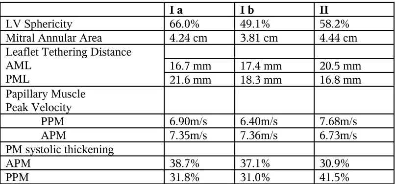
TABLE IV
DETERMINANT OF ISCHEMIC MR
I a I b II
LV Sphericity 66.0% 49.1% 58.2%
Mitral Annular Area 4.24 cm 3.81 cm 4.44 cm
Leaflet Tethering Distance AML
PML 16.7 mm21.6 mm 17.4 mm18.3 mm 20.5 mm16.8 mm Papillary Muscle
Peak Velocity
PPM 6.90m/s 6.40m/s 7.68m/s
APM 7.35m/s 7.36m/s 6.73m/s
PM systolic thickening
APM 38.7% 37.1% 30.9%
PPM 31.8% 31.0% 41.5%
The determinants of ischemic mitral regurgitation namely LV sphericity, MAA, leaflet tethering distance and papillary muscle function are analysed in tableIV.The LV sphericity is higher in group Ia than group Ib and II(66% vs. 49.1% and 58.2%) .Mitral annular area does not show significant difference in all the groups. Posterior leaflet tethering distance is higher in group I whereas anterior leaflet tethering distance is higher in group II. Posterior papillary muscle peak velocity is lower in group I where as anterior muscle is lower in group II (6.4m/s vs.6.73m/s) as systolic thickening of corresponding papillary muscle.
[image:44.612.51.454.158.346.2]
TABLE V
COMPARISON OF DETERMINANT OF ISCHEMIC MR
No MR
1. LV sherecity
I a 63.00% 69.00% <0.01
I b 48.25% 52.50% ns
II 55.50% 55.50% ns
All cases 50.90% 52.50% ns
2 Mitral Annular Area
I a 38.4mm 46.4 mm <0.05
I b 38.5 mm 38.5 mm ns
II 48.5 mm 51.5 mm ns
All cases 41.2 mm 46.8 mm <0.01
3 Leaflet tethering Distance
I a 18.50 mm 25.00 mm <0.01
I b 17.00 mm 23.50 mm <0.05
II 18.60 mm 23.25 mm <0.05
All cases 17.84 mm 24.09 mm 0.01
4 PM function
Systolic Velocity
I a(PPM) 5.98 m/s 7.90 m/s <0.05
I b(PPM) 7.41 m/s 7.10 m/s ns
II(APM) 7.06 m/s 6.35 m/s ns
Systolic thickening
I a(PPM) 24% 40% <0.05
I b(PPM) 36% 33% ns
II(APM) 30% 29% ns
(MAA-Mitral annular area; AML-Anterior mitral leaflet; PML- Posterior mitral leaflet; APM-Anteriolateral papillary muscle; PPM- Posteriomedial papillary muscle; TDI- Tissue Doppler imaging;}
RESULT ANALYSIS
the leaflet tethering distance is high.
MITRAL ANNULAR AREA AND ISCHEMIC MR
0
10
20
30
40
50
60
MAA
I a
I b
II
All
cases
GROUPS
M
it
ra
l
an
n
u
la
r
ar
ea
Mild MR
Mod –sever MR
LEAFLET TETHERING DISTANCE AND MR
SEVERITY
0 5 10 15 20 25 30I a I b II All cases
GROUPS T E T H E R IN G D IS T A N C E Mild MR
This BAR chart depicts the relationship of mitral annular area with severity of ischemic mitral regurgitation. The MAA does not have the consistent correlation with severity of MR.
This bar diagram depicts the relationship between papillary muscle function with
ischemic mitral regurgitation. The papillary muscle systolic peak velocity does not have consistentPapillary muscle function and IMR
0
1
2
3
4
5
6
7
8
9
Group Ia
Group Ib
Group II
correlation with ischemic mitral regurgitation in all groups. In group Ia papillary muscle systolic peak velocity has linear correlation with ischemic mitral regurgitation. In other words normal papillary muscle function is associated with severe MR.
This bar diagram depicts the relationship between leaflet tethering distances with ischemic mitral regurgitation. There is linear relationship between leaflet tethering distance and severity of ischemic mitral regurgitation in all group of patients..
Leaflet tethering distance and IMR
0
5
10
15
20
25
30
I a
I b
II
Groups
le
af
le
t
te
th
er
in
g
d
is
ta
n
ce
m
m
Mild MR
This BAR chart depicts the relationship of LV sphericity with severity of ischemic mitral regurgitation. The LV sphericity has good correlation with severity of MR in group Ia but not in other groups.
LV Sphericity and IMR
0
10
20
30
40
50
60
70
80
Group Ia
Group Ib
Group II
Groups
L
V
S
p
h
er
ic
it
y(
%
)
mild MR
DISCUSSION
Ischemic mitral regurgitation (IMR) is a common complication of both acute and chronic ischemic heart disease and adversely affects the prognosis by doubling the risk of death.8,9 Since its
initial recognition by Burch and Depasquek,25 Ischemic MR has been attributed in both, name and
mechanism, to dysfunction of papillary muscle impairing the normal support of effective cooptation with prolapse of leaflet.
However, isolated papillary muscle dysfunction failed to produce ischemic MR in animal models.59-62 In addition mitral leaflet prolapse which can be caused by Papillary muscle dysfunction is
rare in patients with ischemic MR.63-65
Therefore, the relationship between Papillary muscle dysfunction and ischemic MR has not been established concretely.
Prevailing explanations for such ischemic MR have focused on two factors:
(1) Ischemic distortion of left ventricular geometry, displacing the attachments of the mitral leaflets to the Papillary muscles and annulus and restricting their ability to close effectively at the annular level, 12-19 (2) Decreased left ventricular (LV)-generated force acting to close the leaflets,
particularly when they are under increased tension.18 Both factors would produce the apically tented or
forces-increased tethering and diminished LV contraction-then normal PM contraction, which is also directed toward the apex, should in principle increase apical tethering and augment IMR. Conversely we can propose the hypothesis that in the presence of MR caused by increased tethering, for example, from ischemia with outward distortion of the inferior base of the heart (but not involving the Papillary muscles), adding contractile dysfunction of the Papillary muscles can paradoxically diminish IMR by allowing the leaflets to seat better and approach the annular level more closely to close more
effectively. Tethering may also be reduced by possible stretching of the ischemic papillary muscle towards the annulus by LV force transmitted through the leaflets and chordae. Testing this hypothesis is important not only to sharpen our understanding of mechanism but also practically because abnormal tethering can potentially be addressed mechanically, but restoring Papillary muscle contractile
function may not be possible.
Regional Wall Function by Echocardiography: 66,67
An accurate value of regional myocardial function is systolic thickening (Expressed as a Percentage). Wall thickening is directly related to myocardial function and abnormalities in wall thickening are correlated to well with the extent of Infarct size. However the differentiation of regions with abnormal contractile function from regions with normal function by planer method with coronary artery disease is difficult because of wide range of thickening in normal and abnormal regions. Tissue Doppler Echocardiography velocity can accurately quantify local myocardial function with higher temporal accuracy than any current clinical method.
Papillary Muscle Function by Echocardiography:68,69
echocardiography even though TDI is an angle dependent ultrasound technique and papillary muscle is small intracavitary structure. TDI is better than other conventional methods in assessing the PM function quantitatively. On the other hand left ventricular dilation by left ventricular sphericity, mitral annular area and leaflet tethering distance are also assessed and correlated with degree of IMR in our study.
Mitral Annular Area:
‘Mitral annular dilatation causes functional MR’ concept was created by Dr. Friedberg11
in 1956. This concept was both supported and contradicted by different authors in the following years. In our study mitral annual area is increased in patients with moderate to severe mitral regurgitation than patients with mild mitral regurgitation (46.8mm vs. 41.2mm, p>0.10). However it is significant in patients with increased LV sphericity than patients with normal left ventricle sphericity index (46.4mm Vs 38.4mm p<0.05). But when the LV sphericity is equal mitral annular area does not correlate with severity of MR.
Left ventricular Sphericity:
Is determined by remodeling of different segments of left ventricle. Mitral valve geometry is affected greater in patients with focal LV remodeling than extensive remodeling of left ventricle. In our study the incidence of moderate to severe MR is directly proportional to left ventricular sphericity index. The incidence of MR in patients with increased LV sphericity to normal LV is 50% vs. 20% p<0.01. Our study results correlated well with both experimental work done by Dr.
HARINSABBAHNI et al48 and human work by Dr. SINFYIU et al49 in 2000. In patients with moderate
Leaflet tethering distance:
In our study the most consistent factor that correlated with the severity of Ischemic mitral regurgitation is the mitral leaflet tethering distance. In all groups of patients, the leaflet tethering distance with moderate to severe MR compared to mild MR is 24.09 mm Vs. 17.84 mm [P<0.01]. Mitral leaflet tethering distance is directly proportional to the severity of MR. Our study correlated well with the result of both experimental and human studies in the past by Dr. Robert W Godley, MD, et al51. in 1981. Mitral leaflet tethering distance was proposed by Dr. Fang Zhu, MD et al52 as the
primary mechanism of persistent MR even after mitral Annuloplasty.
PAPILLARY MUSCLE FUNCTION:
“SYNDROME OF PAPILLARY MUSCLE DYSFUNCTON” was first described by DR GE BURCH et al 25 in 1968 and contributed to the pathogenesis of IMR. He proved the above
by autopsy and indirect evidence from echo in the following years. Since the first description is the most controversial subject in cardiology is papillary muscle dysfunction and ischemic MR. In 1991 DR SANJEEVKAUR et al disproved this concept in experimental works with dogs .With advent of tissue doppler study & regional wall assessment by TDI,66,67 the function of papillary muscle was
CLINICAL APPLICATIONS.
The results of the study suggest a central role of leaflet tethering 59-65, as opposed to
Papillary muscle dysfunction, in the mechanism responsible for ischemic MR. Therapeutic approaches to relieve ischemic MR need to be targeted to reduce tethering by LV remodeling. Revascularization of the viable adjacent LV wall is expected to relieve ischemic MR70. The results also support the surgical
approaches targeted at relieving tethering by aneurysm plication and repositioning of Papillary muscles
71-74. In addition, our results suggest that the term “PM dysfunction” be changed to “PM displacement”
to better describe ischemic MR.
STUDY LIMITATIONS.
Ischemic MR includes a wide spectrum of underlying pathophysiologies, such as acute or chronic and global or segmental LV remodeling 59-65. The present study only addressed ischemic MR
due to chronic inferoposterior MI, and chronic anterior wall MI and found an inverse relationship between PM dysfunction and the degree of MR in chronic inferoposterior MI,. Although PM
dysfunction with reduced systolic longitudinal shortening, in theory, is expected to attenuate tethering and MR
In general patients with ischemic MR, such an inverse relationship may not be relevant in many patients with ischemic MR due to different pathophysiologies, such as dilated/ ischemic
cardiomyopathy other entities with only modest variability in Papillary muscle dysfunction and more extensive LV remodeling. Medial and anterolateral Papillary muscle displacement was evaluated by determining Papillary muscle tethering length by 2D echocardiography. Because PM contraction varies according to the spatial direction,75 it is necessary to attempt to establish a standard angle used
CONCLUSIONS
1. Mitral leaflet tethering distance is consistently directly proportional to severity of Ischemic mitral regurgitation.
2. Papillary muscle function is better assessed by tissue Doppler echo than M Mode and 2D echocardiography.
3. Papillary muscle dysfunction is not an independent determinant of ischemic MR in all cases. 4. Papillary muscle dysfunction attenuates ischemic MR in patients with old inferior wall MI with
increased left ventricular sphericity due to focal remodeling.
5. Role of papillary muscle dysfunction in ischemic MR is still elusive and varies depending on factors such as location of myocardial infarction and extent of left ventricular remodeling.