TITLE
Investigating Structural Plasticity In Musicians’ Brains
Using Structural Magnetic Resonance And Diffusion
Tensor Imaging Techniques
Supervisors
DR VANESSA SLUMING
PROFESSOR ANDREJ STANCAK
Thesis submitted in accordance with the requirements of the University of Liverpool for the degree of Doctor in Philosophy
IHSSAN ADEEB ABDUL-KAREEM
DECLARATION
This thesis is the result of my own work. The material contained in this thesis has not been presented, nor is currently being presented, either wholly or in part, for any other degree or qualification.
(IHSSAN ADEEB ABDUL-KAREEM) (DR. VANESSA SLUMING)
DEDICATION
This work is dedicated
To the soul of my mother, GOD bless her
To my beloved wife
ACKNOWLEDGMENTS
I would like to thank all those who helped me throughout the preparation of my thesis. Specifically, I would like to thank my supervisors Dr Vanessa Sluming, Dr Andrej Stancak and Dr Laura Parkes. Special thanks to Dr Vanessa who was very kind, considerate and knowledgeable supervisor.
I would like to thank Jamaan AlGhamdi for his technical support and advice regarding software use and image analysis. I would like to acknowledge Mrs Val Adams, the radiographer at MARIARC for her kind help in subject recruitment ad scanning. I would also like to thank all subjects who volunteered to do this research, their time was most valued.
TABLE OF CONTENTS
TITLE I
DECLARATION II
DEDICATION III
ACKNOWLEDGMENT IV
TABLE OF CONTENTS V
INDEX OF TABLES XIV
INDEX OF FIGURES XV
ABBREVIATIONS XVII
ABSTRACT XIX
CHAPTER ONE - INTRODUCTION 1
1.1 AIM 2
1.7 NEUROPLASTICITY 15 1.7.1 OVERVIEW AND BACKGROUND 15 1.7.2 PRINCIPLES OF EXPERIENCE-DEPENDENT NEUROPLASTICITY 16 1.7.2.1 ENRICHED ENVIRONMENT 16 1.7.2.2 SPECIFICITY 17 1.7.2.3 REPETITION OF STIMULATION 18 1.7.2.4 INTENSITY OF STIMULATION 18 1.7.2.5 EFFECT OF AGE 19 1.7.3 MECHANISMS OF NEUROPLASTICITY AT MICROSCOPIC LEVEL 20 1.7.4 PHYSIOLOGICAL MECHANISMS OF NEUROPLASTICITY 21 1.7.4.1 SYNAPTIC STRENGTHENING: LONG-TERM POTENTIATION 21 1.7.4.2 SYNAPTIC ELIMINATION/WEAKENING: LONG-TERM DEPRESSION 22 1.7.5 STRUCTURAL MECHANISMS OF NEUROPLASTICITY 23 1.7.5.1 SYNAPTOGENESIS AND SYNAPSE REMODELING 23 1.7.5.2 NEUROGENESIS 24 1.7.6 NEUROPLASTICITY AT MACROSCOPIC LEVEL 26
CHAPTER 2 - REVIEW OF LITERATURE 30
2.1 AIM 31
2.5.1 AGE OF STARTING MUSICAL TRAINING 51 2.5.2 DURATION OF MUSICAL TRAINING AND PRACTICE INTENSITY 51 2.5.3 MUSICAL EXPERTISE 52 2.5.4 TYPE OF INSTRUMENT 52 2.6 IS IT POSSIBLE TO TRANSFER NEUROPLASTIC EFFECT OF MUSIC TO NON-
MUSICIANS?
53
2.7 STATEMENT OF THE PROBLEM AND JUSTIFICATION OF THE WORK 54 2.8 SIGNIFICANCE OF THE PRESENT WORK 57 2.9 RESEARCH AIMS AND KEY OBJECTIVES 58 2.10 RESEARCH HYPOTHESES OF THE PRESENT WORK 59 2.10.1 STUDY 1-INVESTIGATING STRUCTURAL PLASTICITY OF POP AND PTR IN MUSICIANS
59
2.10.2 STUDY 2-INVESTIGATING EFFECT OF MUSICAL EXPERTISE ON REGIONAL MORPHOMETRY OF THE CC
60
2.10.3 STUDY 3- INVESTIGATING REGIONAL MACRO- AND MICROSTRUCTURAL PLASTICITY OF THE CC IN MUSICIANS
61
2.10.4 STUDY 4-INVESTIGATING PLASTICITY OF CEREBELLAR WHITE MATTER IN MUSICIANS
62
CHAPTER 3 - GENERAL METHODS 63
3.1 AIM 64
MEASUREMENT
3.9.4 STEREOLOGICAL MEASUREMENTS 76 3.10 MEASUREMENT OF CROSS-SECTIONAL AREA OF CC REGIONS 80 3.10.1 IMAGE PRE-PROCESSING 80 3.10.2 CROSS-SECTIONAL AREA MEASUREMENT 81 3.11 INTRODUCTION TO PRINCIPLES OF DIFFUSION TENSOR IMAGING 84 3.11.1 OVERVIEW OF DIFFUSION TENSOR IMAGING 84 3.12 DIFFUSION OF WATER AND DIFFUSION-WEIGHTED IMAGING 84 3.12.1 DIFFUSION OF WATER 84 3.12.2 ISOTROPIC AND ANISOTROPIC DIFFUSION 85 3.12.3 DIFFUSION-WEIGHTED IMAGE ACQUISITION 87 3.12.4 QUANTITATION OF ADC BY MRI 91 3.12.5 ESTIMATION OF THE DIFFUSION TENSOR 92 3.12.6 COLOUR-CODED MAPS OF THE DIFFUSION TENSOR 93 3.13 QUANTITATIVE DTI-DERIVED METRICS 96 3.13.1 TRACE OF THE TENSOR 96 3.13.2 MEAN DIFFUSIVITY 96 3.13.3 FRACTIONAL ANISOTROPY AND RELATIVE ANISOTROPY 96 3.14 INTERPRETATION OF DTI-DERIVED METRICS 97 3.15 DTI-BASED TRACTOGRAPHY 98 3.16 FIBER TRACKING METHODOLOGIES 99 3.16.1 DETERMINISTIC FIBER TRACKING 99 3.16.1.1 TERMINATION CRITERIA 100 3.16.1.2 PLACEMENT OF THE SEED VOXELS 100 3.16.2 PROBABILISTIC FIBER TRACKING 103 3.17 VALIDATION OF DTI-BASED TRACTOGRAPHY 104 3.18 ANALYSIS OF DTI DATA IN THE PRESENT WORK 105 3.18.1 DTI DATA PRE-PROCESSING 105 3.18.2 TRACTOGRAPHY PARAMETERS 106 3.18.3 TRACTOGRAPHY OF THE CORPUS CALLOSUM, SUPERIOR
CEREBELLAR PEDUNCLE AND MIDDLE CEREBELLAR PEDUNCLE
106
CHAPTER 4 - INVESTIGATING STRUCTURAL PLASTICITY OF PARS
OPERCULARIS & PARS TRIANGULARIS IN MUSICIANS
108
4.1 ABSTRACT 108
4.2 INTRODUCTION 110 4.2.1 SEGREGATION OF PARS OPERCULARIS AND PARS TRIANGULARIS 111 4.2.2 AUTOMATIC-MANUAL CROSS-VALIDATION OF BROCA’S AREA
MORPHOMETRY
4.2.3 HYPOTHESIS 114 4.3 SUBJECTS AND METHODS 115 4.3.1 SUBJECTS 115 4.3.1.1 MUSICIANS’ PROFILES 115 4.3.2 MR DATA ACQUISITION 117
4.4 METHODS 117
4.4.1 MEASUREMENT OF INTRACRANIAL VOLUME 117 4.4.2 MEASUREMENT OF GM/WM VOLUMES OF POP AND PTR 117 4.4.2.1 IMAGE PRE-PROCESSING 117 4.4.2.2 DEFINITION OF POP AND PTR 118 4.4.2.3 STEREOLOGICAL PARAMETERS 118 4.4.2.4 STEREOLOGICAL MEASUREMENT 119 4.4.3 INTER- AND INTRA-RATER RELIABILITY 119 4.4.4 IDENTIFICATION OF THE DIAGONAL SULCUS IN PARS OPERCULARIS 130 4.4.5 MEASUREMENT OF CORTICAL SURFACE AREA 132 4.4.5.1 IMAGE PRE-PROCESSING 132 4.4.5.2 TALAIRACH TRANSFORMATION 132 4.4.5.3 CORTEX-BASED ALIGNMENT 136 4.4.5.4 QUANTITATIVE MEASUREMENT OF CORTICAL SURFACE AREAS 140 4.4.6 STATISTICAL ANALYSIS 143
4.5 RESULTS 144
4.5.1 MEASUREMENT OF INTRACRANIAL VOLUME 144 4.5.2 VOLUME MEASUREMENTS OF POP AND PTR 144 4.5.2.1 EXCLUSION OF CASES FROM VOLUME MEASUREMENT 144 4.5.2.2 COMPARISON OF GM/WM VOLUMES 144 4.5.3 INTRA- AND INTER-RATER STUDY 145 4.5.4 CORTICAL SURFACE AREA MEASUREMENTS OF BA44 AND BA45 145
4.6 DISCUSSION 152
4.6.1 TECHNICAL LIMITATIONS 158 4.6.1.1 MEASUREMENT OF PARS OPERCULARIS AND PARS
TRIANGULARIS VOLUMES
156
4.6.1.2 CORTICAL SURFACE AREA MEASUREMENT OF BA44/45 4.7 CONCLUSION
158 160
CHAPTER 5 - INVESTIGATING THE EFFECT OF MUSICAL EXPERTISE
ON REGIONAL MORPHOMETRY OF THE CORPUS CALLOSUM
161
5.1 ABSTRACT 162
5.2.1 HYPOTHESIS 165 5.3 SUBJECTS AND METHODS 166 5.3.1 SUBJECTS 166 5.3.1.1 MUSICIANS’ PROFILES 166 5.3.2 MR DATA ACQUISITION 107
5.4 METHODS 168
5.4.1 MEASUREMENT OF INTRACRANIAL VOLUME 168 5.4.2 MEASUREMENT OF CROSS-SECTIONAL AREAS OF CC REGIONS 168 5.4.3 INTRA- AND INTER-RATER RELIABILITY 168 5.4.4 STATISTICAL ANALYSIS 168
5.5 RESULTS 170
5.5.1 MEASUREMENT OF INTRACRANIAL VOLUME 170 5.5.2 COMPARISON OF REGIONAL CROSS-SECTIONAL AREAS OF THE CC 170 5.5.3 INTER- AND INTRA-RATER STUDY 170 5.6 DISCUSSION
5.6.1 WITELSON’S VERSUS DTI-BASED PARCELLATION OF CC TOPOGRAPHY 5.6.2 INCREASED CC1 AND CC4 SIZE IN PROFESSIONAL MUSICIANS
5.6.3 METHODOLOGICAL CONSIDERATIONS AND LIMITATIONS OF THE STUDY 5.7 CONCLUSION
174 174 176 180 181
CHAPTER 6 - INVESTIGATING REGIONAL MACRO- &
MICROSTRUCTURAL PLASTICITY OF THE CORPUS CALLOSUM IN
MUSICIANS
182
6.1 ABSTRACT 183
6.2 INTRODUCTION 6.2.1 HYPOTHESIS
185 187 6.3 SUBJECTS AND METHODS 188 6.3.1 SUBJECTS 188 6.3.1.1 MUSICIANS’ PROFILE 188 6.3.2 MR DATA ACQUISITION 190 6.3.2.1 HIGH RESOLUTION STRUCTURAL SCANS AT 3 TESLA 190 6.3.2.2 DIFFUSION-WEIGHTED MR DATA ACQUISITION 190
6.4 METHODS 191
6.4.4 INTRA- AND INTER-RATER RELIABILITY 193 6.4.4.1 MEASUREMENT OF CROSS-SECTIONAL AREAS OF CC REGIONS 193 6.4.4.2 FIBER TRACTOGRAPHY OF THE CC 193 6.4.5 STATISTICAL ANALYSIS 193
6.5 RESULTS 196
6.5.1 MEASUREMENTS OF INTRACRANIAL VOLUME 196 6.5.2 FIBER TRACTOGRAPHY OF THE CC 196 6.5.3 COMPARISON OF REGIONAL MACRO- AND MICROSTRUCTURAL
MEASURES OF THE CC
196
6.5.4 INTRA- AND INTER-RATER STUDY 197 6.5.5 CORRELATION BETWEEN MACROSTRUCTURE (CROSS-SECTIONAL
AREA) AND MICROSTRUCTURE (AVERAGE FA) ACROSS SUBJECTS
197
6.6 DISCUSSION 202
6.6.1 MUSICIANS SHOWED EVIDENCE OF MACRO- AND MICROSTRUCTURAL PLASTICITY IN CC4 REGION
202
6.6.2 LACK OF SIGNIFICANT GENDER EFFECT ON MACRO- AND MICROSTRUCTURE OF THE CC
207
6.6.3 TECHNICAL LIMITATIONS OF THE PRESENT STUDY 208 6.6.3.1 ISSUE OF CROSSING FIBERS 208 6.6.3.2 SENSITIVITY TO SUBJECT MOTION 210 6.6.3.3 EFFECT OF NOISE AND LOW SIGNAL-TO-NOISE RATIO ON DTI 211 6.6.3.4 ISSUE OF MAGNETIC SUSCEPTIBILITY EFFECTS 212 6.6.3.5 ISSUE OF EDDY CURRENTS 213 6.6.3.6 ANALYSIS OF DTI DATA 214 6.6.3.7 INTERPRETATION OF TRACTOGRAPHY RESULTS 215 6.6.3.8 COMPARISON WITH PREVIOUS STUDIES 216 6.6.3.9 VALIDATION OF DTI-BASED TRACTOGRAPHY
6.7 CONCLUSION
216 216
CHAPTER 7 - INVESTIGATING STRUCTURAL PLASTICITY OF
CEREBELLAR WHITE MATTER IN MUSICIANS
218
7.1 ABSTRACT 219
7.2 INTRODUCTION 7.2.1 HYPOTHESIS
7.3.2.1 HIGH RESOLUTION STRUCTURAL SCANS 227 7.3.2.2 DIFFUSION-WEIGHTED MR DATA ACQUISITION 227
7.4 METHODS 228
7.4.1 MEASUREMENT OF INTRACRANIAL VOLUME 228 7.4.2 DTI DATA ANALYSIS 228 7.4.2.1 PRE-PROCESSING OF DTI DATA 228 7.4.2.2 TRACTOGRAPHY PARAMETERS 228 7.4.2.3 FIBER TRACTOGRAPHY OF THE SCP AND MCP 228 7.4.3 INTRA- AND INTER-RATER RELIABILITY 230 7.4.4 MEASUREMENT OF GM AND WM VOLUMES OF CEREBELLUM 230 7.4.5 STATISTICAL ANALYSIS 231
7.5 RESULTS 232
7.5.1 MEASUREMENT OF INTRACRANIAL VOLUME 232 7.5.2 RECONSTRUCTION OF THE SCP AND MCP 232 7.5.2.1 EXCLUSION OF CASES FOR TRACTOGRAPHY 232 7.5.2.2 COMPARISON OF SCP AND MCP TRACTOGRAPHY MEASURES 232 7.5.3 INTRA- AND INTER-RATER RELIABILITY 233 7.5.4 COMPARISON OF GM AND WM VOLUMES OF RIGHT AND LEFT
CEREBELLAR HEMISPHERES
233
7.6 DISCUSSION 241
7.6.1 THE CEREBELLUM IS CRUCIAL FOR MUSICAL PERFORMANCE 242 7.6.2 LACK OF GM DIFFERENCES IN CEREBELLAR HEMISPHERES BETWEEN
GROUPS
245
7.6.3 CEREBELLAR ASYMMETRY 246 7.6.3.1 EFFECT OF HANDEDNESS ON CEREBELLAR ASYMMETRY 247 7.6.3.2 STRUCTURAL ASYMMETRY IN MUSICIANS’ BRAINS 248 7.6.4 TECHNICAL LIMITATIONS OF THE PRESENT STUDY
7.7 CONCLUSION
249 252
CHAPTER 8 - GENERAL DISCUSSION, CONCLUSION AND
RECOMMENDATION
253
8.1 AIM 254
8.2 GENERAL DISCUSSION 254 8.2.1 STUDY 1- INVESTIGATING STRUCTURAL PLASTICITY OF POP AND PTR IN
MUSICIANS
254
8.2.2 STUDY 2- INVESTIGATING EFFECT OF MUSICAL EXPERTISE ON REGIONAL CALLOSAL MORPHOMETRY
254
8.2.3 STUDY 3- INVESTIGATING REGIONAL MACRO- AND MICROSTRUCTURAL PLASTICITY OF THE CC IN MUSICIANS
8.2.4 STUDY 4- INVESTIGATING PLASTICITY OF CEREBELLAR WM IN MUSICIANS
255
8.3 REGIOONAL CC MORPHOMETRY IN MUSICIANS, COMPARISON OF RESULTS IN COHORT 2 AND 3A
256
8.4 FACTORS CONTROLLING NEUROPLASTICITY IN MUSICIANS 259 8.5 ISSUE OF BRAIN DEVELOPMENT 261 8.5.1 THE CEREBELLUM 261 8.5.2 THE CORPUS CALLOSUM 261 8.6 ARE THESE STRUCTURAL CHANGES REPRESENT USE-DEPENDENT PLASTICITY OR GENETIC PREDISPOSITION FOR MUSIC?
263
8.7 CONCLUSION 265
8.8 RECOMMENDATIONS FOR FUTURE STUDIES 266
REFERENCES 268
INDEX OF TABLES
TABLE NO TITLE PAGE NO
Table 2.1 Summary of previous neuroimaging studies comparing musicians and controls
45
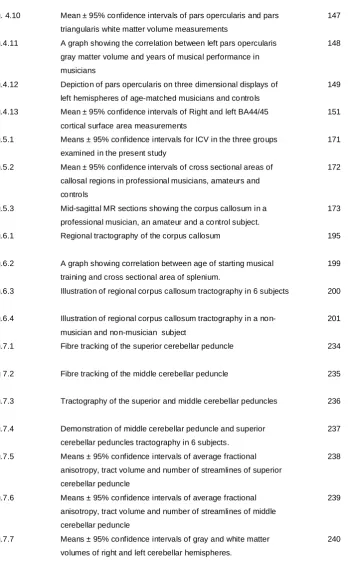
Table 3.1 Summary of image acquisitions performed in the present work 67
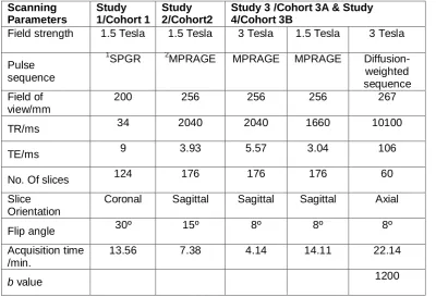
Table 3.2 Summary of cohorts, measurements, and image analysis softwares
69
Table 4.1 Demographics of subjects recruited in this study 116
Table 4.2 Orthogonal views used to localize landmark points for Talairach transformation
133
Table 4.3 The frequency percentage of diagonal sulci in right and left POP in both groups.
150
Table 5.1 Demographics of subjects recruited in the present study 167
Table 5.2 Instrument specialty of all musicians 167
Table 6.1 Demographics of subjects recruited in this study 189
Table 6.2 Means ± standard deviations of cross-sectional areas and tractography derived metrics
198
INDEX OF FIGURES
FIGURE NO TITLE PAGE NO
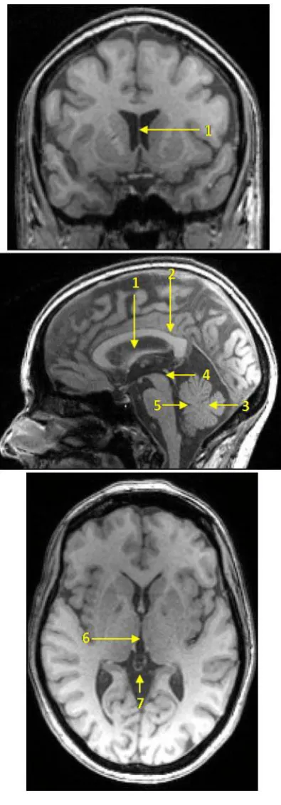
Fig.1.1 A three dimensional (3D) display of the left cerebral hemisphere 11 Fig.1.2 The superior and middle cerebellar peduncles 14 Fig.3.1 The basis of Cavalieri sections method of volume estimation 71 Fig.3.2 The grid system used for estimating slice volumes 73 Fig.3.3 Method of brain realignment according to the bicommissural
plane
75 Fig.3.4 Illustration of Intracranial volume measurement using Cavalieri
method with point counting
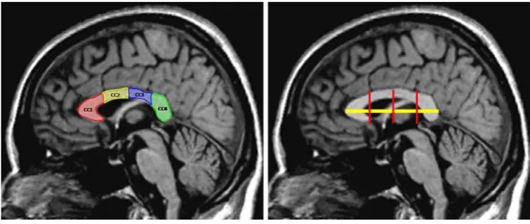
79 Fig.3.5 Criteria for selecting the midsagittal slice 82 Fig.3.6 Method of subdividing the CC on midsagittal T1-weighted MR
image
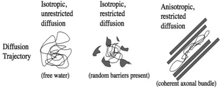
83 Fig.3.7 Illustration of isotropic unrestricted diffusion, isotropic restricted
diffusion, and anisotropic restricted diffusion
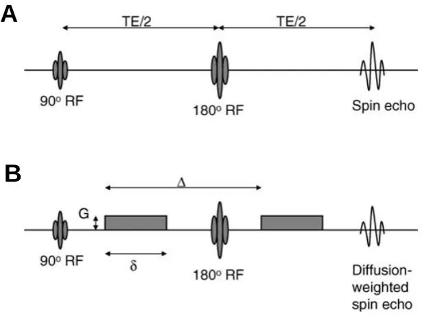
86 Fig.3.8 Illustration of the spin echo, single shot echo-planar imaging 89 Fig.3.9 A diagram demonstrating the relationship between diffusion of
water molecules and gradient applications
92 Fig.3.10 Classes of diffusion tensor ellipsoids 94 Fig.3.11 The Principle of DTI 95 Fig.3.12 A schematic illustration of the FACT algorithm 102 Fig.4.1 Depiction of pars opercularis and pars triangularis on the left
hemisphere of one subject
121 Fig.4.2 Demonstration of pars opercularis and pars triangularis volume
estimation using stereology
122 Fig.4.3 Demonstration of gray matter volume measurement of left POP
using stereology.
126 Fig.4.4 Demonstration of gray matter volume measurement of left PTR
using stereology
129 Fig.4.5 The diagonal sulcus 131 Fig.4.6 Talairach transformation 135 Fig.4.7 Segmentation of the cerebral cortex 138 Fig.4.8 Steps of cortex-based alignment of the left hemisphere of one
subject
142 Fig.4.9 Mean ± 95% confidence intervals of pars opercularis and pars
triangularis gray matter volume measurements
Fig. 4.10 Mean ± 95% confidence intervals of pars opercularis and pars triangularis white matter volume measurements
147 Fig.4.11 A graph showing the correlation between left pars opercularis
gray matter volume and years of musical performance in musicians
148
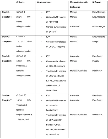
Fig.4.12 Depiction of pars opercularis on three dimensional displays of left hemispheres of age-matched musicians and controls
149 Fig.4.13 Mean ± 95% confidence intervals of Right and left BA44/45
cortical surface area measurements
151 Fig.5.1 Means ± 95% confidence intervals for ICV in the three groups
examined in the present study
171 Fig.5.2 Mean ± 95% confidence intervals of cross sectional areas of
callosal regions in professional musicians, amateurs and controls
172
Fig.5.3 Mid-sagittal MR sections showing the corpus callosum in a professional musician, an amateur and a control subject.
173 Fig.6.1 Regional tractography of the corpus callosum 195 Fig.6.2 A graph showing correlation between age of starting musical
training and cross sectional area of splenium.
199 Fig.6.3 Illustration of regional corpus callosum tractography in 6 subjects 200 Fig.6.4 Illustration of regional corpus callosum tractography in a
non-musician and non-non-musician subject
201 Fig.7.1 Fibre tracking of the superior cerebellar peduncle 234 Fig 7.2 Fibre tracking of the middle cerebellar peduncle 235 Fig.7.3 Tractography of the superior and middle cerebellar peduncles 236 Fig.7.4 Demonstration of middle cerebellar peduncle and superior
cerebellar peduncles tractography in 6 subjects.
237 Fig.7.5 Means ± 95% confidence intervals of average fractional
anisotropy, tract volume and number of streamlines of superior cerebellar peduncle
238
Fig.7.6 Means ± 95% confidence intervals of average fractional anisotropy, tract volume and number of streamlines of middle cerebellar peduncle
239
Fig.7.7 Means ± 95% confidence intervals of gray and white matter volumes of right and left cerebellar hemispheres.
ABBREVIATIONS AP AC ACPC ADC AMPA CC CST DF DTI DTT DWI EEG FA fMRI GM ICV ICC IFG LTD LTP MARIARC MRI MPRAGE MEG MIT MCP Mm MANOVA NMDA PET POP PTR PoCG PC PrCG ROI Absolute pitch Anterior commissure
Anterior commissure-posterior commissure Apparent diffusion coefficient
α-amino-3-hydroxy-5-methyl-4-isoxazolepropionic acid Corpus callosum
Corticospinal tract Degree of freedom Diffusion tensor imaging Diffusion tensor tractography Diffusion- weighted image Electroencephalography Fractional anisotropy
Functional magnetic resonance imaging Gray matter
Intracranial volume
Intraclass correlation coefficient Inferior frontal gyrus
Long-term depression Long-term potentiation
Magnetic Resonance And Image Analysis Research centre Magnetic resonance imaging
Magnetization-Prepared Rapid Gradient-Echo imaging Magnetoencephalography
Melodic intonation therapy Middle cerebellar peduncle Millimetre
Multivariate analysis of variance N-methyl-D-aspartate
Positron emission tomography Pars opercularis
RA SNR SPGR SD SCP T 3D VBM WM
Relative anisotropy Signal to noise ratio Spoiled gradient echo Standard deviation
Superior cerebellar peduncle Tesla
Investigating Structural Plasticity In Musicians’ Brains Using Structural
Magnetic Resonance And Diffusion Tensor Imaging Techniques
IHSSAN ADEEB ABDUL-KAREEM ABSTRACT
N
europlasticity is the ability of the brain to change its structure and/or function in response to environmental stimuli. It is implicated in many processes, such as learning, maturation, skill acquisition, and rehabilitation following brain injury. With the advent of neuroimaging techniques, the study of neuroplasticity and its mechanisms have fascinated researchers given the wide scope with which this process is involved. Musicians have long been considered an ideal model to study neuroplasticity in humans. It has been shown that musicians with their early, intensive, and multimodal skilful practice have structural plasticity in different brain regions. The objective of this work was to extend these structural studies through examining different cohorts of musicians, using a multitude of imaging and morphometric techniques, and performing novel examinations of brain regions essential for enabling high level musical performance such as Broca’s area, corpus callosum (CC), and cerebellum.Three age-, gender- and handedness-matched cohorts were examined. The first cohort included 26 orchestral musicians and 26 non-musicians. High resolution T1-weighted structural MR images were acquired to measure gray and white matter volumes and cortical surface area of Broca’s area subparts: pars opercularis/BA44 and pars triangularis/BA45. The second cohort included 12/12/12 professional musicians/amateur musicians/non-musicians. High resolution T1-weighted MR images were acquired to measure cross-sectional areas of four regions of the midsagittal CC: CC1 (rostrum/ genu/anterior body), CC2 (anterior midbody), CC3 (posterior midbody), and CC4 (isthmus and splenium). In the third cohort, 12/12 musicians and non-musicians were examined. High resolution T1-weighted
structural MR images were acquired to measure cross-sectional areas of CC1-CC4 regions; and diffusion tensor imaging-based tractography was used to measure average fractional anisotropy (FA), mean diffusivity (MD), tract volume, and number of streamlines of the same regions. In a subset (10/10) of this cohort, high resolution structural scans were used to measure gray and white matter volumes of cerebellar hemispheres; and diffusion tensor imaging-based tractography was used to measure average FA, tract volume, and number of streamlines of superior (SCP) and middle (MCP) cerebellar peduncles. Outcome measures were
compared between groups.
1
CHAPTER 1
INTRODUCTION-
2
1.1 AIM
This chapter is divided into three main parts. The first part presents a general overview
of the present work, structure of the thesis, and role of co-authors in published articles.
The second part provides an overview of anatomy of brain structures examined in this
work namely, Broca’s area, corpus callosum (CC), and cerebellum. The third part
provides a detailed description of experience-dependent neuroplasticity including its
principles, microscopic mechanisms, and macroanatomical evidence of neuroplasticity
in the human brain as revealed by neuroimaging studies.
1.2 OVERVIEW OF THE PRESENT WORK
The term ‘neuroplasticity’ describes structural-functional adjustment or adaptation of the
brain to environmental stimuli, such as maturation, learning, and skill acquisition, or a
compensation of some cerebral structures for injured parts (Zilles, 1992, Rauschecker,
1995). This concept, being critically involved in health and disease, has fascinated
scientists to explore its underlying mechanisms and events. Until 1970, the common
belief was that the nervous tissue is static throughout adult life; however, experiments
performed on animals showed that adult brain has the lifelong ability for structural and
functional reorganization induced by learning and skill acquisition. Postmortem
examination of specimens obtained from actively trained animals has shown clear
evidence of microstructural plasticity in the form of dendritic growth and axonal
sprouting (Pascual-Leone et al., 1994, Hallett, 1995, Pascual-Leone et al., 2005), which
could not be demonstrated in inactive animals (Black et al., 1990, Anderson et al., 1996,
Kleim et al., 1998a, Anderson et al., 2002, Swain et al., 2003, Kleim et al., 2004,
Matsuzaka et al., 2007). Furthermore, there is convincing evidence that new neurons
could be generated in response to increased functional demands (Gould et al., 1999b,
Alvarez-Buylla and Garcia-Verdugo, 2002, Duchesne et al., 2002, Fowler et al., 2002,
Rakic, 2002). With the emergence of neuroimaging modalities notably, magnetic
resonance imaging (MRI), neuroscientists were fascinated to investigate
3
with various professions, such as taxi drivers (Maguire et al., 2000, Maguire et al.,
2006b), basket ball players (Park et al., 2006), jugglers (Draganski, 2004), and
mathematicians (Aydin et al., 2007). However, musicians emerged as an ideal model to
study plasticity in the human brain. First, they commonly start their training at an early
age when many brain structures are still vulnerable to plastic changes. Second, their
practice involves the acquisition of multimodal skills, including sensory, motor, auditory,
and visuospatial skills, which should be synchronized and refined to produce the finest
outcome. Third, they continue to practice even outside their working hours unlike
subjects from other professions. In this contest, tens of structural (Schlaug et al., 1995a,
Schlaug et al., 1995b, Keenan et al., 2001, Schmithorst and Wilke, 2002, Sluming et al.,
2002, Gaser and Schlaug, 2003, Hutchinson et al., 2003, Bengtsson et al., 2005, Imfeld
et al., 2009) and functional (Sergent et al., 1992, Elbert et al., 1995, Koeneke et al.,
2004, Vuust et al., 2005, Bangert et al., 2006, Limb et al., 2006, Magne et al., 2006,
Baumann et al., 2007, Sluming et al., 2007, Watanabe et al., 2007) studies were
conducted during the last two decades to investigate neuroplasticity in musicians.
Results revealed structural-functional changes in musicians relative to controls in
different brain regions, including the motor cortex, auditory cortex, prefrontal cortex,
inferior frontal gyrus, superior parietal lobule, cerebellum, CC, and corticospinal tract. In
addition, professional musicians exhibited evidence of cross-modal integration essential
for enabling high level musical performance, such as auditory-somatosensory (Schulz
et al., 2003), auditory-motor (Bangert et al., 2006, Baumann et al., 2007), and
visual-motor integration (Sluming et al., 2007). The correlation of these findings with musical
expertise, age of commencing musical tuition, duration of musical performance, and
instrument specialty has led to the assumption that they represent use-dependent
adaptations imposed by enriched environment in the form of musical practice. In
addition to providing compelling evidence of neuroplasticity in the human brain, results
of these studies may enhance our understanding of the mechanisms of music-brain
interaction, which might have useful applications in health and disease. From the
structural point of view, there is much work to be done to expand our knowledge about
4
regions, which are crucial for musical performance. Thus, I set the goal to extend the
literature through using structural MR and diffusion tensor imaging (DTI) techniques to
perform novel investigations of structural plasticity in these regions. A shared
characteristic of these regions is their considerable connections with several brain parts.
Additionally, recent functional studies have assigned diverse musically-relevant
functions to these regions justifying structural examinations performed in the current
work. Four studies were conducted in the present work:
Study 1- Investigating structural plasticity of Broca’s area subparts: pars opercularis
(POP) and pars triangularis (PTR) in musicians. In this study, gray matter (GM)
volumes, white matter (WM) volumes, and cortical surface areas of POP/BA44 and
PTR/BA45 were compared between musicians and non-musicians. This study is
presented in chapter 4.
Study 2- Investigating the effect of musical expertise on regional morphometry of the
CC. In this study, cross-sectional area measurements of four callosal regions were
compared between professional musicians, amateur musicians, and non-musicians.
This study is presented in chapter 5.
Study 3- Investigating regional macro- and microstructural plasticity of the CC in
musicians. The macrostructure (cross-sectional areas) and microstructure (average
fractional anisotropy (FA), mean diffusivity (MD), tract volume, and number of
streamlines) of four callosal regions were compared between musicians and
non-musicians using structural MR and DTI-based tractography, respectively. This study is
presented in chapter 6.
Study 4- Investigating structural plasticity of cerebellar WM in musicians. In this study,
DTI-based tractography was used to compare average FA, tract volume, and number of
streamlines of SCP and MCP between musicians and non-musicians. In addition, GM
and WM volumes of cerebellar hemispheres were compared between groups. This
study is presented in chapter 7.
To perform these studies, three cohorts of age-, gender-, and handedness-matched
subjects were recruited; the first and third cohorts included musicians and
5
and non-musicians. Cohorts were examined using structural MR (study 1-4) and DTI
techniques (study 3 and 4).
1.3 STRUCTURE OF THE THESIS
The thesis is divided into eight chapters. The first chapter (this chapter) provides an
introduction to the thesis, including a general overview, a brief description of relevant
anatomy, and an introduction to the principles and mechanisms of neuroplasticity. The
second chapter discusses neuroplasticity in musicians’ brains, reviews previous reports
regarding anatomical-functional plasticity in musicians, and summarizes the different
factors that govern this process in musicians. The second chapter concludes by
presenting key objectives and research hypotheses of the present work. The third
chapter presents a summary of recruited cohorts and image acquisitions, in addition
to presenting general principles of methodologies used in the current work. Methods
used in more than one study are mentioned in chapter three, and are referred to when
presenting individual studies in subsequent chapters. The fourth, fifth, sixth, and
seventh chapters present the four studies conducted in the present work, respectively.
To comply with University of Liverpool guidelines, chapters 4-7 are presented in a
‘ready to submit’ style with an abstract, introduction, subjects and methods, results,
discussion, and conclusion sections. A general discussion, conclusion, and
recommendations for future studies are presented in chapter eight. All references are
cited at the end of the thesis under the ‘references’ section using the referencing style
of the ‘Journal of Neuroscience’. The appendix section contains published
peer-reviewed journal articles, conference papers, and abstracts.
1.4 PEER REVIEWED JOURNAL ARTICLES PUBLISHED DURING THE PHD
STUDY
Four articles were published during the PhD study period. Below I present them in a
chronological order:
6
of Salford Postgraduate Annual Research Conference, SPARC. University of
Salford, Manchester.
• Abdul-Kareem IA, Stancak A, Parkes LM, Sluming V 2009: Regional corpus
callosum morphometry: effect of field strength and pulse sequence. J Magn
Reson Imaging, 30 (5):1184-1190.
• Abdul-Kareem IA, Stancak A, Parkes LM, Sluming V 2011: Increased gray
matter volume of left pars opercularis in male orchestral musicians correlate
positively with years of musical performance. J Magn Reson Imaging, 33
(1):24-32.
• Abdul-Kareem IA, Stancak A, Parkes L, Al-Ameen M, AlGhamdi J, Al-Dhafeeri
F, Embleton K, Morris D & Sluming V 2011: Plasticity of the superior and middle
cerebellar peduncles in musicians revealed by quantitative analysis of volume
and number of fibers based on diffusion tensor tractography. Cerebellum, 10:
611-623.
1.5 ROLE OF CO-AUTHORS IN PUBLISHED ARTICLES
• Vanessa Sluming, Andrej Stancak and Laura Parkes - as supervisors, they
participated in reviewing the manuscripts and provided corrections and
suggestions. Vanessa Sluming recruited subjects of the first study.
• May Al-Ameen - performed inter- and intra-rater studies.
• Jamaan AlGhamdi and Faten Aldhafeeri - provided technical help regarding the
use of FreeSurfer software to measure ICV, GM and WM volumes of cerebellar
hemispheres.
• David Morris and Karl Embleton - helped in optimizing the final diffusion-weighted pulse sequence that was used to perform DTI examination of the CC,
7
1.6 RELEVANT ANATOMY
1.6.1 GROSS ANATOMY OF THE BRAIN
The human brain is conventionally divided into three major divisions: The forebrain
(diencephalon and cerebrum), the mid brain, and the hind brain (medulla oblongata,
pons, and cerebellum). The cerebrum (figure 1.1), is the largest part of the brain
consisting of two cerebral hemispheres connected by the CC. The surface of each
hemisphere (the cortex) is composed of GM, which contains neurons supported by
neuroglia. The cerebral cortex is thrown into gyri and sulci, thereby increasing its
surface area. Within the hemisphere is a central core of WM, which is composed of
myelinated nerve fibers of different diameters supported by neuroglia (Williams et al.,
1995, Snell, 2001, Kiernan, 2005).
1.6.2 MICROSCOPIC ANATOMY OF THE BRAIN
The basic functional unit of the brain is the neuron, which is specialized for the
reception of stimuli and the conduction of nerve impulses. Each possesses a cell body
and neuritis; the latter comprise dendrites and axons. Dendrites are responsible for
receiving information and conducting it toward the cell body; they often branch profusely
to increase surface area for the reception of axons from other neurons. The axon is a
single long tubular neurite that conducts impulses away from the cell body. A synapse is
the site where two neurons come in close proximity, and functional inter-neuronal
communications occur. Most neurons make synaptic connections to 1000 or more other
neurons and may receive up to 10,000 connections from other neurons. Communication
at a synapse takes place in one direction only (Snell, 2001). Apart from neurons, the
central nervous system also contains supporting cells, the neuroglia. Their number
exceeds that of neurons about 10-fold and constitutes about half the nervous tissue
mass. The neuroglial cell types in the central nervous system are astroglia,
8 1.6.3 THE BROCA’S AREA
Broca’s area is located in the POP, Brodmann area (BA) 44 and PTR, BA45 of the
inferior frontal gyrus of the dominant hemisphere; it directs the motor area for the
production of speech (figure 1.1B). The homologue of Broca’s area in the right
hemisphere is believed to be involved in emotional aspect of speech (Williams et al.,
1995). Broca’s area is connected to the sensory (receptive) speech area by a bundle of
WM called the arcuate fasciculus. The name Broca was derived from the name of a
French neurologist,
patients who lost the ability to speak after injury to the posterior
the brain (Dronkers et al., 2007). Since then, the approximate region he identified has
become known as Broca’s area, and the deficit in language producti
brain, functions of Broca’s area are not limited to language (to be discussed in details in
chapter 4).
The POP is the posterior part of Broca’s area. It is bounded posteriorly by the inferior
precentral sulcus, which separates it from the precentral gyrus. Superiorly, it is
separated from the middle frontal gyrus by the inferior frontal sulcus, and anteriorly from
the PTR by the anterior ascending ramus of the Sylvian fissure (
1.6.3.1 THE PARS OPERCULARIS
Foundas et al., 1998,
Duvernoy, 1999, Tomaiuolo et al., 1999, Petrides and Pandya, 2004, Petrides, 2006).
The PTR is the anterior part of Broca’s area. It is demarcated posteriorly from the POP
by the anterior ascending ramus of the Sylvian fissure, superiorly from the middle frontal
gyrus by the inferior frontal sulcus, and anteroinferiorly by the anterior horizontal ramus
of the Sylvian fissure (
1.6.3.2 THE PARS TRIANGULARIS
Foundas et al., 1998, Duvernoy, 1999, Tomaiuolo et al., 1999,
9
Cytoarchitectonically, BA 44 and 45 (
1.6.3.3 MICROSCOPIC ANATOMY OF POP AND PTR
Amunts et al., 1999) contain magnopyramidal cells
in lower layer III, medium-large pyramidal cells in upper layer V, and decreased cell
density in lower layer V. There is a size gradient in the cells of layer III, with the smallest
cells at its border with layer II, and the largest near layer IV. The main cytoarchitectonic
difference between areas 44 and 45 is the prominence of layer IV and the total
thickness of the cortex. In particular, layer IV in area 44 is dysgranular, and appears to
be invaded by cells from layers III and V. In area 45, layer IV is granular and more
prominent and creates a clear barrier between layers III and V. The cortex is notably
thinner in area 45 than in area 44 (Amunts et al., 1999).
It has been demonstrated that both BA 44 and 45 have distinct connections with other
cortical regions. Using DTI, it has been shown that the right and left POP are connected
with a large number of clusters in the frontal, parietal, temporal, and occipital lobes. The
right and left PTR were found to have overlapping connectivity in both hemispheres.
They are connected to the left POP, right POP, precentral gyrus, left posterior middle
temporal gyrus, posterior inferior temporal gyrus, left insula, right supramarginal gyrus,
and postcentral gyrus (
1.6.3.4 CONNECTIONS OF POP AND PTR
Xiang et al., 2009). The dorsal pathway (to the parietal lobe and
perisylvian regions in the temporal lobe) is more dominant in POP, whereas for PTR,
tracts into the dorsomedial prefrontal cortex are more prominent.
1.6.4 THE CORPUS CALLOSUM
The CC is the largest WM structure in the brain consisting of approximately 180 million
regions within the two cerebral hemispheres. It is divided into rostrum, genu, body,
isthmus, and splenium. The body is further divided into anterior and posterior parts (de
Lacoste et al., 1985). The rostrum and genu connect mainly prefrontal regions. The
body of the CC connects frontal and parietal regions with fibers originating in the
10
primary motor, somatosensory, and posterior parietal fiber bundles traverse the CC
through the posterior part of the body. The isthmus and splenium connect temporal,
parietal, and occipital cortical regions (de Lacoste et al., 1985, Hofer and Frahm, 2006).
The fibers of the genu curve forward into the frontal lobes forming forceps minor, fibers
of the body extend laterally forming radiation of the CC, while fibers in the splenium
arch backward into the occipital lobe forming forceps major (Snell, 2001).The callosal
fibers interdigitate with association and projection fibers.
There have been few studies that examined the microscopic structure of the human CC.
One of the pioneer studies of the human CC is that of Aboitiz and colleagues (Aboitiz et
al., 1992) who performed postmortem examination of callosa of 20 subjects (10 males
and 10 females). They found that thin fibers are most dense in the genu and decrease
in density posteriorly towards the posterior body, where they reach a minimum. The
density of thin fibers increases again towards the splenium. Large-diameter fibers
showed a pattern complementary to that of thin fibers, having a peak of density in the
posterior body, and a local increase of density in the posterior pole of the CC. There
11
Fig.1.1. A, a three dimensional (3D) display of the left cerebral hemisphere
demonstrating frontal (red), parietal (yellow), temporal (green), and occipital (blue)
lobes. Numbers 1, 2, 3 represent superior, middle and inferior frontal gyri, respectively.
B, a 3D display of the left hemisphere showing Broca’s area and its subparts; POP (red)
and PTR (green). 1, inferior frontal sulcus; 2, Sylvian fissure; 3, precentral sulcus; 4,
anterior horizontal ramus; 5, anterior ascending ramus; PrCG, precentral gyrus; PoCG,
postcentral gyrus. C, a sagittal view showing the medial surface of the cerebral
hemisphere. SCP, superior cerebellar peduncle. Modified from (Netter, 2010).
4 3
12 1.6.5 THE CEREBELLUM
The cerebellum (Latin for little brain), is the largest part of the hind brain. It is located in
the posterior cranial fossa. Anteriorly, the cerebellum is related to the pons and medulla
oblongata. It is connected to the midbrain, pons, and medulla oblongata by the superior
(SCP), middle (MCP), and inferior (ICP) cerebellar peduncles, respectively. The
cerebellum is surfaced with a cortex of GM underneath which lies th
made up largely of
within the WM are four
ventricle. The dentate nucleus is the largest of cerebellar nuclei forming a large
crenated crescent opening toward the SCP (Snell, 2001, Sinnatamby, 2006).
The SCP (figures 1.1C and 1.2B), also called the brachia conjunctiva, are two in
number emerging from the upper and medial part of the white substance of the
hemispheres, and are placed under cover of the upper part of the cerebellum. They are
joined to each other across the midline by the anterior medullary velum. As they ascend
they form the roof of the fourth ventricle. The fibers of the SCP are mainly derived from
the cells of the dentate nucleus of the cerebellum. They continue upward where the two
peduncles undergo a complete decussation ventral to the Sylvian aqueduct (
1.6.5.1 CEREBELLAR PEDUNCLES
Williams et
al., 1995, Snell, 2001). The MCP (figure 1.2A) is the largest cerebellar peduncle, which
is composed entirely of cortico-ponto-cerebellar fibers (Williams et al., 1995, Snell,
2001).
• Cerebral cortex through the cortico-ponto-cerebellar fibers. These fibers arise from nerve cells in the frontal, parietal, temporal, and occipital lobes terminating
in pontine nuclei. The pontine nuclei give rise to transverse fibers of the pons,
which cross the midline and enter the opposite cerebellum as the MCP.
1.6.5.2 CEREBELLAR AFFERENTS
13
• Spinal cord through anterior and posterior spinocerebellar tracts, and cuneocerebellar tract. These tracts convey unconscious muscle joint sense.
Axons of anterior spinocerebellar tract cross to the opposite side in the lateral
column of the spinal cord. They then enter the cerebellum through SCP. Axons
of the posterior spinocerebellar tract ascend in the lateral column of the spinal
cord before entering the cerebellum through the ICP.
• Vestibular nuclei
• Cerebral cortex. The most extensive cerebellar efferent connections are those with the cerebral cortex. The cerebellum connects with the contralateral
cerebral cortex by the dentothalamic tract. Fibers arising from dentate nucleus
travel through the SCP, and cross the midline to the opposite side in the
decussation of the SCP. The fibers end by synapsing with cells in the
contralateral thalamus, and terminate in the primary motor and premotor areas
of the cerebral cortex. By this pathway, the dentate nucleus can coordinate
motor activity on the same side of the body as descending fibers from the
primary motor cortex cross to the opposite side forming the corticospinal tract,
which is the principal motor tract of the body (
1.6.5.3 CEREBELLAR EFFERENTS
14
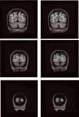
Fig.1.2. The superior and middle cerebellar peduncles. A, a ventral view of the brain
showing the MCP. B, a coronal section through the cerebellum showing the SCP and
dentate nucleus. SCP, superior cerebellar peduncle; MCP, middle cerebellar peduncle.
15
1.7 NEUROPLASTICITY
1.7.1 OVERVIEW AND BACKGROUND
According to Cambridge dictionary, the word plasticity is defined as the quality of being
soft enough to be changed into a new shape (Cambridge dictionaries online,
http://dictionary.cambridge.org/). A defining characteristic of the brain is its remarkable
capacity to undergo activity-dependent morphological and functional remodeling via
mechanisms of plasticity that form the basis of our capacity to encode and retain
memories. The process of neuroplasticity may occur during maturation, learning, skill
acquisition, environmental challenges, or pathology (Zilles, 1992, Lledo et al., 2006).
Through this process, neurons are able to change their function, chemical profile (such
as the amount and type of neurotransmitters), and structure (Woolf and Salter, 2000),
and can get beyond the limitations of their own genome (Pascual-Leone et al., 2005). A
comprehensive understanding of the process of neuroplasticity and its mechanisms is
therefore essential given the wide scope with which this process is implicated. The
great challenge is to manipulate neuroplasticity for optimal outcome (Pascual-Leone et
al., 2005). In rehabilitation medicine for example, the concept of neuroplasticity has
been utilized to develop novel task-oriented therapies that could help in regaining
functions following brain injury (Schneider et al., 2007, Schlaug et al., 2009, Lin et al.,
2010a).
Until around the 1970s, an accepted idea across scientific literature was that the
nervous tissue is essentially static throughout adulthood, both in terms of brain
functions, as well as the idea that it is impossible for new
However, over the past three decades, animal research has established two
phenomena with a profound impact for understanding neuroplasticity. The first is
significant microstructural plasticity in the adult brain, such as increased number of
synapses, increased number of glial cells, and the formation of new blood capillaries,
which account for learning and acquisition of new skills (Black et al., 1990, Anderson et
al., 1996, Kleim et al., 1998a, Anderson et al., 2002, Swain et al., 2003, Kleim et al.,
16
could be stimulated by retraining animals to perform functions impaired by this injury
(Nudo et al., 1996). These findings supported the assumption of a permanently plastic
brain, and sparked an intense interest in studying neuroplasticity in humans.
1.7.2 PRINCIPLES OF EXPERIENCE-DEPENDENT NEUROPLASTICITY
Animal studies have indicated that the structure of the brain is, in part, a function of the
environment in which an animal has been raised. Animals raised in a complex
environment showed structural morphological changes compared to isolated rearing
(
1.7.2.1 ENRICHED ENVIRONMENT
Globus et al., 1973, Greenough and Volkmar, 1973). The concept of an enriched
environment, first described by Hebb (Hebb, 1947), is the classical and most widely
used experimental paradigm to study training-induced plasticity. Using this experimental
paradigm, scientists could compare the behavior and morphological changes of animals
living in an enriched environment with those living under the usual rather plain
conditions of laboratory housing. It has been shown that extended training, which
induces a specific brain function can lead to an enhancement of that function (Kleim
and Jones, 2008), the concept of use it and improve it. In this context, several animal
studies have reported training-induced plasticity in different brain regions (Anderson et
al., 1996, Nudo et al., 1996, Kim et al., 2002, Cuppini et al., 2007, Ferreira et al., 2010,
Real et al., 2010). For instance, monkeys trained to use a rake-shaped tool to retrieve a
piece of food placed out of reach showed stronger and denser temporo-parietal junction
connections in the intraparietal sulcus compared with naive monkeys (Hihara et al.,
2006). Furthermore, rats trained to reach outside of their cage to retrieve food rewards
were shown to have increased distal forepaw representations within the motor cortex
(Kleim et al., 1998a, Kleim et al., 2002). Behavioral developments associated with
environmental enrichment include enhanced skill learning and memory function, and
reduced age-related memory decline (Pysh and Weiss, 1979, Anderson et al., 1996,
Kleim et al., 1998b, Rampon et al., 2000, van Praag et al., 2000, Kim et al., 2002). At
the microscopic level, environmental enrichment results in a range of morphological
17
de novo synaptogenesis (Leuner et al., 2003), and conversion of silent to active
synapses (Markham and Greenough, 2004, Butz et al., 2009). Results of
training-induced plasticity in animals have sparked a considerable interest to study the effect of
environmental enrichment in the human brain using in vivo neuroimaging techniques
(Maguire et al., 1997, Maguire et al., 2000, Mechelli et al., 2004, Maguire et al., 2006a,
Lee et al., 2007, Jancke et al., 2009, Park et al., 2009). Conversely, it has been shown
that neurons not actively participating in neural activity for a long period of time start to
degenerate, the concept of use it or lose it. This concept was first described by Hubel
and Wiesel, 1965 who found that depriving a kitten’s eye of light reduced the number of
neurons in the visual cortex that responded to light (Hubel and Wiesel, 1965). Likewise,
it has been shown that auditory deprivation of cats through cochlear ablation resulted in
loss of auditory cortical sound representation (Reale et al., 1987), and reduction in
synapse number (Perier et al., 1986). The above evidence indicates the presence of
substantial environmental influence on the structure and function of the adult brain;
however, genetic effects (Thompson et al., 2001) and interactions should also be taken
into consideration to understand the true potency of neuroplasticity.
The nature of the learnt skill dictates the pattern of the plasticity (
1.7.2.2 SPECIFICITY
Kleim and Jones,
2008). The literature indicates that the production of significant anatomical-functional
changes in the brain requires learning or skill acquisition, rather than mere use. For
example, it has been shown that rats allocated to an acrobatic training task have more
parallel and climbing fiber synapses, and dendritic growth in the cerebellum than rats
allocated for voluntary exercise condition or an inactive condition (Kleim et al., 1998b,
Kim et al., 2002). Training-induced structural brain changes also show regional
specificity. For example, rats learnt a spatial navigation task had increased dendritic
length and branching in the visual cortex (Greenough et al., 1979, Kolb et al., 2008).
Rats learnt unilateral motor reaching task demonstrated increased dendritic length and
branching in the motor cortex contralateral to the trained limb (Greenough et al., 1985,
18
growth in motor cortices in both cerebral hemispheres (Kolb et al., 2008). Additionally,
classical conditioning in mice using whisker stimuli was shown to specifically expand
cortical representation of trained whiskers (Siucinska and Kossut, 1996).
One of the key factors that drives plasticity is the presence of sufficient repetition of
newly learned behavior (
1.7.2.3 REPETITION OF STIMULATION
Kleim and Jones, 2008). Continuous rehearsal of the acquired
skill is essential to induce lasting structural and functional brain changes resistant to
decay when training ends (Monfils et al., 2005). For example, Trepel and colleagues
(Trepel and Racine, 1998) investigated the effect of daily stimulation on cortical
activation patterns in rats using electrodes implanted into the frontal and parietal
regions of the brain. Lasting long-term potentiation (LTP) of synaptic responses could
be induced by increasing the number of stimulation trains delivered in each session and
by increasing the number of sessions; animals that received 60 trains per day
demonstrated stronger response compared to those that received 5 and 10 trains per
day. In humans, changes in cortical activation patterns induced by short- (20 minutes)
and long-term (5 weeks) piano learning were investigated in musically naive subjects
using auditory and motor tasks (Bangert and Altenmuller, 2003). Although activation
patterns occurred after only 20 minutes, the effect did not reach a peak level until after 5
weeks of training.
Apart from repetition, the intensity of training has a profound impact on driving plasticity.
For example, in the above-mentioned experiment of Trepel and colleagues (
1.7.2.4 INTENSITY OF STIMULATION
Trepel and
Racine, 1998), animals received stimulation trains with different pulse intensities of 16,
32,160, and 500 µA. Results showed that high-intensity stimulation trains of (500 µA)
were sufficient to induce LTP, while animals received 16 and 32 µA trains showed no
significant changes compared to controls. Additionally, microscopic evidence of
structural plasticity in the form of increased dendritic arborization (Kleim et al., 2002)
19
pellets, but not in those trained to perform 60 reaches per day (Luke et al., 2004). In
accord, the total size of the cerebellum in professional musicians was shown to be
positively correlated with the intensity of musical performance (practice time per day)
(Hutchinson et al., 2003).
Early studies suggested that age-related cognitive decline arise primarily from reduction
in the number of neurons; however, several recent studies have indicated that neuronal
number is relatively preserved in the healthy aging brain (
1.7.2.5 EFFECT OF AGE
Peters et al., 1994, Peters and
Sethares, 2002), and that aging is mainly associated with alterations in neuronal
morphology in the form of regression of dendritic spines together with a reduction in the
total number of synapses. Reduced synaptic density has been demonstrated in old rats
(Chen et al., 1995, Long et al., 2009), monkeys (Peters et al., 2008, Hara et al., 2011)
and humans (Masliah et al., 1993), and was shown to be associated with physiological
degradation (Luebke et al., 2004). In accord, in vivo neuroimaging studies in humans
have reported age-related alteration of global morphometric properties, including a
decline in total brain volume (Blatter et al., 1995), GM volume (Jernigan et al., 1991),
and cortical thickness (Salat et al., 2004). These age-related changes were
hypothesized to be the inverse of development; the late maturing regions of the brain
are especially susceptible to atrophic changes (McGinnis et al., 2011). It could thus be
expected that plasticity responses are different in the aged brain (Nieto-Sampedro and
Nieto-Diaz, 2005). Nevertheless, modern neuroscience adopts the assumption of a
permanently plastic brain (Pascual-Leone et al., 2005). Activity-dependent plastic
changes have been demonstrated in the aging brain though brain changes may be less
prominent and/or slower to take place than those observed in younger brains (Green et
al., 1983). For example, it has been demonstrated that exposure to an enriched
environment is associated with enhanced neurogenesis and angiogenesis in aged
animals (Kempermann et al., 2002, Ueda et al., 2005), which was associated with
significant behavioral gain. Regarding humans, several studies have indicated that the
20
cognitive exercise demands (Colcombe et al., 2006, Boyke et al., 2008, Engvig et al.,
2010). For example, elderly subjects who learned to juggle showed increased GM in the
visual cortex, left hippocampus, and in the nucleus accumbens bilaterally (Boyke et al.,
2008). Furthermore, evidence of structural (Schlaug et al., 2009) and functional
neuroplasticity (Schaechter et al., 2002) were reported in elderly stroke patients
following the use of different rehabilitation regimes suggesting that age is not per se a
limiting factor for neuroplasticity.
1.7.3 MECHANISMS OF NEUROPLASTICITY AT MICROSCOPIC LEVEL
A major goal of neuroscience has been to explore the cellular and synaptic mechanisms
that underlie memory function, learning, aging, and lesions. With the advent of electron
microscopy and modern electrophysiological, immunohistochemical, and in vivo
microscopic imaging techniques, considerable progress has been made in describing
cellular mechanisms for learning and plasticity (Denk et al., 1990, Raymond et al., 2002,
Ferguson et al., 2004, Becker et al., 2008). The functional and anatomical malleability of
the nervous system is, to a large extent, the plasticity of the synapses. At the
microscopic level, cellular mechanisms underlying cortical plasticity include
physiological mechanisms (functional reorganization of neurons and synapses) and
structural mechanisms (rewiring of cortical circuits through the formation of new
synapses, removal of old synapses, and morphological changes in the synapses)
(Feldman, 2009). In general, it has been proposed that learning and skill acquisition
induce rapid physiological plasticity through activation of N-methyl-D-aspartate (NMDA)
receptor-dependent LTP and long term depression (LTD). These physiological
mechanisms are followed by slower structural changes of nervous tissue to consolidate
plasticity (Buonomano and Merzenich, 1998, Nieto-Sampedro and Nieto-Diaz, 2005).
Here, I focus on several of the best-studied forms of physiological and structural
21
1.7.4 PHYSIOLOGICAL MECHANISMS OF NEUROPLASTICITY
Long-term potentiation is an enduring enhancement of synaptic transmission following
high frequency electric stimulation. This process was originally suggested by Hebb
(
1.7.4.1 SYNAPTIC STRENGTHENING: LONG-TERM POTENTIATION
Hebb, 1947) who postulated that a synapse used repeatedly is made more efficient,
i.e. reinforced (a hebbian synapse). Once reinforced, synaptic stimulation threshold
becomes lower and the synapse can be activated by stimuli of lower intensity than
originally required; or when activated by the same stimulus, it produces a response of
greater amplitude (Nieto-Sampedro and Nieto-Diaz, 2005). Bliss and colleagues (Bliss
and Gardner-Medwin, 1973, Bliss and Lomo, 1973) were the first to demonstrate LTP
electrophysiologically in the neurons of the hippocampus. Later, LTP was shown to
occur in many cortical regions (Tsumoto, 1992, Maren, 2005, Abidin et al., 2006,
Bruel-Jungerman et al., 2007, Cui et al., 2011, Sheynikhovich et al., 2011, Meis et al., 2012).
Repeated physiological activation of a synapse is translated into a set of morphological
synaptic changes that ensure a record of long duration (from weeks to several months)
(Bruel-Jungerman et al., 2007). Results of the past four decades of research have
provided compelling support for the idea that LTP-induced synaptic changes play an
essential role in adult learning and memory function (for review, see (Martin et al.,
2000)). The primary mechanism for LTP induction is the activation of NMDA receptor
which requires two simultaneous events: the binding of glutamate released from
presynaptic boutons, and a sufficient level of postsynaptic depolarization mediated via
α-amino-3-hydroxy-5-methyl-4-isoxazolepropionic acid (AMPA) receptor activation
(Nieto-Sampedro and Nieto-Diaz, 2005). When these conditions are fulfilled, calcium
flows into the postsynaptic neuron through the NMDA channel, which is the triggering
event in LTP (Bliss and Collingridge, 1993). The rise in intracellular calcium triggers a
host of intracellular events, including activation of several kinases and transcription
factors that ultimately result in modifications of the properties of synaptic receptors and
induction of specific genomic programs (Huang et al., 1996, Kandel, 1997).
22
enrolment of extra-synaptic AMPA receptors (Bruel-Jungerman et al., 2007), while
activation of genomic programs in neurons leads to de novo synthesis of proteins (Frey
and Morris, 1997), and a number of morphological changes in synapses (see below).
These changes result in lasting modifications of the synapse and long-term
consolidation of memories.
Long term depression is an electrophysiological phenomenon equivalent but opposite to
LTP (
1.7.4.2 SYNAPTIC ELIMINATION/WEAKENING: LONG-TERM DEPRESSION
Ito, 1989), which can be induced by low frequency stimulation of a synapse. This
process results in shrinkage or even disappearance of synaptic connections in neural
networks (Zhou et al., 2004). The mechanism of LTD induction is essentially similar to
that of LTP, involving NMDA receptor activation and elevation of intracellular calcium
concentration; however, the increase in calcium concentration is much lower than that
after LTP induction. Unlike LTP, which activates kinases, the mechanism of LTD
involves activating calcineurine, a synaptic phosphatase with high affinity for calcium
(Lisman, 1989). This would trigger several molecular mechanisms, including
dephosphorylation of specific residues on glutamate-containing AMPA receptors and
internalization of AMPA receptors (Collingridge et al., 2004). It was hypothesized that
LTD could serve as a tool to weaken unused connections, thereby facilitating the
emergence of reinforced connections in the network, which enhances the network’s
capacity for information storage (Bruel-Jungerman et al., 2007). Previous studies have
indicated that LTD has a role in the formation of long-term memory and plasticity. For
example, LTD mechanisms were shown to be the dominant form of plasticity process in
the cerebellum (De Zeeuw and Yeo, 2005). In the hippocampus, it was demonstrated
that LTD induction is essential for complex visuospatial learning in rats (Kemp and
Manahan-Vaughan, 2007). Furthermore, it has been shown that induction of genetic
mutations in mice, which impairs LTD in the hippocampus, could result in selective
deficit in spatial memory (Etkin et al., 2006). Since most neural networks could support
both LTP and LTD mechanisms, it was suggested that the main role of LTD in plasticity
23
chronic excitation of neurons and the saturation of neural circuits thereby maintaining
the capacity for learning and memory storage (Moser et al., 1998).
1.7.5 STRUCTURAL MECHANISMS OF NEUROPLASTICITY
Physiological patterns of plasticity may not be sufficient to stabilize neuronal activities.
Structural synaptic changes may thus be required to hardwire functional changes. It has
been shown that LTP and learning could induce remodeling of neural circuits through
de novo synaptogenesis (the physiological process of increasing synapse number and
dendritic complexity)(
1.7.5.1 SYNAPTOGENESIS AND SYNAPSE REMODELING
Chang et al., 1991, Toni et al., 1999) and synapse renewal
(Markham and Greenough, 2004, Nieto-Sampedro and Nieto-Diaz, 2005). Renewal of
synapses includes four main stages triggered by the release of neurotrophins
(Trachtenberg et al., 2002, Feldman, 2009): retraction of dendritic spines and
disconnection of old synapses, growth of new axons and axon sprouting, formation of
new synaptic contacts, and maturation of the new synapses. Neural circuits with higher
capacity for synapse renewal have a greater potential for plastic changes. In addition to
synaptogenesis, environmental enrichment could also result in a host of morphological
changes at existing synapses, such as increased growth, arborization,
surface-to-volume ratio, and contact boutons of dendritic spines together with changes in the
length of post synaptic density, number of synaptic vesicles, and number of vacant
spines (Toni et al., 1999, Geinisman, 2000, Toni et al., 2001, Yuste and Bonhoeffer,
2004, De Paola et al., 2006, Holtmaat et al., 2006, Knott et al., 2006). Increased vacant
synaptic elements, which are either newly formed or arise from the breakdown of
existing synapses, can induce synaptic rewiring through stimulating the production of
neurotrophic factors capable of inducing axonal outgrowth and sprouting (Holland and
Brown, 1980), and re-routing of old axons to different postsynaptic targets (De Paola et
al., 2006). Other morphological changes associated with LTP include changes in
synapse curvature and the appearance of perforations in the synapses (Agnihotri et al.,
1998, Weeks et al., 2001, Connor et al., 2006). It has been postulated that increased
24
possibility of activation of the postsynapse (Bruel-Jungerman et al., 2007). The
appearance of perforated synapses was suggested to precede synapse splitting, giving
rise to multiple morphologically distinct synapses (Luscher et al., 2000). The net effect
of these structural synaptic changes is the increase in stability and strength of
synapses, the emergence of new functional units, and the conversion of dormant to
active synapses (Bruel-Jungerman et al., 2007). In contrast to the above mentioned
morphological synaptic changes associated with LTP, it has been reported that
induction of LTD causes retraction of axonal branches (Chang et al., 1995) and
weakening of synapses and spines (Zhou et al., 2004). Weak synapses are liable to
break again (Becker et al., 2008).
It was classically thought that neurogenesis, the generation of new neurons, is generally
confined to a discrete developmental period; however, this phenomenon was clearly
demonstrated in adult animals (
1.7.5.2 NEUROGENESIS
Kornack and Rakic, 1999, Aberg et al., 2000) and
humans (Eriksson et al., 1998, Eriksson, 2003, Knoth et al., 2010). The process of
neurogenesis has been reported to take place in different brain regions, including the
hippocampus and olfactory bulb (Alvarez-Buylla and Garcia-Verdugo, 2002, Rakic,
2002), neocortex (Gould et al., 1999b), striatum (Fowler et al., 2002), substantia nigra
(Zhao et al., 2003), and amygdala (Duchesne et al., 2002). Adult neurogenesis involves
proliferation of stem cells, morphological and physiological maturation of newborn cells,
and synaptic incorporation into existing neural circuits (Bruel-Jungerman et al., 2007).
Recent studies have indicated that adult neurogenesis can be stimulated by a variety of
factors, including environmental enrichment, physical activity, and learning
(Kempermann et al., 1997, van Praag et al., 1999a, van Praag et al., 1999b, Brown et
al., 2003). For example, animals exposed to an enriched environment showed
increased neurogenesis, which was correlated with enhanced performance in spatial
memory (Nilsson et al., 1999, Dhanushkodi et al., 2007) and novel object recognition
tasks (Bruel-Jungerman et al., 2005). Also, animals enrolled in running exercise
25
ordinary activities (van Praag et al., 1999b). Exercise does not only increase the
number of new neurons but also influences the morphology of individual newly born
cells, i.e., it has qualitative and quantitative effects (Zhao et al., 2006). Theories of
training-induced neurogenesis include modulation of the function of neurotransmitters,
increased expression of growth factors and neurotrophins, and enhanced angiogenesis.
Excitatory neurotransmitters, such as glutaminergic system are up-regulated, whereas
inhibitory neurotransmitters, such as gama-aminobutyric acid system are
down-regulated (Molteni et al., 2002, Kleim et al., 2003a, Kleim et al., 2003b). The crucial role
for adult neurogenesis in learning and memory was also derived from studies showing
that ablation of the process of neurogenesis through irradiation, antimitotic agents, and
genetic ablation, impairs spatial learning and performance in working memory tasks
(Madsen et al., 2003, Winocur et al., 2006).
Although millions of new neurons are generated each day, a large number will not
complete their maturation cycle and die within the first few weeks (Kempermann et al.,
2003). It has been shown that the same factors implicated in neurogenesis, namely
environmental enrichment, physical activity, and learning can rescue young neurons
from death and increase their chance to survive (Gould et al., 1999a, Leuner et al.,
2004, Wu et al., 2008, Epp et al., 2010). These young neurons have a lower threshold
for LTP induction and produce stable LTP more readily than more mature neurons
(Snyder et al., 2001, Saxe et al., 2006). It is thus expected that newly born neurons are
more vulnerable to plastic effect and can increase the overall computational capacity of