Copyright © 2004, American Society for Microbiology. All Rights Reserved.
Efficient Replication by Herpes Simplex Virus Type 1 Involves
Activation of the I
B Kinase-I
B-p65 Pathway
D. Gregory,
1D. Hargett,
2D. Holmes,
2E. Money,
2and S. L. Bachenheimer
1,2*
Curriculum in Genetics and Molecular Biology1and Department of Microbiology and Immunology,2
University of North Carolina School of Medicine, Chapel Hill, North Carolina
Received 10 May 2004/Accepted 28 July 2004
Infection by herpes simplex virus type 1 (HSV-1) induces a persistent nuclear translocation of NFB. To identify upstream effectors of NFB and their effect on virus replication, we employed mouse embryo fibroblast (MEF)-derived cell lines with deletions of either IKK1 or IKK2, the catalytic subunits of the IB kinase (IKK) complex. Infected MEFs were assayed for virus yield, loss of IB␣, nuclear translocation of p65, and NFB DNA-binding activity. Absence of either IKK1 or IKK2 resulted in an 86 to 94% loss of virus yield compared to that of normal MEFs, little or no loss of IB␣, and greatly reduced NFB nuclear translocation. Consistent with reduced virus yield, accumulation of the late proteins VP16 and gC was severely depressed. Infection of normal MEFs, Hep2, or A549 cells with an adenovirus vector expressing a dominant-negative (DN) IB␣, followed by superinfection with HSV, resulted in a 98% drop in virus yield. These results indicate that the IKK-IB-p65 pathway activates NFB after virus infection. Analysis of NFB activation and virus replication in control and double-stranded RNA-activated protein kinase-null MEFs indicated that this kinase plays no role in the NFB activation pathway. Finally, in cells where NFB was blocked because of DNIB expression, HSV failed to suppress two markers of apoptosis, cell surface Annexin V staining and PARP cleavage. These results support a model in which activation of NFB promotes efficient replication by HSV, at least in part by suppressing a host innate response to virus infection.
Cells activate the transcription factor NFB in a wide variety of situations, including responses to stress-inducing insults such as UV irradiation and virus infection or in response to cytokines such as tumor necrosis factor alpha (TNF-␣). NFB has an important role in suppression of apoptosis and regulates the expression of many important antiapoptotic functions (5, 31, 48, 50). NFB, as a p65/p50 heterodimer, is normally se-questered in the cytoplasm in a complex with inhibitor ofB (IB). IB␣is targeted for phosphorylation at serine residues 32 and 36, and IBis targeted for phosphorylation at serine residues 19 and 23 (42, 46, 51), by the multisubunit IB kinase (IKK) (11, 23, 36, 52, 58). This phosphorylation triggers its polyubiquitylation and destruction by the 26S proteosome (6, 7, 12), and, as a result, NFB is translocated to the nucleus (14, 57). Key roles for IKK and IB in NFB signaling were dem-onstrated in studies in which overexpression of a kinase-dead,
trans-dominant form of IKK prevents IB phosphorylation and
inhibits NFB activation (36, 52). IKK is activated by phos-phorylation mediated by mitogen-activated protein (MAP) ki-nase kiki-nase kiki-nases (MAP3Ks) MEKK1, -2, or -3 or NF B-inducing kinase (NIK) (23, 30, 37, 59). Under stress conditions, the double-stranded RNA-activated protein kinase (PKR) has been shown to activate NFB through a pathway dependent on NIK and IKK (57). Distinct roles for the catalytic components of the IKK have been recognized. IKK␣ appears to play a major role in transducing signals for NFB activation during embryonic development (18, 45), while IKKis essential for cytokine and other stress-induced signaling pathways (10, 27,
29). Besides cytoplasmic roles in the activation of NFB, re-cent studies have identified IKK␣and the IKK scaffold protein IKK␥/NEMO in direct regulation of NFB-dependent tran-scription in the nucleus (2, 49, 53). NFB activation is also dependent on distinct signaling pathways which target p65 for phosphorylation (32).
The ability of herpes simplex virus type 1 (HSV-1) to acti-vate NFB has been well documented (1, 15, 40). Beginning at 3 to 5 h postinfection (hpi), HSV-1 induced a strong and persistent nuclear translocation, increased p50/p65-dependent DNA binding activity as measured by electrophoretic mobility shift assay (EMSA), and induced activation of a 3XNF B-luciferase reporter. Persistent NFB activation required virus binding and entry as well as de novo infected cell protein synthesis, including expression of functional viral immediate-early (IE) protein ICP27. Activation was also accompanied by increased IKK activity and loss of both IB␣and IB. Inter-ference with NFB activation occurred following expression of a dominant-negative IB␣(DNIB) containing alanine substi-tutions for serine residues 32 and 36 normally targeted by IKK. The resulting substantial reduction in NFB EMSA activity correlated with a reduction in virus yield. The latter may be related to the reported role of NFB in preventing HSV-1-induced apoptosis (15). The foregoing results argue that the observed persistent NFB activation, rather than being a host response to virus infection, plays a positive role in the promot-ing efficient virus replication.
Toll-like receptors (TLRs) and associated downstream sig-naling components can also mediate activation of NFB (35, 44). While HSV-1 infection stimulated interferon production in mice by a mechanism dependent on TLR9/MyD88 (20) and TLR2 mediated an inflammatory response that contributes to
* Corresponding author. Mailing address: Department of Microbi-ology and ImmunMicrobi-ology, 837 MEJB, University of North Carolina, Chapel Hill, NC 27599-7290. Phone: (919) 966-2445. Fax: (919) 962-8103. E-mail: [email protected].
13582
on November 8, 2019 by guest
http://jvi.asm.org/
lethal encephalitis in a murine model of HSV-1 pathogenesis (22), as yet no direct evidence links TLRs to NFB activation during HSV-1 infection.
The experiments presented here were designed to more fully characterize the activation pathway for NFB in HSV-infected cells and to assess how components of the classical NFB activation pathway (IKK-IB␣-p65/p50) contribute to virus replication. Our approach was to assess virus replication and markers of NFB activation in mouse embryo fibroblasts (MEFs) with targeted deletions in key components of the sig-naling pathway (IKK␣or IKK) or through overexpression of a DNIB␣ and to assess HSV replication and the ability of HSV to suppress markers of apoptosis.
MATERIALS AND METHODS
Viruses and cells.All experiments were performed with the KOS1.1 strain of HSV-1. Adenovirus vectors expressing a dominant-negative IB␣(DNIB␣) or green fluorescent protein (GFP) were obtained from the Virus Vector Core Facility at University of North Carolina (UNC)-Chapel Hill. Spontaneously immortalized mouse embryo fibroblasts derived from normal mice or mice with targeted deletions in IKK␣or IKK (26, 27) were obtained form Tal Kafri (UNC-Chapel Hill). MEFs from normal mice or mice with a targeted deletion of the double-stranded RNA-activated protein kinase PKR (54) were obtained from Bryan Williams (Cleveland Clinic). These cell lines as well as A549 cells were maintained in Dulbecco’s modified Eagle medium (DMEM) supplemented with 10% fetal bovine calf serum, 100 U of penicillin/ml, 1% streptomycin, and 1%L-glutamine (all from Gibco). Hep2 cells were maintained in minimal
essen-tial medium supplemented as described for DMEM.
Plaque assay.Monolayers of cells in 60-mm-diameter dishes were infected with HSV at a multiplicity of infection (MOI) of 5, and cells and medium were harvested at various times postinfection, followed by three cycles of freezing and thawing. Serial 10-fold dilutions of the lysates were assayed in triplicate on monolayers of Vero cells in 12-well dishes. After 1 h, monolayers were covered with DMEM-H containing 2% calf serum and 0.3% methylcellulose. After 3 days of incubation at 37°C, medium was aspirated from the wells and plaques were stained with 0.8% crystal violet in 50% ethanol (EtOH).
Preparation of cell extracts and Western blotting.For preparation of whole-cell extracts, medium was removed and monolayers were rinsed with ice-cold Dulbecco’s phosphate-buffered saline (PBS). Cells were scraped directly into 1⫻
sodium dodecyl sulfate (SDS) sample buffer (3.85 mM Tris base [(pH 6.8], 9.1%
-mercaptoethanol, 1.82% SDS, 4.6% glycerol, and 0.023% bromophenol blue [in 100% EtOH]) and denatured by boiling. Cell-equivalent amounts of lysate were separated by SDS-polyacrylamide gel electrophoresis (SDS-PAGE). Frac-tionated cytoplasmic and nuclear extracts were prepared as described previously (38). Briefly, cells were collected by trypsinization, spun through a cushion of bovine calf serum, washed twice in PBS, resuspended in three packed cell volumes (PCV) of CE buffer (10 mM HEPES, [pH 7.8], 1 mM EDTA, 60 mM KCl, 1 mM phenylmethylsulfonyl fluoride [PMSF], 0.1% NP-40, 25% glycerol, 0.4 mM NaF, 0.4 mM Na3VO4, 10M pepstatin, and Complete Protease Inhib-itor Cocktail [Roche]), and incubated on ice for 10 min. Following a 10-s spin in a benchtop microcentrifuge, the supernatant was removed and the nuclear pellet was resuspended in CW buffer (CE buffer without NP-40 or glycerol) and repelleted. Nuclei were resuspended in 2 PCV of NE buffer (20 mM Tris-HCl [pH 8.0], 420 mM NaCl, 1.5 mM MgCl2, 0.2 mM EDTA, 0.5 mM PMSF, 25% glycerol, and phosphatase and protease inhibitors as described above). After incubation on ice for 10 min, nuclei were pelleted at 60,000 rpm at 4°C for 20 min in a Beckman TLA100.3 rotor. The nuclear extract supernatant was carefully removed, and aliquots were stored at⫺80°C.
Equal amounts of protein were transferred to PolyScreen polyvinylidene di-fluoride (PVDF) membranes (Perkin-Elmer Life Sciences) followed by blocking in TBST (150 mM NaCl, 20 mM Tris [pH 7.6], 0.05% Tween 20) with 5% milk. All probing and washing of membranes was done in TBST. Rabbit polyclonal antibodies for IKK(H-470, sc-7607), IKK␣(H-744, sc-7218), and IB-␣(C-21, sc-371) from Santa Cruz Biotechnology were used at a 1:1,000 dilution overnight at 4°C per the manufacturer’s instructions. Rabbit polyclonal antibody for p65 (100-4165) was from Rockland and was used at a 1:2,000 dilution for 1 h at room temperature. Monoclonal antibodies against HSV IE ICP4 (1101) and ICP27 (1119) were purchased from the Rumbaugh-Goodwin Institute for Cancer Re-search (Plantation, Fla.) and were used at a 1:800 dilution. Polyclonal antibody
for VP16 (Clontech) was used at a 1:5,000 dilution. Polyclonal antibody against ICP8 (3-83) was a generous gift from David Knipe (Harvard University) and was used at a 1:1,000 dilution. Polyclonal antibody against gC (R47), used at 1:5,000, was a generous gift of Gary Cohen (University of Pennsylvania). Rabbit poly-clonal antibody to PARP (H-250, sc-7150) was used at a 1:1,000 dilution. Goat anti-rabbit and anti-mouse secondary antibodies were purchased from Amer-sham Biosciences. The secondary antibody was detected with SuperSignal West Pico Chemiluminescent substrate agent (Pierce). Autoradiographic films were scanned, and images were stored as 8-bit grayscale JPEG files. The density of each band was determined by using Image J (National Institutes of Health). Relative density values were corrected for average background by subtracting the density of a blank portion of the film. The corrected values were then used to calculate fold change relative to the control sample (e.g., the ratio of the mock-infected cell value to the mock-infected cell value).
EMSA.Preparation of nuclear extracts and mobility shift assays for NFB were performed as described previously (17, 38, 40). Nuclear extract was incu-bated with a radiolabeled probe containing aB-binding site, TGGGGATTC CCCA, in buffer containing 10 mM Tris-HCl (pH 7.9), 50 mM NaCl, 0.5 mM EDTA, 10% glycerol, 1 mM dithiothreitol, and 2g of dC) · poly(dI-dC). Following 20 min of incubation at room temperature, aliquots were frac-tionated at 4°C on nondenaturing 4% polyacrylamide gels prepared in 0.25⫻
TBE (1⫻TBE is 10 mM Tris base [pH 8.3], 9 mM boric acid, and 2 mM EDTA). Gels were then placed on 3MM paper, dried under vacuum and heat, and exposed to Kodak BMR film at⫺70°C.
Annexin V staining.Infected Hep2 cells were stained with Annexin V by using the Vybrant Apoptosis Assay kit #2 (Molecular Probes) per the manufacturer’s instructions. Briefly, cells were harvest by trypsin treatment and were resus-pended in DMEM supplemented with 10% fetal bovine calf serum, 100 U of penicillin/ml, 1% streptomycin, and 1%L-glutamine. Following pelleting, cells
were resuspended at 106
/ml in 1⫻binding buffer followed by addition of 50l of Annexin stain/ml and 10l of propidium iodide (100g/ml in 1⫻binding buffer)/ml. Cells were incubated at room temperature for 15 min, and then 400
l of binding buffer was added for a total volume of 500l. Stained cells were kept on ice until Annexin staining was quantified by fluorescence-activated cell sorter (FACS) analysis.
RESULTS
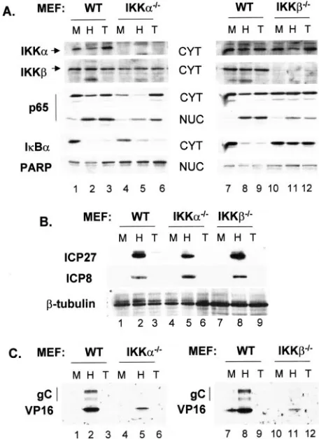
Both IKK␣and IKKcontribute to virus yield and NFB activation in HSV-infected cells.The multisubunit IKK com-plex phosphorylates IB, triggering the latter’s destruction and subsequent nuclear localization of NFB (14, 57). IKK activity was reported to increase following HSV infection (1). To de-termine the importance of IKK catalytic subunits IKK␣and IKK on the replication efficiency of HSV-1, we prepared lysates at 8 hpi from cell lines derived from normal MEFs or MEFs from embryos with targeted deletions of either IKK␣or IKKand assessed the effect of loss of either catalytic com-ponent of IKK on levels of IB␣and nuclear translocation of p65, two markers of NFB activation. The results of Western blots are presented in Fig. 1A. First, cytoplasmic extracts were probed for IKK subunits to confirm their presence or absence in IKKa⫺/⫺ and IKKb⫺/⫺ MEF lines (compare IKK␣ and
IKKin lanes 1 to 3 with 4 to 6 and lanes 7 to 9 with 10 to 12). IKK␣and IKKlevels were used as loading controls for cyto-plasmic samples, and PARP was used as a loading control for nuclear samples. As an assessment of our fractionation proce-dure, we did not detect IKK␣or IKK in nuclear lysates or PARP in the cytoplasm (data not shown). Following infection or TNF-␣ treatment of wild-type (WT), IKKa⫺/⫺, and
IKKb⫺/⫺cells, we observed nuclear translocation of p65
(com-pare lanes 1 to 6 and 7 to 12). Com(com-pared to their correspond-ing mock-infected nuclear samples, increases in nuclear p65 following HSV infection or TNF-␣ treatment were approxi-mately twofold greater in WT cells than in either of the IKK knockout cell types. Importantly, infection of WT and
VOL. 78, 2004 HSV-1 REPLICATION INVOLVES THE IKK-IB-p65 PATHWAY 13583
on November 8, 2019 by guest
http://jvi.asm.org/
IKKa⫺/⫺cells also resulted in two- to threefold reductions in
cytoplasmic p65 compared to that of mock-infected cells, while no reduction in cytoplasmic p65 was detected in IKKb⫺/⫺cells
following infection or TNF-␣treatment. We observed an ap-parent increase in the total amount of p65 after TNF-␣ treat-ment of both WT and IKKa⫺/⫺cells (lanes 3 and 6). Whether
this reflects the ability of this cytokine to activate Sp1 and transcriptionally activate the p65 promoter (47) is presently unknown. Quantification by Image J, as described in Materials and Methods, revealed 29- and 20-fold reductions, respec-tively, in IB␣following HSV infection or TNF-␣treatment of WT cells. Reductions of IB␣in IKKa⫺/⫺cells were 4.4-fold
following HSV infection and 8.7-fold after TNF-␣treatment. Consistent with the reduced nuclear translocation of p65 ob-served in IKKb⫺/⫺cells, IB␣was reduced in these cells only
1.3- and 1.1-fold following infection or TNF-␣treatment. Filters were also probed for several viral proteins represen-tative of different kinetic classes. While IE ICP27 levels were comparable between WT and IKKb⫺/⫺, we observed
de-creased ICP27 accumulation (1.7-fold relative to that of the WT) in IKKa⫺/⫺cells. Delayed early (DE) ICP8 accumulation
was comparable in the three cell types (Fig. 1B, lanes 2, 5, and 8). In contrast, accumulation of the L proteins VP16 and gC was considerably reduced in both knockout cell lines (Fig. 1B, compare lanes 2 and 5 and lanes 8 and 11). Here we observed decreases in VP16 of 5- to 6-fold and decreases in gC of 6- to 10-fold, respectively, in IKKa⫺/⫺ and IKKb⫺/⫺lysates
com-pared to those of WT.
Next, we determined virus yields in cell lines derived from normal MEFs compared to those of IKKa⫺/⫺and IKKb⫺/⫺
MEFs. Cultures were harvested at 16 and 24 hpi, and infec-tious virus was titered on Vero cell monolayers. Results, pre-sented in Table 1, indicated that both IKK␣- and IKK -defi-cient cells were impaired in supporting virus replication, because virus yield at 24 h was reduced 6- to 28-fold compared to that of normal cells. Taken together, the results of yield assays in Table 1 are consistent with results of Western blots indicating that both IKKa⫺/⫺ and IKKb⫺/⫺ cells were
im-paired in nuclear translocation of p65 and in aspects of late viral protein accumulation.
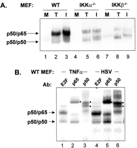
[image:3.585.48.276.71.385.2]We also prepared nuclear extracts from mock-infected, TNF-␣-treated, and HSV-infected cells and performed EMSA to determine the effect of loss of IKK␣ or IKK on NFB DNA-binding activity. In these cell lines we could detect an abundant NFB complex consisting of p50/p65 under condi-tions of TNF-␣treatment or HSV-1 infection (Fig. 2B). Thus, incubation of lysates with a p65 antibody supershifted a rela-tively low-mobility complex, while an incubation with a p50 antibody supershifted both the relatively low-mobility complex and a higher mobility complex. Incubation with an irrelevant antibody to E2F served as a control. While the ability of p65 and p50 antibodies to shift complexes from TNF-␣-treated cells appeared quantitative, a small amount of unshifted p50/ p65 and p50/p50 complexes persisted following incubation of lysates from infected cells with p65 and p50 antibodies. The precise nature of these complexes is presently unknown. The results presented in Fig. 2A indicate that the absence of either of the IKK catalytic subunits resulted in decreased levels of nuclear NFB (p50/p65), consistent with their role in nuclear translocation of p50/p65. Interestingly, the amounts of p50 homodimer activity increased in all three cell types following HSV infection or TNF-␣ treatment (see Fig. 2B for a better example of p50 homodimer activity in WT cells). This latter result suggests that the SCF()-TrCP ubiquitin ligase required to initiate limited proteolysis of p105 to yield p50 is activated following both TNF-␣treatment and HSV-1 infection (39).
FIG. 1. Both IKK␣and IKKcontribute to the activation of NFB in HSV-infected cells. Western blot analysis for proteins involved in NFB activation (A) and for viral proteins (B and C) in murine cell lines. Nuclear and cytoplasmic extracts were prepared from normal (WT), IKKa⫺/⫺, IKKb⫺/⫺, p65⫺/⫺, and p65⫹/⫹cells at 8 hpi, and they were then probed for the indicated cellular and viral proteins as de-scribed in Materials and Methods. M, mock infected; H, HSV infected; T, TNF-␣treated. (B) Nuclear extracts were probed for ICP27 and ICP8, while cytoplasmic extracts were probed for gC and VP16.
TABLE 1. Virus yield assays from normal, IKK␣⫺/⫺, and IKK⫺/⫺MEFsa
Cell type
Results at 16 hpi Results at 24 hpi Yield
(PFU/culture)
Fold decrease
Yield (PFU/culture)
Fold decrease
Normal (3.86⫾1.5)⫻107
(3.74⫾0.36)⫻107 IKK␣⫺/⫺ (7.09⫾3.4)⫻106
5.4 (6.0⫾.37)⫻106 6.2 IKK⫺/⫺ (2.4⫾0.16)⫻106
16.3 (2.62⫾0.33)⫻106 28.5
aCells and culture media from three independent experiments were collected
at the indicated times postinfection, and yields were determined as described in Materials and Methods. Relative fold decrease is the ratio of yield on control cells to knockout cells.
on November 8, 2019 by guest
http://jvi.asm.org/
[image:3.585.301.542.90.156.2]IB␣contributes to efficient replication of HSV. NFB is sequestered in the cytoplasm by IB. Following phosphoryla-tion of IB␣ at serine residues 32 and 36, which triggers its polyubiquitylation and destruction by the 26S proteosome, NFB translocates into the nucleus. DNIB, in which these serines are mutated to alanines, is resistant to cytokine-in-duced turnover, and p50/p65 is retained in the cytoplasm (42). It was previously reported that infection of pooled clones of C33-A cells stably expressing DNIB resulted in reduced nu-clear translocation of NFB and reduced virus yield compared to that of control cells (40). This experimental approach could not control for variable expression of DNIB among individual cells and could potentially accommodate some NFB activa-tion and virus replicaactiva-tion. We therefore employed infecactiva-tion with an adenovirus vector to transiently express DNIB in all cells in the culture. A549 cells were infected with an adenovirus vector expressing GFP (Ad-eGFP) or with adenovirus express-ing DNIB (Ad-DNIB), and after 16 h the cells were super-infected with HSV for 8 h or were treated with TNF-␣for 15 min prior to harvest. Figure 3A represents Western blots for IB␣in cytoplasmic extracts. Infection with HSV or treatment with TNF-␣ of cells infected with Ad-eGFP resulted in the almost complete loss of endogenous IB␣(Fig. 3A, lanes 2 and 3). A 1.45-fold decrease in IB␣was detected in cells infected with Ad-DNIB and superinfected with HSV, consistent with loss of only the endogenous form of the protein. The total level
of IB␣ present following TNF-␣ treatment was unchanged compared to that of mock treatment (Fig. 3A, compare lanes 4 and 6). Whether this reflected minimal loss of endogenous IB␣ following treatment is unclear, but analysis of nuclear extracts by EMSA (Fig. 3B.) indicated efficient suppression of NFB DNA-binding activity in cells infected with Ad-DNIB (compare lanes 1 to 3 with lanes 4 to 6). The effect of DNIB on viral gene expression was also analyzed. Lysates from con-trol and DNIB lysates were probed for the DE protein ICP8 and the L protein gC and were quantified by Image J. While there was a twofold decrease in the amount of ICP8, the level of gC declined 6.5-fold in DNIB-expressing cells compared to that in the control lysate. These results were reminiscent of those presented in Fig. 1 for the effect on viral protein accu-mulation in cells lacking IKK␣or IKK.
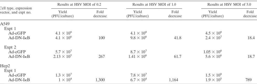
[image:4.585.54.274.69.307.2]We then determined the effect of DNIB expression on virus replication following a similar infection protocol. We infected murine 3T3 cells and the human tumor cell lines A549 and Hep2 with Ad-DNIB, followed by HSV superinfection. Production of progeny virus was determined by a plaque assay on Vero cells, and the results are presented in Tables 2 and 3. Two separate experiments involving murine fibroblasts were performed. Infectious virus was assayed at 16 and 24 hpi fol-lowing low-MOI (0.2) infection or at 20 hpi folfol-lowing infection at an MOI of 0.2 or 1. Virus yields were reduced ⬃15- to 25-fold in DNIB-expressing cells compared to that of Ad-eGFP control cells (Table 2). Following low-MOI (0.2) infec-tion of A549 or Hep2 cells, virus yield was reduced to 1,300-fold (Table 3). When the input MOI of A549 cells was increased to 1, the fold decrease in virus yield was reduced accordingly (42- to 62-fold) and still represented a significant decrease of 18-fold even at an MOI of 5. Replication of HSV in Hep2 cells was extremely sensitive to expression of DNIB. Even at an MOI of 5, virus yield was reduced almost 800-fold (Table 3).
[image:4.585.302.543.71.187.2]FIG. 2. IKK␣and IKKcontribute to NFB activation and effi-cient replication in HSV-infected cells. (A) EMSA for NFB DNA-binding activity in WT, IKKa⫺/⫺, and IKKb⫺/⫺MEFs was performed with extracts prepared from mock-infected cells (M), cells infected for 8 h with HSV (I), or cells treated for 15 min with TNF-␣(T). (B) Ex-tracts prepared from WT MEFs treated for 15 min with TNF-␣ or infected for 8 h with HSV were incubated with antibody to NFB subunits p50 or p65 or an irrelevant antibody against E2F4 prior to incubation with the NFB-specific oligonucleotide. Filled circles to the right of each lane identify supershifted complexes in response to in-cubation with anti-p50 or anti-p65. For both panels, only the portion of the autoradiograph containing specific complexes is shown.
FIG. 3. Characterization of cells expressing DNIB. (A) Western blot for IB␣and viral proteins. Replicate cultures of A549 cells were infected with a 100-ffu/cell concentration of adenovirus vector express-ing enhanced GFP (Ad-eGFP) or DNIB␣(Ad-DNIB). After 16 h, monolayers were mock infected (lanes 1 and 4), infected with HSV-1 at an MOI of 5 for 8 h (lanes 2 and 5), or treated with 50 ng of human recombinant TNF-␣/ml (lanes 3 and 6) for 15 min prior to harvest. Cytoplasmic lysates were prepared and analyzed for IB␣, gC, or ␣-tubulin by Western blot. Nuclear lysates were analyzed for ICP8. (B) EMSA for NFB binding activity. Nuclear lysates were prepared from A549 cells initially infected with either Ad-eGFP or Ad-DNIB, and then they were either mock infected, infected with HSV-1, or treated with TNF-␣as described for cultures analyzed in the experi-ment presented in panel A. NS, nonspecific binding activity.
VOL. 78, 2004 HSV-1 REPLICATION INVOLVES THE IKK-IB-p65 PATHWAY 13585
on November 8, 2019 by guest
http://jvi.asm.org/
PKR is not necessary for NFB activation in HSV-infected cells.Because HSV induces IKK activity (1) and because IKK␣ and IKKboth contribute to NFB activation by HSV-1 (Fig. 1 and 2), we next asked whether PKR, a stress- and virus-induced kinase, is necessary for NFB activation and efficient virus replication. The double-stranded RNA-activated protein kinase, PKR, activates NFB by stimulating IKK activity and inducing turnover of IB␣ and IB (57), though it is not known whether the critical serine residues on IB␣and IB are phosphorylated (21, 33). In fibroblasts from PKR-deficient (PKR0/0) mouse embryos, double-stranded RNA, alpha
inter-feron, and gamma interferon failed to activate NFB, while TNF-␣still functioned as an inducer (57). PKR is known to be activated with late kinetics in cells infected with WT HSV-1 and␥1 34.5 mutants of HSV-1, but it is not known whether this is via autophosphorylation or by a virus-encoded or -induced kinase (8). PKR-dependent phosphorylation of eIF2␣ is re-versed in virus-infected cells through the action of the␥1 34.5 protein (16). Recently, NFB activation in HSV-infected cells was reported to be dependent on PKR, based on infection of PKR⫹/⫹ and PKR0/0 MEFs (43). MEFs from normal mice
(PKR⫹/⫹) or mice with a targeted deletion of PKR (PKR/)
(54) were infected with HSV, and virus yields at 16 and 24 hpi
were determined by plaque assay (Table 4). Virus yields from PKR䡩/䡩 cells were similar at 16 h and were approximately fourfold increased at 24 h relative to yields from PKR⫹/⫹cells.
EMSA for NFB DNA-binding activity from nuclear extracts of PKR⫹/⫹and PKR/cells are presented in Fig. 4A. Little
NFB activity was detectable in the extract from mock-infected PKR⫹/⫹cells, while both TNF-␣treatment and HSV infection
induced NFB activities consisting of p50/p65 or operationally defined as p50 homodimer, based on effects of p65 and p50 antibodies on the mobility of the complexes (data not shown). Mock-infected PKR/ cells constitutively expressed the p50
activity, which increased substantially following HSV infection or TNF-␣treatment. HSV infection also induced p50/p65 ac-tivity comparable to that seen in the PKR⫹/⫹ cells, while
TNF-␣induction was impaired. Finally, we asked whether the absence of PKR affected the turnover of IB␣normally ob-served after virus infection. Equivalent amounts of cell lysate were separated by SDS-PAGE and probed by Western blot (Fig. 4B). Amounts of IB␣were considerably reduced after HSV infection of both PKR⫹/⫹ and PKR/ cells, while the
amount of ICP4 detected was greater in the PKR/ lysate.
[image:5.585.39.541.81.205.2]Together, these results indicate that PKR is not essential for HSV-induced NFB activation, but it does contribute to NFB
TABLE 2. Virus yield in WT MEFsa
Expression vector and expt no.
Results at 16 hpi Results at 20 hpi Results at 24 hpi
Yield (PFU/culture)
Fold decrease
Yield (PFU/culture)
Fold decrease
Yield (PFU/culture)
Fold decrease
Expt 1
Ad-eGFP (HSV MOI⫽0.2) 1.4⫻107 2.5⫻107
Ad-DNIB (HSV MOI⫽0.2) 2.3⫻106 6 1.7⫻106 14.7
Expt 2
Ad-eGFP (HSV MOI⫽0.2) 1.95⫻107
Ad-DNIB (HSV MOI⫽0.2) 7.4⫻105 26.4
Ad-eGFP (HSV MOI⫽0.1) 3.7⫻107
Ad-DNIB (HSV MOI⫽0.1) 2.23⫻106 16.6
aCells and culture media were collected at the indicated time points, and infectious titers were determined as described in Materials and Methods. Relative fold
increase in virus yield is the ratio of yield from control cultures to yield from DNIB-expressing cultures.
TABLE 3. Virus yield in A549 and Hep2 cells expressing DNIBa
Cell type, expression vector, and expt no.
Results at HSV MOI of 0.2 Results at HSV MOI of 1.0 Results at HSV MOI of 5.0
Yield (PFU/culture)
Fold decrease
Yield (PFU/culture)
Fold decrease
Yield (PFU/culture)
Fold decrease
A549 Expt 1
Ad-eGFP 4.1⫻108 4.1⫻108 4.5⫻108
Ad-DN-IB 4.1⫻106 100 9.8⫻106 41.8 2.4⫻107 18.4
Expt 2
Ad-eGFP 5.7⫻107 8.7⫻107 1.05⫻108
Ad-DN-IB 2.13⫻105 267 1.41⫻106 61.7 5.6⫻106 18.7
Hep2 Expt 1
Ad-eGFP 1.3⫻107 7.8⫻107 1.5⫻108
Ad-DN-IB 1⫻104 1,300 6.7⫻104 1,164 1.9⫻105 789
aCells and culture medium were collected, and infectious titers were determined as described in Materials and Methods. Cultures were harvested at 24 hpi. Relative
fold decrease in virus yield is the ratio of yield from control cultures to yield from DN-IB expressing cultures. The analysis of virus yield in Hep2 cells was performed once.
on November 8, 2019 by guest
http://jvi.asm.org/
[image:5.585.45.541.537.699.2]activation by TNF-␣. It remains possible that PKR can have offsetting effects on virus replication. For example, PKR may contribute to NFB activation when present, but in its absence the pathway leading to eIF2␣phosphorylation is disrupted. At this time we cannot rule out the possibility that the greatly increased amount of p50 homodimer also contributes to effi-cient virus replication (see Discussion). The basis for the dif-ferences between our results and those previously published (43) is unclear, though they could reflect differences in HSV strain or the time of harvest postinfection.
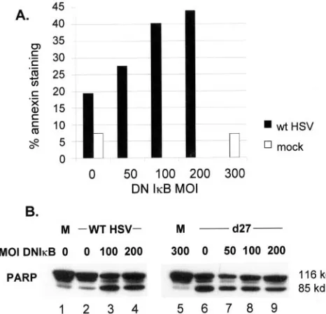
NFB promotes an antiapoptotic response in HSV-infected cells.HSV efficiently suppresses apoptosis (3, 4, 19, 24, 25, 62), and one recent report pointed to activation of NFB as an important mediator of this effect (15). NFB has a well-docu-mented role in suppressing apoptosis under a variety of con-ditions (5, 31, 48, 50). One way in which the NFB activation pathway might be linked to enhanced virus yields would be through the mechanism of apoptosis suppression. To test this model we asked whether expression of DNIB eliminated apo-ptosis suppression by HSV. We assayed for cell surface phos-phoserine by staining with fluorochrome-conjugated Annexin V and for cleavage of PARP (Fig. 5). Parallel cultures of Hep2 cells were infected with Ad-DNIB, followed at 16 h by
super-infection with HSV. Uninfected cultures or cultures receiving Ad-DNIB but not superinfected with HSV served as controls, and all cultures with HSV were harvested at 24 hpi. The frac-tion of HSV-infected cells stained with Annexin V increased from ⬃20 to ⬃43% by 24 hpi as the MOI of Ad-DNIB increased from 0 to 200 focus-forming units (ffu)/cell (Fig. 5A). By contrast, only 7% of cells infected with 300 ffu of Ad-DNIB but not superinfected with HSV displayed cell surface Annexin V staining. In a second experiment, we infected Hep2 cells with different amounts of Ad-DNIB, followed by mock infection, infection with WT HSV-1, or infection with the ICP27 mutant d27-1. The latter virus has been reported to efficiently induce apoptosis in human cells (3, 4). Lysates were prepared and assayed for PARP by Western blot, and the results are shown in Fig. 5B. Whereas unprocessed (⬃116 kDa) PARP was present in all lysates, increased amounts of processed PARP (85-kDa) were present in lysates of WT HSV-infected cells only when DNIB was expressed (lanes 3 to 4) or in lysates from all cultures infected with d27-1 (lanes 6 to 9).
DISCUSSION
[image:6.585.303.536.70.294.2]Previous studies have described NFB activation after HSV infection (1, 15, 40). Results of studies reported here extend this observation, documenting the roles of upstream effectors of NFB in this process as well as in efficient virus replication. Specifically, IKK␣ and IKK as well as IB␣ are critical for
FIG. 4. PKR is not necessary for NFB activation in HSV-infected cells. (A) EMSA for NFB was performed as described in Materials and Methods with nuclear extracts prepared from mock-infected, TNF-␣-treated, or HSV-infected PKR⫹/⫹ and PKR/ MEFs. (B) Western blot of IB␣and IE ICP4 was performed on whole-cell extracts prepared from mock-infected (M) or HSV-infected (I) PKR⫹/⫹and PKR/MEFs.
[image:6.585.43.285.81.142.2]FIG. 5. NFB promotes an antiapoptotic response in HSV-in-fected cells. (A) Replicate monolayers of Hep2 cells were inHSV-in-fected with an adenovirus vector expressing DNIB from a cytomegalovirus pro-moter at the indicated MOI. After 24 h, monolayers were either mock infected or infected with HSV at an MOI of 5. At 24 hpi cells were harvested, stained for Annexin V, and processed for FACS analysis as described in Materials and Methods. (B) Parallel cultures of Hep2 cells were infected with adenovirus vector expressing DNIB at the indicated MOIs. After 24 h, monolayers were mock infected or in-fected with WT HSV-1 or the ICP27 null mutant d27-1 at an MOI of 5. Whole-cell extracts were prepared at 24 hpi for Western blot anal-ysis of PARP.
TABLE 4. Effect of loss of PKR on virus yielda
Cell type
Results at 16 hpi Results at 24 hpi
Yield (PFU/culture)
Fold decrease
Yield (PFU/culture)
Fold increase
PKR⫹/⫹ 1.65⫻107 2.2⫻107
PKR0/0 1.25⫻107 1.3 9.0⫻107 4
a
Monolayers of PKR⫹/⫹and PKR0/0
cells were infected with HSV at an MOI of 5. Cells and culture media were collected at the indicated times postinfection, and infectious titers were determined as described in Materials and Methods. Virus yield on these cell types was analyzed once.
VOL. 78, 2004 HSV-1 REPLICATION INVOLVES THE IKK-IB-p65 PATHWAY 13587
on November 8, 2019 by guest
http://jvi.asm.org/
[image:6.585.45.284.484.659.2]efficient activation, as measured by nuclear localization of p65 and increases in nuclear NFB-dependent EMSA activity. Though activated after HSV infection, no role for PKR in NFB activation was observed. As measured by either accu-mulation of specific viral proteins or production of infectious virus, the intact IKK-IB␣-NFB pathway contributed to effi-cient virus replication. This efficiency of virus replication may in part reflect the suppression of apoptosis by a mechanism requiring activation of the NFB. It was previously reported (40) that nuclear translocation of NFB after HSV infection was not accompanied by NFB-dependent promoter activa-tion. Amici and coworkers (1) reported functional activation of NFB after infection, and the work of Goodkin et al. (15) and work presented here imply that NFB is functional and par-ticipates in the program of apoptosis suppression following virus infection. We attribute the previous inability to detect NFB activity largely to the cell type (C33-A) and the chlor-amphenicol acetyltransferase reporter used in previous studies. We can readily detect functional NFB in HeLa and HEK293T cells following infection by using a 3XNFkB lucif-erase-based reporter (D. Hargett, J. Prince, and S. Bachenhei-mer, unpublished observations).
The requirement for IKK in NFB activation during HSV infection of murine fibroblasts largely mirrors its requirement for activation by TNF-␣. Thus, loss of cytoplasmic IB␣and nuclear accumulation of p65 were either modestly impaired (IKKa⫺/⫺) or largely impaired (IKKb⫺/⫺) relative to normal
cells, and NFB-dependent EMSA activity was greatly re-duced. It has been reported that IKKplays a critical role (27, 29), while IKK␣plays a redundant (18, 26, 45) or distinct (28, 41) role, in cytokine-mediated NFB activation in murine cells. By using an RNA interference approach, both IKK catalytic subunits were shown to play critical roles in cytokine-induced NFB in human cells (44). Future experiments will be directed at understanding whether a requirement for both IKK␣and IKKalso holds for NFB activation by HSV in human cells. Both IKKa⫺/⫺ and IKKb⫺/⫺ cells supported comparable
amounts of IE ICP27 and DE ICP8 accumulation relative to normal cells, but they had greatly reduced expression of L proteins VP16 and gC. These results are consistent with the reduced yield of infectious virus from these cell lines and with previous reports on virus yield (1, 40).
Infection of murine fibroblasts deleted for PKR resulted in NFB-dependent EMSA activity and yield of infectious virus comparable to or greater than that seen following infection of normal cells. We also noted constitutive expression of a p50 homodimer form of NFB and its substantial increase follow-ing either TNF-␣treatment or HSV infection. While p50 ho-modimers would not be expected to have transcriptional trans-activation potential, Bcl-3, an IB␣family member, complexes with and relocates p50 homodimers to the nucleus, where it can also serve as a transcriptional coactivator of p50 ho-modimers (9, 13). Thus, it remains possible that increased p50 homodimer formation in part mediates efficient HSV replica-tion in PKR0/0cells and that preventing p50/p65 activation by,
for example, ectopic expression of DNIB may not reduce HSV replication.
HSV infection induces and then efficiently suppresses apo-ptosis. Consistent with the role of NFB as a cell survival factor, there is a strong correlation between the ability of HSV
to express functional ICP27 and the suppression of apoptosis (3) and between expression of NFB and suppression of apo-ptosis (15). Coupled with the requirement for functional ICP27 to activate NFB (D. Hargett and S. Bachenheimer, submitted for publication), we propose that a delayed-type activation of NFB, dependent on viral gene expression, plays an important role in prolonging functional cell survival and, thus, efficient virus replication. This delayed-type activation has also been observed following infection of cells with human cytomegalo-virus (HCMV) (55) and was termed second-tier activation. Another mechanism of virus-induced NFB activation is in-ferred from experiments which characterized the ability of HSV-1 to both trigger and inhibit apoptosis (4, 25). A key finding in the recognition of a rapid-type activation of NFB stemmed from experiments which demonstrated that ectopic expression of virion glycoproteins gD or gJ could rescue cells from apoptosis following infection with virus deleted for the gD gene (61, 62). Interestingly, the domains of gD involved in blocking apoptosis for the most part do not overlap those domains involved in binding HveC, the ubiquitously expressed gD receptor (60). More recently, either infection with UV-inactivated HSV or exposure of cells to gD was shown to protect cells from Fas-mediated apoptosis by a mechanism that was dependent on NFB activation (34). One inference that could be drawn from both these experiments was that the protective effect of gD occurred at the time of virus binding and entry and, thus, did not have a requirement for de novo viral gene expression. This situation is similar to that of the first-tier activation of NFB observed during infection with HCMV (56) and suggests that two mechanistically distinct mechanisms of NFB activation occur during HSV infection.
ACKNOWLEDGMENTS
D.G. was supported by NIH grant T32 GM-07092. This work was supported by NIH grant AI43314 to S.L.B.
REFERENCES
1.Amici, C., G. Belardo, A. Rossi, and M. G. Santoro.2001. Activation of Ib kinase by herpes simplex virus type 1. A novel target for anti-herpetic therapy. J. Biol. Chem.276:28759–28766.
2.Anest, V., J. L. Hanson, P. C. Cogswell, K. A. Steinbrecher, B. D. Strahl, and A. S. Baldwin.2003. A nucleosomal function for IB kinase-alpha in
NF-B-dependent gene expression. Nature423:659–663.
3.Aubert, M., and J. A. Blaho.1999. The herpes simplex virus type 1 regulatory protein ICP27 is required for the prevention of apoptosis in infected human cells. J. Virol.73:2803–2813.
4.Aubert, M., S. A. Rice, and J. A. Blaho.2001. Accumulation of herpes simplex virus type 1 early and leaky-late proteins correlates with apoptosis prevention in infected human HEp-2 cells. J. Virol.75:1013–1030. 5.Beg, A. A., and D. Baltimore.1996. An essential role for NF-B in preventing
TNF-␣-induced cell death. Science274:782–784.
6.Brockman, J. A., D. C. Scherer, T. A. McKinsey, S. M. Hall, X. Qi, W. Y. Lee, and D. W. Ballard.1995. Coupling of a signal response domain in IB␣to multiple pathways for NF-B activation. Mol. Cell. Biol.15:2809–2818. 7.Chen, Z., J. Hagler, V. J. Palombella, F. Melandri, D. Scherer, D. Ballard,
and T. Maniatis.1995. Signal-induced site-specific phosphorylation targets IB␣to the ubiquitin-proteasome pathway. Genes Dev.9:1586–1597. 8.Chou, J., J. J. Chen, M. Gross, and B. Roizman.1995. Association of a M(r)
90,000 phosphoprotein with protein kinase PKR in cells exhibiting enhanced phosphorylation of translation initiation factor eIF-2 alpha and premature shutoff of protein synthesis after infection with gamma 134.5⫺mutants of herpes simplex virus 1. Proc. Natl. Acad. Sci. USA92:10516–10520. 9.Dechend, R., F. Hirano, K. Lehmann, V. Heissmeyer, S. Ansieau, F. G.
Wulczyn, C. Scheidereit, and A. Leutz.1999. The Bcl-3 oncoprotein acts as a bridging factor between NF-B/Rel and nuclear co-regulators. Oncogene
18:3316–3323.
10.Delhase, M., M. Hayakawa, Y. Chen, and M. Karin.1999. Positive and negative regulation of IB kinase activity through IKKsubunit phosphor-ylation. Science284:309–313.
on November 8, 2019 by guest
http://jvi.asm.org/
11.DiDonato, J. A., M. Hayakawa, D. M. Rothwarf, E. Zandi, and M. Karin.
1997. A cytokine-responsive IB kinase that activates the transcription factor NF-B. Nature388:548–554.
12.Finco, T. S., and A. S. Baldwin.1995. Mechanistic aspects of NF-B regu-lation: the emerging role of phosphorylation and proteolysis. Immunity
3:263–272.
13.Fujita, T., G. Nolan, H. Liou, M. Scott, and D. Baltimore.1993. The candi-date proto-oncogene bcl-3 encodes a transcriptional coactivator that acti-vates through NF-B p50 homodimers. Genes Dev.7:1354–1363. 14.Ghosh, S., M. J. May, and E. B. Kopp.1998. NF-B and Rel proteins:
evolutionarily conserved mediators of immune responses. Annu. Rev. Im-munol.16:225–260.
15.Goodkin, M. L., A. T. Ting, and J. A. Blaho.2003. NF-B is required for apoptosis prevention during herpes simplex virus type 1 infection. J. Virol.
77:7261–7280.
16.He, B., M. Gross, and B. Roizman.1997. The gamma(1)34.5 protein of herpes simplex virus 1 complexes with protein phosphatase 1alpha to de-phosphorylate the alpha subunit of the eukaryotic translation initiation fac-tor 2 and preclude the shutoff of protein synthesis by double-stranded RNA-activated protein kinase. Proc. Natl. Acad. Sci. USA94:843–848. 17.Hilton, M. J., D. Mounghane, T. McLean, N. V. Contractor, J. O’Neil, K.
Carpenter, and S. L. Bachenheimer.1995. Induction by herpes simplex virus of free and heteromeric forms of E2F transcription factor. Virology213:
624–638.
18.Hu, Y., V. Baud, M. Delhase, P. Zhang, T. Deerinck, M. Ellisman, R. Johnson, and M. Karin.1999. Abnormal morphogenesis but intact IKK activation in mice lacking the IKK␣subunit of IB kinase. Science284:316– 320.
19.Jerome, K. R., R. Fox, Z. Chen, A. E. Sears, H. Lee, and L. Corey.1999. Herpes simplex virus inhibits apoptosis through the action of two genes, Us5 and Us3. J. Virol.73:8950–8957.
20.Krug, A., G. D. Luker, W. Barchet, D. A. Leib, S. Akira, and M. Colonna.
2004. Herpes simplex virus type 1 activates murine natural interferon-pro-ducing cells through toll-like receptor 9. Blood103:1433–1437.
21.Kumar, A., J. Haque, J. Lacoste, J. Hiscott, and B. R. Williams.1994. Double-stranded RNA-dependent protein kinase activates transcription fac-tor NF-B by phosphorylating IB. Proc. Natl. Acad. Sci. USA91:6288– 6292.
22.Kurt-Jones, E. A., M. Chan, S. Zhou, J. Wang, G. Reed, R. Bronson, M. M. Arnold, D. M. Knipe, and R. W. Finberg.2004. Herpes simplex virus 1 interaction with Toll-like receptor 2 contributes to lethal encephalitis. Proc. Natl. Acad. Sci. USA101:1315–1320.
23.Lee, F. S., J. Hagler, Z. J. Chen, and T. Maniatis.1997. Activation of the IB␣kinase complex by MEKK1, a kinase of the JNK pathway. Cell88:
213–222.
24.Leopardi, R., and B. Roizman.1996. The herpes simplex virus major regu-latory protein ICP4 blocks apoptosis induced by the virus or by hyperther-mia. Proc. Natl. Acad. Sci. USA93:9583–9587.
25.Leopardi, R., C. Van Sant, and B. Roizman.1997. The herpes simplex virus 1 protein kinase US3 is required for protection from apoptosis induced by the virus. Proc. Natl. Acad. Sci. USA94:7891–7896.
26.Li, Q., Q. Lu, J. Y. Hwang, D. Buscher, K. F. Lee, J. C. Izpisua-Belmonte, and I. M. Verma.1999. IKK1-deficient mice exhibit abnormal development of skin and skeleton. Genes Dev.13:1322–1328.
27.Li, Q., D. Van Antwerp, F. Mercurio, K. F. Lee, and I. M. Verma.1999. Severe liver degeneration in mice lacking the IB kinase 2 gene. Science
284:321–325.
28.Li, X., P. E. Massa, A. Hanidu, G. W. Peet, P. Aro, A. Savitt, S. Mische, J. Li, and K. B. Marcu.2002. IKK␣, IKK, and NEMO/IKK␥are each re-quired for the NF-B-mediated inflammatory response program. J. Biol. Chem.277:45129–45140.
29.Li, Z. W., W. Chu, Y. Hu, M. Delhase, T. Deerinck, M. Ellisman, R. Johnson, and M. Karin.1999. The IKKsubunit of IB kinase (IKK) is essential for nuclear factor B activation and prevention of apoptosis. J. Exp. Med.
189:1839–1845.
30.Ling, L., Z. Cao, and D. V. Goeddel.1998. NF-B-inducing kinase activates IKK-␣by phosphorylation of Ser-176. Proc. Natl. Acad. Sci. USA95:3792– 3797.
31.Liu, Z. G., H. Hsu, D. V. Goeddel, and M. Karin.1996. Dissection of TNF receptor 1 effector functions: JNK activation is not linked to apoptosis while NF-B activation prevents cell death. Cell87:565–576.
32.Madrid, L. V., C. Y. Wang, D. C. Guttridge, A. J. Schottelius, A. S. Baldwin, Jr., and M. W. Mayo.2000. Akt suppresses apoptosis by stimulating the transactivation potential of the RelA/p65 subunit of NF-B. Mol. Cell. Biol.
20:1626–1638.
33.Maran, A., R. K. Maitra, A. Kumar, B. Dong, W. Xiao, G. Li, B. R. Williams, P. F. Torrence, and R. H. Silverman.1994. Blockage of NF-B signaling by selective ablation of an mRNA target by 2–5A antisense chimeras. Science
265:789–792.
34.Medici, M. A., M. T. Sciortino, D. Perri, C. Amici, E. Avitabile, M. Ciotti, E. Balestrieri, E. De Smaele, G. Franzoso, and A. Mastino.2003. Protection by
herpes simplex virus glycoprotein D against Fas-mediated apoptosis: role of nuclear factorB. J. Biol. Chem.278:36059–36067.
35.Medzhitov, R.2001. Toll-like receptors and innate immunity. Nat. Rev. Immunol.1:135–145.
36.Mercurio, F., H. Zhu, B. W. Murray, A. Shevchenko, B. L. Bennett, J. Li, D. B. Young, M. Barbosa, M. Mann, A. Manning, and A. Rao.1997. IKK-1 and IKK-2: cytokine-activated IB kinases essential for NF-B activation. Science278:860–866.
37.Nemoto, S., J. A. DiDonato, and A. Lin.1998. Coordinate regulation of IB kinases by mitogen-activated protein kinase kinase kinase 1 and NF- B-inducing kinase. Mol. Cell. Biol.18:7336–7343.
38.Olgiate, J., G. L. Ehmann, S. Vidyarthi, M. J. Hilton, and S. L. Bachenhei-mer.1999. Herpes simplex virus induces intracellular redistribution of E2F4 and accumulation of E2F pocket protein complexes. Virology258:257–270. 39.Orian, A., H. Gonen, B. Bercovich, I. Fajerman, E. Eytan, A. Israel, F. Mercurio, K. Iwai, A. L. Schwartz, and A. Ciechanover.2000. SCF-TrCP ubiquitin ligase-mediated processing of NF-B p105 requires phosphoryla-tion of its C-terminus by IB kinase. EMBO J.19:2580–2591.
40.Patel, A., J. Hanson, T. I. McLean, J. Olgiate, M. Hilton, W. E. Miller, and S. L. Bachenheimer.1998. Herpes simplex type 1 induction of persistent NF-B nuclear translocation increases the efficiency of virus replication. Virology247:212–222.
41.Sizemore, N., N. Lerner, N. Dombrowski, H. Sakuria, and G. R. Stark.2001. Distinct roles of the IB kinase␣and beta subunits in liberating NFB from IB and in phosphorylating the p65 subunit of NFB. J. Biol. Chem.277:
3863–3869.
42.Sun, S., J. Elwood, and W. C. Greene.1996. Both amino- and carboxyl-terminal sequences within IB␣regulate its inducible degradation. Mol. Cell. Biol.16:1058–1065.
43.Taddeo, B., T. R. Luo, W. Zhang, and B. Roizman.2003. Activation of NF-B in cells productively infected with HSV-1 depends on activated pro-tein kinase R and plays no apparent role in blocking apoptosis. Proc. Natl. Acad. Sci. USA100:12408–12413.
44.Takaesu, G., R. M. Surabhi, K. J. Park, J. Ninomiya-Tsuji, K. Matsumoto, and R. B. Gaynor.2003. TAK1 is critical for IB kinase-mediated activation of the NF-B pathway. J. Mol. Biol.326:105–115.
45.Takeda, K., O. Takeuchi, T. Tsujimura, S. Itami, O. Adachi, T. Kawai, H. Sanjo, K. Yoshikawa, N. Terada, and S. Akira.1999. Limb and skin abnor-malities in mice lacking IKK␣. Science284:313–316.
46.Traenckner, E. B., H. L. Pahl, T. Henkel, K. N. Schmidt, S. Wilk, and P. A. Baeuerle.1995. Phosphorylation of human IB-␣ on serines 32 and 36 controls IB-␣ proteolysis and NF-B activation in response to diverse stimuli. EMBO J.14:2876–2883.
47.Ueberla, K., Y. Lu, E. Chung, and W. A. Haseltine.1993. The NF-B p65 promoter. J. Acquir. Immune Defic. Syndr.6:227–230.
48.Van Antwerp, D. J., S. J. Martin, T. Kafri, D. R. Green, and I. M. Verma.1996. Suppression of TNF-␣-induced apoptosis by NF-B. Science274:787–789. 49.Verma, U. N., Y. Yamamoto, S. Prajapati, and R. B. Gaynor.2004. Nuclear
role of IB kinase-␥/NF-B essential modulator (IKK␥/NEMO) in NF- B-dependent gene expression. J. Biol. Chem.279:3509–3515.
50.Wang, C. Y., M. W. Mayo, and A. S. Baldwin, Jr.1996. TNF- and cancer therapy-induced apoptosis: potentiation by inhibition of NF-B. Science
274:784–787.
51.Whiteside, S. T., M. K. Ernst, O. LeBail, C. Laurent-Winter, N. Rice, and A. Israel.1995. N- and C-terminal sequences control degradation of MAD3/ IB␣in response to inducers of NF-B activity. Mol. Cell. Biol.15:5339– 5345.
52.Woronicz, J. D., X. Gao, Z. Cao, M. Rothe, and D. V. Goeddel.1997. IB kinase⫺: NF-B activation and complex formation with IB kinase⫺and NIK. Science278:866–870.
53.Yamamoto, Y., U. N. Verma, S. Prajapati, Y. T. Kwak, and R. B. Gaynor.
2003. Histone H3 phosphorylation by IKK-alpha is critical for cytokine-induced gene expression. Nature423:655–659.
54.Yang, Y. L., L. F. Reis, J. Pavlovic, A. Aguzzi, R. Schafer, A. Kumar, B. R. Williams, M. Aguet, and C. Weissmann.1995. Deficient signaling in mice devoid of double-stranded RNA-dependent protein kinase. EMBO J.14:
6095–6106.
55.Yurochko, A., T. Kowalik, S. Huong, and E. Huang.1995. Human cytomeg-alovirus upregulates NF-B activity by transactivating the NF-B p105/p50 and p65 promoters. J. Virol.69:5391–5400.
56.Yurochko, A. D., E. S. Hwang, L. Rasmussen, S. Keay, L. Pereira, and E. S. Huang.1997. The human cytomegalovirus UL55 (gB) and UL75 (gH) gly-coprotein ligands initiate the rapid activation of Sp1 and NF-B during infection. J. Virol.71:5051–5059.
57.Zamanian-Daryoush, M., T. H. Mogensen, J. A. DiDonato, and B. R. Wil-liams.2000. NF-B activation by double-stranded-RNA-activated protein kinase (PKR) is mediated through NF-B-inducing kinase and IB kinase. Mol. Cell. Biol.20:1278–1290.
58.Zandi, E., D. M. Rothwarf, M. Delhase, M. Hayakawa, and M. Karin.1997. The IB kinase complex (IKK) contains two kinase subunits, IKK␣and IKK, necessary for IB phosphorylation and NF-B activation. Cell91:243–252. 59.Zhao, Q., and F. S. Lee.1999. Mitogen-activated protein kinase/ERK kinase
VOL. 78, 2004 HSV-1 REPLICATION INVOLVES THE IKK-IB-p65 PATHWAY 13589
on November 8, 2019 by guest
http://jvi.asm.org/
kinases 2 and 3 activate nuclear factor-B through IB kinase-␣and IB kinase-. J. Biol. Chem.274:8355–8358.
60.Zhou, G., E. Avitabile, G. Campadelli-Fiume, and B. Roizman.2003. The domains of glycoprotein D required to block apoptosis induced by herpes simplex virus 1 are largely distinct from those involved in cell-cell fusion and binding to nectin1. J. Virol.77:3759–3767.
61.Zhou, G., V. Galvan, G. Campadelli-Fiume, and B. Roizman.2000.
Glyco-protein D or J delivered in trans blocks apoptosis in SK-N-SH cells induced by a herpes simplex virus 1 mutant lacking intact genes expressing both glycoproteins. J. Virol.74:11782–11791.
62.Zhou, G., and B. Roizman.2001. The domains of glycoprotein D required to block apoptosis depend on whether glycoprotein D is present in the virions carrying herpes simplex virus 1 genome lacking the gene encoding the gly-coprotein. J. Virol.75:6166–6172.