A Single Amino Acid Residue Constitutes the Third
Dimerization Domain Essential for the Assembly and
Function of the Tetrameric Polycystin-2 (TRPP2) Channel
*
Received for publication, October 7, 2010, and in revised form, March 2, 2011Published, JBC Papers in Press, April 7, 2011, DOI 10.1074/jbc.M110.192286
Shuang Feng‡1, Lise Rodat-Despoix§, Patrick Delmas§, and Albert C. M. Ong‡2
From the‡Kidney Genetics Group, Academic Unit of Nephrology, The Henry Wellcome Laboratories for Medical Research,
University of Sheffield Medical School, Beech Hill Road, Sheffield S10 2RX, United Kingdom and the§Centre de Recherche en
Neurophysiologie et Neurobiologie de Marseille, UMR 6231, CNRS, Universite´ de la Me´diterrane´e, CS80011, Bd. Pierre Dramard, 13344 Marseille Cedex 15, France
Autosomal dominant polycystic kidney disease (ADPKD), the most common inherited cause of kidney failure, is caused by mutations in eitherPKD1(85%) orPKD2(15%). The PKD2 pro-tein, polycystin-2 (PC2 or TRPP2), is a member of the transient receptor potential (TRP) superfamily and functions as a nonse-lective calcium channel. PC2 has been found to form oligomers in native tissues, suggesting that similar to other TRP channels, it may form functional homo- or heterotetramers with other TRP subunits. We have recently demonstrated that the homo-dimerization of PC2 is mediated by both N-terminal and C-ter-minal domains, and it is known that PC2 can heterodimerize with PC1, TRPC1, and TRPV4. In this paper, we report that a single cysteine residue, Cys632, mutated in a known PKD2
ped-igree, constitutes the third dimerization domain for PC2. PC2 truncation mutants lacking both N and C termini could still dimerize under nonreducing conditions. Mutation of Cys632 alone abolished dimerization in these mutants, indicating that it was the critical residue mediating disulfide bond formation between PC2 monomers. Co-expression of C632A PC2 mutants with wild-type PC2 channels reduced ATP-sensitive endoplasmic reticulum Ca2ⴙ release in HEK293 cells. The combination of
C632A and mutations disrupting the C-terminal coiled-coil domain (Val846, Ile853, Ile860, Leu867or 4M) nearly abolished dimer
formation and ATP-dependent Ca2ⴙrelease. However, unlike the 4M PC2 mutant, a C632A mutant could still heterodimerize with polycystin-1 (PC1). Our results indicate that PC2 homodimeriza-tion is regulated by three distinct domains and that these events regulate formation of the tetrameric PC2 channel.
Autosomal dominant polycystic kidney disease (ADPKD)3
accounts for⬃10% of patients on renal replacement therapy
and is therefore an important cause of end-stage renal failure
worldwide. It is the most common inherited human renal
dis-ease and results from germ line mutations in eitherPKD1or
PKD2 (1). The cardinal feature of the ADPKD kidney is the
presence of multiple fluid-filled cysts. However, cysts can arise in other epithelial structures such as the liver and pancreas. Variable expression of noncystic manifestations such as cardiac valve abnormalities, diverticular disease, and intracranial aneu-rysms have been described in this condition (2).
Mutations in PKD2 account for 15% of all patients with
ADPKD. The PKD2 protein, polycystin-2 (PC2), is a type II membrane protein with the properties of a high conductance
nonselective Ca2⫹-permeable cation channel (3). PC2 (or
TRPP2) has been included in the TRP (transient receptor potential) superfamily of channels due to its sequence homol-ogy to other TRP channels (4, 5). Its major site of action within the cell has been debated with evidence of ciliary, basolateral, and ER locations reported. In primary cilia, PC2 forms a flow-activated mechanosensitive channel complex in association with PC1 and/or TRPV4 (6, 7). At the basolateral membrane, PC2 (with PC1) could be implicated in cell-cell or cell-matrix adhesion, including possibly in mechanotransduction (8, 9).
PC2 has also been shown to function as an ER Ca2⫹ release
channel, in association with inositol trisphosphate and ryano-dine receptors in different studies (10 –12).
The finding of PC2 oligomers in native tissues indicated that similar to other TRP channels, PC2 channel assembly was likely to involve homodimerization and heterodimerization (8). PC2 interacts with PC1in vitroandin vivoto form a stable heterodi-meric complex (8). Interactions between the two proteins may regulate their trafficking, and there is evidence for reciprocal activation or inhibition of activity in different experimental sys-tems (13–15). PC2 is also likely to function independently of PC1 in determining left-right asymmetry at the embryonic node (16). Interactions between PC2 and two other TRP chan-nel subunits, TRPC1 and TRPV4, have also been reported (5, 7). The physiological function of PC2-TRPC1 channels is unknown, although it has distinct properties from homomeric PC2 and TRPC1 channels (17). A role in cutaneous thermosen-sation has been reported for PC2-TRPV4 channels (7).
In recent papers, we have described two distinct domains involved in PC2 homodimerization (18, 19). The C-terminal coiled-coil domain (CC2) specifically mediates C-terminal homodimerization, and this event is critical for PC1
recogni-*This work was funded by Wellcome Trust Grant GR071201 (to A. C. M. O.)
and a Sheffield Kidney Research Foundation (to A. C. M. O.). Author’s Choice—Final version full access.
1Brayshaw Research Associate and holder of a Ph.D. studentship from the
Sheffield Area Kidney Association.
2Wellcome Trust Research Leave Senior Fellow. To whom correspondence
should be addressed. Tel.: 44-114-271-3402; Fax: 44-114-271-1711; E-mail: [email protected].
3The abbreviations used are: ADPKD, autosomal dominant polycystic kidney
disease; CC2, coiled-coil domain 2; ER, endoplasmic reticulum; PC2, poly-cystin-2; TRP, transient receptor potential.
at University of Sheffield on March 20, 2017
http://www.jbc.org/
tion and binding (19). PC2 CC2 mutants were unable to interact with PC1 but were still able to dimerize via an N-terminal dimerization domain to form tetrameric ATP-sensitive ER
Ca2⫹ release channels (19). In this paper, we report a third
dimerization domain for PC2. Mutation at a single cysteine
residue, Cys632, abolishes disulfide bond-dependent
dimeriza-tion and impairs PC2 channel funcdimeriza-tion.
EXPERIMENTAL PROCEDURES
Materials—All chemicals were purchased from Sigma unless otherwise stated.
Generation of PKD2 Plasmids—Unless otherwise stated, the
PKD2plasmids used in this paper have been reported
previ-ously (18 –21). C-terminal Pk-tagged full-lengthPKD2
trunca-tions were subcloned into pcDNA3 by PCR and ligation using the restriction sites XhoI and XbaI. Three truncation clones
were generated: clone 1 had PKD2(224 –968), clone 2 had
PKD2(469 –968), and clone 3 had PKD2(224 – 679).
Site-spe-cific mutations were introduced into full-lengthPKD2and its
truncations using the QuikChange Site-directed Mutagenesis protocol (Stratagene) as described previously. All changes were verified by nucleotide sequencing.
Immunoblotting and Immunoprecipitation—HEK293 cells were cultured in DMEM supplemented with 10% fetal bovine serum (FBS). Transient transfection was carried out on cells cultured to 60 – 80% confluence using GeneJuice transfection reagent (Novagen) according to the manufacturer’s instruc-tions. Immunoblotting and immunoprecipitation (IP) were performed as described previously using epitope-specific anti-bodies (8). 10g of total protein was loaded per lane, and IP was
performed from 500g of total protein. Samples were
sepa-rated on a 5% SDS-polyacrylamide gel under reducing and nonreducing conditions. The PC2 antibody, p30, generated to
the C-terminal 258 amino acids of human PC2, and thePC1
mAb (7e12) have been reported previously (20, 22). The goat polyclonal PKD2 antibody G20 (sc-10376) was purchased from
Santa Cruz Biotechnology (Santa Cruz, CA).
Oxidation of Cysteine Residues and Disulfide Bond
Forma-tion—HEK293 cells were transfected with the
pcDNA3-Pk-PC2(224 – 679) plasmid and lysates prepared after 48 h. To specifically oxidize cysteine residues, performic acid was freshly
prepared by mixing 1 volume of 30% H2O2with 9 volumes of
100% formic acid and incubated at room temperature for 1 h (23). Performic acid was then added to HEK293 lysates at a 1:80
ratio (v/v) and neutralized with 5 MNaOH at a similar ratio
prior to loading in nonreducing buffer. In parallel, cell lysates
were treated with the reducing agents DTT (2 mM) or
-mer-captoethanol (5%), respectively, for 30 min to disrupt disulfide
bonds. 15g of protein of each sample was resolved on 7.5%
SDS-PAGE prior to immunodetection with an anti-Pk tag antibody.
Transfection of HEK293T Cells and Calcium Imaging— HEK293T cells were cultured as described previously (19). Briefly, HEK293T cells were maintained in DMEM-HEPES
(GlutaMAXTM-l, 4,500 mg/liter glucose, 25 mMHEPES buffer)
supplemented with 10% decomplemented fetal bovine serum, 1% MEM nonessential amino acids, and 1% sodium pyruvate. Cells at 60% confluence were transfected by Jet PEI (Polyplus
transfection) or Lipofectamine 2000 (Invitrogen) in 0.7-cm2
wells coated with poly-L-lysine (5g/ml) or in 100-mm dishes.
Coverslips with HEK293T cells were loaded with fura-PE3/AM (2M) for 30 min (37 °C, 5% CO2) in culture medium. Glass
coverslips were then individually inserted in a specific chamber (Harvard Apparatus) and placed on the stage of an inverted epifluorescence microscope (Olympus IX71) equipped with a X40, UApo/340 –1.15 W water-immersion objective. Fura-PE3 was alternately excited at 345 and 380 nm, and ratios of the resulting images (345/380) were produced every 5 s. The source of excitation light was a xenon arc lamp, and excitation wave-length was selected by a fast excitation filter wheel (Illumina-tion Systems MT20; Olympus). Digital images were sampled at 12-bit resolution by a fast-scan, cooled charge-coupled device (B/W CCD) digital camera (Orca-ER, Hamamatsu). All images were background-subtracted and controlled by Cell-R software (Olympus).
The 340/380 ratios of emitted fura-PE3/AM fluorescence were calibrated using the standard equation of Grynkiewicz (24), [Ca2⫹]⫽K
d⫻⫻((R⫺Rmin)/(Rmax⫺R)), whereRis the
fluorescence ratio recorded at the two excitation wavelengths (F340andF380),Kdrepresents the apparent dissociation
con-stant of fura-PE3/AM for Ca2⫹(266 nM),R
minandRmaxare the
fluorescence ratios under Ca2⫹-free and Ca2⫹-saturating (5
mM) conditions and⫽F380, zero Ca2⫹/F
380, saturating Ca 2⫹
(24). The calibration was performed by perfusing HEK293T
cells with 5Mionomycin during 15 min before bath perfusion
of solutions containing no calcium (0 mM Ca2⫹ and 5 mM
EGTA) or 5 mMCa2⫹(with no EGTA added). Data calibration
was then confirmed by measuring the emitted fura-PE3/AM
fluorescence in the presence of known Ca2⫹ concentrations
ranging from 0 to 39M(data not shown) (Molecular Probes).
RESULTS
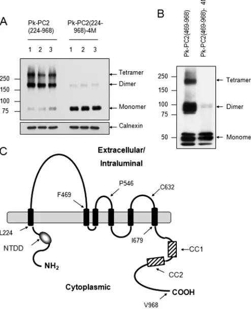
Identification of a Third Dimerization Domain for PC2—We recently reported the existence of two separate N- and C-ter-minal dimerization domains for PC2 important for channel assembly and activity (18, 19). To clarify the relative role of each domain for PC2 self-association, we initially deleted the entire
N terminus from full-length PC2 (Pk-PC2(224 –968)) (Fig. 1A).
Surprisingly, this showed a very similar pattern to wild-type PC2 with prominent dimer and tetramer formation. The intro-duction of four substitution mutations (Val846, Ile853, Ile860,
Leu867or 4M) in the C-terminal CC2 has been shown to
spe-cifically disrupt C-terminal dimerization and abolish PC2
binding to PC1 (19). As shown in Fig. 1A, CC2 mutations also
markedly reduced PC2 oligomerization to a predominant monomeric form, as seen with wild-type PC2. Deletion of the N-terminal domain did not completely abolish dimer forma-tion in this protein. Furthermore, the combined deleforma-tion of both N- and C-terminal domains (Pk-PC2 (224 – 679)) permit-ted the formation of dimers under nonreducing conditions (Fig. 2B). These results indicated the existence of a third dimeriza-tion domain within the transmembrane region of PC2 between amino acids 224 and 679.
Deletion of the First Extracellular Loop Does Not Abolish Dimer Formation—In view of the sensitivity of PC2 dimeriza-tion to reducing agents, we hypothesized that cysteine residues
at University of Sheffield on March 20, 2017
http://www.jbc.org/
within the putative extracellular loops of plasma membrane located PC2 (or the intraluminal loops for ER localized PC2) were most likely to be mediating PC2 dimerization through the formation of disulfide bonds. The first loop (amino acids 245– 468) contains three cysteine residues at cysteines 331, 344, and 437.
We initially deleted the entire N-terminal region including the first extracellular loop (amino acids 1– 468) in combination with a deletion of the C-terminal domain (amino acids 679 – 968). However, this mutant protein was highly unstable when expressed in HEK293 cells (data not shown). We therefore revised our strategy to generate a deletion construct omitting the N-terminal and first loop deletion (Pk-PKD2(469 –968)) only. As shown in Fig. 1B, deletion of the first loop did not alter dimer or tetramer formation. Once again, introduction of CC2 mutations (4M) markedly reduced but did not abolish dimer formation under nonreducing conditions though tetramers were not visualized (Fig. 1B). These results indicate that the first loop is not involved in mediating homodimerization.
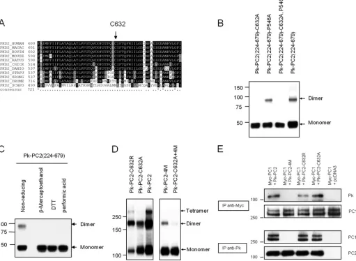
A Single Cysteine Residue in the Third Extracellular Loop Constitutes the Third Dimerization Domain—We next sought to investigate the role of other cysteine residues in either the
second (amino acids 527–550) or third (amino acids 620 – 658) extracellular loops. There is a single cysteine residue in the third loop (Cys632) which has been reported to be mutated in a PKD2 pedigree (C632R) (25). Of relevance, this residue is highly
conserved between human and worm PC2 protein (Fig. 2A). It
also lies within the predicted pore forming region although no functional assays had been previously performed.
We generated a site-specific C632A mutation in a construct with both N- and C-terminal domains deleted (Pk-PKD2(224 – 679)). Deletion of both domains had no effect, but the C632A mutation completely abolished dimer formation in the absence of both N- and C-terminal domains under nonreducing condi-tions (Fig. 2B). In contrast, mutation of an irrelevant residue (P546A) in the second extracellular loop had no effect (Fig. 2B). Incubation of the Pk-PKD2(224 – 679)) protein with oxidizing or reducing agents confirmed that dimer formation was medi-ated by disulfide bonds between two cysteine residues (Fig. 2C). Introduction of the C632A mutation into full-length PC2 shifted the ratio between monomers and dimers to the
mono-meric form with no tetramers seen (Fig. 2D). Under the same
conditions, wild-type PC2 showed prominent dimers and
tetramers (Fig. 2D). However, a C632R mutant had an
oligo-merization pattern intermediate between wild type and C632A. The reason for the difference between the C632A and C632R mutants is unclear. CC2 mutations (4M) had a more marked effect than C632A on dimer formation. Dimer formation was almost completely abolished in the combined mutant
(C632A⫹4M), although a faint dimer band presumably due to
the N-terminal dimerization domain could still be visualized on prolonged exposure (Fig. 2D).
Mutations of Cys632Do Not Alter Formation of the PC1-PC2
Complex—Mutations of CC2 (4M) have been shown to abolish PC1 recognition and formation of a functional PC1-PC2 recep-tor-ion channel complex (19). We examined whether a C632A or C632R mutation would affect PC1 binding. As shown in Fig.
2D, PC1 bound normally to PC2 C632A and C632R mutants
but not to PC2– 4M in co-IP assays. These results confirm that binding to PC1 is specifically determined by C-terminal dimerization through the CC2 (Fig. 2E). It seems likely that the role of Cys632-mediated dimerization is in the assembly of
tetra-meric PC2 channels rather than the heterotetra-meric PC1-PC2 channel complex.
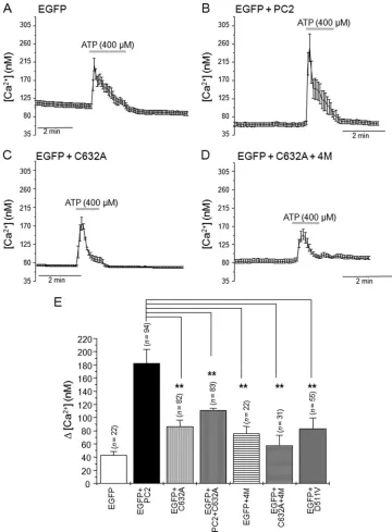
Mutations of Cys632Reduce ATP-sensitive Ca2⫹Release from ER-located PC2 Channels—We have recently shown that PC2 channels can function as receptor-operated ER located
homo-meric Ca2⫹release channels independently of PC1 (19). Based
on these findings, we compared Ca2⫹release from intracellular
stores in response to ATP (400M) in cells expressing wild-type
PC2 (PC2wt) and cells expressing PC2-C632A, PC2wt⫹
PC2-C632A, PC2– 4M, and PC2-C632A⫹4M mutants (Fig. 3).
Compared with cells expressing wild-type PC2 (⌬[Ca2⫹],
182.06⫾21.18,n⫽94), ATP-mediated Ca2⫹transients had
significantly (p⬍0.01) smaller amplitudes in cells expressing
PC2-C632A (⌬[Ca2⫹], 86.66 ⫾9.20, n⫽ 82), PC2wt⫹
PC2-C632A (⌬[Ca2⫹], 111.39⫾4.60,n⫽83), PC2– 4M (⌬[Ca2⫹],
76.04 ⫾ 10.74, n ⫽ 22), and PC2-C632A⫹4M (⌬[Ca2⫹],
57.72⫾15.68,n⫽31). We confirmed that Ca2⫹release was
[image:3.594.40.287.63.367.2]mediated by ER PC2 by expressing the channel-dead mutant
FIGURE 1.Evidence of a third dimerization domain for PC2.A, expression
pattern of epitope-tagged mutant PC2 on nonreducing SDS-PAGE. The trun-cation mutant PC2(224 –968), which lacks the N terminus (1–223), is still able
to form homodimers and tetramers.Mutation of CC2 (4M) in this construct
results in a predominant monomeric pattern with faint but detectable
dimers. Calnexin was used as an endogenous control for loading.B, deletion
of the N terminus and first extracellular loop (469 –968) shows a migration pattern similar to that of PC2(224 –968) under nonreducing conditions. Muta-tion of CC2 (4M) results in a predominant monomeric pattern with faint
dimers.C, diagram of PC2 showing its likely topology and key residues. The
N-terminal dimerization domain (NTDD), coiled coil domains 1 (CC1) and 2 (CC2) within the C terminus have been described previously.
at University of Sheffield on March 20, 2017
http://www.jbc.org/
PC2-D511V. In these cells, the ATP-induced Ca2⫹ rise (⌬[Ca2⫹], 83.37⫾16.25,n⫽55) was not significantly different
from ATP responses obtained in cells expressing EGFP alone (⌬[Ca2⫹], 43.24⫾5.68,n⫽22). These data indicate that
resi-due Cys632has a key role in the formation of functional ER
tetrameric PC2 channels.
DISCUSSION
Mutations inPKD1andPKD2give rise to ADPKD whose
clinical phenotype is characterized by the formation of fluid-filled cysts in the kidney and other organs as well as the pres-ence of noncystic manifestations such as vascular aneurysms and cardiac valve defects. PC1 and PC2 have been shown to interact via their C-terminal domains (26, 27), and a native PC1-PC2 complex has been identified (8). It seems likely there-fore that signals resulting from the interaction between both
proteins are critical to the maintenance of normal kidney struc-ture and function.
[image:4.594.42.553.67.441.2]PC2 is known to form oligomers in native cells and tissues (8), and there is evidence that PC2 can function independently of PC1 at the embryonic node in the determination of body axis patterning (16). It is unclear whether this PC2-specific function is mediated by PC2 ciliary complexes or by PC2-ER complexes because there is evidence for the involvement of both in differ-ent studies (16, 19, 28). Unusually for TRP channels, PC2 has also been reported to interact with subunits from other TRP subfamilies such as TRPC1 and TRPV4 which co-localize to primary cilia (7, 17). PC2 interacts via its C terminus with the C termini of TRPC1 and TRPV4 (Fig. 4). In addition, PC2 and TRPC1 reportedly interact through their transmembrane regions, although it is unclear whether this interaction is direct (5). The physiological significance of these ciliary and/or
FIGURE 2.A conserved cysteine residue in the third extracellular loop mediates PC2 dimerization.A, sequence alignment of Cys632showing its complete
evolutionary conservation from human toCaenorhabditis elegansPC2.B, expression pattern of epitope-tagged mutant PC2 on nonreducing SDS-PAGE.
Deletion of both N- and C-terminal domains, PC2(224 – 679), does not abolish dimer formation. Mutation at Cys632but not Pro546abolishes the formation of
dimer formation in the absence of the two known dimerization domains.C, cell lysates of PC2(224 – 679) resolved on SDS-PAGE. Lysates were either incubated
under nonreducing conditions, reducing agents (-mercaptoethanol, DTT), or performic acid prior to loading. Immunoblotting was with anti-Pk tag antibody. D, expression pattern of epitope-tagged full-length PC2 on nonreducing SDS-PAGE. Wild-type PC2 migrates most prominently with dimeric and tetrameric
species. The C632A mutation abolished tetramer bands but with an equal ratio of monomers and dimers. The double mutation (C632A⫹4M) almost
com-pletely abolished dimer formation. The pattern observed with C632R was intermediate between wild type and C632A.E, immunoprecipitation assays with
epitope-tagged full-length PC1 and PC2. Mutation at C632A or C632R did not affect the PC1-PC2 interaction, whereas CC2 mutations (4M) abolished PC1-PC2 interactions.
at University of Sheffield on March 20, 2017
http://www.jbc.org/
plasma membrane complexes in kidney function is not yet clear. Mice deficient in TRPC1 and TRPV4 do not develop kid-ney cysts.
Functional interactions between PC2 and other major ER Ca2⫹release channels such as the inositol trisphosphate
recep-tor and the type II ryanodine receprecep-tor (in cardiac tissue) have also been reported (11, 12, 29). PC2 binds via its C terminus to both receptors, although possibly to the ryanodine receptor in its open state (Fig. 4). An additional N-terminal binding site for PC2 to the ryanodine receptor has been described (12). Again, the functional significance of these ER complexes is uncertain.
In addition, the precise stoichiometry of the various PC2 com-plexes has not been resolved.
[image:5.594.115.475.57.546.2]In this paper, we report that PC2 homodimerization is regu-lated by a third dimerization domain. In recent studies, we reported the existence of a new N-terminal dimerization domain (18) and with others, a previously unrecognized C-ter-minal CC2 for PC2 (19, 30, 31). Although there is disagreement as to whether the latter leads to dimerization or trimerization of the PC2 C terminus, oligomerization of PC2 appears to be an absolute requirement for PC1 binding (19, 30). The specificity of CC2 in mediating PC2 dimerization for PC1 recognition is
FIGURE 3.The Cys632mutation abrogates ATP-dependent PC2 Ca2ⴙrelease channel activity.A–D, averaged calcium transients in response to ATP
application (400M) in HEK293T cells expressing EGFP (A), EGFP⫹PC2 (B), EGFP⫹PC2-C632A (C), and EGFP⫹PC2-C632A⫹4M (D). Thegray horizontal bars
indicate duration of ATP application.E, histogram showing the average rise in [Ca2⫹]
iinduced by ATP in cells expressing EGFP (0.5g of cDNA), EGFP⫹PC2 (0.5
g of cDNA), EGFP⫹PC2-C632A, EGFP⫹PC2⫹PC2-C632A, EGFP⫹PC2– 4M, EGFP⫹PC2-C632A⫹4 M and EGFP⫹PC2-D511V. Bars represent mean⫾S.E.
*,p⬍0.05; **,p⬍0.01.
at University of Sheffield on March 20, 2017
http://www.jbc.org/
confirmed by the lack of effect of the Cys632mutants in this
study.
Previously, we showed that PC2 mutants unable to undergo C-terminal dimerization (PC2– 4M) were still able to form
ER-based Ca2⫹release channels and could partially rescue the LR
defect seen in zebrafish embryos though not the cystic pro-nephric kidney (19). These results indicated that although C-terminal dimerization is specific to the formation of PC1-PC2 heteromers, other upstream dimerization regions (includ-ing the N-terminal domain) are critical to the formation of homomeric PC2 complexes. Our results demonstrate that
PC2-C632A mutants have reduced ER PC2-mediated Ca2⫹release
similar to the PC2– 4M mutants and predict that they will be similarly functionally effective in rescuing the laterality defect. In conclusion, we demonstrate that PC2 homodimerization is regulated by three distinct domains and that these events regulate formation of the tetrameric PC2 channel (Fig. 5). Our
results define a single cysteine residue, Cys632, known to be
mutated in a PKD2 pedigree, as a key functional residue regu-lating PC2 channel activity (25). Cys632lies in a putative third
extracellular loop (for plasma membrane channel) or third intraluminal loop (for the ER channel) which constitutes part of the predicted channel pore region for PC2. Our studies indicate that it mediates disulfide bonding between PC2 monomers to form homodimers critical for the formation of tetrameric chan-nels (Fig. 5). Co-expression of a C632A mutant suppressed the effect of wild-type PC2 in ER Ca2⫹release assays, proving the
functional importance of this interaction. Of interest, a
com-bined mutant (C632A⫹4M) retaining the N-terminal domain
had almost absent ATP-sensitive PC2-dependent Ca2⫹release.
In a previous study, we found that expression of the N-terminal dimerization domain as a surface-anchored protein in mamma-lian cells could inhibit plasma membrane PC2 channel activity in mIMCD3 cells (18). These results imply that the N-terminal dimerization domain is not critical for the function of ER PC2 channels but is required for the function of surface PC2 chan-nels. In LLCPK-1 cells, surface PC2 can be activated via mem-brane depolarization or EGF stimulation. This effect appears to be mediated by the unblocking of PC2 by mDia-1 through its activation by RhoA (32). It seems likely that distinct regulatory pathways, possibly through altering key protein-protein inter-actions, regulate the function of the ER and surface PC2 channels.
REFERENCES
1. Calvet, J. P., and Grantham, J. J. (2001)Semin. Nephrol.21,107–123 2. Wilson, P. D. (2004)N. Engl. J. Med.350,151–164
3. Mochizuki, T., Wu, G., Hayashi, T., Xenophontos, S. L., Veldhuisen, B., Saris, J. J., Reynolds, D. M., Cai, Y., Gabow, P. A., Pierides, A., Kimberling, W. J., Breuning, M. H., Deltas, C. C., Peters, D. J., and Somlo, S. (1996)
Science272,1339 –1342
4. Montell, C., Birnbaumer, L., Flockerzi, V., Bindels, R. J., Bruford, E. A., Caterina, M. J., Clapham, D. E., Harteneck, C., Heller, S., Julius, D., Kojima, I., Mori, Y., Penner, R., Prawitt, D., Scharenberg, A. M., Schultz, G., Shi-mizu, N., and Zhu, M. X. (2002)Mol. Cell9,229 –231
5. Tsiokas, L., Arnould, T., Zhu, C., Kim, E., Walz, G., and Sukhatme, V. P. (1999)Proc. Natl. Acad. Sci. U.S.A.96,3934 –3939
6. Nauli, S. M., Alenghat, F. J., Luo, Y., Williams, E., Vassilev, P., Li, X., Elia, A. E., Lu, W., Brown, E. M., Quinn, S. J., Ingber, D. E., and Zhou, J. (2003)
Nat. Genet.33,129 –137
7. Ko¨ttgen, M., Buchholz, B., Garcia-Gonzalez, M. A., Kotsis, F., Fu, X., Doerken, M., Boehlke, C., Steffl, D., Tauber, R., Wegierski, T., Nitschke, R., Suzuki, M., Kramer-Zucker, A., Germino, G. G., Watnick, T., Prenen, J., Nilius, B., Kuehn, E. W., and Walz, G. (2008)J. Cell Biol.182,437– 447 8. Newby, L. J., Streets, A. J., Zhao, Y., Harris, P. C., Ward, C. J., and Ong,
A. C. (2002)J. Biol. Chem.277,20763–20773
9. Streets, A. J., Newby, L. J., O’Hare, M. J., Bukanov, N. O., Ibraghimov-Beskrovnaya, O., and Ong, A. C. (2003) J. Am. Soc. Nephrol. 14,
1804 –1815
10. Koulen, P., Cai, Y., Geng, L., Maeda, Y., Nishimura, S., Witzgall, R., Eh-rlich, B. E., and Somlo, S. (2002)Nat. Cell Biol.4,191–197
11. Li, Y., Wright, J. M., Qian, F., Germino, G. G., and Guggino, W. B. (2005)
J. Biol. Chem.280,41298 – 41306
12. Anyatonwu, G. I., Estrada, M., Tian, X., Somlo, S., and Ehrlich, B. E. (2007)
Proc. Natl. Acad. Sci. U.S.A.104,6454 – 6459
13. Hanaoka, K., Qian, F., Boletta, A., Bhunia, A. K., Piontek, K., Tsiokas, L., Sukhatme, V. P., Guggino, W. B., and Germino, G. G. (2000)Nature408,
990 –994
14. Xu, G. M., Gonza´lez-Perrett, S., Essafi, M., Timpanaro, G. A., Montalbetti, N., Arnaout, M. A., and Cantiello, H. F. (2003) J. Biol. Chem. 278,
1457–1462
15. Delmas, P., Nauli, S. M., Li, X., Coste, B., Osorio, N., Crest, M., Brown, D. A., and Zhou, J. (2004)FASEB J.18,740 –742
[image:6.594.40.289.55.197.2]16. McGrath, J., Somlo, S., Makova, S., Tian, X., and Brueckner, M. (2003)Cell
FIGURE 4.Diagrammatic representation of PC2 homophilic and
hetero-philic interaction domains.The various interaction domains between PC2 and a number of key calcium channel proteins are shown. Apart from PC2, PC1, and inositol trisphosphate receptor (IP3R), the key residues mediating
other interactions have not been fully defined.RYR, ryanodine receptor.
FIGURE 5.Model of PC2 in a lipid bilayer showing the three dimerization
domains.PC2 is displayed as a homotetramer with the transmembrane domains (TM) embedded in a lipid bilayer. C-terminal (CT) homodimerization
mediated by the C-terminal CC2 (Ser835-Ala873) is indicated by ablack line.
CC2-mediated dimerization has a specific role in PC1 recognition and forma-tion of a PC1-PC2 heteromeric complex. N-terminal (NT) dimerizaforma-tion
medi-ated by the N-terminal dimerization domain (Gly199-Glu207) is indicated by a
black line. Disulfide bonding between PC2 monomers mediated by Cys632is
indicated by ayellow line.
at University of Sheffield on March 20, 2017
http://www.jbc.org/
[image:6.594.41.293.259.407.2]114,61–73
17. Bai, C. X., Giamarchi, A., Rodat-Despoix, L., Padilla, F., Downs, T., Tsio-kas, L., and Delmas, P. (2008)EMBO Rep.9,472– 479
18. Feng, S., Okenka, G. M., Bai, C. X., Streets, A. J., Newby, L. J., DeChant, B. T., Tsiokas, L., Obara, T., and Ong, A. C. (2008)J. Biol. Chem.283,
28471–28479
19. Giamarchi, A., Feng, S., Rodat-Despoix, L., Xu, Y., Bubenshchikova, E., Newby, L. J., Hao, J., Gaudioso, C., Crest, M., Lupas, A. N., Honore´, E., Williamson, M. P., Obara, T., Ong, A. C., and Delmas, P. (2010)EMBO J. 29,1176 –1191
20. Ong, A. C., Ward, C. J., Butler, R. J., Biddolph, S., Bowker, C., Torra, R., Pei, Y., and Harris, P. C. (1999)Am. J. Pathol.154,1721–1729
21. Streets, A. J., Moon, D. J., Kane, M. E., Obara, T., and Ong, A. C. (2006)
Hum. Mol. Genet.15,1465–1473
22. Ong, A. C., Harris, P. C., Davies, D. R., Pritchard, L., Rossetti, S., Biddolph, S., Vaux, D. J., Migone, N., and Ward, C. J. (1999) Kidney Int. 56,
1324 –1333
23. Hirs, C. H. W. (1967) inMethods in Enzymology(Colowick, S. P., and Kaplan, N. O., eds) vol. 11, pp. 198 –199, Academic Press, New York 24. Grynkiewicz, G., Poenie, M., and Tsien, R. Y. (1985)J. Biol. Chem.260,
3440 –3450
25. Magistroni, R., He, N., Wang, K., Andrew, R., Johnson, A., Gabow, P., Dicks, E., Parfrey, P., Torra, R., San-Millan, J. L., Coto, E., Van Dijk, M., Breuning, M., Peters, D., Bogdanova, N., Ligabue, G., Albertazzi, A., Hate-boer, N., Demetriou, K., Pierides, A., Deltas, C., St. George-Hyslop, P., Ravine, D., and Pei, Y. (2003)J. Am. Soc. Nephrol.14,1164 –1174 26. Tsiokas, L., Kim, E., Arnould, T., Sukhatme, V. P., and Walz, G. (1997)
Proc. Natl. Acad. Sci. U.S.A.94,6965– 6970
27. Qian, F., Germino, F. J., Cai, Y., Zhang, X., Somlo, S., and Germino, G. G. (1997)Nat. Genet.16,179 –183
28. Fu, X., Wang, Y., Schetle, N., Gao, H., Pu¨tz, M., von Gersdorff, G., Walz, G., and Kramer-Zucker, A. G. (2008)J. Am. Soc. Nephrol.19,1342–1351 29. Sammels, E., Devogelaere, B., Mekahli, D., Bultynck, G., Missiaen, L., Parys, J. B., Cai, Y., Somlo, S., and De Smedt, H. (2010)J. Biol. Chem.285,
18794 –18805
30. Yu, Y., Ulbrich, M. H., Li, M. H., Buraei, Z., Chen, X. Z., Ong, A. C., Tong, L., Isacoff, E. Y., and Yang, J. (2009)Proc. Natl. Acad. Sci. U.S.A.106,
11558 –11563
31. Celiæ, A., Petri, E. T., Demeler, B., Ehrlich, B. E., and Boggon, T. J. (2008)
J. Biol. Chem.283,28305–28312
32. Bai, C. X., Kim, S., Li, W. P., Streets, A. J., Ong, A. C., and Tsiokas, L. (2008)
EMBO J.27,1345–1356
at University of Sheffield on March 20, 2017
http://www.jbc.org/
Shuang Feng, Lise Rodat-Despoix, Patrick Delmas and Albert C. M. Ong
Channel
Essential for the Assembly and Function of the Tetrameric Polycystin-2 (TRPP2)
doi: 10.1074/jbc.M110.192286 originally published online April 7, 2011 2011, 286:18994-19000.
J. Biol. Chem.
10.1074/jbc.M110.192286
Access the most updated version of this article at doi:
Alerts:
When a correction for this article is posted
•
When this article is cited
•
to choose from all of JBC's e-mail alerts
Click here
http://www.jbc.org/content/286/21/18994.full.html#ref-list-1
This article cites 32 references, 18 of which can be accessed free at
at University of Sheffield on March 20, 2017
http://www.jbc.org/