The impact of alpha lipoic acid on developmental
competence of mouse vitrified pre-antral follicles in
comparison to those isolated from vitrified ovaries
Sahar Hatami1 M.Sc., Saeed Zavareh1, 2 Ph.D., Mojdeh Salehnia3 Ph.D., Taghi Lashkarbolouki1,2 Ph.D., Mohammad Taghi Ghorbanian1, 2 Ph.D., Isaac Karimi4 Ph.D.
1.School of Biology, Damghan University, Damghan, Iran. 2.Institute of Biological Sciences,
Damghan University, Damghan, Iran.
3.Department of Anatomy, Tarbiat Modares University, Tehran, Iran.
4.Laboratory of Molecular and Cellular Biology, Department of Basic Veterinary Sciences, School of Veterinary Medicine, Razi University, Kermanshah, Iran.
Corresponding Author: Saeed Zavareh, School of Biology and Institute of Biological Sciences, Damghan University, Ceshme Ali St., Damghan, Iran. Email: [email protected]; [email protected]
Tel: (+98) 232 5247146
Received: 3 June 2013 Revised: 29 August 2013 Accepted: 15 September 2013
Abstract
Background: Cryopreservation of ovarian tissues and pre-antral follicles is a promising prospect for preservation of women fertility.
Objective: The aim of this study was to evaluate the in vitro developmental competence of mouse vitrified antral follicles in comparison to isolated pre-antral follicles derived from vitrified ovaries in the presence of alpha lipoic acid (ALA).
Materials and Methods: Pre-antral follicles derived from fresh, vitrified-warmed ovarian tissues and vitrified–warmed pre-antral follicles were cultured individually with or without ALA, followed by adding hCG to induce ovulation. The follicle growth, oocyte maturation, and embryo development were assessed.
Results: The diameter and development of follicles, oocyte maturation and embryo development rates were significantly higher in ALA supplemented groups compared to the respective ALA-free conditions groups. Aforementioned parameters were significantly higher in vitrified-warmed follicles in comparison to follicles derived from vitrified-warmed ovaries.
Conclusion: These findings support a superior performance of pre-antral follicles when vitrified rather than when isolated from vitrified ovaries with regard to increasing the rates of developmental parameters. Moreover, ALA improves the in vitro maturation of pre-antral follicles in vitrified and non-vitrified samples.
Key words: Vitrification, Ovary, Preantral follicles, Alpha Lipoic Acid.
This article extracted from M.Sc. thesis. (Sahar Hatami)
Introduction
n recent years, preservation of female fertility in cancer patients who are facing chemotherapy or radiotherapy has been
done in several ways such as
cryopreservation of ovarian tissue, oocytes and embryos. Cryopreservation of ovarian tissue rather than cryopreservation of oocytes and embryos has many advantages (1). It will preserve the ovarian functional and structural integrity which in turn leads to the restoration of both steroidogenic and gametogenic functions (2). Furthermore, in order to prevent delay in cancer treatment, ovarian tissue can be collected from a patient regardless of the menstrual cycle at any time (3).
This method is also appropriate for pre-pubertal girls and patients with premature ovarian failure (3, 4). In recent decades, the conventional slow freezing of ovarian tissue
has been performed successfully nevertheless it also associated with some disadvantages (4). Vitrification as an alternative approach is achieved by direct plunging into liquid nitrogen
(LN2) without subsequent cryodamage
resulting from ice formation (5, 6). High cooling and thawing rate are necessary to reduce adverse effects of crystallization in vitrification. With increasing sample size, the sample is surrounded by vapor and not LN2 so that the cooling rate decreases which in turn leads to dramatic decline of the survival rates of vitrified samples (5). Ovarian follicles can also be isolated from fresh ovarian tissue and then vitrified (7).
This procedure not only increases cooling rate through reducing sample size, but also reduces the risk of transmission of cancer cells from vitrified ovarian tissue back into recipients and also provides the assessment of follicles quality during culture period after
I
warming (8). Nowadays, several culture systems have been proven to cultivate isolated follicles and used to achieve an alive progeny (7, 9, 10).
However, cryopreservation seems to
trigger biological events not responsible for normal cell function and follicular development (11). Increased production of reactive oxygen species (ROS) is one of the consequences of crypreservation and thawing of semen and ovarian tissue (9, 12, 13). It could be due to a hypoxic condition that leads to excess electron production which in turn results in formation of ROS (12). It has been
demonstrated that reduced oxygen
environment prompts damaging effects on maturation of squirrel monkey's oocytes (14).
In the in vivo condition, generation of ROS is
equilibrated by enzymatic and no enzymatic antioxidant defense systems of cells (15, 16).
However, the lack of this physiological
defense system in the in vitro condition under
high pressure of oxygen can lead to oxidative stress (17, 18). Alpha-lipoic acid (ALA), as a
coenzyme of mitochondrial multienzyme
complexes, is well known for its antioxidant properties which acts directly on scavenging of ROS, and indirectly on intracellular recycling of other antioxidants (19, 20). It has
been revealed that ALA improves the in vitro
development of follicles via decreasing ROS concentration and increasing total antioxidant capacity (TAC) level of follicles throughout culture period (21).
Furthermore, it has been shown that ALA supplemented maturation medium improves developmental competence and modifies oxidative status of oocytes through inhibiting the expression of apoptotic activators and accelerate synthesize glutathione (GSH),
glutathione peroxidase 4 (GPx4) and
superoxide dismutase (SOD) (22). To date there is no study indicating the effects of ALA on the developmental competence of vitrified pre-antral follicle and isolated pre-antral
follicles derived from vitrified ovaries.
Therefore, the objective of the present study
was to compare follicle growth and
development, oocyte maturation and embryo development rates of mouse vitrified-warmed pre-antral follicles and isolated pre-antral follicles derived from whole vitrified-warmed ovaries during in vitro culture in the presence of ALA.
Materials and methods
Reagents
All reagents were purchased from Sigma-Aldrich, UK unless otherwise stated and all media were made with Mili-Q water.
Animals
In this experimental animal study, female National Medical Research Institute (NMRI) mice were purchased from Pasteur Institute, Iran. Animals were cared for and used according to the Animal Ethics Committee. The mice were housed and bred in the Animal House of Damghan University under light and temperature controlled conditions (12 h of light
and 12 h of darkness; and 24oC) and they
provided with food and water ad libitum.
For all experiments, mice aged 14-15 days old were killed by cervical dislocation and their ovaries were placed in α-minimal essential medium (α-MEM; Gibco, UK) supplemented with 10% fetal bovine serum (FBS; Gibco, UK), 2.2 g/l sodium bicarbonate, 25 mM HEPES, 100 IU/ml penicillin and 75 µg/ml streptomycin. The ovaries were randomly divided into 3 groups: 1) those that not vitrified (control), 2) those that vitrified and 3) those which their follicles were isolated and then vitrified. Isolated pre-antral follicles of each group were cultured in the presence or absence of 100 µM of ALA.
Pre-antral follicles isolation
The preantral follicles of fresh and vitrified-warmed ovaries were isolated mechanically by using a 29 gauge needle under a stereomicroscope (Nikon, Japan) at 10X magnification. Isolated follicles were selected according to the following criteria: rounded follicular structure with 140-150 µm diameters, containing visible centrally located healthy oocyte surrounded with intact several layers of
granulosa cells, an intact basement
membrane, at least one layer of theca cells and no antral cavity. During the operating procedures the culture medium was always kept at 37oC.
Vitrification and warming of ovarian tissue and pre-antral follicles
The vitrification procedure was based on a method described previously with some
modifications. Exposure time to the equilibrium solution (ES) and vitrification solution (VS) for ovarian tissue and pre-antral follicles were also based on the methods described previously (23, 24).
In brief, ovarian tissues and isolated pre-antral follicles were initially incubated for 10 and 5 minutes respectively in ES containing 7.5% (v/v) ethylene glycol (EG) and 7.5% (v/v)
dimethylsulfoxide (DMSO) in Dulbecco
phosphate-buffered saline medium (DPBS, Gibco, UK) with 20% fetal bovine serum (FBS), then incubated in vs. (15% EG, 15% DMSO and 0.5M sucrose in DPBS+20% FBS) for another 2 min and 30 sec for ovarian
tissue and isolated pre-antral follicles,
respectively. After dehydration, the ovarian
tissues and pre-antral follicles were
immediately drawn into a Pasteur pipette with minimum volume of VS (<0.1µl) and were individually placed on top of a polypropylene strip of cryotop carrier (Kitazato, Japan) and immediately immersed into LN2.
The thin strip of cryotop was covered with cap and stored in LN2 tank for at least 1 week. All equilibration and vitrification steps were carried out at room temperature. For warming, the cryotop cap was removed while it was still immersed in LN2, then the strip
directly submerged in DPBS medium
containing 1M sucrose. The pre-antral follicles and ovarian tissue were left in the warming solution (1M sucrose in DPBS) for 30 sec and 5 min respectively and transferred into droplets of DPBS medium containing 0.5 and 0.25M sucrose at an interval of 5 min and 3 min respectively at room temperature. After this, ovarian tissue and pre-antral follicles were pipetted into fresh α-MEM medium
supplemented with 10% FBS at 37oC for
another 10 min before placing in culture medium.
In vitro culture of isolated pre-antral follicles
In vitro culture of the isolated fresh and
vitrified/warmed pre-antral follicles was
performed according to our previous
methodology (21). Briefly, fresh and
vitrified/warmed pre-antral follicles were
cultured individually in 20 µl droplets of α-MEM (pH= 7.2) supplemented with 0.23 mM sodium pyruvate, 5% FBS, 100 mIU/ml
recombinant follicle stimulating hormone
(rFSH; Gonal-f Switzerland), 10 mg/mL insulin, 5.5 mg/mL transferrin and 0.67 mg/ml selenium (ITS; Gibco, UK), 20 ng/ml murine recombinant epidermal growth factor, 100 IU/ml penicillin, 75 µg/ml streptomycin, 2.2g/l sodium bicarbonate and 100 µM ALA according to experimental design under mineral oil in a humidified atmosphere of 5%
CO2 in air at 37oC in Falcon Petri dishes
(60×15 mm) for up to 12 days. At 48h intervals, 10 µl of culture medium from each drop was replenished by fresh medium.
Evaluation of morphological and developmental parameters of cultured pre-antral follicles
The survival rate of the pre-antral follicles was assessed microscopically based on the morphology of the pre-antral follicle every other day under inverted microscope during culturing period and they were compared at the end of the study as described previously (7). Briefly, a follicle was considered normal when it possessed a centrally located
spherical and homogeneous oocyte
surrounded by complete and compact layers of granulosa cells with no noticeable damage to the basement membrane. Follicles with partially or completely naked oocytes or those
that showed morphological signs of
degeneration, such as darkness of oocytes and surrounding cumulus cells or those with
misshapen oocytes, were graded as
degenerated.
Antral-like cavity formation was considered as visible lucent area in the granulosa cell mass around the oocyte. Follicles diameter was measured only in healthy follicles on day 2 and 4 of culturing period, mean follicle diameter was assessed by measuring and averaging two perpendicular cross sectional diameters of each follicle with pre-calibrated ocular micrometer under inverted microscope (Nikon, Tokyo, Japan; 100× magnification) (21).
In vitro ovulation induction
In the twelfth day of culture, ovulation and final oocyte maturation were induced by
substitution of culture media with fresh
maturation medium supplemented with 1.5 IU/ml human chronic gonadotropin (hCG;
IBSA, Switzerland). 24 h after hCG addition in all groups, released oocytes were classified as germinal vesicle (GV), germinal vesicle breakdown (GVBD) when the GV was absent, and metaphase II oocytes (MII) when the first polar body was extruded.
In vitro fertilization
The fertile NMRI male mice were sacrificed by cervical dislocation and their caudae epididymides dissected and placed into a 500-µl drop of modified Tyrods medium (mT6) supplemented with 5% bovine serum albumin (BSA) under mineral oil. The caudae epididymides were ripped with the aid of 28 gauge needle and the spermatozoa squeezed out gently by using forceps and incubated for
90 min at 37oC in 5% CO2 to allow the
spermatozoa to swim out and capacitate. Capacitated sperm suspension was added to MII oocyte to give the final motile sperm
concentration of 1-2×10⁶ /ml.
MII oocytes and spermatozoa were co-incubated in modified KSOM-AA medium at
37oC under 5% CO2 for 6h. Oocytes
presenting two pronuclei (2PN) were
considered normally fertilized. Fertilized
oocytes were washed and gently pipetted to remove spermatozoa and attached cumulus cells. Zygotes were then transferred to drops of fresh KSOM-AA medium supplemented and
cultured at 37oC under 5% CO2 with maximum
humidity until day 5. Day of insemination was considered as day 0.
Embryos were observed on the heated
stage (37oC) of an inverted microscope
(Nikon, TE2000-U with Hoffman modulation contrast). The numbers of zygotes that reached to two cells, morula and blastocytes were counted.
Statistical analysis
Each experiment was carried out at least four replicates. The data are presented as the mean±SD. Statistical analysis was performed using SPSS-ver.16 software package (SPSS Inc., Chicago, IL, USA). All proportional data
were analyzed after arcsine of sqtr
transformation. Differences in groups were evaluated by one-way analysis of variance
(ANOVA) and Tukey's HSD was used as post
hoc test. Differences were considered
significant at a level of p<0.05.
Results
The morphological and maturational
parameters of the cultured vitrified and non-vitrified isolated pre-antral follicles with or without ALA are summarized in table I and II. By day 12 of culture, with respect to the follicle diameter and rates of survival, antrum formation and oocyte maturation, there were significantly differences in both vitrified and non-vitrified ALA supplemented samples compared with ALA-free conditions groups (Table I & II(. After the fourth day of culture, granulosa cells proliferated and grew through basal membrane to form the irregular and diffuse appearance that was impossible to measure follicle diameter. The mean follicle diameter at the beginning of the culture (147.9±5.9 µm) was not significantly different among all groups (p=0.83). During the culture
period, the follicle diameter increased
progressively.
As show in table I, the diameter of fresh pre-antral follicles on day 2 and 4 (223.9±17.1 and 323.3±11.6 µm respectively) of cultivation period in the presence of supplementation with ALA was significantly higher than the other groups. Also, the mean diameter of the vitrified-warmed pre-antral follicles on day 2 and 4 in the presence of ALA (180.5±7.2 and 272.1±18.4 µm respectively) were significantly higher than those which were isolated from vitrified-warmed ovaries. There were no significant differences between diameters of vitrified-warmed follicles and those which were derived from vitrified-warmed ovaries that were cultured in the presence of ALA (Table I).
The survival (94.3%) and antrum formation (92.3%) rates were significantly higher in fresh pre-antral follicles supplemented with ALA than the other groups (Table II). The rates of survival (68.3%) and antrum formation (62.8%) in the vitrified-warmed pre-antral follicles were significantly higher than those which were isolated from vitrified-warmed ovaries (57.3% and 46.0%, respectively). There was no significant difference between these rates of vitrified-warmed pre-antral follicles and pre-antral follicles derived from vitrified-warmed ovaries which were cultured in the presence of ALA (66.7% and 60.6%, respectively).
Table II shows the percentage of GV, MI and MII oocytes from the follicles after addition of the hCG induction on day 12. Higher percentages of MII oocytes were also obtained in vitrified-warmed pre-antral follicles than those which were isolated from
vitrified-warmed ovaries (28.3% and 15.0%
respectively, p=0.001), although both were statistically lower than control group (42.9%). The percentage of MII oocytes was similar between cultured isolated pre-antral follicles derived from vitrified-warmed ovaries (26.7%) with ALA and those of vitrified-warmed pre-antral follicles (28.3%); however, both rates were significantly lower in comparison to control group.
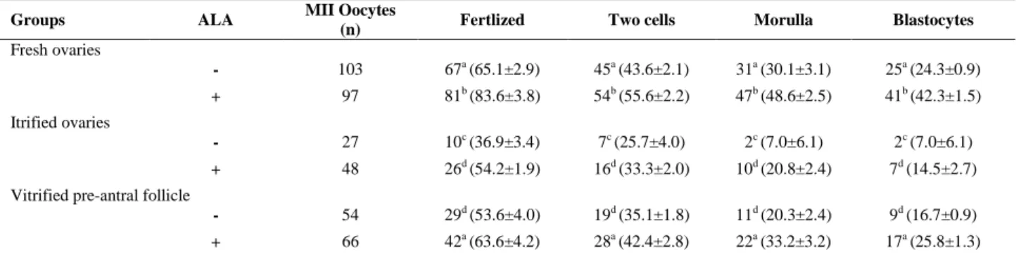
Nevertheless, the differences were all significant compared to that of pre-antral follicles which were cultured in the presence of ALA (53.9%). Fertilization and embryo developmental rates of MII oocytes derived from cultured isolated follicles in non-vitrified
and vitrified groups in the presence of ALA are shown in table III. There were significant differences in the fertilization rate and blastocyst formation rate in between the respective ALA-free and ALA supplemented groups both in vitrified and non-vitrified groups.
Significant higher rates of fertilization and blastocyte were observed in vitrified-warmed pre-antral follicles group in comparison with vitrified-warmed ovaries groups; however both were statistically lower than control group. There were no significant differences in the rates of fertilization and blastocyst formation between vitrified-warmed pre-antral follicles samples and pre-antral follicles derived from vitrified-warmed ovaries which were cultured in the presence of ALA. Also, these situations were observed between cultured vitrified-warmed pre-antral follicles samples in the presence of ALA and ALA-free fresh pre-antral follicles samples.
Table I. Diameter of cultured preantral follicles derived from fresh ovaries, vitrified ovaries and vitrified follicles in the presence or
absence of alpha lipoic acid (ALA)
Groups ALA NO. of follicles Follicle diameter (µm ± SD)
0th day 2th day 4th day
Fresh ovaries
- 240 148.2 ± 5.3 189.6 ± 11.4a 290.5 ± 26.3a
+ 180 147.5 ± 6.6 223.9 ±17.1b 323.3 ± 11.6b
Vitrified ovaries
- 180 147.6 ± 5.2 170.5 ± 13.1d 255.3 ± 37.3d
+ 180 147.3 ± 6.5 176.5 ± 4.8c 262.9 ± 4.7c
Vitrified pre-antral follicles
- 180 148.3 ± 5.0 175.3 ± 7.2c 264.2 ± 6.7c
+ 180 148.6 ± 5.7 180.5 ± 7.2a 272.1± 18.4a
In all cases at least 4 experimental replicates were performed. Different superscripts in the same column reflect different levels of significant difference (p<0.05)
Table II. Maturation rates of cultured pre-antral follicles derived from fresh ovaries, vitrified ovaries and vitrified follicles in the
presence or absence of alpha lipoic acid (ALA)
Groups ALA N Survived Degenerated Antrum
formation
Maturation stages of oocyte
GV MI MII
Fresh ovaries
- 240 204a (85.0±1.9) 36a (15.0±1.9) 184a (76.7±5.6) 30 (12.5±3.5) 51 (21.2±3.7) 103a (42.9±2.8)
+ 180 169 b (94.3±1.2) 11b (5.7±1.2) 166b (92.3±4.0) 16 (9.5±2.4) 34 (18.9±5.1) 97b (53.9±4.2)
Vitrified ovaries
- 180 102c (57.3±3.5) 72c (43.3±2.9) 83c (46.0±4.2) 27 (15.0±1.7) 22 (12.2±3.5) 27c (15.0±1.7)
+ 180 120d (66.7±3.3) 60d (33.3±3.3) 111d (60.6±2.5) 20 (11.1±2.5) 31 (20.6±4.2) 48d (26.7±1.7)
Vitrified pre-antral follicle
- 180 123 d (68.3±6.0) 57d (31.7±6.0) 113d (62.8±4.2) 21 (11.7±1.7) 20 (11.1±2.5) 54d (28.3±1.7)
+ 180 150a (83.3±3.3) 30a (16.7±3.3) 134a (74.4±4.2) 20 (11.1±2.5) 21 (11.7±1.7) 66a (36.7±3.3)
Data were presented as number (% ± SD) in all columns. In all cases at least 4 experimental replicates were performed.
Different superscripts in the same column reflect different levels of significant difference (p<0.05) GV: germinal vesicle stage oocyte; MI: metaphase I oocyte; MII: metaphase II oocyte.
Table III. Development rates of derived embryos from cultured pre-antral follicles in the presence or absence of alpha lipoic acid (ALA)
Groups ALA MII Oocytes
(n) Fertlized Two cells Morulla Blastocytes
Fresh ovaries
- 103 67a (65.1±2.9) 45a (43.6±2.1) 31a (30.1±3.1) 25a (24.3±0.9)
+ 97 81b (83.6±3.8) 54b (55.6±2.2) 47b (48.6±2.5) 41b (42.3±1.5)
Itrified ovaries
- 27 10c (36.9±3.4) 7c (25.7±4.0) 2c (7.0±6.1) 2c (7.0±6.1) + 48 26d (54.2±1.9) 16d (33.3±2.0) 10d (20.8±2.4) 7d (14.5±2.7)
Vitrified pre-antral follicle
- 54 29d (53.6±4.0) 19d (35.1±1.8) 11d (20.3±2.4) 9d (16.7±0.9)
+ 66 42a (63.6±4.2) 28a (42.4±2.8) 22a (33.2±3.2) 17a (25.8±1.3)
Data were presented as number (% ± SD) in all columns. In all cases at least 4 experimental replicates were performed.
Different superscripts in the same column reflect different levels of significant difference (p<0.05) MII: metaphase II oocyte.
Discussion
In the present study, vitrification of isolated mouse pre-antral follicle was more effective in comparison with vitrification of whole ovarian tissue due to improving follicular survival, antrum formation and oocyte maturation rates. This difference in the cryobiological features of vitrified-warmed antral follicles and pre-antral follicles derived from vitrified-warmed ovarian tissue may be attributed to their morphological differences. In this regard, it seems that vitrification of ovarian tissue compared to vitrification of pre-antral follicles is facing to some obstacles such as the high volume ratio to low surface area of a whole ovary, which will slow down the cooling and warming rates (25).
This feature of ovarian tissue causes temperature transition from the central region of the ovary to be slowly driven, which in turn will lead to increased possibility of ice crystal formation thus be at a greater risk of severe damage (3, 26). Furthermore, ovarian tissue
thickness prevents permeation of
cryoprotectant to the entire tissue within a given exposure time to equilibration and vitrification solution thus resulting in ice formation (25). In order to overcome this problem, the exposure time to equilibration and vitrification solution should be increased which in turn can lead to toxicity in surface areas (2).
Thus, it appears that differences between maturational competence of vitrified-warmed pre-antral follicles and pre-antral follicles obtained from vitrified-warmed ovarian tissue are due to the small size of the pre-antral follicles and increased surface to volume ratio,
which in turn will overcome obstacles mentioned above. Effect of cryopreservation on ultra-structural properties of vitrified ovaries has been investigated previously (27-29). In this regard, ultra-structural evaluation by transmission electron microscopy showed that the low developmental competence of oocyte after vitrification and warming is partially due to diffused and fragmented mitochondria (29).
Also, according to the ultra-structure
investigations, it was shown that oocytes of follicles derived from vitrified-warmed ovarian tissues reveales increased numbers of swollen mitochondria (27, 28).
Our explanation for different developmental competence among vitrified groups and fresh samples is that, mitochondrial disorders that are occurred throughout cryopreservation could increase ROS generation in vitrified samples and it seems that these disorders were minor in vitrified-warmed pre-antral follicles than pre-antral follicles derived from vitrified-warmed ovary, because development rate was higher in vitrified-warmed pre-antral follicles than those of pre-antral follicles dived
from vitrified-warmed ovaries. Several
defense mechanisms against ROS are present including non-enzymatic antioxidants and enzymatic defense mechanisms within cells (30).
We reported useful impact of ALA as a potent antioxidant on the developmental competence of isolated pre-antral follicles in fresh samples (21). However, this is the first
study to show the effects of ALA on in vitro
maturation of vitrified-warmed pre-antral
follicles and pre-antral follicles derived from vitrified-warmed ovaries. The present study showed that ALA could improve the rates of
survival, antrum formation and MII oocytes of vitrified-warmed and fresh pre-antral follicles
after long-term in vitro culture.
Several hypotheses can be considered for the role of ALA on improving the development of vitrified-warmed and fresh pre-antral follicles. Some investigators attributed the efficiency of ALA to unique antioxidant properties of lipoate/ dihydrolipoate system, i.e. ROS scavenging ability (21, 22, 31). This feature of ALA has been attributed partially to suppress TNF-alpha-induced ROS generation
and 6-hydroxydopamine induced ROS
generation (32). Also it has been known that ALA acts as a coenzyme of pyruvate dehydrogenase complex that catalyses the consumption of pyruvate in mouse follicles and oocytes (35).
To test this hypothesis, further studies must
be designed to determine monergic
coenzymatic role or possible synergic
coenzymatic roles of ALA combined with the rest of coenzymes of pyruvate dehydrogenase complex like thiamine pyrophosphate, FAD,
NAD+, and coenzyme A. In this sense, sodium
pyruvate routinely is a constant component of culture media like KSOM-AA medium (see Materials and Methods). ALA may be involved in improving development of cultured pre-antral follicles via metal chelation, antioxidant recycling, and gene expression (19). The other explanation for effects of ALA in improving IVM conditions may be related to inhibition of cell death (36). Also, it has been shown that ALA increases the catalase (CAT) activity and glutathione peroxidase (GPx), activity (37).
CAT and GPx are two fundamental enzymes can be used as a biomarker of oxidative stress (38). Also it has been suggested that activation of GPx in follicular
fluid were clearly associated with
developmental competence of oocyte (39). In
sum, the results of present study
demonstrated that vitrification of isolated pre-antral follicles is more effective method to increase follicular survival, antrum formation and oocyte development rates rather than vitrification of whole ovarian tissue. In addition to, this study showed that culture medium
supplementation with ALA improves
development of cultured pre-antral follicles derived from fresh and vitrified samples.
Future mechanistic studies on the
identification of metabolic role of ALA involved
in follicular maturation beside its antioxidative activity in assisted reproduction techniques should therefore be conducted.
Acknowledgments
This study was supported in part by grants from the Damghan University and Institute of Biological Sciences of Damghan University. The authors are grateful for Dr. Mehdi Khorshidi, for providing support and laboratory facilities.
Conflict of interest
There is no conflict of interest in this research.
References
1. Amorim CA, Goncalves PB, Figueiredo JR. Cryopreservation of oocytes from pre-antral follicles.
Hum Reprod Update 2003; 9: 119-129.
2. Posillico S, Kader A, Falcone T, Agarwal A. Ovarian Tissue Vitrification: Modalities, Challenges and
Potentials. Curr Women's Health Rev 2010; 6:
352-366.
3. Huang L, Mo Y, Wang W, Li Y, Zhang Q, Yang D. Cryopreservation of human ovarian tissue by
solid-surface vitrification. Eur J Obstet Gynecol Reprod
Biol 2008; 139: 193-198.
4. Isachenko V, Lapidus I, Isachenko E,
Krivokharchenko A, Kreienberg R, Woriedh M, et al.
Human ovarian tissue vitrification versus
conventional freezing: morphological,
endocrinological, and molecular biological
evaluation. Reproduction 2009; 138: 319-327.
5. Liebermann J, Nawroth F, Isachenko V, Isachenko E, Rahimi G, Tucker MJ. Potential importance of
vitrification in reproductive medicine. Biol Reprod
2002; 67: 1671-1680.
6. Zavareh S, Salehnia M, Saberivand A. Comparison
of Different Vitrification Procedures on
Developmental Competence of Mouse Germinal Vesicle Oocytes in the Presence or Absence of
Cumulus Cells. Int J Fertil Steril 2009; 3: 111-118.
7. Desai N, AbdelHafez F, Ali MY, Sayed EH, Abu-Alhassan AM, Falcone T, et al. Mouse ovarian follicle
cryopreservation using vitrification or slow
programmed cooling: assessment of in vitro development, maturation, ultra-structure and meiotic
spindle organization. J Obstet Gynaecol Res 2011;
37: 1-12.
8. Amorim CA, Van Langendonckt A, David A, Dolmans MM, Donnez J. Survival of human pre-antral follicles after cryopreservation of ovarian tissue, follicular isolation and in vitro culture in a calcium alginate
matrix. Hum Reprod 2009; 24: 92-99.
9. Abedelahi A, Salehnia M, Allameh AA, Davoodi D. Sodium selenite improves the in vitro follicular development by reducing the reactive oxygen species level and increasing the total antioxidant
capacity and glutathione peroxide activity. Hum Reprod 2010; 25: 977-985.
10. Oktem O, Oktay K. The role of extracellular matrix and activin-A in in vitro growth and survival of
murine preantral follicles. Reprod Sci 2007; 14:
358-366.
11. Liu HC, He Z, Rosenwaks Z. Mouse ovarian tissue cryopreservation has only a minor effect on in vitro
follicular maturation and gene expression. J Assist
Reprod Genet 2003; 20: 421-431.
12. Wang AW, Zhang H, Ikemoto I, Anderson DJ, Loughlin KR. Reactive oxygen species generation by
seminal cells during cryopreservation. Urology 1997;
49: 921-925.
13. Rahimi G, Isachenko E, Sauer H, Isachenko V, Wartenberg M, Hescheler J, et al. Effect of different vitrification protocols for human ovarian tissue on
reactive oxygen species and apoptosis. Reprod Fertil
Dev 2003; 15: 343-349.
14. Yeoman RR, Williams LE, Abee CR. Low oxygen inhibits but complex high-glucose mediumfacilitates in vitro maturation of squirrel monkey
oocyte-granulos a cell complexes. J Assist Reprod Genet
1999; 16: 102-107.
15. Agarwal A, Said TM, Bedaiwy MA, Banerjee J, Alvarez JG. Oxidative stress in an assisted
reproductive techniques setting. Fertil Steril 2006;
86: 503-512.
16. Agarwal A, Gupta S, Sharma R. Oxidative stress and its implications in female infertility- a clinician's
perspective. Reprod Biomed Online 2005; 11:
641-650.
17. Sajal Gupta, Lucky Sekhon, Kim Y, Ashok Agarwal. The Role of Oxidative Stress and Antioxidants in
Assisted Reproduction. Women’s Health Rev 2010;
6: 227-238.
18. Bedaiwy MA, Falcone T, Mohamed MS, Aleem AA, Sharma RK, Worley SE, et al. Differential growth of human embryos in vitro: role of reactive oxygen
species. Fertil Steril 2004; 82: 593-600.
19. Packer L, Witt EH, Tritschler HJ. Alpha-lipoic acid as
a biological antioxidant. Free Radic Biol Med 1995;
19: 227-250.
20. Packer L, Tritschler HJ, Wessel K. Neuroprotection
by the metabolic antioxidant alpha-lipoic acid. Free
Radic Biol Med 1997; 22: 359-378.
21. Talebi A, Zavareh S, Kashani MH, Lashgarbluki T, Karimi I. The effect of alpha lipoic acid on the developmental competence of mouse isolated
preantral follicles. J Assist Reprod Genet 2012; 29:
175-183.
22. Zhang H, Wu B, Liu H, Qiu M, Liu J, Zhang Y, et al. Improving development of cloned goat embryos by supplementing alpha-lipoic acid to oocyte in vitro
maturation medium. Theriogenology 2013; 80:
228-233.
23. Kuwayama M, Vajta G, Kato O, Leibo SP. Highly efficient vitrification method for cryopreservation of
human oocytes. Reprod Biomed Online 2005; 11:
300-308.
24. Chen SU, Chien CL, Wu MY, Chen TH, Lai SM, Lin
CW, et al. Novel direct cover vitrification for
cryopreservation of ovarian tissue increase follicle
viability and pregnancy capability in mice. Hum
Reprod Update 2006; 21: 2794-2800.
25. Taghavi A. Comparison of apoptosis, viability and maturation in mouse preantral follicle after slow freezing and cryotop vitrification. Tehran, Tarbiat Modares; 2009.
26. Bagchi A, Woods EJ, Critser JK. Cryopreservation
and vitrification: recent advances in fertility
preservation technologies. Exp Rev Med Dev 2008;
5: 359-370.
27. Courbiere B, Caquant L, Mazoyer C, Franck M, Lornage J, Salle B. Difficulties improving ovarian functional recovery by microvascular transplantation
and whole ovary vitrification. Fertil Steril 2009; 91:
2697-2706.
28. Chen SU, Chien CL, Wu MY, Chen TH, Lai SM, Lin CW, et al. Novel direct cover vitrification for cryopreservation of ovarian tissues increases follicle
viability and pregnancy capability in mice. Hum
Reprod 2006; 21: 2794-2800.
29. Salehnia M, Abbasian Moghadam E, Rezazadeh Velojerdi M. Ultrastructure of follicles after vitrification
of mouse ovarian tissue. Fertil Steril 2002; 78:
644-645.
30. Nagai S, Mabuchi T, Hirata S, Shoda T, Kasai T,
Yokota S, et al. Correlation of abnormal
mitochondrial distribution in mouse oocytes with
reduced developmental competence. Tohoku J Exp
Med 2006; 210: 137-144.
31. Combelles CM, Gupta S, Agarwal A. Could oxidative stress influence the in-vitro maturation of oocytes?
Reprod Biomed Online 2009; 18: 864-880.
32. Cakatay U. Pro-oxidant actions of alpha-lipoic acid
and dihydrolipoic acid. Med Hypotheses 2006; 66:
110-117.
33. Byun CH, Koh JM, Kim DK, Park SI, Lee KU, Kim GS. Alpha-lipoic acid inhibits TNF-alpha-induced
apoptosis in human bone marrow stromal cells. J
Bone Miner Res 2005; 20: 1125-1135.
34. Fujita H, Shiosaka M, Ogino T, Okimura Y, Utsumi T, Sato EF, et al. Alpha-lipoic acid suppresses 6-hydroxydopamine-induced ROS generation and apoptosis through the stimulation of glutathione synthesis but not by the expression of heme
oxygenase-1. Brain Res 2008; 1206: 1-12.
35. Johnson MT, Freeman EA, Gardner DK, Hunt PA. Oxidative metabolism of pyruvate is required for
meiotic maturation of murine oocytes in vivo. Biol
Reprod 2007; 77: 2-8.
36. Voloboueva LA, Liu J, Suh JH, Ames BN, Miller SS.
(R)-alpha-lipoic acid protects retinal pigment
epithelial cells from oxidative damage. Invest
Ophthalmol Vis Sci 2005; 46: 4302-4310.
37. Akpinar D, Yargicoglu P, Derin N, Aliciguzel Y, Agar A. The effect of lipoic acid on antioxidant status and lipid peroxidation in rats exposed to chronic restraint
stress. Physiol Res 2008; 57: 893-901.
38. Luberda Z. The role of glutathione in mammalian
gametes. Reprod Biol 2005; 5: 5-17.
39. Paszkowski T, Traub AI, Robinson SY, McMaster D. Selenium dependent glutathione peroxidase activity
in human follicular fluid. Int J Clin Chem 1995; 236:
173-180.