Activation of the Chicken Anemia Virus Apoptin Protein by Chk1/2
Phosphorylation Is Required for Apoptotic Activity and Efficient Viral
Replication
Thomas J. Kucharski,a,bTimothy F. Ng,dDavid M. Sharon,a,dPedram Navid-Azarbaijani,dMahvash Tavassoli,c Jose G. Teodoroa,b,d
Goodman Cancer Research Centre, McGill University, Montréal, QC, Canadaa
; Department of Biochemistry, McGill University, Montréal, QC, Canadab
;Department of Molecular Oncology, King’s College London, London, UKc
; Department of Microbiology and Immunology, McGill University, Montréal, QC, Canadad
ABSTRACT
Chicken anemia virus (CAV) is a single-stranded circular DNA virus that carries 3 genes, the most studied of which is the gene
encoding VP3, also known as apoptin. This protein has been demonstrated to specifically kill transformed cells while leaving
normal cells unharmed in a manner that is independent of p53 status. Although the mechanistic basis for this differential
activ-ity is unclear, it is evident that the subcellular localization of the protein is important for the difference. In normal cells, apoptin
exists in filamentous networks in the cytoplasm, whereas in transformed cells, apoptin is present in the nucleus and appears as
distinct foci. We have previously demonstrated that DNA damage signaling through the ataxia telangiectasia mutated (ATM)
pathway induces the translocation of apoptin from the cytoplasm to the nucleus, where it induces apoptosis. We found that
apo-ptin contains four checkpoint kinase consensus sites and that mutation of either threonine 56 or 61 to alanine restricts apoapo-ptin
to the cytoplasm. Furthermore, treatment of tumor cells expressing apoptin with inhibitors of checkpoint kinase 1 (Chk1) and
Chk2 causes apoptin to localize to the cytoplasm. Importantly, silencing of Chk2 rescues cancer cells from the cytotoxic effects of
apoptin. Finally, treatment of virus-producing cells with Chk inhibitor protects them from virus-mediated toxicity and reduces
the titer of progeny virus. Taken together, our results indicate that apoptin is a sensor of DNA damage signaling through the
ATM-Chk2 pathway, which induces it to migrate to the nucleus during viral replication.
IMPORTANCE
The chicken anemia virus (CAV) protein apoptin is known to induce tumor cell-specific death when expressed. Therefore,
un-derstanding its regulation and mechanism of action could provide new insights into tumor cell biology. We have determined
that checkpoint kinase 1 and 2 signaling is important for apoptin regulation and is a likely feature of both tumor cells and host
cells producing virus progeny. Inhibition of checkpoint signaling prevents apoptin toxicity in tumor cells and attenuates CAV
replication, suggesting it may be a future target for antiviral therapy.
C
ircoviruses are a diverse group of nonenveloped, icosahedral
viruses containing circular, single-stranded DNA genomes (
1
,
2
). These viruses have been shown to infect a wide range of hosts,
and they are the causative agents of several serious diseases in
animals. In particular, chicken anemia virus (CAV), a member of
the genus
Gyrovirus
, is the etiological agent of chicken infectious
anemia. CAV infects several bone marrow-derived cells, resulting
in severe anemia and immunosuppression in young chickens and
compromised immune response in older birds (
3
,
4
). CAV can
lead to considerable economic loss during intensive chicken
farm-ing, and control of the virus through vaccination is currently
stan-dard practice. Recently, a novel circovirus with partial homology
to CAV isolated from a skin swab was designated human gyrovirus
(HGyV) (
5
). The identification of HGyV indicates a distinct
po-tential for circovirus human pathogenesis, warranting further
in-vestigation.
The negative-sense CAV genome consists of 2,319 nucleotides
and is replicated by a rolling-circle mechanism, although the
packaging and egress of viral particles are poorly characterized (
2
,
6
). It encodes multiple overlapping open reading frames (
7
) that
are translated into three distinct polypeptides. CAV viral protein 1
(VP1) is the major structural protein, while VP2 is a replicase and
has dual-specificity phosphatase activity (
8
). VP3, also called
apo-ptin, is a nonstructural protein implicated in the induction of
apoptosis and viral cytotoxicity in host cells. It is currently
be-lieved that the induction of apoptosis via apoptin by host cell
factors represents a late step in the viral replication cycle whereby
the resultant cell death facilitates the egress of novel viral particles,
perhaps via association with apoptotic bodies.
Apoptin has attracted considerable interest due to its ability to
mediate cell death selectively in cells that have undergone an
on-cogenic transformation (
9–11
). Preclinical
in vitro
and
in vivo
proof-of-concept studies performed with apoptin have
demon-strated some success, with several small-animal models showing
effective tumor regression while displaying minimal systemic
tox-icity (reviewed in reference
12
).
Apoptin is a compact, 14-kDa polypeptide consisting of 121
Received12 May 2016Accepted3 August 2016
Accepted manuscript posted online10 August 2016
CitationKucharski TJ, Ng TF, Sharon DM, Navid-Azarbaijani P, Tavassoli M, Teodoro JG. 2016. Activation of the chicken anemia virus apoptin protein by Chk1/2 phosphorylation is required for apoptotic activity and efficient viral
replication. J Virol 90:9433–9445.doi:10.1128/JVI.00936-16.
Editor:L. Banks, International Centre for Genetic Engineering and Biotechnology Address correspondence to Jose G. Teodoro, [email protected]. T.J.K. and T.F.N. contributed equally to this work.
Copyright © 2016, American Society for Microbiology. All Rights Reserved.
on November 7, 2019 by guest
http://jvi.asm.org/
amino acids enriched in basic residues (
13
). An N-terminal
leu-cine-rich sequence (LRS) mediates nuclear export and
multim-erization of apoptin into globular complexes that interact with
multiple cellular partners, notably the promyelocytic leukemia
(PML) protein (
9
,
14
). The C-terminal domain of apoptin
con-tains both a bipartite nuclear localization sequence (NLS) and a
secondary nuclear export sequence (NES), and together, these
motifs confer the nucleocytoplasmic shuttling activity of the
pro-tein (
9
,
15
). The extreme C-terminal residues of apoptin
consti-tute a functional protein interaction motif mediating interaction
with the anaphase-promoting complex or cyclosome (APC/C)
ubiquitin E3 ligase. The APC/C is a 1.5-MDa protein complex that
is essential for mitotic progression by ubiquitinating securin,
cy-clins, and other proteins that prevent mitotic exit (
16
).
Interest-ingly, this inhibitory interaction has been shown to mediate
p53-independent G
2/M cell cycle arrest and apoptosis in cancer cells by
means of APC/C dissociation and degradation within PML
nu-clear bodies (PML-NB) (
17
,
18
).
The differential subcellular localization of apoptin has been
implicated as the major mechanism mediating tumor-specific
cy-totoxicity (
9
,
19
). In transformed cells, apoptin predominantly
localizes to the nucleus, whereas this accumulation is impaired in
normal cells. In the host setting of viral replication, CAV induces
marked toxicity with tropism for cells of the hematopoietic lineage
(
20
). These cell types undergo rapid proliferation and hence are
prime candidates for exhibiting replication-associated stress
re-sponses. Stress responses result from the generation of aberrant
DNA replication structures and exposure of the viral
single-stranded genome. Similarly, activation of oncogenes in human
cancer promotes deregulated firing of origins of replication,
fur-ther contributing to replication stress and facilitating genomic
instability (
21–24
). Collectively, these lesions induce a DNA
dam-age response (DDR) mediated by the phosphatidylinositol
3-ki-nase-like (PIKK) ataxia telangiectasia mutated
(ATM)-check-point kinase 2 (Chk2) and ATM-Rad3 related (ATR)-Chk1 axes
(
25–27
).
Our previous studies have shown that DDR signaling regulates
the subcellular localization of apoptin, requiring both ATM and
DNA-dependent protein kinase (DNA-PK) (
28
). However, the
primary structure of apoptin lacks consensus motifs for ATM or
DNA-PK [p(Ser/Thr)-Gln], suggesting that a downstream
com-ponent activated in response to ATM or DNA-PK could act as a
functional apoptin kinase in human and avian host cells. In
sup-port of this notion, we identified four Chk1 and Chk2
phosphor-ylation consensus motifs [Arg-X-X-p(Ser/Thr)] within apoptin.
In this study, we observed that functional inhibition of Chk1/2
activity was accompanied by impaired apoptin nuclear
localiza-tion and concomitant apoptosis. Moreover, we demonstrate that
apoptin constitutes a veritable checkpoint kinase substrate in
can-cer cells. Two phosphorylation site mutants in the N-terminal
domain of apoptin (T56A and T61A) showed significant
impair-ment in nuclear accumulation, suggesting that these residues are
the principal sites of Chk-mediated phosphorylation.
Impor-tantly, inhibition of the Chk proteins resulted in reduced
cyto-pathic effect (CPE) in virus-producing cells, as well as reduced
production of progeny virus. In summary, the regulation of
apo-ptin described in this study provides mechanistic insight necessary
for the establishment of a cohesive model of apoptin function and
contributes to a broader understanding of dysregulated DNA
damage response signaling in human cancer.
MATERIALS AND METHODS
Cell lines, viruses, and drugs.H1299 non-small-cell lung adenocarci-noma cells were obtained from the ATCC and were maintained in Dul-becco’s modified Eagle medium (Wisent Inc., QC, Canada) supple-mented with 10% fetal bovine serum (FBS) (Sigma) and 0.1% gentamicin (Wisent Inc., QC, Canada) at 37°C with 5% CO2. MDCC MSB-1 cells (CLS Cell Lines Services, Eppelheim, Germany), a chicken lymphoblast line immortalized with Marek’s disease virus, were cultured in 1⫻RPMI 1640 (Wisent Inc., QC, Canada) supplemented with 10% FBS and 50
g/ml gentamicin at 41°C under 5% CO2. The adenoviruses encoding apoptin (Ad-Apwt) or LacZ (Ad-LacZ) were described previously (17) and used to infect H1299 cells at a multiplicity of infection (MOI) of 35. The Chk1/2 inhibitors AZD7762 and CHIR-124 were purchased from Selleck (Houston, TX) and used at the indicated concentrations. Pacli-taxel (originally named taxol) was used at 100 nM. Cells were irradiated using a Radsource RS-2000 irradiator.
Site-directed mutagenesis and plasmid construction.Flag-apoptin point mutants were generated by the QuikChange (Stratagene) protocol using synthetic primers (Sigma) and PCR amplification of a p3⫻ Flag-myc-CMV-26 expression vector encoding wild-type (WT) apoptin. The N-terminal green fluorescent protein (GFP)-apoptin expression vector was subcloned from Flag vectors as described previously (17). To generate N-terminal hemagglutinin (HA)-tagged apoptin fusion proteins, wild-type and mutant apoptin open reading frames were amplified using PCR, digested by restriction endonucleases, and ligated into the pcDNA3-HA vector. Mutant clones were confirmed by sequencing.
Transfection.H1299 cells were seeded and transfected with the indi-cated plasmids the following day at 70% confluence using Lipofectamine 2000 (Invitrogen) following the manufacturer’s protocol. Silencing of Chk1 and Chk2 was carried out by overnight transfection of H1299 cells seeded at low confluence in 6-well plates or 60-mm dishes using 50 nmol of scrambled small interfering RNA (siRNA) (5=-AAU UCU CCG AAC GUG UCA CGU dTdT-3=), Chk1-1 (5=-GCG UGC CGU AGA CUG UCC AdTdT-3=), Chk1-2 (5=-GCA ACA GUA UUU CGG UAU A dTdT-3=), Chk2-1 (5=-GAA CCU GAG GAC CAA GAA C dTdT-3=), or Chk2-2 (5=-AAC GCC GUC CUU UGA AUA ACA dTdT-3=) siRNA duplexes (Sigma) and 5l Lipofectamine 2000 (Invitrogen). The cells were allowed to proliferate for 18 h prior to infection with Ad-LacZ or Ad-Apwt.
Western blotting.SDS-PAGE and Western blotting were performed with standard protocols using the following antibodies at the indicated dilutions: 1:1,000 mouse monoclonal anti-Flag M2 (Sigma), 1:1,000 rab-bit monoclonal HA (BioLegend), 1:2,000 mouse monoclonal anti-GFP (Clontech), 1:2,000 monoclonal rat anti-pSer10 histone H3 (Sigma), 1:500 monoclonal anti-pSer1981 ATM (Santa Cruz), 1:1,000 monoclonal rabbit anti-ATM (Cell Signaling), 1:1,000 monoclonal rabbit anti-Chk1 (Cell Signaling), 1:50 rabbit polyclonal anti-APC1 (described in reference
17), 1:1,000 mouse monoclonal anti-Cdc20 (Santa Cruz), 1:500 mouse monoclonal Cdh1 (NeoMarkers), 1:1,000 mouse monoclonal anti-53BP1 (BD), 1:500 polyclonal rabbit anti-pSer25 anti-53BP1 (Bethyl), 1:1,000 rabbit monoclonal anti-pThr68 Chk2 (Cell Signaling), 1:500 mouse monoclonal anti-Chk2 (Santa Cruz), 1:1,000 rabbit polyclonal anti-APC2 (BioLegend), 1:500 mouse monoclonal anti-Cdc27 (BD), 1:1,000 mouse polyclonal anti-APC7 (a gift of A. S. Turnell), 1:1,000 rabbit polyclonal anti-APC8 (Santa Cruz), 1:1,000 rabbit polyclonal anti-APC5 (a gift of A. S. Turnell), and 1:1,000 rabbit polyclonal antibodies against apoptin and phosphoapoptin (T108), which have been previously described (29), and 1:1,000 rabbit polyclonal anti-actin (Sigma). The secondary antibod-ies used were 1:5,000 goat polyclonal anti-mouse and mouse anti-rabbit horseradish peroxidase (HRP) (Jackson ImmunoResearch, Inc.). The blots were developed with Western Lightning Plus ECL (PerkinElmer) or SuperSignal West Femto (Thermo Scientific) chemiluminescent sub-strates and exposed on film. The secondary antibodies used for immuno-fluorescence assays were Alexa Fluor 488- and 594-conjugated goat poly-clonal anti-mouse, -rat, and -rabbit (ThermoFisher Scientific).
on November 7, 2019 by guest
http://jvi.asm.org/
Immunofluorescence.Cells grown on glass coverslips or 6-well plates were fixed in ice-cold methanol at⫺20°C for 20 min. The cells were washed twice with phosphate-buffered saline (PBS) and blocked in 10% antibody dilution buffer (ADB) (10% FBS, 0.05% Triton X-100 in PBS) for 15 min. Staining was carried out by incubation with the primary an-tibody in 10% ADB for 1 h at room temperature. The cells were washed 3 times for 5 min each time with PBS, followed by incubation with Alexa Fluor-conjugated secondary antibodies diluted 1:500 in 10% ADB for 1 h at room temperature. The cells were washed again, counterstained with DAPI (4=-6-diamidino-2-phenylindole; 1g/ml), and mounted on slides using ImmuMount mounting medium (Thermo Scientific Shanton). The cells were analyzed using a Zeiss AxioVert fluorescence microscope with AxioVision and cropped for presentation with ImageJ software.
Cell extract preparation.H1299 cells were seeded on 10-cm dishes prior to transfection with the indicated Flag-apoptin constructs or infec-tion with adenoviral vectors and treatment with dimethyl sulfoxide (DMSO) or AZD7762. Immunoprecipitation experiments were per-formed using 1,000g of whole-cell extract. For whole-cell extract prep-aration, cells were harvested as described above and resuspended in 100l of NETN buffer (50 mM Tris-Cl, pH 7.5, 2 mM MgCl2, 150 mM NaCl, 1% NP-40) per 1⫻106cells, vortexed briefly, and lysed on ice for 30 min. The
lysates were then centrifuged at maximum speed for 15 min at 4°C, and 4⫻Laemmli buffer was added to 60l of lysate (⬃200g) and set aside as input for immunoblot analysis. The supernatants were incubated with 10
l of packed EZview red anti-Flag M2 affinity gel (Sigma) for 2 h at 4°C on an end-over-end rotator. Following incubation, the supernatant was dis-carded and the affinity gel was washed five times with NETN, eluted in 60
l 1⫻Laemmli buffer, and boiled for 5 min prior to use. Subcellular fractionation was performed as described previously (28). All the buffers were supplemented with one Complete Mini protease inhibitor cocktail tablet (Roche) per 10 ml.32P metabolic labeling was performed on apop-tin as previously described (30). Following detection of the32P signal on film, the same gel was rehydrated and then stained with Coomassie to demonstrate equal expression of apoptin.
In vitro kinase assay.H1299 cells were seeded on 10-cm dishes prior to transfection with 5g empty vector or apoptin constructs and 12.5l Lipofectamine 2000 in 5 ml Opti-MEM for 4 h. At 48 h posttransfection, the cells were harvested and then lysed in 1 ml modified RIPA buffer (50 mM HEPES, pH 7.5, 150 mM NaCl, 1% Triton X-100, 0.1% sodium deoxycholate, 0.1% SDS) supplemented with protease inhibitors. Flag-apoptin was then immunoprecipitated as described above. Following five washes with RIPA buffer, the resin was washed three times with wash
Merge
Flag (Apoptin) DAPI
DMSO
12.5 μM 6.3 μM 3.1 μM 1.6 μM 0.8 μM
0
0.8 1.6 3.1 6.3 12.5 0
50 100
P
e
rcen
t o
f Ap
o
p
tin
in
t
h
e Nu
cl
eu
s (
%)
Concentration of AZD7762 (µM)
* * **
******
A
B
3XFLAG-ApWT 3XF
NES NLS
T20 T56 T61 T114
1 121
C
D
CAV Apoptin: 17-R P P T* ---R A P T* L R S A T*---R P R T*-114
Cdc25C: 210-G L Y R S P S* M P E N K K-222
Rif1: 2199-N K V R R V S* F A D P I Y-2211
Map4: 919-R L S R L A T* N T S A P D-931
EIF4A
Flag Chk2
Ad-LacZ Ad-ApWT
0 3.1 6.3
C N C N C N C N
0 AZD7762 (µM)
E
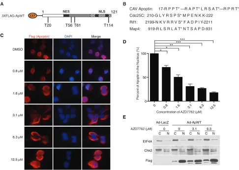
FIG 1Treatment with the pan-Chk inhibitor AZD7762 impairs nuclear localization of apoptin in a dose-dependent manner. (A) Schematic of 3⫻Flag-apoptin showing possible Chk1 and Chk2 phosphorylation sites (not drawn to scale). (B) Comparison of the amino acid sequence of apoptin with those of known checkpoint kinase substrates. Asterisks indicate that the preceding amino acid is the one that is phosphorylated (i.e., the threonine); hyphens indicate a stretch of irrelevant amino acids between the sequences that are shown. (C) H1299 cells were infected with Ad-Apwt and treated with DMSO or the indicated concentrations of AZD7762. At 24 h postinfection, the cells were processed for Flag immunofluorescence. (D) Quantification of cells displaying nuclear apoptin was determined by scoring at least 100 Flag-positive cells per condition. *,Pⱕ0.05; **,Pⱕ0.01; ***,Pⱕ0.0005. The data are presented as means and standard errors of the mean (SEM). (E) Cells were prepared as for panel C but fractionated into nuclear (N) and cytoplasmic (C) fractions and then analyzed by SDS-PAGE and Western blotting. Numbers indicate the concentrations of AZD7762 with which the cells were treated.
on November 7, 2019 by guest
http://jvi.asm.org/
[image:3.585.41.527.65.410.2]buffer (25 mM HEPES, pH 7.5, 50 mM NaCl, 0.1% Triton X-100) and then finally washed twice with kinase assay buffer (5 mM MOPS [mor-pholinepropanesulfonic acid], pH 7.2, 2.5 mM glycerol 2-phosphate, 5 mM MgCl2,1 mM EGTA, 0.4 mM EDTA, 0.25 mM dithiothreitol [DTT]). The resin was then incubated with 100 ng Chk2 (Biovision) in kinase assay buffer and 5l ATP cocktail {575l kinase buffer, 15l 10 mM cold ATP, and 10 l of 32P-labeled 10-mCi/ml (3,000-Ci/mmol) ␥-ATP ([␥ -32P]ATP)} for 15 min at 30°C. The total reaction volume was 25l.
Re-sidual ATP was then washed out, and 4⫻sample buffer was added to the resin slurry. The samples were then resolved by SDS-PAGE. The gel was stained with Coomassie to determine apoptin expression levels and finally dried and exposed to film to capture apoptin phosphorylation.
Two-dimensional gel electrophoresis.H1299 cells were seeded on 15-cm dishes prior to infection with Ad-Apwt and treatment with DMSO or AZD7762 (6.3M). Cells were harvested as described above, and whole-cell extracts were prepared using 100l of modified RIPA buffer. Flag-apoptin was purified by immunoprecipitation as described above, with washing performed using RIPA buffer. Proteins were eluted with 60
l DeStreak rehydration solution (GE) and boiled for 5 min prior to loading on an Ettan IPGPhor I isoelectric-focusing system (Amersham Biosciences) using Immobiline DryStrip gels, pH 6 to 11 (GE). The gels were rehydrated with DeStreak rehydration solution in ceramic strip holders overnight prior to isoelectric focusing following the manufactur-er’s recommendation. The gel strips were then incubated sequentially in
two aliquots of equilibration buffer (50 mM Tris-Cl, pH 8.8, 6 M urea, 2% SDS, 30% glycerol) supplemented with 2% DTT and 2.5% iodoacet-amide, respectively, on a rotating platform for 15 min each time at room temperature. The equilibrated strips were installed into standard SDS-PAGE cassettes and sealed with 0.5% agarose. The proteins were then resolved in the second dimension, transferred to nitrocellulose mem-branes, and probed by Flag immunoblotting.
Flow cytometry.H1299 cells were seeded at low confluence in 6-well plates or 60-mm dishes and transfected overnight with siRNA duplexes. The cells were then infected with Ad-LacZ or Ad-Apwt as described above. For cell cycle analysis, cells were trypsinized at 24 h postinfection, counted, and resuspended in ice-cold PBS prior to overnight fixation of 1⫻106cells in 70% ethanol at⫺20°C. The cells were subsequently washed and resuspended in 500l of propidium iodide (PI) staining buffer (200g/ml PI, 0.5 mg/ml RNase A, 0.1% Triton X-100 in PBS) and incubated for 15 min at 37°C in the dark. For apoptosis quantification, cells were harvested at 24, 48, or 72 h postinfection; washed twice; and resuspended at 1⫻106cells/ml in ice-cold 1⫻annexin V binding buffer (10 mM HEPES, pH 7.4, 140 mM NaCl, 2.5 mM CaCl2). One hundred microliters of cell suspension (1⫻105cells) was incubated with 5l phycoerythrin (PE)-conjugated annexin V (BD BioSciences) and 5g 7-aminoactinomycin D (7-AAD) (AG Scientific) for 30 min at room tem-perature in the dark. Samples were diluted with 400l 1⫻binding buffer prior to data acquisition. The stained cell preparations were analyzed
Merge
Flag (Apoptin) DAPI
DMSO
5.0 μM 2.5 μM 1.25 μM 0.62 μM
A
0.31 μM
B
Control
0.15 0.31 0.62 1.25 2.5 5.0 0
50 100 150
Concentration of CHIR-124 (μM)
P
ercen
t o
f A
popt
in
in t
h
e
N
u
c
le
us
(
%
)
**** **** ****
ns
ns *
CHIR-124
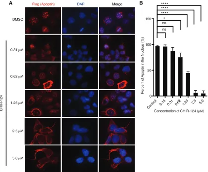
FIG 2Treatment with a selective Chk1 inhibitor impairs nuclear localization of apoptin in a dose-dependent manner. (A) H1299 cells were infected with Ad-Apwt and treated with DMSO or the indicated concentrations of CHIR-124 (Chk1-i). At 24 h postinfection, the cells were processed for Flag immuno-fluorescence. (B) Quantification of cells displaying nuclear apoptin was determined by scoring at least 100 Flag-positive cells per condition. The data are presented as means and SEM (n⫽3). Statistical significance was tested for treatment against the DMSO control (Student’sttest; *,P⬍0.05; ****,P⬍0.0001).
on November 7, 2019 by guest
http://jvi.asm.org/
[image:4.585.84.504.67.416.2]immediately on a FACSCalibur flow cytometer. Appropriate unstained and single-stained controls were prepared for determination of compen-sation using CellQuest Pro software (BD). Detection of mitotic cells was performed by staining for pSer10 histone H3 as previously described (31). The data were subsequently processed with FlowJo software.
Transfections and preparation of replicative CAV DNA.The full-length CAV replicon used in these studies has been previously described (7). Viral genomes were recircularized with T4 DNA ligase to generate replicative-form (RF) viral genomes. MSB-1 cells were electroporated us-ing the Amaxa Nucleofector system kit T (Lonza) accordus-ing to the man-ufacturer’s instructions. MSB-1 cells (2.0⫻106) and 400 ng of RF viral DNA were used in each transfection. Following electroporation, cells were allowed to recover for 1 h before being washed three times in PBS to eliminate residual RF viral DNA. The cells were then suspended at 2.5⫻ 105cells/ml in growth medium.
Quantification of viral DNA.Viral DNA was harvested from MSB-1 cells using the QiaAmp Viral RNA minikit (Qiagen) according to the manufacturer’s instructions. The purified DNA was then digested with DpnI to minimize background generated by the RF viral DNA, derived from bacteria, used in the initial electroporation. Quantitative PCR (qPCR) amplification was carried out using the QuantiFast SYBR green qPCR kit. Primers targeting the Vp1 gene were designed so that the prod-uct spanned a DpnI restriction site. The cycling parameters were as fol-lows: 95°C for 5 min, 95°C for 15 s, 60°C for 30 s, 95°C for 15 s, and 60°C for 20 min; melting curve, 95°C. The primers used to detect CAV were as follows: FW, ATGACCCTGCAAGACATGGG; RV, CTTTTTGCCACCG GTTCTGG.
Statistical analysis.Quantitative data are presented as means and standard errors of the mean. Where indicated, statistical analyses were performed using the Student two-tailedttest with data collected from 3 or more biological repeats.
RESULTS
Chemical inhibition of Chk1/2 induces cytoplasmic
accumula-tion of apoptin in tumor cells.
Regulation of apoptin
nucleocy-toplasmic shuttling by posttranslational modifications,
princi-pally phosphorylation, has been proposed to regulate the balance
between NES and NLS activities, maintaining apoptin in the
nu-clei of transformed cells. We previously demonstrated that
inhi-bition of ATM and DNA-PK in H1299 non-small-cell lung
ade-nocarcinoma cells efficiently impaired the nuclear localization of
apoptin (
28
). Given that the primary structure of apoptin lacks
ATM or DNA-PK consensus motifs, we hypothesized that
down-stream checkpoint kinases, namely, Chk1 and Chk2, could
phos-phorylate apoptin and regulate its subcellular localization. In
sup-port of this notion, we identified four Chk consensus motifs
within the primary structure of apoptin [Arg-X-X-p(Ser/Thr)] at
threonine residues 20, 56, 61, and 114 (
Fig. 1A
and
B
). These
motifs are very similar to those in known Chk substrates (
32
,
33
)
(
Fig. 1B
). In order to assess the involvement of checkpoint kinases
in apoptin localization within transformed cells, we employed the
small-molecule ATP-competitive kinase inhibitor AZD7762 in
control taxo l
5 GY I R
AZD7762
AZD7762+5GY IR 0
10 20 30
P
ercen
ta
g
e
o
f mi
to
tic cel
ls
**** ***
****
ATM
+
-p-Ser1981 ATM
53BP1
p-Thr68 Chk2
Chk2 p-Ser25 53BP1
actin Chk1
12.5 6.3 3.1 12.5 6.3
μM AZD7762 0 0 3.1
5 GY IR - - - + + +
A
B
FIG 3Treatment with the pan-Chk inhibitor AZD7762 impairs checkpoint kinase activity, but not ATM kinase activity. ATM S1981, 53BP1 S25, and Chk2 T68 are characterized ATM consensus sites. (A) H1299 cells were pretreated for 30 min with the indicated concentrations of AZD7762 (Chk-i) and then irradiated with 5 Gy ionizing radiation (IR). At 6 h posttreatment, the cells were harvested and processed for SDS-PAGE. (B) Quantification of mitotic cells by flow cytometry measurement of pSer10 histone 3-positive cells following the same treatment as for panel A. The data are presented as means and SEM (n⫽3). Statistical significance was tested for treatment against the control (Student’sttest; ****,P⬍0.0001; ***,P⫽0.0001).
on November 7, 2019 by guest
http://jvi.asm.org/
[image:5.585.73.522.62.387.2]H1299 cells expressing Flag-tagged apoptin. To examine the
ef-fects of AZD7762 on apoptin localization, cells were infected and
treated with various concentrations of AZD7762 or dimethyl
sul-foxide (DMSO) prior to processing for Flag immunofluorescence.
Examination of the cells revealed mostly cytoplasmic localization
of apoptin in treated cells (
Fig. 1C
). Quantification of the cells
revealed a significant dose-dependent increase in cytoplasmic
apoptin, consistent with what is shown in representative
micro-graphs (
Fig. 1D
). We then confirmed these observations by
bio-chemical fractionation of apoptin-expressing cells treated with
AZD7762. We found that apoptin accumulated in the cytoplasm
in response to treatment with the inhibitor (
Fig. 1E
). We also
tested a second inhibitor, CHIR-124, that primarily inhibits Chk1
and found that it efficiently inhibited nuclear accumulation of
apoptin, further confirming the role of these kinases in regulating
apoptin localization (
Fig. 2A
and
B
).
We next checked the effects of AZD7762 treatment on DNA
damage signaling in H1299 cells and found that treatment
in-duced DNA damage signaling through ATM, likely due to
aber-rant DNA replication during S phase (
34
). Treatment of irradiated
H1299 cells with AZD7762 did not markedly affect ATM signaling
(
Fig. 3A
), confirming the specificity of the inhibitor. Since a
func-tion of the ATM-Chk2 pathway is to induce G
2arrest and block
mitotic entry in response to DNA damage, we tested the efficacy of
the inhibitor by measuring the percentage of mitotic cells in
irra-diated cells treated with the inhibitor. We found that treatment
with AZD7762 completely abolished the cell cycle arrest induced
by Chk2 in response to irradiation (
Fig. 3B
). The accumulation of
mitotic cells in response to treatment with the
microtubule-stabi-lizing drug paclitaxel served as a positive control for the assay.
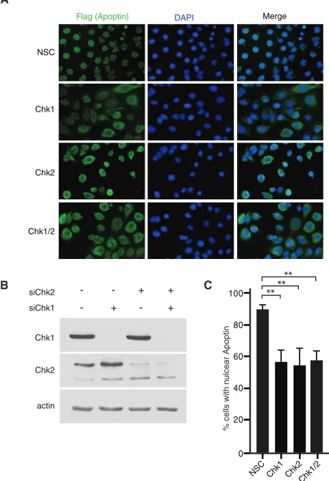
Silencing of Chk1/2 expression via siRNA induces
cytoplas-mic accumulation of apoptin in tumor cells.
In order to
deter-mine if Chk1 or Chk2 affects localization of apoptin, we designed
siRNAs to specifically knock down expression of the kinases.
Im-munofluorescence staining of apoptin showed that specific
target-ing of Chk1 and Chk2 alone or in combination resulted in an
increase in cytoplasmic accumulation of apoptin comparable to
that with a nontargeting control sequence (
Fig. 4A
and
C
). Protein
levels of Chk1 and Chk2 were analyzed by immunoblotting to
verify the knockdown efficiency (
Fig. 4B
). Furthermore,
com-bined knockdown using both targeting sequences did not yield
further impairment of nuclear localization. Also, knockdown of
Chk1 and Chk2 with a second set of siRNA duplexes yielded nearly
identical results (data not shown). Thus, apoptin phosphorylation
by individual checkpoint kinases may represent interdependent
events, raising the possibility that Chk1 and Chk2 may regulate
activation of distinct apoptin functions in a context-dependent
manner.
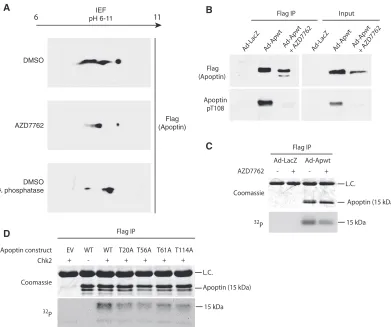
Apoptin is a checkpoint kinase substrate.
To confirm that the
regulation of apoptin subcellular localization by the checkpoint
kinases is a result of phosphorylation, we performed
two-dimen-sional gel electrophoresis to analyze Flag-tagged apoptin peptides
in the presence or absence of inhibitor. Flag-apoptin was
intro-duced by adenoviral infection into H1299 cells with concurrent
treatment with the Chk1/2 inhibitor AZD7762 or DMSO (vehicle
control). Flag-apoptin was immunoprecipitated under stringent
conditions and analyzed in the first dimension by isoelectric
fo-cusing and then by standard SDS-PAGE. The migration of
apop-tin peptides at a higher isoelectric point can be observed in the
presence of AZD7762 relative to control treatment (
Fig. 5A
). Since
phosphorylated peptides migrate at a lower isoelectric point,
ow-ing to the negative charge of phosphoryl groups at physiological
pH, the compaction of peptides is consistent with a loss of
phos-phorylation. The migration of apoptin in the presence of
AZD7762 was comparable to that of Flag-apoptin incubated with
lambda phosphatase prior to isoelectric focusing.
Previous studies have implicated the phosphorylation of T108
as a critical mediator of apoptin localization and function (
35
). To
address whether impairment of Chk1 and Chk2 alter the
phos-phorylation status of T108, we isolated Flag-apoptin complexes
from H1299 cells by immunoprecipitation and performed
immu-noblot analysis with a phosphospecific antibody for apoptin T108.
Interestingly, there was a marked reduction in T108
phosphory-lation following treatment with AZD7762 (
Fig. 5B
). Moreover,
chemical inhibition of Chk1 and Chk2 affected the migration of
Flag-apoptin on SDS-PAGE gels, so that a faster-migrating,
lower-molecular-weight band is clearly visible (
Fig. 5B
). Our data
sug-gest that inhibition of Chk1 and Chk2 results in cytoplasmic
relo-Merge
Flag (Apoptin) DAPI
NSC
Chk1
Chk2
Chk1/2
0 20 40 60 80
100 ****
** A
B C
% cells with n
u
lcear Apoptin
NSC Chk1 Chk2 Chk1/2 Chk1
Chk2
actin siChk1
siChk2 +
+ +
-+
-FIG 4RNAi knockdown of Chk1/2 induces cytoplasmic localization of wild-type apoptin. (A) H1299 cells were transfected at low confluence with the indicated siRNA duplexes and allowed to proliferate for 16 h. The cells were then infected with Ad-Apwt and processed for Flag immunofluorescence at 24 h postinfection. (B) Immunoblot analysis of the experiment shown in panel A verifying knockdown efficiency and specificity. (C) Quantification of cells dis-playing nuclear apoptin was determined by scoring at least 100 Flag-positive cells per condition. **,Pⱕ0.01. NSC indicates a nonsilencing control.
on November 7, 2019 by guest
http://jvi.asm.org/
[image:6.585.299.539.73.424.2]calization of apoptin and loss of phosphorylation at the T108 site
by nuclear cyclin-Cdk2 complexes, although we cannot dismiss
the possibility that Chk1 or Chk2 acts directly on the
nonconsen-sus T108 residue (
36
).
To confirm that inhibition of Chk1 and Chk2 directly alters
apoptin phosphorylation, we performed [
32P]orthophosphate
metabolic labeling in the presence and absence of AZD7762,
fol-lowed by immunoprecipitation of apoptin under stringent
condi-tions. We found that
32P labeling of apoptin was reduced in the
presence of AZD7762, indicating that apoptin is less
phosphory-lated in treated cells. Moreover, the apoptin immunoprecipitated
from the treated cells migrated faster in the gel, consistent with a
loss of phosphorylation (
Fig. 5C
).
Finally, to determine if Chk2 directly phosphorylates apoptin,
recombinant apoptin was immunoprecipitated from H1299 cells
and added to purified Chk2 in the presence of [
␥
-
32P]ATP.
Figure
5D
shows that Chk2 was indeed able to directly phosphorylate
apoptin.
In vitro
kinase reactions were also performed using
apo-ptin mutants in which each of the four predicted Chk1/2
phos-phorylation sites was changed to alanines (T20A, T56A, T61A,
and T114A). Each of the four mutants displayed less
32P
incorpo-ration than wild-type apoptin, suggesting that at least
in vitro
, all
four sites are phosphorylated by Chk2 (
Fig. 5D
). Taken together,
our observations support the hypothesis that Chk-mediated
phos-phorylation is a regulatory mechanism of apoptin activation.
Mutation of N-terminal threonine residues impairs nuclear
localization of apoptin in tumor cells.
To determine which of the
possible phosphorylation sites on apoptin are regulated by the
Chk proteins
in vivo
, we transiently transfected the Chk1/2
phos-phorylation site mutants into H1299 cells and compared their
localization patterns with that of wild-type apoptin. The T56A and
T61A mutants displayed the greatest defect in nuclear
accumula-tion, with cells displaying filamentous apoptin located in the
cy-toplasm (
Fig. 6A
and
B
). Therefore, our data suggest that
phos-phorylation at N-terminal threonine residues facilitates nuclear
localization of apoptin. Given that the T56A and T61A mutants
failed to localize to the nucleus, we determined if these mutants
were otherwise functional. We therefore tested whether the
apo-Flag IP Input
Flag (Apoptin)
Apoptin pT108
Ad-LacZ A d-Apwt
Ad-Apwt + AZD7762 DMSO
AZD7762
DMSO λ phosphatase
IEF pH 6-11
6 11
Flag (Apoptin)
A
B
Ad-LacZ A d-Apwt
Ad-Apwt + AZD7762
C
Ad-Apwt Ad-LacZ
Coomassie
32 P
AZD7762 - + - +
L.C. Flag IP
Coomassie
32 P
Chk2 + - + + + + +
EV WT WT T20A T56A T61A T114A Apoptin construct
D
L.C.
Apoptin (15 kDa)
15 kDa
Apoptin (15 kDa)
15 kDa Flag IP
FIG 5Apoptin is a checkpoint kinase substrate. (A) H1299 cells were infected with Ad-Apwt and treated with DMSO or 6.3M AZD7762 (Chk-i). At 24 h postinfection, Flag-apoptin was purified by immunoprecipitation under stringent conditions and resolved in the first dimension with a pH 6 to 11 gradient and then analyzed by SDS-PAGE in the second dimension. Immunoblotting for Flag was performed to visualize Flag-apoptin peptides. Lambda-phosphatase treatment of purified Flag-apoptin served as a positive control. IEF, isoelectric focusing. (B) H1299 cells were treated with DMSO or 6.3M Chk-i and infected with Ad-Apwt or Ad-LacZ for 24 h. Whole-cell extracts were prepared for immunoprecipitation (IP) of Flag complexes and immunoblot analysis with the indicated antibodies (pT108, phospho-specific antibody [Ab] for phosphorylated Thr108). (C) H1299 cells were infected with Ad-Apwt or Ad-LacZ and treated with DMSO or 6.3M AZD7762. At 23 h postinfection, the medium was changed to include 250 mCi/ml [32P]orthophosphate. Following 1 h of labeling, Flag-apoptin was purified by immunoprecipitation under stringent conditions and analyzed by SDS-PAGE. (D) H1299 cells were transfected with the indicated constructs. At 48 h posttransfection, Flag-apoptin was immunoprecipitated with anti-Flag resin. The resin was then incubated with 100 ng Chk2 and [␥-32P]ATP for 15 min at 30°C. Residual␥-ATP was washed out, and 4⫻sample buffer was added to the samples, which were then analyzed by SDS-PAGE. L.C. indicates IgG light chain.
on November 7, 2019 by guest
http://jvi.asm.org/
[image:7.585.96.487.65.392.2]ptin mutants had defects in multimerization activity, which was
previously shown to be essential for induction of apoptosis (
9
).
The aggregation of monomers and interaction of apoptin with
PML bodies has been shown to require the N-terminal
leucine-rich motif (
14
,
18
). We cotransfected H1299 cells with HA-tagged
apoptin and mutant Flag-apoptin and tested the interaction of
differentially tagged apoptin. Immunoprecipitation of
Flag-apo-ptin captured both wild-type and mutant apoFlag-apo-ptin, indicating that
interaction between apoptin monomers was unaffected (
Fig. 6C
).
Similarly, we determined if the phosphorylation mutants were
still able to interact with the anaphase-promoting complex.
Con-sistent with previous studies (
17
), wild-type apoptin was able to
pull down both core (APC1, APC2, APC3 or Cdc27, APC5, and
APC8) and coactivator (Cdc20 and Cdh1) components of the
complex under the conditions employed in our assay (
Fig. 6D
).
T56A and T61A mutants immunoprecipitated APC/C subunits
with efficiency similar to that of wild-type apoptin, indicating that
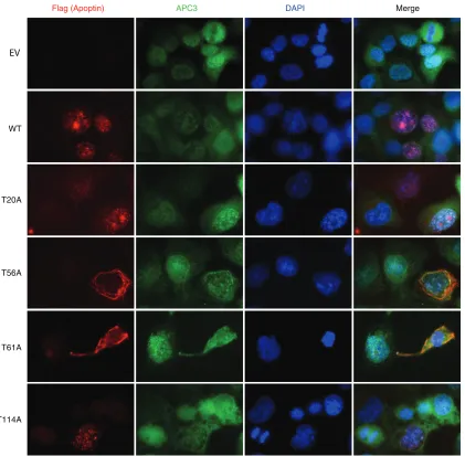
these mutants are otherwise functional. We confirmed these
re-sults by staining cells for both APC3 and WT or mutated apoptin.
We found that the apoptin T56A and T61A mutants continued to
colocalize with APC3 despite being cytoplasmic (
Fig. 7
). Together,
these results indicate that residues T56 and T61 are important for
regulating the localization of apoptin but do not affect other
ac-tivities of the protein, including multimerization and interaction
with the APC/C.
Chk1/2 activity regulates apoptin-induced G
2/M arrest and
apoptosis.
If checkpoint kinases regulate the subcellular
localiza-tion of apoptin, chemical inhibilocaliza-tion or knockdown of Chk1 and
Chk2 would be predicted to impair apoptin functions in the
nu-cleus. The interaction of apoptin with the APC/C has been shown
to result in the sequestration of the APC/C to PML bodies,
result-ing in G
2/M arrest (
17
). To determine whether Chk1 and Chk2
MergeFlag (Apoptin) DAPI
WT
T20A
T56A
T61A
T114A
Flag-Ap HA-Ap
-WT WT WT T56A T61A
FLAG
HA
FLAG
HA Flag IP
Input
-+
+
+
+
+
+
+
+
Flag IP Input
APC1
APC2
Cdc27
Cdh1 APC5
APC8
Flag
construct WT T56A T61A
HC EV
WT
T20A T56A T61A
T114A
0 20 40 60 80
Flag-Apoptin
****
A
B
C
% cells with n
ulcear Apoptin
D
WT T56A T61A EV
FIG 6Mutation of Thr56 and Thr61 impairs nuclear localization of apoptin in H1299 cells. (A) H1299 cells were transfected with Flag-tagged WT or mutant apoptin. The cells were allowed to proliferate for 24 h prior to processing for Flag immunofluorescence with nuclear staining (DAPI). (B) Quantification of cells displaying nuclear apoptin was determined by scoring at least 100 Flag-positive cells per condition. **,Pⱕ0.01. The error bars indicate SEM. (C and D) H1299 cells were transfected with the indicated constructs for 24 h. Whole-cell extracts were prepared for immunoprecipitation of Flag complexes and immunoblot analysis with the indicated antibodies. E.V. indicates empty vector.
on November 7, 2019 by guest
http://jvi.asm.org/
[image:8.585.113.472.67.471.2]action is required for apoptin-induced cell cycle arrest, we
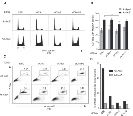
per-formed cell cycle analysis of H1299 cells transfected with siRNAs
targeting Chk1 and/or Chk2 and transduced with an
apoptin-expressing vector. Individual or combined knockdown of Chk1
and Chk2 alone did not alter the cell cycle, as evaluated by control
LacZ-expressing-cell profiles (
Fig. 8A
). Analysis of cell cycle
pro-files from apoptin-expressing cells showed a significant decrease
in cells with 4 N DNA content upon transfection with siRNA
targeting Chk2 (
Fig. 8B
).
We then investigated the kinetics of apoptin-induced cell
death. H1299 cells were treated as described above for cell cycle
analysis, and cell death was assessed at various time points by
annexin V/7-AAD staining (
Fig. 8C
and
D
). In accordance with
previous studies (
17
), at 72 h postinfection, approximately 54% of
apoptin cells were apoptotic in the nonsilencing controls.
Nota-bly, we observed a significant decrease in late apoptotic cells in
cells treated with siRNA targeting either Chk1 or Chk2 (
Fig. 8C
and
D
). These data show that silencing of Chk1 and Chk2
func-tionally delays apoptin-induced cell death, presumably by
im-paired accumulation of apoptin in the nucleus so that it cannot
execute its proapoptotic functions.
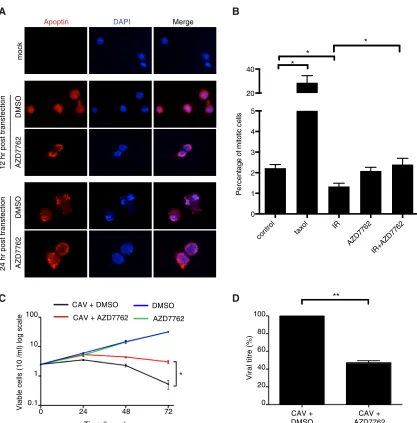
Inhibition of Chk activity in CAV-infected cells attenuates
virus production and cytopathic effect.
Given the importance of
the Chk proteins in regulating apoptin, we asked whether this
regulation plays a role during CAV replication. To accomplish
this, we used a chicken lymphoid cell line transformed with
Marek’s disease virus (MSB-1) that permits CAV replication (
37
).
We first determined if AZD7762 was able to affect the localization
of apoptin in the context of viral replication. MSB-1 cells were
infected with wild-type CAV, and the localization of apoptin was
examined by immunofluorescence using an apoptin
anti-body (
29
).
Figure 9A
shows that apoptin localized to the nuclei of
CAV-infected cells but that addition of AZD7762 resulted in
cy-toplasmic retention of the protein. We then checked if AZD7762
Merge
Flag (Apoptin) DAPI
WT
T20A
T56A
T61A
T114A
APC3
EV
FIG 7Mutation of Thr56 and Thr61 does not affect colocalization of the APC/C with apoptin. H1299 cells were transfected with Flag-tagged WT or mutant apoptin. The cells were allowed to proliferate for 24 h prior to processing for Flag and APC3 immunofluorescence with nuclear staining (DAPI). E.V. indicates empty vector.
on November 7, 2019 by guest
http://jvi.asm.org/
[image:9.585.85.506.65.478.2]was able to abolish the cell cycle arrest normally induced by the
Chk proteins in response to ionizing radiation, as we did in H1299
cells. We found that while the irradiation caused cell cycle arrest in
untreated cells, treatment with AZD7762 abolished this arrest
in MSB-1 cells (
Fig. 9B
). These experiments confirmed that
AZD7762 is able to inhibit chicken Chk1/2 and that inhibition of
the Chk proteins can affect localization of apoptin during viral
replication. CAV is known to induce a potent CPE (
7
), and we
therefore determined if AZD7762 had an effect on CPE mediated
by viral replication.
Figure 9C
shows that MSB-1 cells infected
with CAV rapidly decreased in viability while mock-infected cells
continued to grow normally. Interestingly, inhibition of Chk
pro-teins with AZD7762 was able to significantly protect
in-fected cells from death. We next asked whether treatment of
CAV-producing cells with the inhibitor would have an effect on viral
replication. When fresh cells were infected with CAV and treated
with AZD7762, we observed an almost 60% decrease in virus
pro-duction in the treated population (
Fig. 9D
). Taken together, these
data show that Chk signaling affects apoptin activity during viral
infection and is required for effective CAV replication.
DISCUSSION
Subcellular localization has been implicated as the main
charac-teristic contributing to the selective mechanism of action of
apo-ptin (
9
,
19
). Although the importance of apoptin localization in
apoptosis has been well characterized, the mechanism of
regula-tion and how it relates to viral replicaregula-tion has yet to be addressed.
In the current study, we show that chemical inhibition or RNA
interference (RNAi)-mediated silencing of Chk1 and Chk2
abro-gates nuclear accumulation of apoptin in transformed cells.
Con-sistent with the localization data, we demonstrated that cell cycle
arrest and apoptosis induced by apoptin were impaired in cells
with Chk1 and Chk2 knockdown. Our data show that two
threo-nine residues (T56 and T61) adjacent to the N-terminal NES motif
are sites of Chk phosphorylation, providing a mechanistic basis
for regulation of apoptin localization. Importantly, we show that
2n 4n
DNA content (PI)
NSC siChk1 siChk2 siChk1/2
Ad-lacZ
Ad-Apwt 24hpi
0 10 20 30 40 50
*
% of cells with 4N DNA content
siRNA
NSC
siChk1 siChk2 siChk1/2
A
B
0 20 40 60
%
o
f C
e
lls
w
ith
L
a
te
A
p
o
p
to
ti
c
m
a
rk
e
rs Ad-Apwt
Ad-lacZ
siRNA NSC
siChk1 siChk2 siChk1/2
Annexin V (PE)
7.13 5.21 3.49 15.7
54 12.9 15.3 9.42
NSC siChk1 siChk2 siChk1/2
7-AAD
Ad-lacZ
Ad-Apwt 72hpi
C
D
Ad-Apwt Ad-lacZ
FIG 8RNAi knockdown of Chk2 impairs apoptin-induced G2/M arrest and apoptosis. (A) H1299 cells were transfected at low confluence with the indicated siRNA duplexes prior to infection with Ad-Apwt or Ad-LacZ. At 24 h postinfection (hpi), the cells were fixed and stained with PI for flow cytometry. (B) Quantification of cells containing 4 N (G2/M phase) DNA content derived from replicate experiments, as shown in panel A. *,Pⱕ0.05. The error bars indicate SEM. (C) Representative flow cytometric profiles of cells treated as for panel A and harvested and stained at 72 hpi with 7-AAD and PE annexin V. Percentages of PE- and 7-AAD-double-positive cells corresponding to the late apoptotic population are boxed. (D) Quantification of late apoptotic cells at 72 hpi performed by manual gating analysis.
on November 7, 2019 by guest
http://jvi.asm.org/
[image:10.585.84.509.63.436.2]inhibition of Chk1/2-mediated apoptin phosphorylation and
nu-clear localization impairs viral replication.
Although the subcellular localization of apoptin appears to
be a major mechanism of regulation, there is evidence that
other factors may also be required. For example, it has been
demonstrated that forced localization of apoptin to the nucleus
of primary cells by fusing a canonical nuclear localization
sig-nal is insufficient to induce apoptosis (
9
,
15
). Previous studies
have shown that threonines 107 and 108 on apoptin are
phos-phorylated in transformed cells and that this modification is
important in regulating apoptotic activity (
35
,
38
,
39
). Neither
of these sites has the consensus sequence of a Chk1/2 target,
and others have suggested that they may be targeted by a
nu-clear kinase, such as HIPK2 (
40
). Full activation of apoptin may
therefore require a 2-step process whereby DDR signaling promotes
nuclear localization and a nuclear kinase, such as HIPK2, then
phos-phorylates T-108. Consistent with this model, we observed that
T-108 phosphorylation was reduced in cells treated with Chk
inhib-itors (
Fig. 3B
). Once apoptin becomes localized to the nucleus,
addi-tional modifications, such as sumoylation, may also contribute to full
functional activation of the protein (
18
).
The notion that Chk1 and Chk2 phosphorylation may
reg-ulate apoptin function by altering localization may explain the
transformation-specific localization of the protein. Cancer
cells have been shown to have elevated basal levels of DDR
signaling and therefore to also have elevated Chk activity (
24
,
MergeApoptin DAPI
DMSO
AZD7762
12 hr post transfection
CAV + AZD7762 CAV +
DMSO
Viral titre (%)
Time (hours)
Viable cells (10
/ml) log scale
mock
A
B
DMSO
AZD7762
24 hr post transfection
CAV + DMSO
CAV + AZD7762 AZD7762 DMSO
C
control taxo l
IR
AZD7762 IR+AZD7762 0
1 2 3 4 5 20 40
P
ercen
tag
e o
f mi
to
tic cel
ls
* *
*
D
0 20 40 60 80 100 100
10
1
0.1
24 48 72
0
**
FIG 9Inhibition of Chk activity in CAV-producing cells attenuates virus production and cytopathic effect. (A) MSB-1 cells were transfected with CAV and treated with AZD7762 (350 nM) or DMSO. The cells were then fixed and stained for immunofluorescence as indicated. (B) MSB-1 cells were pretreated with AZD7762 (350 nm) for 30 min and then irradiated with 3 Gy ionizing radiation. Mitotic cells were quantified by flow cytometry measurement of pSer10 histone 3-positive cells. (C) MSB-1 cells were transfected with CAV and treated with AZD7762 (350 nm) or DMSO. Cellular viability was monitored over time by trypan blue staining. (D) MSB-1 cells were transfected with CAV and treated with AZD7762 (350 nm) or DMSO. Newly synthesized viral genomes were extracted 72 h posttransfection and quantified by quantitative PCR. Statistical significance was tested for treatment against the control (Student’sttest; *,P⬍0.05; **,P⬍0.01).
on November 7, 2019 by guest
http://jvi.asm.org/
[image:11.585.84.501.62.485.2]41
,
42
). In the context of viral infection, the insights into
selec-tive activation of apoptin presented in our study may aid in the
clarification of apoptin function in viral replication and
patho-genesis. It is likely that during early stages of viral infection in
normal (nontransformed) host cells, apoptin remains
cyto-plasmic and inactive. During rolling-circle replication of the
CAV genome, single-stranded DNA is generated and serves as a
trigger for DNA damage sensors, including RPA and other
components involved in ATR activation (
3
). Abnormal
repli-cation fork structures subsequently generate double-strand
breaks (DSB) and induce recruitment and activation of ATM
(
25
,
26
). In response to signaling events emanating from these foci,
Chk1 and Chk2 subsequently initiate activity on downstream
sub-strates, mediating appropriate repair responses. The abundance of
apoptin and activated Chk1 and Chk2 would steadily increase upon
the onset of viral DNA replication, eventually reaching a threshold
analogous to that of transformed cells, whereby apoptin-induced cell
death is initiated. This model is consistent with evidence indicating
that nuclear apoptin is associated with productive CAV replication
and cytopathogenicity (
43
).
Our group previously characterized an inhibitory
conse-quence of nuclear apoptin for DDR focus formation,
princi-pally mediated by proteasomal degradation of the essential
DSB mediator MDC1 (
28
). It is possible that apoptin senses
ATM-Chk2 and ATR-Chk1 signaling by direct
phosphoryla-tion events, promoting subsequent impairment of the host
re-sponse and thus preventing it from interfering with viral
rep-lication. The inhibitory role of apoptin is similar in principle to
reported functions of several viral effectors, including the herpes
sim-plex virus (HSV) immediate-early protein ICP0. HSV ICP0 is a
func-tional RING ubiquitin E3 ligase inducing the degradation of RNF8
and RNF168, which are host factors downstream of MDC1 that
fa-cilitate DDR mediator recruitment at DSB-induced foci (
27
,
44
,
45
).
Additionally, other DNA tumor viruses, including adenovirus,
polyomaviruses, and human papillomavirus, encode multiple
pro-teins that regulate the DDR (
46–48
).
Despite our identification of the mechanistic basis for
apo-ptin activation by DDR signaling, the mechanism by which
nuclear apoptin induces apoptosis remains incompletely
de-fined. Previous work by our group and others has suggested
that interaction with the APC/C results in mitotic catastrophe
and cell death (
9
,
17
,
49
). Inhibition of APC/C function is a
common mechanism exploited by several metazoan viruses,
presumably to maintain favorable cellular conditions for viral
replication (
50
,
51
). Interestingly, in light of essential functions
of DDR kinases and mediators in the coordination of cell cycle
checkpoints, the described interplay of apoptin with the DDR may
represent a strategy common to many viruses. Indeed, understanding
the mechanism of apoptin may contribute to the elucidation of novel
links between the DNA damage response, the spindle assembly
checkpoint, and virus replication.
ACKNOWLEDGMENTS
We thank Mathieu Noteborn for the CAV replicon.
This work was supported by grants from the Canadian Institute of Health Research (CIHR), MOP-179122, and the Natural Science and Engineering Research Council (NSERC) of Canada to J.G.T. T.J.K. was supported by studentships from the Fonds de la Recherche en Santé du Québec (FRSQ) and CIHR. T.F.N. was supported by an NSERC un-dergraduate studentship research award.
FUNDING INFORMATION
This work, including the efforts of Jose G. Teodoro, was funded by Gou-vernement du Canada | Natural Sciences and Engineering Research Council of Canada (NSERC) (RGPIN 355976-09). This work, including the efforts of Jose G. Teodoro, was funded by Gouvernement du Canada | Canadian Institutes of Health Research (CIHR) (MOP-179122).
REFERENCES
1.Fauquet CM, Fargette D.2005. International Committee on Taxonomy of Viruses and the 3,142 unassigned species. Virol J2:64.http://dx.doi.org /10.1186/1743-422X-2-64.
2.Cheung AK.2012. Porcine circovirus: transcription and DNA replication. Virus Res164:46 –53.http://dx.doi.org/10.1016/j.virusres.2011.10.012. 3.Schat KA.2009. Chicken anemia virus. Curr Top Microbiol Immunol
331:151–183.
4.Todd D.2000. Circoviruses: immunosuppressive threats to avian spe-cies: a review. Avian Pathol 29:373–394. http://dx.doi.org/10.1080 /030794500750047126.
5.Sauvage V, Cheval J, Foulongne V, Gouilh MA, Pariente K, Manu-guerra JC, Richardson J, Dereure O, Lecuit M, Burguiere A, Caro V, Eloit M.2011. Identification of the first human gyrovirus, a virus related to chicken anemia virus. J Virol85:7948 –7950.http://dx.doi.org/10.1128 /JVI.00639-11.
6.Prasetyo AA, Kamahora T, Kuroishi A, Murakami K, Hino S.2009. Replication of chicken anemia virus (CAV) requires apoptin and is com-plemented by VP3 of human torque teno virus (TTV). Virology385:85– 92.http://dx.doi.org/10.1016/j.virol.2008.10.043.
7.Noteborn MH, de Boer GF, van Roozelaar DJ, Karreman C, Kranen-burg O, Vos JG, Jeurissen SH, Hoeben RC, Zantema A, Koch G, van Ormondt H, van der Eb AJ.1991. Characterization of cloned chicken anemia virus DNA that contains all elements for the infectious replication cycle. J Virol65:3131–3139.
8.Peters MA, Jackson DC, Crabb BS, Browning GF.2002. Chicken anemia virus VP2 is a novel dual specificity protein phosphatase. J Biol Chem
277:39566 –39573.http://dx.doi.org/10.1074/jbc.M201752200. 9.Heilman DW, Teodoro JG, Green MR.2006. Apoptin
nucleocytoplas-mic shuttling is required for cell type-specific localization, apoptosis, and recruitment of the anaphase-promoting complex/cyclosome to PML bod-ies. J Virol80:7535–7545.http://dx.doi.org/10.1128/JVI.02741-05. 10. Jeurissen SH, Wagenaar F, Pol JM, van der Eb AJ, Noteborn MH.1992.
Chicken anemia virus causes apoptosis of thymocytes after in vivo infec-tion and of cell lines after in vitro infecinfec-tion. J Virol66:7383–7388. 11. Los M, Panigrahi S, Rashedi I, Mandal S, Stetefeld J, Essmann F,
Schulze-Osthoff K.2009. Apoptin, a tumor-selective killer. Biochim Bio-phys Acta1793:1335–1342.http://dx.doi.org/10.1016/j.bbamcr.2009.04 .002.
12. Backendorf C, Visser AE, de Boer AG, Zimmerman R, Visser M, Voskamp P, Zhang YH, Noteborn M.2008. Apoptin: therapeutic poten-tial of an early sensor of carcinogenic transformation. Annu Rev Pharma-col ToxiPharma-col48:143–169.http://dx.doi.org/10.1146/annurev.pharmtox.48 .121806.154910.
13. Leliveld SR, Dame RT, Rohn JL, Noteborn MH, Abrahams JP.2004. Apoptin’s functional N- and C-termini independently bind DNA. FEBS Lett557:155–158.http://dx.doi.org/10.1016/S0014-5793(03)01465-0. 14. Leliveld SR, Zhang YH, Rohn JL, Noteborn MH, Abrahams JP.2003.
Apoptin induces tumor-specific apoptosis as a globular multimer. J Biol Chem278:9042–9051.http://dx.doi.org/10.1074/jbc.M210803200. 15. Danen-Van Oorschot AA, Zhang YH, Leliveld SR, Rohn JL, Seelen MC,
Bolk MW, Van Zon A, Erkeland SJ, Abrahams JP, Mumberg D, Note-born MH.2003. Importance of nuclear localization of apoptin for tumor-specific induction of apoptosis. J Biol Chem278:27729 –27736.http://dx .doi.org/10.1074/jbc.M303114200.
16. Peters J-M.2006. The anaphase promoting complex/cyclosome: a ma-chine designed to destroy. Nat Rev Mol Cell Biol7:644 – 656.
17. Teodoro JG, Heilman DW, Parker AE, Green MR. 2004. The viral protein Apoptin associates with the anaphase-promoting complex to in-duce G2/M arrest and apoptosis in the absence of p53. Genes Dev18:1952– 1957.http://dx.doi.org/10.1101/gad.1198404.
18. Janssen K, Hofmann TG, Jans DA, Hay RT, Schulze-Osthoff K, Fischer U.2007. Apoptin is modified by SUMO conjugation and targeted to pro-myelocytic leukemia protein nuclear bodies. Oncogene26:1557–1566.
http://dx.doi.org/10.1038/sj.onc.1209923.
on November 7, 2019 by guest
http://jvi.asm.org/
19. Danen-Van Oorschot AA, Fischer DF, Grimbergen JM, Klein B, Zhuang S, Falkenburg JH, Backendorf C, Quax PH, Van der Eb AJ, Noteborn MH.1997. Apoptin induces apoptosis in human transformed and malignant cells but not in normal cells. Proc Natl Acad Sci U S A
94:5843–5847.http://dx.doi.org/10.1073/pnas.94.11.5843.
20. Kuscu B, Gurel A.2008. Lesions in the thymus and bone marrow in chicks with experimentally induced chicken infectious anemia disease. J Vet Sci9:15–23.http://dx.doi.org/10.4142/jvs.2008.9.1.15.
21. Hills SA, Diffley JF.2014. DNA replication and oncogene-induced rep-licative stress. Curr Biol24:R435–R444.http://dx.doi.org/10.1016/j.cub .2014.04.012.
22. Negrini S, Gorgoulis VG, Halazonetis TD. 2010. Genomic instabili-ty—an evolving hallmark of cancer. Nat Rev Mol Cell Biol11:220 –228.
http://dx.doi.org/10.1038/nrm2858.
23. Halazonetis TD, Gorgoulis VG, Bartek J.2008. An oncogene-induced DNA damage model for cancer development. Science319:1352–1355.
http://dx.doi.org/10.1126/science.1140735.
24. Gorgoulis VG, Vassiliou LV, Karakaidos P, Zacharatos P, Kotsinas A, Liloglou T, Venere M, Ditullio RA, Jr, Kastrinakis NG, Levy B, Kletsas D, Yoneta A, Herlyn M, Kittas C, Halazonetis TD. 2005. Activation of the DNA damage checkpoint and genomic instability in human precancerous lesions. Nature434:907–913.http://dx.doi.org /10.1038/nature03485.
25. Zeman MK, Cimprich KA.2014. Causes and consequences of replication stress. Nat Cell Biol16:2–9.http://dx.doi.org/10.1038/ncb2897. 26. Ciccia A, Elledge SJ.2010. The DNA damage response: making it safe to
play with knives. Mol Cell40:179 –204.http://dx.doi.org/10.1016/j.molcel .2010.09.019.
27. Lukas J, Lukas C, Bartek J.2011. More than just a focus: the chromatin response to DNA damage and its role in genome integrity maintenance. Nat Cell Biol13:1161–1169.http://dx.doi.org/10.1038/ncb2344. 28. Kucharski TJ, Gamache I, Gjoerup O, Teodoro JG.2011. DNA damage
response signaling triggers nuclear localization of the chicken anemia vi-rus protein Apoptin. J Virol85:12638 –12649.http://dx.doi.org/10.1128 /JVI.05009-11.
29. Jiang J, Cole D, Westwood N, Macpherson L, Farzaneh F, Mufti G, Tavassoli M, Gaken J.2010. Crucial roles for protein kinase C isoforms in tumor-specific killing by apoptin. Cancer Res70:7242–7252.http://dx.doi .org/10.1158/0008-5472.CAN-10-1204.
30. Lu Y, Kucharski TJ, Gamache I, Blanchette P, Branton PE, Teodoro JG.
2014. Interaction of adenovirus type 5 E4orf4 with the nuclear pore sub-unit Nup205 is required for proper viral gene expression. J Virol88:
13249 –13259.http://dx.doi.org/10.1128/JVI.00933-14.
31. Juan G, Darzynkiewicz Z.2004. Detection of mitotic cells. Curr Pro-toc Cytom Chapter 7:Unit 7.24.http://dx.doi.org/10.1002/0471142956 .cy0724s28.
32. O’Neill T, Giarratani L, Chen P, Iyer L, Lee CH, Bobiak M, Kanai F, Zhou BB, Chung JH, Rathbun GA.2002. Determination of substrate motifs for human Chk1 and hCds1/Chk2 by the oriented peptide library approach. J Biol Chem277:16102–16115.http://dx.doi.org/10.1074/jbc .M111705200.
33. Blasius M, Forment JV, Thakkar N, Wagner SA, Choudhary C, Jackson SP.2011. A phospho-proteomic screen identifies substrates of the check-point kinase Chk1. Genome Biol12:R78.http://dx.doi.org/10.1186/gb -2011-12-8-r78.
34. Buisson R, Boisvert JL, Benes CH, Zou L.2015. Distinct but concerted roles of ATR, DNA-PK, and Chk1 in countering replication stress during S phase. Mol Cell59:1011–1024.http://dx.doi.org/10.1016/j.molcel.2015 .07.029.
35. Rohn JL, Zhang YH, Aalbers RI, Otto N, Den Hertog J, Henriquez NV, Van De Velde CJ, Kuppen PJ, Mumberg D, Donner P, Noteborn MH.
2002. A tumor-specific kinase activity regulates the viral death protein Apoptin. J Biol Chem277:50820 –50827.http://dx.doi.org/10.1074/jbc .M208557200.
36. Maddika S, Panigrahi S, Wiechec E, Wesselborg S, Fischer U, Schulze-Osthoff K, Los M.2009. Unscheduled Akt-triggered activation of cyclin-dependent kinase 2 as a key effector mechanism of apoptin’s anticancer toxicity. Mol Cell Biol 29:1235–1248. http://dx.doi.org/10.1128/MCB .00668-08.
37. Goryo M, Suwa T, Matsumoto S, Umemura T, Itakura C.1987. Serial propagation and purification of chicken anaemia agent in MDCC-MSB1 cell line. Avian Pathol16:149 –163.http://dx.doi.org/10.1080 /03079458708436360.
38. Lee YH, Cheng CM, Chang YF, Wang TY, Yuo CY.2007. Apoptin T108 phosphorylation is not required for its tumor-specific nuclear localization but partially affects its apoptotic activity. Biochem Biophys Res Commun
354:391–395.http://dx.doi.org/10.1016/j.bbrc.2006.12.201.
39. Lanz HL, Florea BI, Noteborn MH, Backendorf C.2012. Development and application of an in vitro apoptin kinase assay. Anal Biochem421:68 – 74.http://dx.doi.org/10.1016/j.ab.2011.10.030.
40. Poon IK, Oro C, Dias MM, Zhang J, Jans DA.2005. Apoptin nuclear accumulation is modulated by a CRM1-recognized nuclear export signal that is active in normal but not in tumor cells. Cancer Res65:7059 –7064.
http://dx.doi.org/10.1158/0008-5472.CAN-05-1370.
41. DiTullio RA, Jr, Mochan TA, Venere M, Bartkova J, Sehested M, Bartek J, Halazonetis TD.2002. 53BP1 functions in an ATM-dependent check-point pathway that is constitutively activated in human cancer. Nat Cell Biol4:998 –1002.http://dx.doi.org/10.1038/ncb892.
42. Bartkova J, Horejsi Z, Koed K, Kramer A, Tort F, Zieger K, Guldberg P, Sehested M, Nesland JM, Lukas C, Orntoft T, Lukas J, Bartek J.2005. DNA damage response as a candidate anti-cancer barrier in early human tumori-genesis. Nature434:864 – 870.http://dx.doi.org/10.1038/nature03482. 43. Renshaw RW, Soine C, Weinkle T, O’Connell PH, Ohashi K, Watson S,
Lucio B, Harrington S, Schat KA.1996. A hypervariable region in VP1 of chicken infectious anemia virus mediates rate of spread and cell tropism in tissue culture. J Virol70:8872– 8878.
44. Chaurushiya MS, Lilley CE, Aslanian A, Meisenhelder J, Scott DC, Landry S, Ticau S, Boutell C, Yates JR III, Schulman BA, Hunter T, Weitzman MD.2012. Viral E3 ubiquitin ligase-mediated degradation of a cellular E3: viral mimicry of a cellular phosphorylation mark targets the RNF8 FHA domain. Mol Cell 46:79 –90. http://dx.doi.org/10.1016/j .molcel.2012.02.004.
45. Panier S, Durocher D.2013. Push back to respond better: regulatory inhibition of the DNA double-strand break response. Nat Rev Mol Cell Biol14:661– 672.http://dx.doi.org/10.1038/nrm3659.
46. Carson CT, Orazio NI, Lee DV, Suh J, Bekker-Jensen S, Araujo FD, Lakdawala SS, Lilley CE, Bartek J, Lukas J, Weitzman MD. 2009. Mislocalization of the MRN complex prevents ATR signaling during adenovirus infection. EMBO J28:652– 662.http://dx.doi.org/10.1038 /emboj.2009.15.
47. Moody CA, Laimins LA.2009. Human papillomaviruses activate the ATM DNA damage pathway for viral genome amplification upon differ-entiation. PLoS Pathog 5:e1000605. http://dx.doi.org/10.1371/journal .ppat.1000605.
48. Turnell AS, Grand RJ.2012. DNA viruses and the cellular DNA-damage response. J Gen Virol 93:2076 –2097. http://dx.doi.org/10.1099/vir.0 .044412-0.
49. Lanz HL, Zimmerman RME, Brouwer J, Noteborn MHM, Backendorf C.2013. Mitotic catastrophe triggered in human cancer cells by the viral protein apoptin. Cell Death Dis4:e487.http://dx.doi.org/10.1038/cddis .2013.2.
50. Mo M, Shahar S, Fleming SB, Mercer AA.2012. How viruses affect the cell cycle through manipulation of the APC/C. Trends Microbiol20:440 – 448.http://dx.doi.org/10.1016/j.tim.2012.05.007.
51. Smolders L, Teodoro JG.2011. Targeting the anaphase promoting com-plex: common pathways for viral infection and cancer therapy. Expert Opin Ther Targets15:767–780.http://dx.doi.org/10.1517/14728222.2011 .558008.
on November 7, 2019 by guest
http://jvi.asm.org/