Acute Replication in Mice and VICE Domain Formation
Heba H. Mostafa, David J. Davido
Department of Molecular Biosciences, University of Kansas, Lawrence, Kansas, USA
The herpes simplex virus 1 (HSV-1) immediate-early protein, infected cell protein 22 (ICP22), is required for efficient replication in restrictive cells, for virus-induced chaperone-enriched (VICE) domain formation, and for normal expression of a subset of viral late proteins. Additionally, ICP22 is important for optimal acute viral replicationin vivo. Previous studies have shown that the US1 gene that encodes ICP22, produces an in-frame, N-terminally truncated form of ICP22, known as US1.5. To date, studies conducted to char-acterize the functions of ICP22 have not separated its functions from those of US1.5. To determine the individual roles of ICP22 and US1.5, we made viral mutants that express either ICP22 with an M90A mutation in the US1.5 initiation codon (M90A) or US1.5 with three stop codons introduced upstream of the US1.5 start codon (3ⴛstop). Our studies showed that, in contrast to M90A, 3ⴛstop was unable to replicate efficiently in the eyes and trigeminal ganglia of mice during acute infection, to efficiently establish a latent infection, or to induce VICE domain formation and was only mildly reduced in its replication in restrictive HEL-299 cells and murine embryonic fibroblasts (MEFs). Both mutants enhanced the expression of the late viral proteins virion host shutoff (vhs) and glycoprotein C (gC) and inhibited viral gene expression mediated by HSV-1 infected cell protein 0 (ICP0). When we tested our mutants’ sensitivity to type I interferon (beta interferon [IFN-]) in restrictive cells, we noticed that the plating of the ICP22 null (d22) and 3ⴛstop mutants was reduced by the addition of IFN-. Overall, our data suggest that US1.5 partially complements the functions of ICP22.
H
erpes simplex virus 1 (HSV-1) infects a majority of theworld’s population, causing diseases that range from vesicu-lar eruptions around the mouth and keratitis to life-threatening
encephalitis (1). During its life cycle, the virus switches between a
lytic replication cycle and a latent form of infection (2). The lytic
replication cycle is characterized by an ordered gene cascade that begins with immediate-early (IE) genes, followed by the early (E)
and then late (L) genes (3). The latent form of infection,
estab-lished when the virus reaches the sensory neurons innervating the primary site of infection, is characterized by an overall lack of lytic gene expression, with the exception of the latency-associated
tran-scripts (LATs) (4). This latent infection is lifelong and can be
reactivated via different stress stimuli.
Of the five IE proteins, infected cell protein 22 (ICP22) is the least studied, and its functions during viral replication are not fully understood. This is partially due to early reports that showed that
ICP22 is dispensable for HSV-1 replication in Vero cells (5).
Fur-ther studies demonstrated that ICP22 is required for efficient rep-lication in a subset of cell lines, including primary human cell lines
and rodent cells, which are designated restrictive cells (6,7). In
addition, ICP22 was shown to be necessary for optimal acute
oc-ular and neuronal replication and the establishment of latency (7,
8). Another well-characterized function of ICP22 is the
modifica-tion of the phosphorylamodifica-tion status of the C terminus of the large
subunit of the host RNA polymerase II (9–11). Although the
sig-nificance of this function in viral replication is not clear, it has been proposed that the altered modification of RNA polymerase II either enhances HSV-1 genome transcription or helps suppress
cellular transcription (12). Recently, ICP22 was also shown to be
required for the formation of virus-induced chaperone-enriched
(VICE) domains (13). These domains were initially described to
form in response to viral infection (14,15) and contain many
chaperone and polyubiquitinated proteins. Consequently, VICE domains are thought to have a role in nuclear protein quality
control in viral infection (16).
ICP22 is encoded by the US1 gene and is composed of 420
amino acids. This protein has been shown to be extensively mod-ified posttranslationally and appears to be phosphorylated, at least
in part, by the virus-encoded kinase UL13 (17). Examining the
functions of ICP22 has been complicated by the coexpression of
an in-frame N-terminally truncated form, termed US1.5 (18). The
US1.5 protein was initially proposed to be the product of a
tran-script which is distinct from that of ICP22 and whose translation initiates at the codon corresponding to methionine (M)147 of the
ICP22 open reading frame (ORF) (18). When the expression of
US1.5 was examined by Bowman and Schaffer, the authors
re-ported that they were unable to detect a second transcript encoded
by the US1 gene (19). Furthermore, mutagenesis studies
estab-lished that expression or translation of the US1.5 protein
origi-nated from an alternate codon (i.e., M90) in the ICP22 ORF (19).
None of the studies conducted to characterize ICP22 has
sep-arated its functions from those of US1.5. Although viral mutants
have been made to express only the US1.5 protein, these mutants
were engineered based on the previously reported initiation
codon (i.e., M147) of US1.5 (20,21). The goal of the current study
was to separate the functions of ICP22 from those of US1.5.
Con-sequently, we generated mutant viruses from plasmids by
mutat-ing the mapped US1.5 initiation codon M90 to alanine (M90A) so
that only ICP22 is expressed or by introducing three stop codons
upstream of the initiation codon of US1.5 (3⫻stop) (19) so that
only US1.5 is expressed. We then tested these mutant viruses for
acute replication and establishment of latency in the mouse ocular
Received22 August 2013 Accepted27 September 2013
Published ahead of print2 October 2013
Address correspondence to David J. Davido, [email protected]. Copyright © 2013, American Society for Microbiology. All Rights Reserved. doi:10.1128/JVI.02424-13
on November 7, 2019 by guest
http://jvi.asm.org/
model and for virus replication and VICE domain formation in cell culture-based assays. Moreover, we examined the inhibitory effect of both proteins on the transactivated gene expression of the HSV-1 IE protein, ICP0, and whether they contributed to the plating efficiency of HSV-1 in the presence and absence of type I
interferon (beta interferon [IFN-]). Our data indicate that
al-though both ICP22 and US1.5 are able to enhance the maximum
expression of the late viral proteins virion host shutoff (vhs) and glycoprotein C (gC) and inhibit ICP0-mediated gene expression in cell culture, only ICP22 is required for efficient acute viral
rep-lication and the establishment of latencyin vivoand for the
for-mation of VICE domains. Additionally, our data demonstrate a new function for ICP22 in counteracting the antiviral effects of
IFN-.
MATERIALS AND METHODS
Cells and viruses.Vero cells and HEL-299 cells were obtained from the American Type Culture Collection (ATCC). Vero cells were grown in Dulbecco’s modified Eagle’s medium (DMEM) supplemented with 5% fetal bovine serum (FBS), 2 mML-glutamine, 100g/ml penicillin, and 100 U/ml streptomycin. HEL-299 cells were grown in minimum essential medium (MEM) alpha supplemented with 10% FBS,L-glutamine, and antibiotics as described for Vero cells. CD-1 mouse embryonic fibroblasts (MEFs) (kindly provided by Kristi Neufeld) and 129 MEFs (prepared in our laboratory under a protocol approved by the University of Kansas Institutional Animal Care and Use Committee) were grown in the same medium as Vero cells except that the DMEM was supplemented with 15% FBS. The wild-type HSV-1 strain KOS (passage 11) was used in our study. All the viruses were propagated in and titers were determined on Vero cells.
Construction of mutant and marker rescue (MR) viruses.To make our mutants in a KOS background, we initially made the ICP22 null mu-tant, d22, by marker transfer. For the construction of this virus, Vero cells were seeded on 60-mm dishes at 5⫻105cells/plate. Twenty-four hours later, cells were cotransfected with 1g of KOS viral DNA and 2.5g of AgeI-linearized pUCNS:lacZ plasmid (22) (kindly provided by Stephen Rice). Cells were transfected with Fugene HD (Roche) at a ratio of 3:1 (l of transfection reagent tog of DNA) according to the manufacturer’s recommendations. Mutants were identified by blue/white selection in the presence of 5-bromo-4-chloro-3-indolyl--D-galactopyranoside (X-Gal). Blue plaques were picked and plaque purified at least three times. Correct insertion of the mutation was confirmed by BamHI digestion of viral DNA and Southern blot analyses (Fig. 1B). To create the M90A and 3⫻stop mutant viruses, pAlter-M90A and pAlter-3⫻stop plasmids (kindly provided by Priscilla Schaffer’s lab) (19) were digested with EcoRI and KpnI and cotransfected with d22 viral DNA. White plaques were picked and purified at least three times. Correct insertion of the M90A mutation was confirmed with BamHI and PciI digests and Southern blot analyses (Fig. 1B). Correct insertion of the 3⫻stop mutation was con-firmed with SacII digests and Southern blot analyses (Fig. 1B). M90AMR and 3⫻stopMR viruses were made by cotransfecting the corresponding mutant viral DNA with the wild-type pAlter-ICP22 plasmid cut with EcoRI and KpnI. Plaques were picked randomly, and the rescuants were initially identified by PCR and restriction enzyme digestion analyses; res-cue by the wild-type ICP22 sequences was confirmed by Southern blot analyses (H. H. Mostafa and D. J. Davido, unpublished data).
Acute viral replication in mice.Infections were performed as previ-ously described (23). Briefly, CD-1 outbred female mice (6 to 7 weeks old) were purchased from Charles Rivers Laboratories (Shrewsbury, MA) and cared for according to Guide for the Care and Use of Laboratory Animals (24). The protocol for using these mice was approved by the University of Kansas Institutional Animal Care and Use Committee. Mice were anes-thetized by intraperitoneal injection of ketamine (75 to 100 mg/kg of body weight) and xylazine (10 mg/kg of body weight). Corneas of mice were
scarified with a 26-gauge needle and infected with KOS, d22, M90A, 3⫻stop, or each MR virus at 2⫻105PFU of virus per eye in 5l of medium. To determine acute ocular titers, the eyes of infected mice were swabbed at 4 h and on days 1, 3, and 5 postinfection using moistened FIG 1Construction of ICP22 and US1.5 mutant viruses. (A) Diagram of the HSV-1 genome showing the US1 gene, which encodes ICP22 and US1.5 protein. The open boxes denote the repeated sequences flanking the unique long (UL) and unique short (US) segments. The ICP22 null mutant, d22, has thelacZgene (which codes for-galactosidase) inserted in place of the ICP22 ORF. M90A is mutated such that only ICP22, but not US1.5, is produced as the initiation codon of US1.5 is mutated to alanine. 3⫻stop produces only US1.5 and not ICP22 as this mutant has three stop codons inserted upstream of the initiation codon of US1.5. (B) The restriction enzyme digestion patterns with the fragment lengths (indicated in base pairs) on the left (B, BamHI; P, PciI; S, SacII) and Southern blots shown on the right. The M90A and 3⫻stop mutations were designed to eliminate a PciI site and add an SacII site, respectively. Lanes 1 and 2, KOS and d22, respectively, cut with BamHI; lanes 3 and 4, KOS and M90A, respectively, cut with PciI; lanes 5 and 6, KOS and 3⫻stop, respectively, cut with SacII. The star in the second blot denotes a 1.9-kb BamHI fragment of the opposite US/RSjunction (21). (C) ICP22 and US1.5 protein expression of KOS (lanes 1 and 5), d22 (lanes 2 and 6), M90A (lanes 3 and 7), and 3⫻stop (lanes 4 and 8) mutant viruses at 8 h postinfection in HEL-299 cells or Vero cells, as determined by Western blot analyses.
on November 7, 2019 by guest
http://jvi.asm.org/
[image:2.585.321.521.67.487.2]cotton-tipped swabs, and samples were placed in 500l of Vero cell growth medium. To determine acute replication in the trigeminal ganglia (TG), mice were euthanized by CO2asphyxiation at days 3 and 5 postin-fection, and TG were removed and placed in 500l of 1% FBS growth medium and 100l of 1-mm glass beads. Samples were homogenized with a Mini-Beadbeater 8 (BioSpec). Virus titers for all samples were determined on Vero cells, and differences in virus titers were evaluated using a Student’sttest.
Viral pathogenicity scoring.At day 8 postinfection, mice were scored for the appearance of gross lesions based on the following scoring system: 0, no signs of infection; 1, swollen eyelids; 2, less than 20% removal of hair around the eyes; 3, 20 to 70% removal of hair between the eyes; and 4, more than 70% removal of hair between the eyes. Statistical analyses were performed using a Student’sttest.
Latent viral genome loads in TG.At day 28 postinfection, TG were collected, and DNA was isolated from each TG as previously described (25). PCR primers specific for the HSV-1 UL50 gene were used to measure viral DNA loads (26), and primers for the mouse adipsin gene were used as a loading control (27). Real-time PCR was performed in a total volume of 25l per sample of FastStart SYBR green Master (Roche, Indianapolis, IN) and primers in a StepOnePlus Real-Time PCR System (Applied Bio-systems). For UL50 PCR samples, 125 ng of DNA was used per reaction mix, and for adipsin PCR samples, 10 ng of DNA was used per reaction mix. All samples were analyzed in duplicate. Standard curves for each PCR condition were carried out as described previously (27) to quantify the amount of viral DNA present in each sample relative to the adipsin gene using the 2⫺⌬⌬CT(whereC
Tis threshold cycle) method (28). Statistical
analyses were performed using a one-way analysis of variance (ANOVA) test.
Viral yield assays.HEL-299 cells or MEFs were plated at 1⫻105cells per well in 12-well plate. Twenty-four hours later, cells were infected at a multi-plicity of infection (MOI) of 0.1 with each virus. One hour after addition of the virus, cells were washed with acid wash buffer for 1 min to inactivate any unabsorbed virus, then washed with phosphate-buffered saline (PBS), and supplemented with HEL-299 or MEF growth medium. Twenty-four hours postinfection, cells were harvested, and viral yields were determined by stan-dard plaque assays. For viral yield assays in the presence of IFN-, the same protocol was followed as above, with the exception that HEL-299 cells and MEFs were pretreated with human IFN-(catalog number 11410-2; PBL InterferonSource) or mouse IFN-(catalog number 12400-1; PBL Interfer-onSource), respectively, at 1,000 units per ml for 16 h before the addition of virus. The same concentration of IFN-was added to PBS washes and media during the course of infection.
Western blot analysis.HEL-299 cells and MEFs were plated on 12-well plates at 1⫻105cells per well. Twenty-four hours postplating, cells were infected at an MOI of 2 for each virus. Samples were harvested at 8 or 24 h postinfection in 50l of 1⫻Laemmli buffer (100°C) supplemented with 1⫻protease inhibitors (1g/ml leupeptin, 1g/ml aprotinin, 1 mM phenylmethylsulfonyl fluoride [PMSF]). Samples were heated at 95°C for 5 min, vortexed, and centrifuged. Ten microliters per sample was resolved on a 6% SDS-PAGE gel. Proteins were transferred to nitrocellulose mem-branes using a semidry transfer unit (catalog number TE77; GE Health-care). Each membrane was blocked in 5% bovine serum albumin (BSA) in 0.1% Tween 20 diluted in Tris-buffered saline (TBS-T) for 1 h at room temperature. The following primary antibodies were used: for ICP22, rab-bit polyclonal antibody 413 produced by Bethyl Laboratories (19), diluted 1:5,000; for-actin, a rabbit polyclonal antibody (catalog number sc-1616; Santa Cruz Biotechnology), diluted 1:1,000; for vhs, a rabbit poly-clonal antibody described in Read et al. (29), diluted 1:100; and for gC, a mouse monoclonal antibody (catalog number sc-56982; Santa Cruz Bio-technology), diluted 1:250. The membranes were washed three times in TBS-T, and probed with peroxidase-conjugated goat antirabbit (Jackson ImmunoResearch) diluted 1:1,000 and peroxidase-conjugated goat anti-mouse (Jackson ImmunoResearch) diluted 1:1,000 at room temperature for 1 h. Membranes were washed three times in TBS-T and developed
using SuperSignal West Pico chemiluminescent substrate (Thermo Fisher Scientific). Images were captured with a Kodak 4000R image station, and the band intensities were measured by densitometry using ImageJ.
Immunofluorescence studies.HEL-299 cells or Vero cells were plated on glass coverslips in 24-well plates for 24 h (5⫻104per well). Cells were then infected at an MOI of 0.1. After 24 h of infection, cells were fixed with 4% formaldehyde in phosphate-buffered saline (PBS) for 15 min at room temperature, washed with PBS, and blocked for 1 h in 5% normal goat serum and 0.2% Triton X-100 diluted in PBS at 37°C. The primary anti-bodies used were ICP22 rabbit polyclonal 413, described above in our Western blot studies, diluted 1:500, and Hsc70 rat monoclonal (catalog number SPA-815; Stressgen), diluted 1:500. Cells were incubated in pri-mary antibodies for 1 h at 37°C and then washed at least three times. Each secondary antibody was added for 1 h at 37°C. The secondary antibodies used were goat anti-rabbit dy488 (Jackson ImmunoResearch), diluted 1:500, and goat anti-rat Alexa-Fluor 568 (Invitrogen),diluted 1:1,000. Cells were then washed three times with PBS, mounted with a Prolong Antifade Kit (Invitrogen), and examined by fluorescence microscopy (Nikon Eclipse TE-2000-U4).
Luciferase assays.Vero cells were plated at 5⫻104cells per well in 24-well plates for 24 h. Cells were cotransfected with 50 ng of the reporter plasmid (VP16 promoter-luciferase construct) (30) alone or with 100 ng of pAlter-1⫹ICP0 (pICP0), or pAlter-1 that expresses both ICP22 and US1.5 (pICP22), only ICP22 (pM90A) (19), or only US1.5 (p3⫻stop) (19), all driven by their native promoters, or with a combination of 100 ng of pICP0 and 100 ng of pICP22, pM90A, or p3⫻stop. In all cases, salmon sperm DNA was added for a total of 1g per well. Transfections were performed according to the manufacturer’s protocol using Fugene HD at a ratio of 3:1. Cells were transfected for 48 h, wells were washed with PBS, and each well was then harvested in 100l of 1⫻passive lysis buffer (PLB) (catalog number E1941; Promega) by rocking at room temperature for 15 min. Samples were harvested in microcentrifuge tubes, vortexed for 30 s, and briefly centrifuged at room temperature. Fifty microliters of each sample was then added to 50l of luciferase assay buffer (125 mM sodium MES [2-(N-morpholino)ethanesulfonic acid] pH 7.8, 25 mM magnesium acetate, 2 mg of ATP per ml) as previously described (31). Luciferase assays were conducted using the Promega Luciferase Assay System 1000; 50l of 1 mMD-luciferin was added per sample, and there was a 2-s delay before a 10-s read (HT Multicode Microplate Reader; Biotec Synergy). Light units are displayed as percentages normalized to the pICP0 values, and statistical analyses were performed using a Mann-WhitneyUtest.
Plaque reduction assays.HEL-299 cells or MEFs were plated in 24-well plates at 5⫻104cells per well for 24 h. Cells were then mock treated or pretreated with human IFN-as described above for viral yield assays. Sixteen hours later, cells were infected with serially diluted KOS, d22, M90A, or 3⫻stop virus. At 1 h postinfection, cells were overlaid with 0.5% methylcellulose in the appropriate cell culture medium. Three days later, cells were fixed in 4% formaldehyde in PBS for 15 min at room tempera-ture, washed with PBS, and stained for 1 h at room temperature with an anti-HSV-1 antibody (catalog number B0114; Dako), diluted 1:350, at room temperature with shaking. Plates were washed once with PBS and incubated with horseradish peroxidase-conjugated antibody (Jackson ImmunoResearch), diluted 1:500, for 1 h at room temperature. Plates were washed once with PBS, and plaques were visualized using an amino-ethylcarbazole (AEC) peroxidase substrate kit (Vector Laboratories) ac-cording to the manufacturer’s recommendations. Pictures were taken with a scanner, and the number of pixels within a given plaque (represent-ing its area) was measured by Adobe Photoshop. Twenty plaques were examined for each virus.
RESULTS
Construction of ICP22 mutant viruses.In order to separate the
functions of ICP22 and the US1.5 protein during viral infection,
we constructed two mutant viruses. The M90A mutant (Fig. 1A
andB) was generated by homologous recombination as
on November 7, 2019 by guest
http://jvi.asm.org/
ously described (19), except that we used an ICP22 deletion virus
(d22) that we generated in a KOS background (Fig. 1A,B, andC).
M90A contains a methionine-to-alanine mutation in codon 90 of
ICP22, which is the initiating codon for US1.5 (Fig. 1AandB).
Consequently, this mutant expresses ICP22 and not US1.5 (Fig.
1C) (19). The 3⫻stop mutant virus was generated in a manner
similar to that used for the M90A mutant virus. This mutant in-troduces three stop codons upstream of the initiation codon of
US1.5 (Fig. 1AandB) and expresses the US1.5 protein and not
ICP22 (Fig. 1C).
ICP22 but not US1.5 is required for efficient acute viral rep-licationin vivo.Genetic studies have demonstrated the
impor-tance of ICP22 for acute replicationin vivowhen the productive
replication of various ICP22 mutant viruses was examined in
dif-ferent animals models (7,8,32). In order to test the individual
roles of ICP22 and the US1.5 protein during acute viral replication
in vivo, we used a mouse ocular model of HSV-1 infection. The
eyes of mice were infected with KOS, d22, M90A, or 3⫻stop and,
in the case of M90A and 3⫻stop, with their corresponding marker
rescue (MR) viruses. Eye swabs were taken at 4 h and at days 1, 3, and 5 postinfection to monitor ocular replication, or trigeminal ganglion homogenates were prepared on days 3 and 5
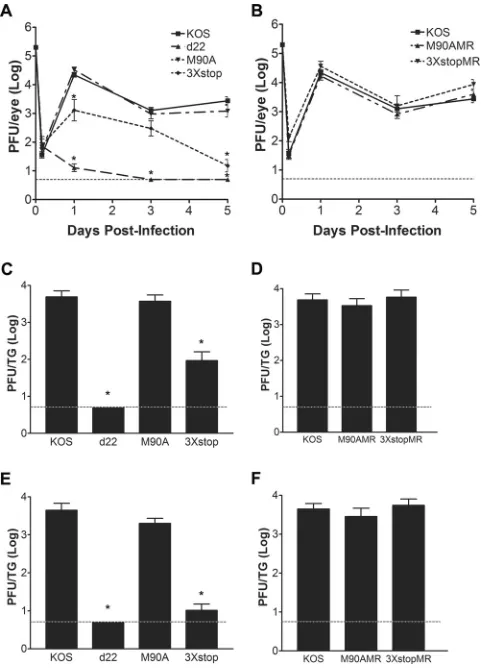
postinfec-tion to monitor acute replicapostinfec-tion in neurons. As shown inFig. 2A,
the M90A mutant virus replicated to levels comparable to the level of KOS in the eyes of mice for all time points tested. In contrast,
the replication of the 3⫻stop mutant in the eyes was significantly
reduced relative to that of KOS on day 1 (17-fold;ttest,P⫽0.01)
and day 5 (184-fold;ttest,P⫽6.8⫻10⫺7) postinfection. The
replication of d22 in the eyes of mice was significantly reduced at days 1, 3, and 5 postinfection by 1722-, 255-, and 554-fold,
respec-tively (ttest,Pvalues of 5.9⫻10⫺11, 1.7⫻10⫺19, and 6.5⫻10⫺18,
respectively). The replication of M90AMR and 3⫻stopMR viruses
in the eyes was comparable to that of KOS (Fig. 2B). When acute
replication of the M90A and 3⫻stop mutants in TG was
exam-ined, only the 3⫻stop mutant showed decreased growth at day 3
(59-fold;ttest,P⫽3.6⫻10⫺5) (Fig. 2C) and day 5 (429-fold;t
test,P⫽2.3⫻10⫺10) (Fig. 2E). The M90A mutant replicated as
efficiently as KOS on both days (Fig. 2CandE). We were unable to
detect progeny virus in the TG of d22-infected mice on days 3 and
5 postinfection (ttest,P⫽6.7⫻10⫺16andP⫽3.3⫻10⫺18,
respectively) (Fig. 2CandE). The M90AMR and 3⫻stopMR
vi-ruses replicated at levels similar to the level of KOS in TG on both
days (Fig. 2DandF). Thus, ICP22, not US1.5, is the major
con-tributor to acute ocular and, consequently, neuronal replication.
ICP22 but not US1.5 is required for external pathological le-sions in mice.We scored the pathological lesions caused by the
ocular infection of mice with KOS, d22, M90A, 3⫻stop,
M90AMR, and 3⫻stopMR at day 8 postinfection. The severity of
infection was ranked on a scale of 0 to 4 for each group, where 0 indicates no signs of infection and 4 indicates more than 70% loss of hair from between the eyes, which is the result of scratching or
secondary inflammation. As shown inFig. 3, the infection with the
3⫻stop mutant was similar to that with d22 in that it did not
produce any signs of disease, with both groups of mice having
pathology scores of 0 (ttest,P⫽9⫻10⫺5). Infection with the
M90A mutant, on the other hand, was similar to that with KOS and the MR viruses, where inoculated mice showed obvious signs
of infection (scores of 2.5 ⫾ 0.5 for the M90A, KOS, and
3⫻stopMR viruses and 2.33 ⫾ 0.47 for the M90AMR virus
FIG 2Acute replication of M90A and 3⫻stop mutants and MR virusesin vivo. (A and B) Acute ocular replication of M90A and 3⫻stop mutants and MR viruses in mice. CD-1 mice were infected with 2⫻105PFU per eye. At the indicated time points, the eyes of mice were swabbed, and viral titers were determined by standard plaque assays. (C to F) Acute TG replication of M90A and 3⫻stop mutants and MR viruses in mice. CD-1 mice were infected with 2⫻105PFU per eye. On day 3 (C and D) or day 5 (E and F) postinfection, mice were sacrificed, TG were removed and homogenized, and viral titers were determined by standard plaque assays. *,P⬍0.05, compared to KOS (Stu-dent’sttest). Error bars represent the standard errors of the means (n⫽8 samples/virus/time point). In all cases, the dashed line is the limit of detection.
FIG 3Gross pathological effects of M90A and 3⫻stop mutants and MR vi-ruses. CD-1 mice were infected with 2⫻105PFU per eye. A representative picture from each group of infected mice at 8 days postinfection is shown.
on November 7, 2019 by guest
http://jvi.asm.org/
[image:4.585.43.285.66.400.2] [image:4.585.319.522.66.217.2][means⫾standard deviations;n⫽5 to 6 mice per virus]) (Fig. 3). Our data indicate that, in contrast to ICP22, the expression of
US1.5 is insufficient to induce signs of viral pathogenesis in mice.
ICP22 is required for efficient establishment of latency.To examine the ability of our mutants to establish a latent infection, TG from infected mice were collected at 28 days postinfection, and viral DNA loads were determined using real-time PCR. As shown inFig. 4, relative to KOS, the 3⫻stop mutant was significantly
reduced for viral DNA levels by 11.5-fold (one way ANOVA,P⬍
0.05). The M90A mutant, on the other hand, was able to establish
latency comparable to the levels of the KOS and MR viruses (Fig.
4). The fold reduction of the d22 mutant was comparable to that
of the mock-infected mice (ⱖ568-fold; one way ANOVA,P⬍
0.05) (H. H. Mostafa and D. J. Davido, unpublished data). The defect in establishing an efficient latent infection with both d22
and 3⫻stop is likely attributed to their impaired acute replication
phenotypes in the eyes and TG, as has been observed with other
HSV-1 mutants (33,34).
ICP22 or US1.5 is sufficient for HSV-1 replication in restric-tive cells.It has been previously established that ICP22 is required for efficient viral growth in certain cell lines, which have been
termed restrictive cell lines (7). In order to test the roles of ICP22
and US1.5 in viral replication in restrictive cells, we performed a
viral yield assay in HEL-299 cells (7). HEL-299 cells were infected
at an MOI of 0.1 with KOS, d22, M90A, or the 3⫻stop mutant. At
24 h postinfection, cells and progeny virus were collected, and viral yields were determined with a standard plaque assay. As
shown inFig. 5, the M90A mutant replicated to levels that were
comparable to the level of KOS; however, d22 replication was
significantly reduced (83-fold;ttest,P⫽2.5⫻10⫺7). Replication
of the 3⫻stop mutant was only modestly impaired relative to that
of KOS (4.5-fold;t test,P⫽0.003) (Fig. 5). Furthermore, the
replication of the 3⫻stop mutant was significantly reduced
rela-tive to that of the d22 mutant (18.4-fold;ttest,P⫽5.5⫻10⫺8).
Similar results were seen in mouse embryo fibroblasts (MEFs) of CD-1 mice (Mostafa and Davido, unpublished). These data indi-cate that ICP22 is sufficient to enhance viral growth in HEL-299
and MEF cells; however, the US1.5 protein can, to a great extent,
complement the function of full-length ICP22.
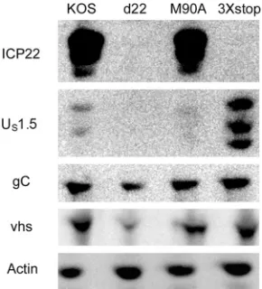
Both ICP22 and US1.5 enhance the expression of late viral proteins in cell culture.ICP22 has been demonstrated to enhance the expression of a subset of late viral proteins, including viral
glycoprotein C (gC) and virion host shutoff (vhs) (6,7,9,35). In
order to test the individual roles of ICP22 and US1.5 in the
expres-sion of late viral proteins, we infected HEL-299 cells with KOS,
d22, M90A, and 3⫻stop for 24 h and examined vhs and gC protein
levels by Western blotting. Compared to levels in KOS-infected samples, the expression levels of gC and vhs were reduced in
d22-infected cells but not in M90A- or 3⫻stop-infected cells (Fig. 6).
Similar results were also observed at 12 h postinfection (Mostafa and Davido, unpublished). Our data show that both ICP22 and
US1.5 are capable of stimulating late viral protein expression.
Analogous results were observed in MEFs (CD-1 strain) and Vero cells (Mostafa and Davido, unpublished).
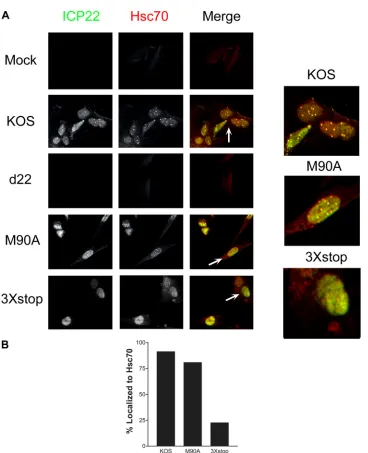
US1.5 does not induce VICE domains.ICP22 has been shown
to be required for the formation of VICE domains (13) during
productive HSV-1 infection. To test the individual contributions
of ICP22 and US1.5 in the formation of VICE domains, HEL-299
FIG 4Establishment of latency of M90A and 3⫻stop mutants and MR vi-ruses. CD-1 mice were infected with 2⫻105PFU per eye. At 28 days postin-fection, mice were sacrificed, TG were removed, and genomic DNA was iso-lated from samples. The amount of HSV-1 DNA present in each sample was quantified by real-time PCR (n⫽10 to 12 TG per group). Results shown are the fold reductions compared to KOS levels. The fold reduction for d22 was comparable to that of the mock-infected TG (ⱖ568-fold).
FIG 5Growth of M90A and 3⫻stop mutant viruses on 299 cells. HEL-299 cells were infected at an MOI of 0.1 with the indicated viruses. At 24 h postinfection, cells were harvested, and viral titers were determined by a stan-dard plaque assay. *,P⬍0.05 compared to KOS (Student’sttest). Error bars represent the standard errors of the means.
FIG 6Examination of late viral gene expression for M90A and 3⫻stop mu-tant viruses. HEL-299 cells were infected at an MOI of 2 with the indicated viruses. At 24 h postinfection, cells were harvested, and protein levels from cell extracts were determined by Western blot analysis.
on November 7, 2019 by guest
http://jvi.asm.org/
[image:5.585.78.247.63.218.2] [image:5.585.337.503.66.224.2] [image:5.585.349.494.523.683.2]cells were mock infected or infected with KOS, d22, M90A, or
3⫻stop for 24 h, and the localization of ICP22 and the VICE
domain marker, Hsc70, was examined by immunofluorescence. Our experiments showed that the M90A mutant was able to form
VICE domains that appeared identical to those of KOS (Fig. 7A
andB), in which ICP22 colocalized with Hsc70 in the nucleus. The
3⫻stop mutant, however, was unable to induce Hsc70 puncta
(Fig. 7AandB). Although the US1.5 protein localized to the
nu-cleus, it failed to form the speckled pattern of ICP22 staining
ob-served with KOS- and M90A-infected nuclei (Fig. 7A). Notably,
the staining pattern of Hsc70 in the presence of US1.5 localized to
the nucleus, which was different from that of mock- and
d22-infected cells (Fig. 7A). This indicates that although US1.5 fails to
induce the formation of VICE domains, it is still capable of
relo-calizing Hsc70 to the nucleus. Similar results were observed in Vero cells (Mostafa and Davido, unpublished).
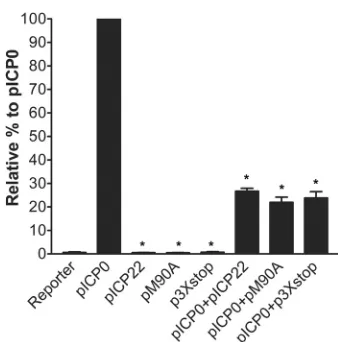
M90A and 3ⴛstop inhibit ICP0-directed transactivation of an HSV promoter-luciferase reporter.The IE protein ICP0 is a
promiscuous transactivator of all classes of HSV-1 genes (36). One
published report demonstrated that ICP22 represses ICP0-trans-activated expression of reporter genes in transient transfections
(37), which could be either directly through interfering with
ICP0’s transactivation activity or indirectly by reducing ICP0’s
expression (38,39). To investigate if this inhibitory function is
carried out by ICP22, US1.5, or both, we performed a luciferase
assay to test the effect of both proteins on ICP0-mediated gene expression. ICP0 expression led to the maximum activity of the
VP16 promoter and was given the 100% value (Fig. 8). Neither the
FIG 7ICP22 but not US1.5 induces VICE domain formation. (A) HEL-299 cells were mock infected or infected with the indicated viruses at an MOI of 0.1. At 24 h postinfection, cells were fixed and stained for ICP22 and Hsc70 and examined by fluorescence microscopy. (B) At least 100 cells that expressed ICP22 and/or US1.5 were examined for each virus, and the percentages of these cells that colocalized with Hsc70 are shown.
on November 7, 2019 by guest
http://jvi.asm.org/
[image:6.585.111.479.58.511.2]pICP22 plasmid (which expresses both ICP22 and US1.5),
pM90A, or p3⫻stop was able to activate VP16 promoter.
Com-bining pICP22, pM90A, or p3⫻stop with pICP0 led to a
signifi-cant inhibitory effect on ICP0’s transactivation of the VP16
pro-moter (Mann Whitney U-test, P ⫽ 0.002). Our results
demonstrate that both ICP22 and US1.5 have significant and
com-parable inhibitory effects on the ICP0-directed transactivation in reporter gene experiments.
ICP22 but not US1.5 counteracts the effects of beta interferon (IFN-) in restrictive cell lines.In order to understand the
reduc-tion in the ability of the 3⫻stop mutant to replicate acutelyin vivo,
we wanted to test if there is a relationship between ICP22 or US1.5
and the type I IFN response. An earlier publication examined the plaquing of an ICP22 null mutant in IFN-treated Vero cells, a cell
line permissive for ICP22 mutant replication (5), and concluded
that ICP22 is not required to counteract an established type I IFN
response (40). Consequently, we decided to perform plaque
re-duction assays in a restrictive cell line, HEL-299 cells. Interest-ingly, we found a noticeable reduction in the number of plaques
formed by d22 in the presence of IFN-relative to KOS (Table 1).
The overall plaque size for d22 was smaller than that of KOS. Furthermore, the reduction in plaque size for d22 was further
enhanced by IFN-compared to that for KOS (46-fold versus
10-fold reduction) (Fig. 9). For M90A, the reduction in the
num-ber of plaques in the presence of IFN-was comparable to that for
KOS (Table 1), and the plaque sizes with (9-fold reduction) and
without IFN-were similar for the two viruses. Interestingly,
al-though the reduction in the number of plaques formed by 3⫻stop
after the addition of IFN-was comparable to that for KOS (Table
1), the plaques appeared smaller than those of KOS only after the
addition of IFN-(37-fold versus 10-fold reduction) (Fig. 9). In
order to correlate this observation to thein vivoacute replication
data, we decided to repeat the same experiment in CD-1 MEFs.
Our results showed that the size of d22 and 3⫻stop plaques was
markedly smaller than that of the wild-type plaques, a phenotype
that was enhanced in IFN--treated cells (34-fold and 21-fold
reductions, respectively, versus a 2-fold reduction with KOS) (Fig. 9). Consistent with these experiments, there was a reduction
in the number of plaques for d22 and 3⫻stop that exceeded the
decreases observed for KOS and M90A (Table 1). When we
re-peated the same experiments on MEFs of strain 129, we observed
a further reduction in the number of plaques for d22 and 3⫻stop
(Table 1). To determine if the plaque reduction phenotype corre-lated with reductions in viral replication, we performed yield
as-says in the absence or presence of IFN-. As shown inFig. 10A,
reductions in viral replication after the addition of IFN-were
similar for KOS, d22, M90A, and 3⫻stop mutants (⬃50-fold
de-crease) on HEL-299 cells, and similar results were noticed on 129
MEFs (Mostafa and Davido, unpublished). In CD-1 MEFs (Fig.
10B), IFN-further impaired d22 replication (140-fold compared
to 6-fold reduction of KOS), while IFN-only slightly reduced the
growth of M90A and 3⫻stop compared to that of KOS (19- and
27-fold reductions, respectively). These data indicate that ICP22
but not US1.5 is required for efficient plaquing in the restrictive
cell lines, HEL-299 and MEFs, in the presence of a preestablished interferon response and that the degree of this requirement is cell type dependent.
DISCUSSION
The HSV-1 genome contains⬃90 genes, and, with some
excep-tions, each gene encodes a single protein. The US1 gene is one such
exception as it codes for two proteins: ICP22 and its N-terminally
truncated form, US1.5 (18). To date, reports that have examined
the functions of ICP22 have largely not separated its biological
activities from those of US1.5. The goal of this study was to
char-acterize the individual contributions of ICP22 and US1.5 in the
HSV-1 life cycle by generating mutant viruses (i.e., M90A and
3⫻stop) that can express only one of the two proteins. Our data
show that the US1.5 protein is dispensable for acute replication in
mice and for the induction of VICE domains. Also, our data indi-cate that either protein can functionally enhance the expression of late viral proteins in cell culture and inhibit ICP0-mediated gene expression in transient-transfection assays. Additionally, we iden-tified a new role for ICP22 in enhancing viral plaquing in the
presence of IFN-, a function that was not fully compensated by
US1.5 alone. Lastly, the M90A mutant behaved like KOS in our
assays, indicating that the methionine-to-alanine amino acid sub-stitution at residue 90 did not impact the functional activities of ICP22 tested in our study.
We observed that the expression of US1.5 alone (expressed
[image:7.585.77.246.62.233.2]FIG 8Inhibition of ICP0’s transactivated gene expression by vectors that express either ICP22 or US1.5. Vero cells were transfected with an HSV-1 reporter plasmid (50 ng; pGL3-VP16) and a plasmid expressing wild-type ICP0 (pICP0), both ICP22 and US1.5 (pICP22), ICP22 alone (pM90A), or US1.5 alone (p3⫻stop) or a combination of pICP0 and pICP22, pM90A, or p3⫻stop for 48 h. Cell extracts were analyzed in luciferase assays to monitor ICP0’s transactivating activity. *,P⬍0.05 compared to pICP0 (Mann-Whit-neyUtest). The error bars indicate the standard errors of the means.
TABLE 1Plaque reduction assays of KOS and ICP22 mutants in the absence and presence of IFN-
Virus
No. of plaques without IFN-/no. of plaques with IFN-in:a
HEL-299 cells CD-1 MEFs 129 MEFs
Expt 1 Expt 2 Expt 1 Expt 2 Expt 1 Expt 2
KOS 6.7 15 6.7 2 37.5 20
d22 50 133 45.5 20 1,100 1,400
M90A 6.7 6.2 4 6.7 27 35
3⫻stop 20 13 14 20 125 175
aHEL-299 cells or MEF cells were infected with serially diluted viruses in the absence or
presence of 1,000 U/ml of IFN-. At 3 days postinfection, cells were fixed and immunostained for plaque formation. Plaques were counted for all samples, and the ratios of plaque numbers between untreated and treated plates were determined. The data shown are results from two independent experiments for each cell line.
on November 7, 2019 by guest
http://jvi.asm.org/
[image:7.585.300.545.593.679.2]from 3⫻stop) was marginally reduced for viral replication in re-strictive HEL-299 and MEF cells; however, its acute replication in the eyes and TG of mice was remarkably reduced relative to KOS.
The diminished replication of 3⫻stop in TG may be attributed to
its reduced ocular replication (34), given that the acute TG titers of
3⫻stop at days 3 and 5 followed a similar trend for replication in
the eyes at days 1 and 5 postinfection (Fig. 2A,C, andE). This
impaired acute replication phenotype both in the eyes and TG
likely explains 3⫻stop’s defect in establishing an efficient latent
infection as similar results have been observed with other mutants
of HSV-1 (33,34). This decrease in the establishment of latency by
3⫻stop would be expected to negatively impact reactivation. For
this reason, we did not performex vivoreactivation studies with
this mutant.
The deficiency of the 3⫻stop mutant in acute replicationin
vivosuggests that the US1.5 protein lacks a critical function of
ICP22. What could this function be? A potential answer is that it is
unable to induce VICE domains (Fig. 7AandB). Notably, our
results provide a finer map of the region of ICP22 required for VICE domain formation, reducing it from the first 146
N-termi-nal residues as previously described (13) to the first 89 N-terminal
amino acids. Because the functional significance of VICE domains in the HSV-1 life cycle is not yet fully understood, it is possible that the requirement of VICE domains in viral growth could be cell
type dependent and specifically required forin vivoreplication.
Thus, this defect in the VICE domains observed with the 3⫻stop
virus may contribute to its impaired acute replicationin vivo.
Ad-ditional studies will be needed to determine the role VICE do-mains play in HSV-1 replication and how ICP22 facilitates their
formation. While VICE domain formation is affected in 3⫻
stop-infected cells, US1.5 is expected to modify the C terminus of the
large subunit of the host RNA polymerase II based on ICP22
map-FIG 9Plaque reduction assays. HEL-299 cells or CD-1 MEFs cells were infected with serial dilutions of the indicated viruses in the absence or the presence of 1,000 U/ml IFN-. At 3 days postinfection, cells were fixed and immunostained for plaque formation.
FIG 10Growth of M90A and 3⫻stop with and without IFN-. HEL-299 cells (A) or CD-1 MEFs (B) were infected at an MOI of 0.1 in the presence or in the absence of 1,000 U/ml IFN-with the indicated viruses. At 24 h postinfection, cells were collected, and the viral titers were determined by a standard plaque assay. *P⬍0.05, compared to KOS (Student’sttest). Error bars represent the standard errors of the means.
on November 7, 2019 by guest
http://jvi.asm.org/
[image:8.585.138.452.68.311.2] [image:8.585.138.453.546.694.2]ping studies by Bastian and Rice as residues 147 to 420 of ICP22
are sufficient to modify RNA polymerase II (21). Although the
mechanism by which ICP22 alters RNA polymerase II to enhance viral gene expression is unknown, the expected presence of this
activity in US1.5 may explain the intermediate levels of acute
oc-ular and neuronal replication observed with 3⫻stop in mice
com-pared to levels with KOS and d22.
Although the 3⫻stop mutant’s expression level of US1.5
pro-tein might be lower than that of ICP22 expressed from either KOS
or M90A (Fig. 1Cand6), the protein is still able to fully carry out
some functions of ICP22; these functions include enhancing late viral protein expression, inhibiting ICP0 transactivated gene ex-pression, and largely enabling efficient viral replication in cell
cul-ture. Additionally, we also tested if overexpression of US1.5 using
the human cytomegalovirus (HCMV) promoter (19) might lead
to VICE domain formation in transiently transfected HEL-299 cells. In contrast to results for ICP22, we did not detect VICE domains in these experiments (Mostafa and Davido, unpub-lished). Thus, while it may be argued that the expression levels of
US1.5 dictate its ability to complement ICP22’s functions, these
experiments do not support this possibility.
Another plausible explanation for the impaired replication of
the 3⫻stop mutant is related to the host’s antiviral IFN response.
Specifically, our results demonstrate that the 3⫻stop and d22
mu-tants are hypersensitive to IFN-in cell culture. Thus, we have
identified a new function of ICP22 in that it counteracts the effects of this type I IFN. Interestingly, this phenotype correlated very well with the impaired replication phenotype in the restrictive cell
line HEL-299 and MEFs of the CD-1 mouse strain (Fig. 5and10)
and in MEFs of strain 129 and the neuronal cell line SK-N-SH (Mostafa and Davido, unpublished). On the other hand, the
hy-persensitivity of both mutants to IFN-was not detected in the
permissive cell line, Vero cells (40; also Mostafa and Davido,
un-published), which are known to have a defect in IFN production
(41). This could explain, in part, the replication phenotypes of
3⫻stop and d22 in mice. Because the decrease in the plaque sizes
observed in plaque reduction assays (Fig. 9) was not accompanied
by a similar reduction in viral growth (Fig. 10) after addition of
IFN-, there is the possibility that the observed phenotype is due
to a defect in cell-to-cell spread rather than a defect in viral repli-cation. Whether this function of ICP22 is mediated directly or indirectly through other viral factors will require more studies to determine. Related to this possibility, the reduction in the levels of the late viral protein vhs might partially contribute to this pheno-type in the case of the d22 mutant. vhs is an endoribonuclease that degrades viral and cellular mRNA and is capable of interfering
with an interferon-induced antiviral state (42). This mechanism is
not applicable to the 3⫻stop phenotype, which is able to enhance
wild-type levels of vhs protein (Fig. 6). Additionally, there could
be a potential association between the formation of VICE domains and the interferon response based on published reports that have
linked the cellular chaperone machinery to type I IFN (43–45).
It is notable that the acute replication of the ICP22 null mutant, d22, was greatly reduced in the eyes relative to the level of the
3⫻stop mutant both at day 1 and at day 3. Consequently, d22 was
unable to acutely replicate in TG or establish a detectable latent infection in our experiments. The absence of both ICP22 and
US1.5 in d22 significantly attenuated lytic and latent infections in
mice, a result which mirrors thein vivophenotypes of other ICP22
mutants (8,20,32). Many of these mutants do not express optimal
levels of late viral gene products, including vhs, US11, and/or gC. As previously mentioned, it has been hypothesized that the ex-pression of these late genes is dependent on VICE domain forma-tion and/or the altered modificaforma-tion of the C terminus of the host
RNA polymerase II (46). Ultimately, this defect in late viral gene
expression likely reduces d22 growth in mice. Additionally, a re-duction in gC levels could thwart the ability of HSV-1 to
counter-act complement-mediated neutralization (47–50), which might
further contribute to the defective acute replication phenotype of d22in vivo. Lastly, the composition of virion proteins in the ICP22
mutant 22/n199 is altered, and this mutant replicates poorly in
mice (8). It was hypothesized that 22/n199 virions are less stable
than wild-type virions and that this contributes to itsin vivo
phe-notype. Consequently, it is plausible that the composition of d22 virions is similarly altered and that this negatively impacts its rep-lication.
Our results did not define a unique function for US1.5 or a
defect in HSV-1 replication in its absence. This raises the question
of why HSV-1 has evolved to express the US1.5 protein. One
pos-sibility might be that the viral requirement for US1.5 is conditional
and is enhanced only under certain circumstances during the
HSV-1 life cycle. Moreover, while US1.5 is not required for HSV-1
replication in our assays, a role for US1.5 in the HSV-1 life cycle
may be apparent in other model systems or in its natural host, humans. Finally, since we find that many, if not most, of the
known functions of ICP22 can be carried out by US1.5, it may be
that US1.5 can serve as a biological backup for ICP22 should
ribo-some readthrough be blocked at the translation initiation codon of ICP22.
ACKNOWLEDGMENTS
This work was supported by the University of Kansas.
The content of this article is solely the responsibility of the authors and does not necessarily represent the official views of the University of Kan-sas.
We thank members of the Davido lab and Stephen Rice for discussions related to this project and critical reading of the manuscript. We also thank Kristi Neufeld for cells and reagents.
REFERENCES
1.Roizman R, Knipe DM, Whitley RJ. 2007. Herpes simplex viruses, p 2501–2601.InKnipe DM, Howley PM (ed), Fields virology, 5th ed. Lip-pincott Williams & Wilkins, Philadelphia, PA.
2.Knipe DM, Cliffe A.2008. Chromatin control of herpes simplex virus lytic and latent infection. Nat. Rev. Microbiol.6:211–221.
3.Roizman B, Kozak M, Honess RW, Hayward G.1975. Regulation of herpesvirus macromolecular synthesis: evidence for multilevel regulation of herpes simplex 1 RNA and protein synthesis. Cold Spring Harbor Symp. Quant. Biol.39:687–701.
4.Stevens JG.1987. Defining herpes simplex genes involved in neuroviru-lence and neuroinvasiveness. Curr. Eye Res.6:63– 67.
5.Post LE, Roizman B.1981. A generalized technique for deletion of spe-cific genes in large genomes: alpha gene 22 of herpes simplex virus 1 is not essential for growth. Cell25:227–232.
6.Poffenberger KL, Raichlen PE, Herman RC.1993. In vitro characteriza-tion of a herpes simplex virus type 1 ICP22 delecharacteriza-tion mutant. Virus Genes 7:171–186.
7.Sears AE, Halliburton IW, Meignier B, Silver S, Roizman B. 1985. Herpes simplex virus 1 mutant deleted in the␣22 gene: growth and gene expression in permissive and restrictive cells and establishment of latency in mice. J. Virol.55:338 –346.
8.Orlando JS, Balliet JW, Kushnir AS, Astor TL, Kosz-Vnenchak M, Rice SA, Knipe DM, Schaffer PA.2006. ICP22 is required for wild-type com-position and infectivity of herpes simplex virus type 1 virions. J. Virol. 80:9381–9390.
on November 7, 2019 by guest
http://jvi.asm.org/
9.Rice SA, Long MC, Lam V, Schaffer PA, Spencer CA.1995. Herpes simplex virus immediate-early protein ICP22 is required for viral modifi-cation of host RNA polymerase II and establishment of the normal viral transcription program. J. Virol.69:5550 –5559.
10. Durand LO, Advani SJ, Poon AP, Roizman B. 2005. The carboxyl-terminal domain of RNA polymerase II is phosphorylated by a complex containing cdk9 and infected-cell protein 22 of herpes simplex virus 1. J. Virol.79:6757– 6762.
11. Fraser KA, Rice SA.2007. Herpes simplex virus immediate-early protein ICP22 triggers loss of serine 2-phosphorylated RNA polymerase II. J. Vi-rol.81:5091–5101.
12. Rice SA.2011. Multiple roles of immediate-early protein ICP22 in HSV-1 replication.InWeller SK (ed), Alphaherpesviruses: molecular virology. Caister Academic Press, Norfolk, United Kingdom.
13. Bastian TW, Livingston CM, Weller SK, Rice SA.2010. Herpes simplex virus type 1 immediate-early protein ICP22 is required for VICE domain formation during productive viral infection. J. Virol.84:2384 –2394. 14. Burch AD, Weller SK.2004. Nuclear sequestration of cellular chaperone
and proteasomal machinery during herpes simplex virus type 1 infection. J. Virol.78:7175–7185.
15. Burch AD, Weller SK.2005. Herpes simplex virus type 1 DNA polymer-ase requires the mammalian chaperone hsp90 for proper localization to the nucleus. J. Virol.79:10740 –10749.
16. Livingston CM, Ifrim MF, Cowan AE, Weller SK.2009. Virus-induced chaperone-enriched (VICE) domains function as nuclear protein quality control centers during HSV-1 infection. PLoS Pathog.5:e1000619. doi:10 .1371/journal.ppat.1000619.
17. Purves FC, Roizman B.1992. The UL13 gene of herpes simplex virus 1 encodes the functions for posttranslational processing associated with phosphorylation of the regulatory protein␣22. Proc. Natl. Acad. Sci. U. S. A.89:7310 –7314.
18. Carter KL, Roizman B.1996. The promoter and transcriptional unit of a novel herpes simplex virus 1␣gene are contained in, and encode a protein in frame with, the open reading frame of the␣22 gene. J. Virol.70:172– 178.
19. Bowman JJ, Schaffer PA.2009. Origin of expression of the herpes simplex virus type 1 protein U(S)1.5. J. Virol.83:9183–9194.
20. Ogle WO, Roizman B.1999. Functional anatomy of herpes simplex virus 1 overlapping genes encoding infected-cell protein 22 and US1.5 protein. J. Virol.73:4305– 4315.
21. Bastian TW, Rice SA.2009. Identification of sequences in herpes simplex virus type 1 ICP22 that influence RNA polymerase II modification and viral late gene expression. J. Virol.83:128 –139.
22. Long MC, Leong V, Schaffer PA, Spencer CA, Rice SA.1999. ICP22 and the UL13 protein kinase are both required for herpes simplex virus-induced modification of the large subunit of RNA polymerase II. J. Virol. 73:5593–5604.
23. Mostafa HH, Thompson TW, Kushnir AS, Haenchen SD, Bayless AM, Hilliard JG, Link MA, Pitcher LA, Loveday E, Schaffer PA, Davido DJ. 2011. Herpes simplex virus 1 ICP0 phosphorylation site mutants are at-tenuated for viral replication and impaired for explant-induced reactiva-tion. J. Virol.85:12631–12637.
24. National Research Council.2011. Guide for the care and use of laboratory animals, 8th ed. National Academies Press, Washington, DC.
25. Halford WP, Schaffer PA. 2000. Optimized viral dose and transient immunosuppression enable herpes simplex virus ICP0-null mutants to establish wild-type levels of latency in vivo. J. Virol.74:5957–5967. 26. Schrimpf JE, Tu EM, Wang H, Wong YM, Morrison LA. 2011. B7
costimulation molecules encoded by replication-defective, vhs-deficient HSV-1 improve vaccine-induced protection against corneal disease PLoS One6:e22772. doi:10.1371/journal.pone.0022772.
27. Strand SS, Leib DA.2004. Role of the VP16-binding domain of vhs in viral growth, host shutoff activity, and pathogenesis. J. Virol.78:13562– 13572.
28. Livak KJ, Schmittgen TD.2001. Analysis of relative gene expression data using real-time quantitative PCR and the 2⫺⌬⌬CTmethod. Methods25: 402– 408.
29. Read GS, Karr BM, Knight K.1993. Isolation of a herpes simplex virus type 1 mutant with a deletion in the virion host shutoff gene and identifi-cation of multiple forms of thevhs(UL41) polypeptide. J. Virol.67:7149 – 7160.
30. Kushnir AS, Davido DJ, Schaffer PA.2010. Role of nuclear factor Y in stress-induced activation of the herpes simplex virus type 1 ICP0 pro-moter. J. Virol.84:188 –200.
31. Flory E, Weber CK, Chen P, Hoffmeyer A, Jassoy C, Rapp UR.1998. Plasma membrane-targeted Raf kinase activates NF-B and human im-munodeficiency virus type 1 replication in T lymphocytes. J. Virol.72: 2788 –2794.
32. Poffenberger KL, Idowu AD, Fraser-Smith EB, Raichlen PE, Herman RC.1994. A herpes simplex virus type 1 ICP22 deletion mutant is altered for virulence and latency in vivo. Arch. Virol.139:111–119.
33. Katz JP, Bodin ET, Coen DM. 1990. Quantitative polymerase chain reaction analysis of herpes simplex virus DNA in ganglia of mice infected with replication-incompetent mutants. J. Virol.64:4288 – 4295. 34. Leib DA, Coen DM, Bogard CL, Hicks KA, Yager DR, Knipe DM, Tyler
KL, Schaffer PA.1989. Immediate-early regulatory gene mutants define different stages in the establishment and reactivation of herpes simplex virus latency. J. Virol.63:759 –768.
35. Purves FC, Ogle WO, Roizman B.1993. Processing of the herpes simplex virus regulatory protein␣22 mediated by the UL13 protein kinase deter-mines the accumulation of a subset of alpha and gamma mRNAs and proteins in infected cells. Proc. Natl. Acad. Sci. U. S. A.90:6701– 6705. 36. Everett RD.1984. Transactivation of transcription by herpes virus
prod-ucts: requirement for two HSV-1 immediate-early polypeptides for max-imum activity. EMBO J.3:3135–3141.
37. Bowman JJ, Orlando JS, Davido DJ, Kushnir AS, Schaffer PA.2009. Transient expression of herpes simplex virus type 1 ICP22 represses viral promoter activity and complements the replication of an ICP22 null virus. J. Virol.83:8733– 8743.
38. Guo L, Wu WJ, Liu LD, Wang LC, Zhang Y, Wu LQ, Guan Y, Li QH. 2012. Herpes simplex virus 1 ICP22 inhibits the transcription of viral gene promoters by binding to and blocking the recruitment of P-TEFb. PLoS One7:e45749. doi:10.1371/journal.pone.0045749.
39. Cun W, Guo L, Zhang Y, Liu L, Wang L, Li J, Dong C, Wang J, Li Q. 2009. Transcriptional regulation of the herpes simplex virus 1␣-gene by the viral immediate-early protein ICP22 in association with VP16. Sci. China C Life Sci.52:344 –351.
40. Mossman KL, Saffran HA, Smiley JR.2000. Herpes simplex virus ICP0 mutants are hypersensitive to interferon. J. Virol.74:2052–2056. 41. Emeny JM, Morgan MJ.1979. Regulation of the interferon system:
evi-dence that Vero cells have a genetic defect in interferon production. J. Gen. Virol.43:247–252.
42. Chee AV, Roizman B.2004. Herpes simplex virus 1 gene products oc-clude the interferon signaling pathway at multiple sites. J. Virol.78:4185– 4196.
43. Yang M, Wang C, Zhu X, Tang S, Shi L, Cao X, Chen T.2011. E3 ubiquitin ligase CHIP facilitates Toll-like receptor signaling by recruiting and polyubiquitinating Src and atypical PKC. J. Exp. Med.208:2099 – 2112.
44. Shang L, Tomasi TB.2006. The heat shock protein 90-CDC37 chaperone complex is required for signaling by types I and II interferons. J. Biol. Chem.281:1876 –1884.
45. Yang K, Shi H, Qi R, Sun S, Tang Y, Zhang B, Wang C.2006. Hsp90 regulates activation of interferon regulatory factor 3 and TBK-1 stabiliza-tion in Sendai virus-infected cells. Mol. Biol. Cell17:1461–1471. 46. Rice SA, Davido DJ.2013. HSV-1 ICP22: hijacking host nuclear functions
to enhance viral infection. Future Microbiol.8:311–321.
47. Hook LM, Lubinski JM, Jiang M, Pangburn MK, Friedman HM.2006. Herpes simplex virus type 1 and 2 glycoprotein C prevents complement-mediated neutralization induced by natural immunoglobulin M anti-body. J. Virol.80:4038 – 4046.
48. Friedman HM, Cohen GH, Eisenberg RJ, Seidel CA, Cines DB.1984. Glycoprotein C of herpes simplex virus 1 acts as a receptor for the C3b complement component on infected cells. Nature309:633– 635. 49. Hung SL, Peng C, Kostavasili I, Friedman HM, Lambris JD, Eisenberg
RJ, Cohen GH.1994. The interaction of glycoprotein C of herpes simplex virus types 1 and 2 with the alternative complement pathway. Virology 203:299 –312.
50. Kostavasili I, Sahu A, Friedman HM, Eisenberg RJ, Cohen GH, Lambris JD.1997. Mechanism of complement inactivation by glycoprotein C of herpes simplex virus. J. Immunol.158:1763–1771.