R E S E A R C H
Open Access
Overexpression of BID in thyroids of transgenic
mice increases sensitivity to iodine-induced
autoimmune thyroiditis
Su He Wang
1,2*, Yongyi Fan
1and James R Baker Jr
1,2*Abstract
Background:BID functions as a bridge molecule between death-receptor and mitochondrial related apoptotic
pathways to amplify apoptotic signaling. Our previous studies have demonstrated a substantial increase in BID expression in primary normal thyroid epithelia cells treated with inflammatory cytokines, including the combination of IFNγand IL-1βor IFNγand TNFα. The aim of this study was to determine whether an increase in BID expression in thyroid can induce autoimmune thyroiditis.
Methods:A transgenic mouse line that expresses human BID in thyroid cells was established by fusing a mouse thyroglobulin (Tg) promoter upstream of human BID (Tg-BID). We tested whether the increased expression of pro-apoptotic BID in thyroid would induce autoimmune thyroiditis, both in the presence and absence of 0.3% iodine water.
Results:Our data show that Tg-BID mice in a CBA/J (H-2 k) background do not spontaneously develop autoimmune
thyroiditis for over a year. However, upon ingestion of iodine in the drinking water, autoimmune thyroiditis does develop in Tg-BID transgenic mice, as shown by a significant increase in anti-Tg antibody and mononuclear cell infiltration in the thyroid glands in 30% of mice tested. Serum T4 levels, however, were similar between iodine-treated Tg-BID transgenic mice and the wild type mice.
Conclusions:Our data demonstrate that increased thyroid expression of BID facilitates the development of
autoimmune thyroiditis induced by iodine uptake. However, the overexpression of BID itself is not sufficient to initiate thyroiditis in CBA/J (H-2 k) mice.
Keywords:BID, Iodine, Autoimmune thyroiditis, Mice, Transgene
Background
Autoimmune (Hashimoto’s) thyroiditis is characterized by lymphocytic infiltration of the parenchyma, causing a dense accumulation of lymphocytes, plasma cells, and macrophages with germinal center formation and thyroid enlargement. Although the pathogenesis of autoimmune thyroiditis is not entirely clear, studies have demonstrated that increasing the local production of inflammatory cytokines in the thyroid microenvironment plays a critical role in facilitating apoptosis in thyrocytes, leading to
autoimmune thyroiditis [1-3]. Among various inflam-matory cytokines, IFN-γ, TNF-α and IL-1βappear to be critical since the combination of two such cytokines can significantly facilitate the development of experimental autoimmune thyroiditis (EAT) in mice [1,3,4], a well-recognized animal model of autoimmune thyroiditis.
BH3 interacting-domain death agonist (BID) is a pro-apoptotic Bcl-2 family member that functions as a bridge molecule between two classic apoptotic pathways to augment apoptotic signaling. Our previous studies showed that the expression of BID in primary normal thyroid cells was significantly increased by inflammatory cytokines in vitro [3]. The pretreatment of those inflam-matory cytokines can also sensitize thyroid epithelia cells to death-receptor mediated apoptosis [2,3]. This finding * Correspondence:[email protected];[email protected]
1Michigan Nanotechnology Institute for Medicine and Biological Sciences,
Ann Arbor, Michigan, USA
2
Department of Internal Medicine, University of Michigan, Ann Arbor, Michigan, USA
autoimmune thyroiditis.
It has also been shown that there is connection bet-ween iodine ingestion and autoimmune thyroiditis. Indeed, iodine administration has been implicated in increasing incidence and/or severity of spontaneous thyroiditis in BB/W rats, CS chickens, hamsters and NOD-H-2 h4 mice [8]. CBA/J (H-2 k) mice, however, are genetically susceptible to EAT induced by Tg but are resistant to EAT induced by iodine intake alone [8-10]. In this study, we employed CBA/J (H-2 k) mice to exa-mine whether the overexpression of BID can overcome this resistance, and sensitize these mice to iodine-induced autoimmune thyroiditis.
Methods
Tg-BID transgenic mice
To produce a transgenic mouse line that overexpresses human BID specifically in the thyroid, a rat thyroglobu-lin (Tg) promoter was fused with the human BID gene. To accomplish this, the rat Tg promoter region was amplified by PCR using rat genomic DNA as a template and primers with internal Afl II/KpnI sites (Forward pri-mer 5′ata tac tta ctt aag ctg cag aca agc agg cat gc-3′; Reverse primer 5′tta act ata ggt acc tac tca aat gat ggg gta gga g-3′). After digestion, the fragment was fused upstream of 0.9 kb of BID cDNA sequence (accession no.AF042083). Then, Tg-BID transgene was cloned into pCMV or pcDNA3.1 (Invitrogen). After confirming the correct sequence for the Tg promoter and insertion site, a 2515 bp Tg-BID transgene fragment released by Bg1II/ NaeI was used to develop transgenic mice in the Trans-genic Core of our institute. The Tg-BID transgene was microinjected into eggs from C57BL/6 J x CBA/J (H-2 k) female mice. The Tg-BID positive C57BL/6 J x CBA/J (H-2 k) mice were confirmed by PCR. CBA/J (H-2 k) mice, which have a genetic predisposition to develop EAT [8,9], were crossed with the Tg-BID positive C57BL/6 J x CBA/J (H-2 k) mice to produce C1 mice. Subsequent generations (C2-C8) were produced with similar backcrosses. All animal experiments were con-ducted following a protocol approved by the University of Michigan Committee on the Use and Care of Animals (UCUCA) (Approval Ref No. PRO00005076).
RT-PCR from the same RNA samples and was used as an internal control. mRNA expression was quantified using the comparative CT (cross threshold, the PCR cycle num-ber that crosses the signal threshold) method as previously described [4].
Protein isolation and detection
Mouse tissue protein was isolated from Trizol homoge-nized tissues after extraction of RNA, according to the manufacturer’s protocol (Invitrogen). Western blots were performed according to the previous procedures [1-4]. BID expression was also detected by immunohistochemis-try. Briefly, mouse primary thyroid cells cultured in 8-well plates were stained with mouse anti-human BID antibody (IgG1, BD Transduction Laboratories), then stained with goat anti-mouse IgG1-Cy2 and DAPI.
[image:2.595.304.538.631.677.2]Induction and evaluation of autoimmune thyroiditis 8-week old, female BID transgenic mice and age matched wild type mice were given iodine water (0.3% of Sodium iodide) for 8 weeks. Evaluation of thyroiditis was done by measuring serum anti-Tg antibody and examining thyroid histopathology in BID transgenic mice and wild type mice. 10 mice were included for each group. Serum Tg anti-body was determined by solid-phase ELISA as previous described [11]. Briefly, plastic microtiter plates were coated with 100μl of murine Tg (10μg/ml), and sera from individual mice in 1:50, 1:100, and 1:200 dilutions were added to each well. An alkaline phosphatase-conjugated, sheep anti-mouse IgG (Sigma) was added to determine anti-Tg IgG. Thyroid histopathology was examined as de-scribed in our previous publications [1,4].
Statistical analysis
The values given are presented as mean ± SD. Statistical analysis was performed using one-way analysis of va-riance (ANOVA) followed by Student’s t test. p < 0.05 was considered as significant.
Results
Establishment of transgenic mice with BID overexpression in thyroid gland
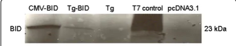
To generate Tg-BID transgenic mice, the rat Tg promoter region was fused upstream of the Bid transgene and cloned into pCMV to form CMV-BID. Sequence analysis was used to confirm that the Tg-BID plasmid had the correct sequence for the Tg promoter and insertion site. To verify expression of BID from the Tg-BID construct, the TNT-coupled transcription/translation system was used according to the manufacturer’s protocol. Under
in vitro transcription/translation conditions, the Tg-BID construct expressed a BID protein of the predicted size (23 kD), which was the same size produced by the CMV-BID (Figure 1). It is obvious that CMV promoter is much stronger than the Tg promoter. This verified that the BID protein translated from these transgenes can be recog-nized by anti-BID antibody.
The expression of the transgene has also been validated in transfected FRTL-5 cells. The pcDNA3.1-Tg-BID trans-gene was efficiently expressed in FRTL-5 cells transfected with pcDNA3.1-Tg-BID, but not in FRTL-5 cells trans-fected with control pcDNA3.1-Tg plasmid (Figure 2A). In a second set of experiments, FRTL-5 cells transfected with pcDNA3.1-Tg-BID were cultured in complete media with-out TSH for one week, followed by the addition of 0–0.625 mU/ml of TSH for 72 hours. It is known that TSH is able to activate Tg promoter. Figure 2B shows that BID protein expression was regulated by TSH, confirming the functionality of the Tg promoter.
Assessment of spontaneous thyroiditis in Tg-BID CBA/J (H-2 k) mice
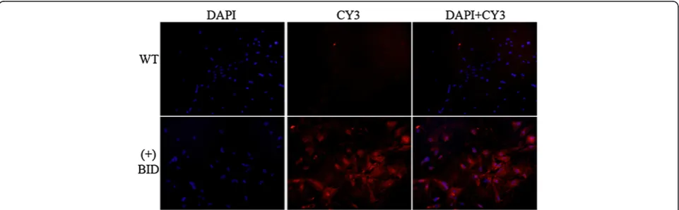
[image:3.595.55.540.90.217.2]Tg-BID positive C57BL/6 J X CBA/J (H-2 k) mice were crossed with CBA/J (H-2 k) mice, which are a strain genetically susceptible to EAT [8-10], to produce C1 mice, and subsequent generations were made with simi-lar backcrosses. RNAs extracted from the C4 transgenic mice thyroids demonstrated message for Tg-BID positive mice, but not in DNA negative mice (Table 1), nor in other tissues in the transgene positive mice (data not shown). The expression of BID protein was examined in the thyroid from the founder to C4 transgenic mice using Western blot and found that a considerable num-ber of mice expressed BID (Figure 3). Mice with high, medium and low levels of Tg-BID were defined based on the relative level of Tg-BID protein as compared to the actin control. Clones with very high levels of Tg-BID relative to actin controls were considered “high” (e.g., clones 55 and 205 in Figure 3); clones with Tg-BID levels that were similar to the actin control were considered “medium” (e.g., clones 534 and 102); and clones with very low levels of Tg-BID relative to the control were considered “low” (e.g., clones 66 and 118). Clones positive for BID by Western blot were confirmed by immunofluorescent staining of cultured primary mouse thyrocytes (Figure 4). It was noted that the levels of BID protein in the positive clones were various, which Figure 2Transgene BID protein expressed in FRTL-5 cells and FRTL-5 cells treated with TSH.Total protein was isolated from FRTL-5 cells transfected with pcDNA3.1-Tg-BID and subjected to Western blot for the transgene Bid protein(A). Total protein from FRTL-5 cells transfected with pcDNA3.1-TgBID and treated with a series of diluted TSH for 72 hours was also isolated and subjected to Western blot to detect BID
(B). GAPDH was used as a loading control.
Table 1 Quantification of Tg-BID mRNA expression in mouse thyroid by RT-PCR
Mice Subgroup n Tg-BID level
Wild type 8 0
Transgenic Low 8 5
Transgenic Medium 8 37
[image:3.595.305.540.663.733.2]enabled us to divide the transgenic mice into high, medium and low Tg-BID expression group for the fur-ther function study.
Though the levels of Tg-BID proteins were highly vari-able in those transgenic mice, no anti-Tg antibody was detectable in sera. In addition, serum T4 levels were not different between Tg-BID transgenic mice and wild-type mice. Furthermore, the gross appearance and micro-structure of thyroid glands in all transgenic mice ap-peared similar to that of the wild-type mice, suggesting that the overexpression of BID itself has no impact on thyroid function in terms of T4, and does not cause a notable autoimmune response in the thyroid gland.
Iodine induced autoimmune thyroiditis in Tg-BID transgenic CBA/J (H-2 k) mice
[image:4.595.55.541.89.277.2]As demonstrated above, no spontaneous thyroiditis was observed in Tg-BID transgenic CBA/J (H-2 k) mice. To in-vestigate whether BID can cooperate with other thyroiditis risk factors to facilitate the development of autoimmune thyroiditis, we treated mice with iodine, a known risk fac-tor for thyroiditis [10,12,13]. Mice treated with 0.3% iodine begin to show an increase in anti-Tg antibody at Week 4 in Tg-BID transgenic mice compared with wild type CBA/J (H-2 k) mice, and the level of anti-Tg antibody was signifi-cantly higher at Week 8 (* p < 0.01, Figure 5). Consistent with this increase in anti-Tg antibody, 3 of 10 Tg-BID Figure 3Expression of transgene Tg-BID in the thyroid of the founder (A), C1 (B) and C2 (C) mice.Mouse thyroid cells were isolated and cell lysates prepared and subjected to Western blot for BID. Actin was detected as a control for equal loading. The number represents different clones of transgenic mice. WT: wild type.
[image:4.595.58.539.545.694.2]transgenic CBA/J (H-2 k) mice showed thyroid gland mononuclear cell infiltration whereas no thyroids of the wild type CBA/J (H-2 k) had this phenotype (Figure 6). Despite the increase of serum anti-Tg antibody and mono-nuclear cell infiltration, serum T4 levels were similar bet-ween iodine-treated Tg-BID transgenic CBA/J (H-2 k) mice and wild-type CBA/J (H-2 k) mice. These findings suggest that the thyroid function of the Tg-BID transgenic mice ap-pears within the normal range after receiving 8-week iodine in the drinking water, even though iodine uptake does re-sult in a certain degree of autoimmune responses such as autoantibody production and mononuclear cell infiltration in Tg-BID transgenic mice.
Discussion
Our previous work demonstrated that the Fas pathway in thyrocytes can be activated by Fas ligand in the pres-ence of combinations of pro-inflammatory cytokines such as IL-1β, IFNγ and TNFα [1,2]. Further experi-ments revealed that the induction of apoptosis in thyro-cytes is correlated with an increase of BID and Bak levels but a decrease of p44/42 mitogen-activated pro-tein kinase activity [2,14,15]. These findings suggest that the Fas apoptotic pathway activity may be further ampli-fied by the bridge pro-apoptotic molecule BID, leading to the release of Bak from the mitochondria and activa-tion of caspases. Thus, apoptosis of thyrocytes in EAT or autoimmune thyroiditis is dependent on both cell death receptors and mitochondrial elements, in which BID plays a critical role [16,17]. In contrast to most
apoptotic activity, apoptotic cell death in EAT or auto-immune thyroiditis is not anti-inflammatory [18]. In-stead, apoptotic thyroid cells and secondary necrotic cells induce a pro-inflammatory environment, which may provide sufficient levels of self-antigens to intensify a deregulated immune response [18,19]. The occurrence of inflammation is now known to be closely associated with a BID-related pathway that is either dependent or independent of apoptosis [20-22].
[image:5.595.309.540.89.424.2]To test whether the genetic changes in BID play a role in the development of autoimmune thyroiditis, we suc-cessfully developed a transgenic mouse line in which BID is specifically over-expressed in the thyroid. We ob-served Tg-BID transgenic mice for 15 months, and du-ring this period these mice remained healthy. These mice showed no obvious difference in activity and ap-pearance compared with the control mice. In addition, no difference in thyroid histology and morphology were observed. We found that regardless of the level of BID Figure 5Elevation of serum thyroid autoantibody anti-Tg in
Tg-BID transgenic CBA/J mice treated with iodine.Sera were collected from the mice before iodine administration (pre-treatment), 4 weeks after iodine administration and 8 weeks after iodine administration. Anti-Tg antibody was measured by solid-phase ELISA and results were expressed as OD 405 ± SD of 1:200 dilution of serum from ten individual mice per group.
Figure 6Thyroid histology.Representative hematoxylin and eosin staining of thyroid glands of the wild type CBA/J (H-2 k) mice
[image:5.595.57.293.89.278.2]tally unexpected. Autoimmune thyroiditis is a disorder with multiple causative factors including genetic, envi-ronmental, and immunological elements [18,23]. Among these elements, iodine is able to exert pleiotropic effects on either metabolic or immunological processes of thy-roid. While iodine is an indispensable constituent of the two major thyroid hormones T3 and T4 [24], it contri-butes to the development of autoimmune thyroiditis by enhancing the antigenicity of thyroglobulin and reducing regulatory T cells [13,25]. Though iodine is known to contribute to the development of autoimmune thyroi-ditis [8-10], it may not be able to do so without other co-factors. For example, iodine is able to induce thyroid-itis in nearly 100% of NOD.H-2 h4 mice that express I-Ak on the NOD genetic background, but cause none in mice such as CBA/J (H-2 k) (H-2 k) and NOD.SWR (H-2(q)) that do not carry I-Ak [8]. It is unknown whether mice with BID overexpression would sensitize thyroiditis induced by iodine. To this end, we employed iodine-resistant CBA/J (H-2 k) mice in this study. We show that upon iodine administration, Tg-BID trans-genic CBA/J (H-2 k) mice do indeed develop auto-immune thyroiditis in about 8 weeks, which is evident by a significant increase in serum anti-Tg autoantibody and 30% of Tg-BID transgenic CBA/J (H-2 k) mice hav-ing mononuclear cell infiltration into the thyroid glands. The timing of iodine-induced autoimmune thyroiditis in Tg-BID transgenic CBA/J (H-2 k) mice appears to be quite similar to that in NOD-H-2 h4 mice [8].
Conclusions
Our study has demonstrated that the increasing BID ex-pression specifically in thyroid does not cause autoimmune thyroiditis. However, mice with thyroid-specific BID over-expression are at high risk of development of autoimmune thyroiditis induced by known pathogenic factors such as iodine. These findings support the common concept that autoimmune thyroiditis is a multi-factorial disease, result-ing from interplay of genetic, environmental, and en-dogenous factors [18,23].
Abbreviations
BID:BH3 interacting-domain death agonist; EAT: Experimental autoimmune thyroiditis; Tg: Thyroglobulin.
Received: 17 March 2014 Accepted: 12 June 2014 Published: 23 June 2014
References
1. Wang SH, Bretz JD, Phelps E, Mezosi E, Arscott PL, Utsugi S, Baker JR Jr:A unique combination of inflammatory cytokines enhances apoptosis of thyroid follicular cells and transforms nondestructive to destructive thyroiditis in experimental autoimmune thyroiditis.J Immunology2002,
168:2470–2474.
2. Mezosi E, Wang SH, Utsugi S, Bajnok L, Bretz JD, Gauger PG, Thompson NW, Baker JR Jr:Induction and regulation of Fas-mediated apoptosis in human thyroid epithelial cells.Mol Endocrinology2005,19:804–811. 3. Wang SH, Van Antwerp M, Kuick R, Gauger PG, Doherty GM, Fan YY, Baker
JR Jr:Microarray analysis of cytokine activation of apoptosis pathways in the thyroid.Endocrinology2007,148:4844–4852.
4. Wang SH, Chen GH, Fan YY, Van Antwerp M, Baker JR Jr:TRAIL inhibits experimental autoimmune thyroiditis by the expansion of CD4 + CD25+ regulatory T cells.Endocrinology2009,150:2000–2007.
5. Paunovic V, Carter NA, Thalhamer T, Blair D, Gordon B, Lacey E, Michie AM, Harnett MM:Immune complex-mediated co-ligation of the BCR with FcγRIIB results in homeostatic apoptosis of B cells involving Fas signalling that is defective in the MRL/Lpr model of systemic lupus erythematosus.J Autoimmun2012,39:332–346.
6. Askari N, Correa RG, Zhai D, Reed JC:Expression, purification, and characterization of recombinant NOD1 (NLRC1): A NLR family member. J Biotechnol2012,157:75–81.
7. Midgley A, Mayer K, Edwards SW, Beresford MW:Differential expression of factors involved in the intrinsic and extrinsic apoptotic pathways in juvenile systemic lupus erythematosus.Lupus2011,20:71–79. 8. Braley-Mullen H, Sharp GC, Medling B, Tang H:Spontaneous autoimmune
thyroiditis in NOD.H-2 h4 mice.J Autoimmun1999,12:157–165. 9. Fang Y, Zhao L, Yan F:Chemokines as novel therapeutic targets in
autoimmune thyroiditis.Recent Pat DNA Gene Seq2010,4:52–57. 10. Li HS, Carayanniotis G:Induction of goitrous hypothyroidism by dietary
iodide in SJL mice.Endocrinology2007,148:2747–2752.
11. Wang SH, Cao Z, Wolf JM, Van Antwerp M, Baker JR Jr:Death ligand tumor necrosis factor-related apoptosis-inducing ligand inhibits experimental autoimmune thyroiditis.Endocrinology2005,146:4721–4726.
12. Rose NR, Bonita R, Burek CL:Iodine: an environmental trigger of thyroiditis.Autoimmun Rev2002,1:97–103.
13. Xue H, Wang W, Shan Z, Li Y, Li Y, Teng X, Gao Y, Fan C, Teng W:Dynamic changes of CD4 + CD25 + regulatory T cells in NOD.H-2 h4 mice with iodine-induced autoimmune thyroiditis.Biol Trace Elem Res2011,
143:292–301.
14. Wang SH, Mezosi E, Wolf JM, Cao Z, Utsugi S, Gauger PG, Doherty GM, Baker JR Jr:IFNγsensitization to TRAIL-induced apoptosis in human thyroid carcinoma cells by up-regulating Bak expression.Oncogene2004,
23:928–935.
15. Mezosi E, Wang SH, Utsugi S, Bretz JD, Thompson NW, Gauger PG, Baker JR Jr:The combination of IL-1βand TNFαsensitizes human thyroid epithelial cells to TRAIL by a complex mechanism involving Bid and p42/44 MAPK.J Clin Endo Metab2004,89:250–257.
16. Song G, Chen GG, Hu T, Lai PB:Bid stands at the crossroad of stress-response pathways.Curr Cancer Drug Targets2010,10:584–592. 17. Kantari C, Walczak H:Caspase-8 and bid: caught in the act between
death receptors and mitochondria.Biochim Biophys Acta1813,
18. Wang SH, Baker JR Jr:Thyroiditis. InImmunoendocrinology: Scientific and Clinical Aspects.1st edition. Edited by Eisenbarth GS. New York, NY: Humana Press; 2011:443–457.
19. Salzano M, Russo E, Postiglione L, Guerra A, Marotta V, Esposito S, Vitale M:
Interferon-γinhibits integrin-mediated adhesion to fibronectin and survival signaling in thyroid cells.J Endocrinol2012,215:439–444. 20. Sebastian BM, Roychowdhury S, Tang H, Hillian AD, Feldstein AE, Stahl GL,
Takahashi K, Nagy LE:Identification of a cytochrome P4502E1/Bid/C1q-dependent axis mediating inflammation in adipose tissue after chronic ethanol feeding to mice.J Biol Chem2011,286:35989–35997.
21. Yeretssian G, Correa RG, Doiron K, Fitzgerald P, Dillon CP, Green DR, Reed JC, Saleh M:Non-apoptotic role of BID in inflammation and innate immunity.Nature2011,474:96–99.
22. Kang HR, Cho SJ, Lee CG, Homer RJ, Elias JA:Transforming growth factor (TGF)-beta1 stimulates pulmonary fibrosis and inflammation via a Bax-dependent, Bid-activated pathway that involves matrix metalloproteinase-12.J Biol Chem2007,282:7723–7732.
23. Wang SH, Baker JR:The role of apoptosis in thyroid autoimmunity.Thyroid
2007,17:975–979.
24. Schomburg L, Köhrle J:On the importance of selenium and iodine metabolism for thyroid hormone biosynthesis and human health. Mol Nutr Food Res2008,52:1235–1246.
25. Barin JG, Talor MV, Sharma RB, Rose NR, Burek CL:Iodination of murine thyroglobulin enhances autoimmune reactivity in the NOD.H2 mouse. Clin Exp Immunol2005,142:251–259.
doi:10.1186/1479-5876-12-180
Cite this article as:Wanget al.:Overexpression of BID in thyroids of transgenic mice increases sensitivity to iodine-induced autoimmune thyroiditis.Journal of Translational Medicine201412:180.
Submit your next manuscript to BioMed Central and take full advantage of:
• Convenient online submission
• Thorough peer review
• No space constraints or color figure charges
• Immediate publication on acceptance
• Inclusion in PubMed, CAS, Scopus and Google Scholar
• Research which is freely available for redistribution